Review
Natural Compounds as Target Biomolecules in Cellular
Adhesion and Migration: From Biomolecular Stimulation to Label-Free Discovery and Bioactivity-Based Isolation
Beatrix Péter
1,* , Imre Boldizsár
2,3, Gábor M. Kovács
2,4, Anna Erdei
5,6, Zsuzsa Bajtay
5,6, Alexandra Vörös
1, Jeremy J. Ramsden
7, Ildikó Szabó
8,9, Szilvia B ˝osze
8,9and Robert Horvath
1
Citation: Péter, B.; Boldizsár, I.;
Kovács, G.M.; Erdei, A.; Bajtay, Z.;
Vörös, A.; Ramsden, J.J.; Szabó, I.;
B˝osze, S.; Horvath, R. Natural Compounds as Target Biomolecules in Cellular Adhesion and Migration:
From Biomolecular Stimulation to Label-Free Discovery and Bioactivity-Based Isolation.
Biomedicines2021,9, 1781. https://
doi.org/10.3390/biomedicines9121781
Academic Editor: Sharon Marx
Received: 25 October 2021 Accepted: 22 November 2021 Published: 26 November 2021
Publisher’s Note:MDPI stays neutral with regard to jurisdictional claims in published maps and institutional affil- iations.
Copyright: © 2021 by the authors.
Licensee MDPI, Basel, Switzerland.
This article is an open access article distributed under the terms and conditions of the Creative Commons Attribution (CC BY) license (https://
creativecommons.org/licenses/by/
4.0/).
1 Nanobiosensorics Group, Research Centre for Energy Research, Institute for Technical Physics and Materials Science, Konkoly-Thege u 29-33, 1120 Budapest, Hungary; aavoros@gmail.com (A.V.);
r74horvath@gmail.com (R.H.)
2 Department of Plant Anatomy, Institute of Biology, Eötvös Loránd University, 1117 Budapest, Hungary;
boldizsarimi@gmail.com (I.B.); gaborm.kovacs@ttk.elte.hu (G.M.K.)
3 Department of Pharmacognosy, Semmelweis University, Üll˝oiút 26, 1085 Budapest, Hungary
4 Plant Protection Institute, Centre for Agricultural Research, Hungarian Academy of Sciences, 1022 Budapest, Hungary
5 Department of Immunology, Eötvös Loránd University, 1117 Budapest, Hungary;
anna8erdei@gmail.com (A.E.); bajtay.zsuzsanna@ttk.elte.hu (Z.B.)
6 MTA-ELTE Immunology Research Group, Eötvös Loránd Research Network (ELKH), Eötvös Loránd University, 1117 Budapest, Hungary
7 Clore Laboratory, University of Buckingham, Buckingham MK18 1EG, UK;
jeremy.ramsden@buckingham.ac.uk
8 MTA-ELTE Research Group of Peptide Chemistry, Eötvös Loránd Research Network (ELKH), Institute of Chemistry, Eötvös Loránd University, 1117 Budapest, Hungary; szaboi8@gmail.com (I.S.);
szilvia.bosze@gmail.com (S.B.)
9 National Public Health Center, Albert Flóriánút 2-6, 1097 Budapest, Hungary
* Correspondence: peter.beatrix@energia.mta.hu
Abstract:
Plants and fungi can be used for medical applications because of their accumulation of special bioactive metabolites. These substances might be beneficial to human health, exerting also anti-inflammatory and anticancer (antiproliferative) effects. We propose that they are mediated by influencing cellular adhesion and migration via various signaling pathways and by directly inactivating key cell adhesion surface receptor sites. The evidence for this proposition is reviewed (by summarizing the natural metabolites and their effects influencing cellular adhesion and migration), along with the classical measuring techniques used to gain such evidence. We systematize existing knowledge concerning the mechanisms of how natural metabolites affect adhesion and movement, and their role in gene expression as well. We conclude by highlighting the possibilities to screen natural compounds faster and more easily by applying new label-free methods, which also enable a far greater degree of quantification than the conventional methods used hitherto. We have systemati- cally classified recent studies regarding the effects of natural compounds on cellular adhesion and movement, characterizing the active substances according to their organismal origin (plants, animals or fungi). Finally, we also summarize the results of recent studies and experiments on SARS-CoV-2 treatments by natural extracts affecting mainly the adhesion and entry of the virus.
Keywords:
natural compound; cell adhesion; movement; CAM; integrin; viability; biosensors;
preparation; isolation; SARS-CoV-2
1. Introduction
Natural medicines, extracted from herbs and other living sources such as serpent venoms, have been used by humans since the earliest times. In contrast, the use of mineral substances as medicines was an innovation of Paracelsus introduced as recently as the
Biomedicines2021,9, 1781. https://doi.org/10.3390/biomedicines9121781 https://www.mdpi.com/journal/biomedicines
16th century AD. Today, there is renewed interest in natural substances for curing illnesses, both directly and as inspirations for manufactured pharmaceuticals (for example, an anti- malaria agent extracted from the sweet wormwood plant [1]). Many people prefer to take mixtures of herbs as alternatives to industrial medicine, which often causes deleterious side effects [2]. Plants (and fungi) synthesize a large number of specific compounds called secondary metabolites (SMs). The function of SMs in plants and fungi is not fully understood; however, many of these compounds can be used for medicinal purposes. They have special significance in cancer therapy: among compounds introduced since the 1940s, almost 50% were isolated, purified SMs or their semi-synthetic derivatives [3].
The study of the mode of action of natural substances at the cellular and molecular levels only began a few years ago with the advent of modern techniques. Compared to manufactured pharmaceuticals systematic scientific evidence for the efficacy and safety of natural substances is generally still lacking [2]. When studying SMs, the major challenge is to elucidate their effective targets, which are responsible for the medicinal effects of these natural compounds. In contrast with synthetic drugs the target is usually preselected, and the challenge is to find a molecule that binds to the target and not the others.
We suggest that these substances influence primarily at the cellular level. The general approach has been to apply labeling techniques to investigate effects on cell adhesion, mi- gration, motility etc. in vitro [1]. We shall review new, label-free techniques for monitoring cell adhesion and spreading directly, without label-induced perturbation.
Cell adhesion is crucial for the assembly of individual cells into tissues [4]. It is responsible for the overall architecture of the tissue [4]. Monitoring cell adhesion and spreading is important because these processes maintain the multicellular tissue structure, and individual cell migration, survival, proliferation, differentiation, gene expression, cell- cell communication and immunity, and cancer metastasis [1]. Thus, studying the adhesion and spreading of the treated cells by natural compounds helps to understand their effects on metabolism, development and physiology.
In this review we summarize the effects of natural compounds from plants, fungi and snake venom on cellular adhesion and movement, and the methods applied to reveal these effects. Finally, we mention some possible ways to prevent or reduce symptoms of COVID-19 by applying natural compounds according to the recent literature in this topic.
2. Relationship between Adhesion, Movement and Inflammation
Inflammation is typified by the accumulation of leukocytes and other mesenchymal cells in response to attractant molecules at sites of injury or infection [5–7]. Leukocytes become activated after being exposed to chemoattractants and are capable of adhering tightly to the endothelium [7]. Cytokines and endotoxins stimulate the endothelium to become more adhesive for leukocytes [5,7,8]. The general classes of cell adhesion molecules (CAM) are integrins, selectins, the immunoglobulin superfamily of cell adhesion molecules and cadherins [5,7,9]. Integrins and selectins on circulating leukocytes mediate their adhesion to the endothelium, whereas selectins and members of the immunoglobulin superfamily on the endothelium mediate their affinity for leukocytes [7,9]. CAMs are known to play a critical role in the recruitment of cells into various tissues and in the maintenance and regulation of the integrity of the tissues [5].
CAM expression is tightly regulated in normal tissue environment; however, inappro- priate expression of CAMs disrupts normal cell-cell and cell-matrix interactions, facilitating tumour formation.
Integrins are prime regulators of communication between cells and their microenvi- ronment. These evolutionarily old cellular adhesion receptors play an important role in physiological and pathological processes. These large molecules are responsible for the at- tachment of cells to the extracellular matrix (ECM) components and cell-to-cell interactions.
Integrin heterodimers are composed of noncovalently bound α and β subunits.
In vertebrates the integrin family is composed of 18 α subunits and 8 β subunits that form 24 different heterodimeric complexes [10] (Figure 1). The integrins can be grouped into subgroups based on their subunit composition and ligand-binding specificity.
A subgroup of integrins (8 out of 24) recognize proteins (such as fibronectin and vitronectin) that contain the Arg-Gly-Asp (RGD) sequence. The collagen binding integrins α 1 β 1, α 2 β 1, α 10 β 1, and α 11 β 1 are able to recognize the triple-helical GFOGER collagen sequence. The laminin receptors α3β1, α6β1 and α7β1 mediate adhesion to basement membrane laminins. Vertebrates also have leukocyte-specific integrins that mediate cell- cell adhesion. The β 2 integrins are present on the cell surface in a normally inactive state, in which they do not bind ligands. An activation signal like an inflammatory stimulus results in a conformational change of the integrin, increasing its ability to bind the ligand. Activated β 2 integrins play an important role in cellular adhesion, migration and phagocytosis.
The lack of the β subunit can block preimplantation development (β1) and perinatal lethality ( β 8) and is involved in various defects of leukocyte function ( β 2, β 7), inflammation ( β 6), hemostasis and angiogenesis ( β 3). Integrins frequently intercommunicate; that is, they are able to activate or inhibit each other’s function [10].
The CD11/CD18 β 2 integrins are specifically expressed on circulating leukocytes, and play a significant role in fast adhesion to endothelial cells [7,9]. The intercellular adhe- sion molecule-1 (ICAM-1) and vascular cellular adhesion molecule-1 (VCAM-1) are well- characterized endothelial cell ligands for CD11/CD18 [7,11]. The endothelial cell surface quantity of molecules like ICAM-1 is increased with the release of some proinflammatory cytokines from lymphocytes; an environment is thereby generated in which the leukocytes are more chemoreceptive to arterial walls during inflammatory processes [5]. Recruitment of arterial leukocytes is a significant step in the progression of different inflammatory diseases, such as rheumatism [12,13], liver inflammation [13,14], atherosclerosis [13,14], and inflammatory bowel disease [13]. Medical plant extracts may alter pathological mech- anisms via the modulation of adhesion molecules [5]; this general observation will be analyzed and systematized below.
All morphogenetic processes are affected by cell migration, which contributes to many illnesses, including cardiovascular disease and cancer [15]. In general, cell move- ment begins with extension of the membrane followed by the formation of new adhesive protrusions at the front, which link the actin cytoskeleton to the substratum, generating traction forces that move the cell forwards; adhesive protrusions at the rear are simultane- ously dismantled [15]. The cycle of forming and dismantling of adhesive structures drives migration [15]. Rho GTPases have an important role in this process; they regulate actin polymerization and myosin II activity and, therefore, adhesion dynamics [15].
The adhesion molecules driving migration are the same as those involved in the inflam-
matory response, but their expression and ligand-binding capacity depend on the stimuli.
Figure 1. Integrin family members and their ligands. Integrins are transmembrane heterodimer molecules containing α and β subunits. The figure illustrates the association of these subunits occurring in mammalian cells. As shown, 18 α and 8 β subunits form 24 different, distinct integrins.
These can be grouped in subfamilies based on evolutionary relationships (different colors of α subunits), ligand specificity and, in the case of β2 and β7 integrins, restricted expression on white blood cells. Integrins can be grouped into two larger classes that bind to cell surface cell adhesion molecules (CAMs) and ECM ligands. They can be further classified as collagen-binding integrins (α1β1, α2β1, α10β1, and α11β1), RGD-recognizing integrins (α5β1, αVβ1, αVβ3, αVβ5, αVβ6, αVβ8, and αIIbβ3), laminin-binding integrins (α3β1, α6β1, α7β1, and α6β4), and leukocyte integrins (αLβ2, αMβ2, αXβ2, and αDβ2). The β2 integrin subunit (CD18) can pair with one of the four α subunits (αL-CD11a, αM-CD11b, αX-CD11c, and αD-CD11d) [16]. The α4β1 and α9β1 integrins recognize fibronectin and VCAM-1. The β2 and β7 integrins are restrictedly expressed by leukocytes). Asterisks show the alternatively spliced cytoplasmic domains. This figure is based on the study of Hynes [10] and Yue et al. [17]. (RGD, Arg-Gly-Asp sequence; VCAM-1, a vascular cellular adhesion molecule-11).
3. Mechanisms of Action of Natural Compounds 3.1. Prestimulation with Cytokines
To demonstrate the inhibitory effect of a natural compound on cellular adhesion, cells, typically from the human umbilical vein endothelial cell line (HUVEC), are usually first treated with certain cytokines to stimulate the expression of CAM (Figure 2). In vivo, lipopolysaccharide (LPS, from Gram-negative bacteria) stimulates the immune response by interacting with its leukocyte membrane receptor, the Pattern Recognition Receptor (PRR) CD14 (with TLR4-MD2), to induce the generation of cytokines such as tumour necrosis factor α (TNF-α), interleukin-1 and -6 (IL-1, IL-6) (Figure 3). TNF-α is also involved in systemic inflammation [2,18]. It is primarily produced by activated monocytes or macrophages [19]. Note, cytokine generation, increase of expression of cytokines can also be stimulated by certain plant extracts (for example, among others, garlic (Allium sativum) decreases the level of IL-1α, IL-2, IL-6, IL-12, TNF- α and IFN-γ, however,
Figure 1.
Integrin family members and their ligands. Integrins are transmembrane heterodimer molecules containing
αand
βsubunits. The figure illustrates the association of these subunits occurring in mammalian cells. As shown, 18
αand 8
βsubunits form 24 different, distinct integrins.
These can be grouped in subfamilies based on evolutionary relationships (different colors of
αsubunits), ligand specificity and, in the case of
β2 andβ7 integrins, restricted expression on whiteblood cells. Integrins can be grouped into two larger classes that bind to cell surface cell adhesion molecules (CAMs) and ECM ligands. They can be further classified as collagen-binding integrins (α
1β1,
α2β1,
α10β1, and
α11β1), RGD-recognizing integrins (α
5β1,
αVβ1,
αVβ3,
αVβ5,
αVβ6,
αVβ8, and
αIIbβ3), laminin-binding integrins (α
3β1,
α6β1,
α7β1, and
α6β4), and leukocyte integrins (α
Lβ2,
αMβ2,
αXβ2, and
αDβ2). The
β2integrin subunit (CD18) can pair with one of the four
αsubunits (α
L-CD11a,
αM-CD11b,
αX-CD11c, and
αD-CD11d) [16]. The
α4β1 andα9β1 integrins recognizefibronectin and VCAM-1. The
β2 andβ7 integrins are restrictedly expressed by leukocytes). Asterisksshow the alternatively spliced cytoplasmic domains. This figure is based on the study of Hynes [10]
and Yue et al. [17]. (RGD, Arg-Gly-Asp sequence; VCAM-1, a vascular cellular adhesion molecule-11).
3. Mechanisms of Action of Natural Compounds 3.1. Prestimulation with Cytokines
To demonstrate the inhibitory effect of a natural compound on cellular adhesion,
cells, typically from the human umbilical vein endothelial cell line (HUVEC), are usually
first treated with certain cytokines to stimulate the expression of CAM (Figure 2). In vivo,
lipopolysaccharide (LPS, from Gram-negative bacteria) stimulates the immune response by
interacting with its leukocyte membrane receptor, the Pattern Recognition Receptor (PRR)
CD14 (with TLR4-MD2), to induce the generation of cytokines such as tumour necrosis
factor α (TNF- α ), interleukin-1 and -6 (IL-1, IL-6) (Figure 3). TNF- α is also involved
in systemic inflammation [2,18]. It is primarily produced by activated monocytes or
macrophages [19]. Note, cytokine generation, increase of expression of cytokines can also
be stimulated by certain plant extracts (for example, among others, garlic (Allium sativum)
decreases the level of IL-1α, IL-2, IL-6, IL-12, TNF- α and IFN-γ, however, increases IL-10),
as summarized by Spelman, et al. [20]. After stimulation of the endothelial cells, the plant
(or venom) extract is then added to them. The natural compound may downregulate expression of the adhesion molecules, resulting in the diminution of cell adhesion, and the compound has an antiinflammatory effect in consequence. The ICAMs and VCAMs are the most researched adhesion molecules [5]. We elaborate on this in the next section.
increases IL-10), as summarized by Spelman, et al. [20]. After stimulation of the endothelial cells, the plant (or venom) extract is then added to them. The natural compound may downregulate expression of the adhesion molecules, resulting in the diminution of cell adhesion, and the compound has an antiinflammatory effect in consequence. The ICAMs and VCAMs are the most researched adhesion molecules [5].
We elaborate on this in the next section.
Figure 2. Adhesion molecule stimulation and mechanisms in endothelial cells. Inhibitory effect of a
natural compound on cellular adhesion, typically of the human umbilical vein endothelial cell line (HUVEC), which are usually first treated with certain cytokines to stimulate the expression of CAMs.
Figure 3. Stimulation mechanism for cytokine modulation in lymphocytes. In vivo,
lipopolysaccharide (LPS, from Gram-negative bacteria) stimulates the immune response by interacting with its leukocyte membrane receptor, CD14 (with TLR4-MD2), to induce the generation of cytokines such as TNF- α, IL-1, IL-1 α, IL-6.
3.2. Inhibition of CAMs by Suppression of Their Expression (Downregulation)
The phytochemicals of olive oil (from Olea europaea) and red wine (from Vitis vinifera), oleuropein (monoterpene, seco-iridoid glucoside), hydroxytyrosol (phenylethanoid), tyrosol (phenylethanoid), elenolic acid (monoterpene, seco-iridoid) and resveratrol (stilbene) at nutritionally relevant concentrations have been shown to inhibit endothelial adhesion molecule expression (Figure 2). This provides a strongly suggestive basis for the atheroprotective property of the so-called “Mediterranean diet” [21].
Figure 2.
Adhesion molecule stimulation and mechanisms in endothelial cells. Inhibitory effect of a natural compound on cellular adhesion, typically of the human umbilical vein endothelial cell line (HUVEC), which are usually first treated with certain cytokines to stimulate the expression of CAMs.
increases IL-10), as summarized by Spelman, et al. [20]. After stimulation of the endothelial cells, the plant (or venom) extract is then added to them. The natural compound may downregulate expression of the adhesion molecules, resulting in the diminution of cell adhesion, and the compound has an antiinflammatory effect in consequence. The ICAMs and VCAMs are the most researched adhesion molecules [5].
We elaborate on this in the next section.
Figure 2. Adhesion molecule stimulation and mechanisms in endothelial cells. Inhibitory effect of a
natural compound on cellular adhesion, typically of the human umbilical vein endothelial cell line (HUVEC), which are usually first treated with certain cytokines to stimulate the expression of CAMs.
Figure 3. Stimulation mechanism for cytokine modulation in lymphocytes. In vivo,
lipopolysaccharide (LPS, from Gram-negative bacteria) stimulates the immune response by interacting with its leukocyte membrane receptor, CD14 (with TLR4-MD2), to induce the generation of cytokines such as TNF- α, IL-1, IL-1 α, IL-6.
3.2. Inhibition of CAMs by Suppression of Their Expression (Downregulation)
The phytochemicals of olive oil (from Olea europaea) and red wine (from Vitis vinifera), oleuropein (monoterpene, seco-iridoid glucoside), hydroxytyrosol (phenylethanoid), tyrosol (phenylethanoid), elenolic acid (monoterpene, seco-iridoid) and resveratrol (stilbene) at nutritionally relevant concentrations have been shown to inhibit endothelial adhesion molecule expression (Figure 2). This provides a strongly suggestive basis for the atheroprotective property of the so-called “Mediterranean diet” [21].
Figure 3.
Stimulation mechanism for cytokine modulation in lymphocytes. In vivo, lipopolysaccha- ride (LPS, from Gram-negative bacteria) stimulates the immune response by interacting with its leukocyte membrane receptor, CD14 (with TLR4-MD2), to induce the generation of cytokines such as TNF-
α, IL-1, IL-1α, IL-6.3.2. Inhibition of CAMs by Suppression of Their Expression (Downregulation)
The phytochemicals of olive oil (from Olea europaea) and red wine (from Vitis vinifera), oleuropein (monoterpene, seco-iridoid glucoside), hydroxytyrosol (phenylethanoid), ty- rosol (phenylethanoid), elenolic acid (monoterpene, seco-iridoid) and resveratrol (stilbene) at nutritionally relevant concentrations have been shown to inhibit endothelial adhesion molecule expression (Figure 2). This provides a strongly suggestive basis for the atheropro- tective property of the so-called “Mediterranean diet” [21].
Walnut (Juglans regia) extract and its principal active component ellagic acid decreased
the stimulated endothelial expression of ICAM-1 and VCAM-1, indicating a mechanism
for the known antiatherogenic and osteoblastic activity of the substance [18]. A walnut- enriched diet may therefore indeed be cardioprotective and inhibit osteoporosis [18].
Curcumin (diphenylheptanoid) from the Curcuma longa rhizome also downregulated the expression of adhesion molecules and, hence, monocyte adhesion [13]. Saponin (triter- pene) astagaloside IV from Mongolian milkvetch (Astagalus membranaceus) decreased the LPS-induced expression of VCAM-1 and E-selectin on the surface of HUVEC, hence this Chinese traditional medicinal herb is predicted to have anti-inflammatory efficacy; however, ICAM-1 was not affected [22].
Tripertygium wilfordii is a vine-like plant that grows in south China, and in the Chinese pharmacopoeia the extract from its root is prescribed for treating long-term rheumatoid arthritis and systemic lupus erytnematosus [7,8]. Chang et al., applied IL-1 α to stimulate HUVEC cells; treatment with a high concentration (50 ng/mL) of the herb extract (contain- ing wilforonide, alkaloids, diterpenes, triterpenes, b-sitosterol, daucosterol, dulcitol and glycosides [7,23]) had a significant inhibitory effect on both the expression and secretion of the cellular adhesion molecules, and thus may be a potential therapeutic agent for the treatment of inflammatory diseases [7].
3.3. Mechanism of Downregulation
The downregulation of CAMs by natural products is achieved by inhibiting their gene expression.
The molecular details involve the uptake of the natural product by the cytoplasm followed by interaction between the compound and transcription factors for adhesion molecule genes. Activation of the transcription factor nuclear factor κB (NF-κB), is mediated by the proinflammatory cytokines mentioned above, for instance TNF- α , and triggers gene expression of adhesion molecules (Figure 4). NF- κ B binding sites are found in the promoter region of E-selectin, ICAM-1 and VCAM-1 [13,24–27]. NF- κ B is in the cytoplasm in inactive form, complexed to IκB “nuclear factor of κ light polypeptide gene enhancer in B-cells inhibitor” [13]. When cells are stimulated with TNF-α, the IκB is phosphorylated, ubiquitinated, and degraded. The thereby activated NF-κB translocates to the nucleus and transcriptionally up-regulates cytokine receptors as well as the adhesion molecules [13,26,28,29].
Walnut
(Juglans regia) extract and its principal active component ellagic aciddecreased the stimulated endothelial expression of ICAM-1 and VCAM-1, indicating a mechanism for the known antiatherogenic and osteoblastic activity of the substance [18].
A walnut-enriched diet may therefore indeed be cardioprotective and inhibit osteoporosis [18].
Curcumin (diphenylheptanoid) from the Curcuma longa rhizome also downregulated the expression of adhesion molecules and, hence, monocyte adhesion [13]. Saponin (triterpene) astagaloside IV from Mongolian milkvetch (Astagalus membranaceus) decreased the LPS-induced expression of VCAM-1 and E-selectin on the surface of HUVEC, hence this Chinese traditional medicinal herb is predicted to have anti- inflammatory efficacy; however, ICAM-1 was not affected [22].
Tripertygium wilfordii is a vine-like plant that grows in south China, and in the
Chinese pharmacopoeia the extract from its root is prescribed for treating long-term rheumatoid arthritis and systemic lupus erytnematosus [7,8]. Chang et al., applied IL-1α to stimulate HUVEC cells; treatment with a high concentration (50 ng/mL) of the herb extract (containing wilforonide, alkaloids, diterpenes, triterpenes, b-sitosterol, daucosterol, dulcitol and glycosides [7,23]) had a significant inhibitory effect on both the expression and secretion of the cellular adhesion molecules, and thus may be a potential therapeutic agent for the treatment of inflammatory diseases [7].
3.3. Mechanism of Downregulation
The downregulation of CAMs by natural products is achieved by inhibiting their gene expression.
The molecular details involve the uptake of the natural product by the cytoplasm followed by interaction between the compound and transcription factors for adhesion molecule genes. Activation of the transcription factor nuclear factor κB (NF-κB), is mediated by the proinflammatory cytokines mentioned above, for instance TNF-α, and triggers gene expression of adhesion molecules (Figure 4). NF-κB binding sites are found in the promoter region of E-selectin, ICAM-1 and VCAM-1 [13,24–27]. NF-κB is in the cytoplasm in inactive form, complexed to IκB “nuclear factor of κ light polypeptide gene enhancer in B-cells inhibitor” [13]. When cells are stimulated with TNF-α, the IκB is phosphorylated, ubiquitinated, and degraded. The thereby activated NF-κB translocates to the nucleus and transcriptionally up-regulates cytokine receptors as well as the adhesion molecules [13,26,28,29].
Figure 4. The NF-κB pathway in endothelial cells. The compounds shown to have anti-inflammatory effects in vivo completely annulled LPS- and TNF-α-triggered nuclear translocation of NF-κB and
Figure 4.
The NF-κB pathway in endothelial cells. The compounds shown to have anti-inflammatory effects in vivo completely annulled LPS- and TNF-α-triggered nuclear translocation of NF-κB and NF-κB DNA-binding activity in endothelial cells, thus the production of cytokine receptors and ICAM, VCAM is inhibited.
The expression and function of integrins on various immune cells are summarized in
Table 1.
Table 1.
Ligands and functions of different integrins of human leukocytes (RGD, Arg-Gly-Asp sequence; VCAM-1, vascular cellular adhesion molecule-1; ICAM-1, Intercellular adhesion molecule-1) [10,30–33].
Integrin Ligands Functions
β1 RGD, VCAM-1, E-cadherin Adhesion
β2 iC3b, fibrinogen, ICAM-1 Adhesion, phagocytosis, apoptotic cell clearance
β6 RGD, fibronectin Adhesion, endocytosis, inflammation
β 7 RGD, VCAM-1 Adhesion, inflammation
Kawasaki and co-workers demonstrated that hot-water extract of Curcuma longa also suppressed the phosphorylation and degradation of IκBα in endothelial cells [13].
Another extract, the triterpene saponin astragaloside IV (3-O-β-D-xylopyranosyl-6-O-β-D- glucopyranosylcycloastragenol) from the Chinese herb Astragalus membranaceus, shown to have anti-inflammatory effects in vivo, completely annulled LPS- and TNF- α -triggered nuclear translocation of NF- κ B and NF- κ B DNA-binding activity in endothelial cells [22]
(Figure 4), furthermore, it has shown that it has antioxidative stress, antiapoptosis, and antifibrosis activities, both in vitro and in vivo [34,35].
Recent studies have shown that astragaloside IV (AS-IV) administration ameliorates diabetic neuropathy in streptozotocin (STZ)-induced diabetic rats via an anti-inflammatory mechanism [36], inhibits endoplasmic reticulum stress [37], and protects podocytes [34,38].
However, the effect and mechanism of AS-IV on diabetic neuropathy induced by type 2 diabetes remain unknown [34]. Flavonoid quercetin attenuates TNF-α-induced ICAM-1 and MMP-9 expression in ARPE-19 cells via the MEK1/2–ERK1/2 and PKCδ–JNK1/2–
c-Jun or NF- κ B pathways [39]. Flavonoid apigenin significantly suppressed the TNF- α -stimulated upregulation of VCAM-1-, ICAM-1-, and E-selectin-mRNA to the basal levels [40]. Simple phenolic compound salicin inhibits IL-1 β -induced production of pro- inflammatory cytokines such as TNF- α , IL-6, and monocyte chemoattractant protein-1 (MCP-1), vascular adhesion molecules such as (ICAM-1 and VCAM-1, and high-mobility group protein 1 (HMGB-1)) [41].
Lignan-type active compounds manassantin A and B, dineolignan compounds, inhib- ited the PMA-induced ICAM-1/LFA-1-mediated homotypic aggregation of the HL-60 cells without cytotoxicity, with MIC values of 1.0 and 5.5 nM, respectively. Even though these compounds did not affect the adhesion of ICAM-1 to LFA-1, they inhibited PMA-induced ICAM-1 expression in HL-60 cells in a dose-dependent fashion. These results suggest that manassantin A and B inhibit cell aggregation through downregulation of ICAM-1 expression [42].
HUVECs treated with sesquiterpene α -iso-cubebene showed markedly suppressed TNF- α -induced mRNA expression of VCAM-1 and E-selectin, but little alteration in ICAM- 1 mRNA expression. α -iso-cubebene treatment also significantly decreased the TNF- α-induced cell surface and total protein expression of VCAM-1 and E-selectin without affecting ICAM-1 expression [43].
Diterpene andrographolide significantly reduced E-selectin expression of activated endothelial cells, and inhibited E-selectin expression at the mRNA level [44].
In vitro, triterpene saponine dioscin decreased monocyte adhesion to TNF- α -treated HUVECs by reducingvascular cell adhesion molecule-1 (VCAM-1) and intercellular adhe- sion molecule 1 (ICAM-1) expression and inhibiting endothelial lipase (EL) expression in TNF-α-treated HUVECs and macrophages by blocking the NF-kB pathway [45].
Ethyl 3
0,4
0,5
0-trimethoxycinnamate and piperine are the two active principles of Piper
longum. Using primary human umbilical vein endothelial cells, Kumar et al. [46] evaluated
the activities of ethyl 3
0,4
0,5
0-trimethoxycinnamate on TNF- α -induced expression of cell
adhesion molecules, ICAM-1, VCAM-1 and E-selectin, which play key roles in controlling
Biomedicines2021,9, 1781 8 of 56
various inflammatory diseases. Both compounds inhibited the TNF-α-induced expression of ICAM-1 [46].
Ganoderma lucidum, a medicinal mushroom, has been used in traditional Chinese medicine to prevent and treat various diseases, for example cancer [47]. A polysaccharide derived from the fungus interacted with cell surface proteins and β 1-integrin expression was diminished, while β -actin expression was not affected [47].
We summarize the different modes of pathway intervention involving gene expression in Table 2.
Table 2.
Different modes of pathway intervention involving gene expression by natural compounds.
Mode Action
P1 Inhibiting the dephosphorylation of I κB and, hence, the activation of NF-κB P2 Inhibiting translocation of activated NF-κB into the nucleus P3 Inhibition of binding of activated NF- κ B to promoter sites for
CAM expression
3.4. Intervention at the ECM
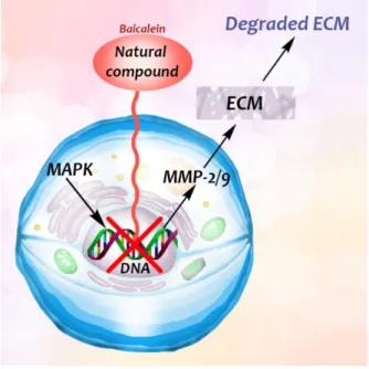
Flavonoid baicalein, derived from the root of Scutelaria baicalensis, a widely used Chi- nese herbal medicine that has been used in anti-cancer and anti-inflammatory therapy [48], has an inhibitory effect on the expression of matrix metalloproteinases (MMPs) [48], which are involved in the degradation of the extracellular matrix. Destruction of basement mem- branes and stromal extracellular matrix is critical for favoring metastasis and invasion of malignant cells [48]. The MMPs have therefore a role in promoting tumour growth, invasion and metastasis. Treatment of human breast carcinoma (MDA-MB-231) cells with baicalein inhibited the expression of MMP-2/9, which is a result of the mitogen-activated protein kinase (MAPK) signaling pathway [48] (Figure 5).
adhesion molecule 1 (ICAM-1) expression and inhibiting endothelial lipase (EL) expression in TNF-α-treated HUVECs and macrophages by blocking the NF-kB pathway [45].
Ethyl 3′,4′,5′-trimethoxycinnamate and piperine are the two active principles of Piper longum. Using primary human umbilical vein endothelial cells, Kumar et al. [46] evaluated the activities of ethyl 3′,4′,5′-trimethoxycinnamate on TNF-α-induced expression of cell adhesion molecules, ICAM-1, VCAM-1 and E-selectin, which play key roles in controlling various inflammatory diseases. Both compounds inhibited the TNF-α-induced expression of ICAM-1 [46].
Ganoderma lucidum, a medicinal mushroom, has been used in traditional Chinese medicine to prevent and treat various diseases, for example cancer [47]. A polysaccharide derived from the fungus interacted with cell surface proteins and β1-integrin expression was diminished, while β-actin expression was not affected [47].
We summarize the different modes of pathway intervention involving gene expression in Table 2.
Table 2. Different modes of pathway intervention involving gene expression by natural
compounds.
Mode Action P1 Inhibiting the dephosphorylation of I κB and, hence, the activation of NF-κB P2 Inhibiting translocation of activated NF-κB into the nucleus
P3 Inhibition of binding of activated NF-κB to promoter sites for CAM expression
3.4. Intervention at the ECM
Flavonoid baicalein, derived from the root of Scutelaria baicalensis, a widely used Chinese herbal medicine that has been used in anti-cancer and anti-inflammatory therapy [48], has an inhibitory effect on the expression of matrix metalloproteinases (MMPs) [48], which are involved in the degradation of the extracellular matrix. Destruction of basement membranes and stromal extracellular matrix is critical for favoring metastasis and invasion of malignant cells [48]. The MMPs have therefore a role in promoting tumour growth, invasion and metastasis. Treatment of human breast carcinoma (MDA-MB-231) cells with baicalein inhibited the expression of MMP-2/9, which is a result of the mitogen- activated protein kinase (MAPK) signaling pathway [48] (Figure 5).
Figure 5.
The interaction between the ECM, MMPs and natural compounds in breast carcinoma.
Baicalein blocked the expression of MMP-2/9, thus the degradation of the extracellular matrix is also inhibited.
Flavonoid (chalcone) butein (3,3,2
0,4
0-tetrahydroxychalcone) is an active substance found
in several plants, such as Semecarpus anacardium, Dalbergia odorifera, Caragana jubata and
Rhus verniciflua [49]. It has been demonstrated that it decreased leukocyte adhesion to A549
cells through the inhibition of TNF-α-induced ICAM-1 and VCAM-1 expression by inhibiting the NF-κB/MAPK/Akt signaling pathway. Butein also inhibits ROS generation [49], and may prevent TNF-α-induced airway inflammation [49] (Figure 4).
3.5. Inhibition of CAM Binding by Blocking Specific Cell–Surface Receptor Sites
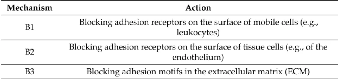
We can distinguish three types of mechanisms inhibiting cell adhesion by blocking (B) specific receptor sites (Table 3).
Table 3.
Three types of mechanisms inhibiting cell adhesion by blocking specific receptor sites.
Mechanism Action
B1 Blocking adhesion receptors on the surface of mobile cells (e.g., leukocytes)
B2 Blocking adhesion receptors on the surface of tissue cells (e.g., of the endothelium)
B3 Blocking adhesion motifs in the extracellular matrix (ECM)
In contrast to intervention at the level of protein expression, natural compounds can also specifically block cell recognition motifs, such as the amino acid triplet RGD. For example, the polyphenol EGCG from green tea has been shown to block RGD motifs (B) (Arg-Gly-Asp) and, hence, inhibit adhesion [50].
We recall that integrins are transmembrane heterodimers, a family of plasma mem- brane receptors that mediate adhesion of leukocytes to ECM [51]. Integrins are also involved in pathophysiological processes as well, such as genetic and autoimmune dis- eases, metastasis and thrombosis and, thus, integrins are important therapeutic target structures [51,52]. Blocking or disruption of binding to integrin receptors is therefore an important topic in industrial drug discovery [51,53]. Serpent venom disintegrins, a family of low molecular weight proteins, typically contain the RGD motif, and are known to block integrin activities by binding with high affinity to the integrins [51,54]. For example, rhodostomin, a snake venom from Calloselasma rhodostoma, blocked the integrin ανβ3 and also affected pp125 FAK phosphorylation and the actin cytoskeleton [55]. Rhodostomin contains an RGD motif that specifically inhibits the integrin-binding function [56]. It can be produced in Pichia pastoris (methylotrophic yeast) as well and it inhibits platelet aggregation with a K(I) of 78 nM as potent as native rhodostomin [56]. However, its D51E mutant blocks platelet aggregation with a K(I) of 49 mM [56]. Structural analysis of rhodostomin and its D51E mutant showed that they have the same tertiary fold with three two-stranded antiparallel beta-sheets [56]. Two minor differences between them were inferred from their backbone dynamics and 3D structures [56]. The docking of rhodostomin into integrin α IIb β 3 showed that between the backbone amide and carbonyl groups of the D51 residue were formed hydrogen bonds with the integrin residues R216 and R214, respectively [56].
In contrast, these hydrogen bonds were absent in the D51E mutant-integrin complex [56].
Another serpent venom, echistatin from Echis carinatus, inhibited integrin-mediated cell adhesion via selective recognition by α IIb β 3, α 5 β 1, ανβ 3 integrins [51], preventing their adhesion to the ECM.
Not only snake venom affects integrin-mediated adhesion, but herbs as well.
Epigallocatechin-gallate (EGCG) (flavonoid ester) is the main polyphenol of green tea (Camellia sinensis). Many studies have shown its beneficial effect on human health [1].
The majority of them demonstrated direct effects on cell adhesion and movement. In
a previous study we showed that EGCG indirectly affects HeLa cell adhesion: the
cells cannot adhere onto EGCG-pre-treated model ECM coatings [50]. The polyphe-
nol formed multilayers in poly-L-lysine polyethylene-glycol-RGD (PLL-g-PEG-RGD)
chains, and blocked the RGD motifs [50]. EGCG alters the properties of mucin as
well; EGCG-mucin mixtures showed that discrete particles are formed and their size
increases with the ratio of EGCG to mucin [57]. Another natural compound, cistifolin
(benzofuran derivative) from the rhizome of the gravel root (Eupatorium purpureum), known as an anti-rheumatic herbal drug, was identified as a potent inhibitor of β1 and β2 integrin-mediated cell adhesion and, thus, has therapeutic potential for diseases where integrin adhesion molecules play a significant role [58].
4. Measurement Techniques for Monitoring Cellular Functions, Adhesion and Viability
4.1. Classical Techniques for Measuring Cell Viability
Experimental natural compounds are added to stimulated cell cultures to induce a CAM response, which is commonly measured by labeling methods, mainly the enzyme- linked immunosorbent assay (ELISA), Western blot, and flow cytometry. ELISA uses antibodies linked to enzymes that create a color change (e.g., by altering a dye) to identify the examined substances [13]. The Western blot is a widely used technique in biology to detect specific proteins in a sample by letting animal-derived or synthetic antibodies bind to them [13]. ELISA and Western blot techniques cannot be used for measuring cell viability directly; however, for example, Western blot can be applied as a complementary method to study the mechanism of cell death (apoptosis, autophagy markers, etc.), thus it provides a lot of information about the mechanism. Flow cytometry is usually a laser-based technique for cell sorting and counting. A wide range of fluorophores can be used in flow cytometry measurements. Fluorophores are fluorescent labels that can attach to the antibody that recognizes the target molecule of the cell [2,59]. However, impedance-based flow cytometers also exist, known as Coulter counters, which are well established label-free methods for sizing and counting cells and particles [60].
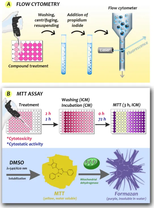
Some workers have used staining to reveal the effect of natural compounds on cell viability, mainly the MTT (3-(4,5-dimethylthiazol-2-yl)-2,5-diphenyltetrazolium bromide) assay and the trypan blue exclusion test. However, Wang et al. in 2010 showed that the MTT- and MTS (3-(4,5-dimethylthiazol-2-yl)-5-(3-carboxymethoxyphenyl)-2-(4-sulfophenyl)-2H- tetrazolium)-based assays (Figure 6) underestimate the antiproliferative effect of EGCG [1,61].
The terms “cell viability” or “compound cytotoxicity” have broad meanings in drug discovery. For in vitro monolayer cell cultures, a compound is considered to be cytotoxic if the compound interferes with cellular attachment, or significantly alters cellular morphol- ogy, cell growth and cell viability. A variety of assay methods can be used to estimate the number of viable eukaryotic cells after exposure of the investigated compounds. These cell-based assays are often used for screening collections of compounds to determine if the compounds have effects on cell proliferation or show cytotoxic and cytostatic effects.
Cell-based assays are also widely used for monitoring organelle function. These screening methods have been devised to examine a broad variety of parameters associated with biochemical events necessary for sustaining viability, especially as evinced by membrane integrity. The quantities emerging from metabolism (especially ATP-based viability) assays are proportional to viable cell number. Cytotoxicity assays determine parameters propor- tional to the degree of cell death. The fundamental difference between the approaches depends on the length of exposure to the compound (short-term exposures (4 h or less) may adversely affect metabolism markers or ATP content before measurable membrane integrity changes, and long-term exposures (24 h or more), particularly after early primary necrosis, may lead to underestimation of cytotoxicity owing to degradation of marker enzyme activity after its release into the extracellular environment [62–65].
Most cell viability and cytotoxicity assays can be divided into three categories: those
that (i) exploit the loss of membrane integrity; (ii) directly measure metabolic markers or
ATP content; and (iii) assess metabolic activity. Other forms of detection exist. Crystal
violet staining can reveal the adherence of cells and thus be used to measure the viability
of adherent cells [66–68]. Determination of the loss of membrane integrity: these assays
rely on the breakdown/disintegration of the cell membrane to allow different molecules to
enter the cell, or allow intracellular compounds to be secreted to the extracellular area.
Figure 6. Schematic illustration of the measurement steps of flow cytometry (A) and tetrazolium-
based colorimetric MTT viability tests (B). (A) Propidium iodide is a popular red-fluorescent counterstain. Living cells are not permeable to it, but dead cells are (hence acquire dark coloring) and can be detected in the population after exposure treatment of the cells. (B) The basis of the MTT assay is that the yellow, water-soluble tetrazole becomes purple, insoluble formazan by the action of mitochondrial dehydrogenase of living cells. Cytotoxic and cytostatic activities can be determined from the optical density of the control and treated cells (ICM: incomplete medium, CM: complete medium). The formazan can be conveniently extracted by DMSO for colorimetric measurement.
The terms “cell viability” or “compound cytotoxicity” have broad meanings in drug discovery. For in vitro monolayer cell cultures, a compound is considered to be cytotoxic if the compound interferes with cellular attachment, or significantly alters cellular morphology, cell growth and cell viability. A variety of assay methods can be used to estimate the number of viable eukaryotic cells after exposure of the investigated compounds. These cell-based assays are often used for screening collections of
Figure 6.Schematic illustration of the measurement steps of flow cytometry (A) and tetrazolium- based colorimetric MTT viability tests (B). (A) Propidium iodide is a popular red-fluorescent coun- terstain. Living cells are not permeable to it, but dead cells are (hence acquire dark coloring) and can be detected in the population after exposure treatment of the cells. (B) The basis of the MTT assay is that the yellow, water-soluble tetrazole becomes purple, insoluble formazan by the action of mitochondrial dehydrogenase of living cells. Cytotoxic and cytostatic activities can be determined from the optical density of the control and treated cells (ICM: incomplete medium, CM: complete medium). The formazan can be conveniently extracted by DMSO for colorimetric measurement.
Metabolic assays primarily focus on measuring ATP levels, or the reduction of tetra- zolium salts, or resazurin dyes inside living cells.
Cellular proliferation causes a change in the ratios of certain metabolites e.g., NADPH/
NADP, FADH/FAD, FMNH/FMN, and NADH/NAD. These metabolic intermediates are
then capable of reducing tetrazolium salts to formazan product, which can be detected.
Resazurin (7-hydroxy-3H-phenoxazin-3-one 10-oxide) is a non-fluorescent redox dye, which when reduced to resorufin becomes a red compound, so the color change can be detected.
The most common method for assaying live cell proliferation is measuring the amount of DNA synthesis, which is done by adding a labeled DNA analog called BrdU (5-bromo-2
0- deoxyuridine (BrdU), which is incorporated into the DNA instead of thymidine. To assess the incorporation of BrdU into the DNA colorimetric ELISA or immunohistochemical staining methods are used. A newer approach is to detect the incorporation of the alkyne containing thymidine analog EdU. The incorporation can be detected by a copper catalyzed azide-alkyne cycloaddition.
4.2. Limitations and Considerations When Using Membrane Integrity or Metabolic Assays The listed substrates (Tables 4–7) all have distinct advantages and disadvantages when compared to each other. Assay sensitivity, noise-to-signal ratio, ease of use, and also reagent stability are all factors that have to be considered. Metabolic assays also need to consider that the reduction of said substrates are impacted by other intracellular metabolic activity, and have no direct effect on the cells viability or cytotoxicity of a studied compound.
In fixed samples such as animal tissues and cell population, proliferation can still be measured, but only by immunostaining for specific proliferative markers. Ki-67 is a nuclear protein that is associated with cell proliferation and ribosomal RNA transcription.
Traditional antibodies for Ki-67 can only be used to stain frozen (not paraffin embedded) samples. MIB-1 antibodies however target a different epitope of Ki-67, and thus can be used to stain formalin and paraffin fixed samples, making the use of Ki-67 as a proliferative marker easier. Another commonly used marker is the proliferating cell nuclear antigen.
This protein expedites DNA synthesis by holding the polymerase to the DNA, so it is
expressed widely in the nucleus during DNA synthesis, making it an effective marker of
cell proliferation. Other markers that can be used include MCM2 also. It has to be noted
that all such assays measuring DNA synthesis directly or indirectly are sensitive to the
stage of the cell in the cell cycle at the time the measurement is carried out.
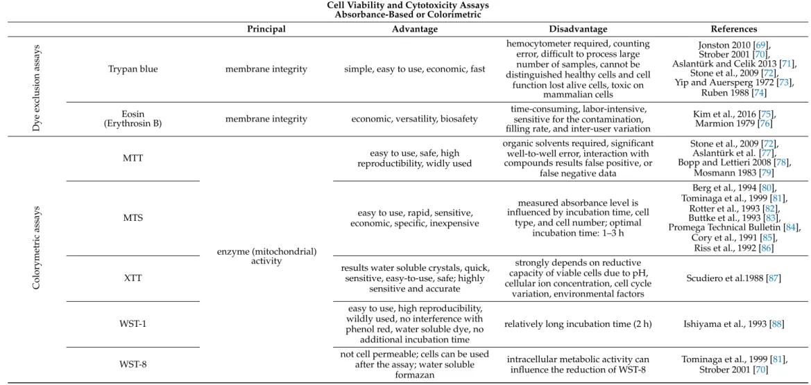
Table 4.
Absorbance-based and colorimetric cell viability and cytotoxicity assays
1.
Cell Viability and Cytotoxicity AssaysAbsorbance-Based or Colorimetric
Principal Advantage Disadvantage References
Dye exclusion assays Trypan blue membrane integrity simple, easy to use, economic, fast
hemocytometer required, counting error, difficult to process large number of samples, cannot be distinguished healthy cells and cell
function lost alive cells, toxic on mammalian cells
Jonston 2010 [69], Strober 2001 [70], Aslantürk and Celik 2013 [71],
Stone et al., 2009 [72], Yip and Auersperg 1972 [73],
Ruben 1988 [74]
Eosin
(Erythrosin B) membrane integrity economic, versatility, biosafety
time-consuming, labor-intensive, sensitive for the contamination, filling rate, and inter-user variation
Kim et al., 2016 [75], Marmion 1979 [76]
Colorymetric assays
MTT
enzyme (mitochondrial) activity
easy to use, safe, high reproductibility, widly used
organic solvents required, significant well-to-well error, interaction with compounds results false positive, or
false negative data
Stone et al., 2009 [72], Aslantürk et al. [77], Bopp and Lettieri 2008 [78],
Mosmann 1983 [79]
MTS easy to use, rapid, sensitive,
economic, specific, inexpensive
measured absorbance level is influenced by incubation time, cell
type, and cell number; optimal incubation time: 1–3 h
Berg et al., 1994 [80], Tominaga et al., 1999 [81],
Rotter et al., 1993 [82], Buttke et al., 1993 [83], Promega Technical Bulletin [84],
Cory et al., 1991 [85], Riss et al., 1992 [86]
XTT
results water soluble crystals, quick, sensitive, easy-to-use, safe; highly
sensitive and accurate
strongly depends on reductive capacity of viable cells due to pH, cellular ion concentration, cell cycle
variation, environmental factors
Scudiero et al.1988 [87]
WST-1
easy to use, high reproducibility, wildly used, no interference with phenol red, water soluble dye, no
additional incubation time
relatively long incubation time (2 h) Ishiyama et al., 1993 [88]
WST-8
not cell permeable; cells can be used after the assay; water soluble
formazan
intracellular metabolic activity can influence the reduction of WST-8
Tominaga et al., 1999 [81],
Strober 2001 [70]
Table 4.
Cont.
Cell Viability and Cytotoxicity Assays Absorbance-Based or Colorimetric
Principal Advantage Disadvantage References
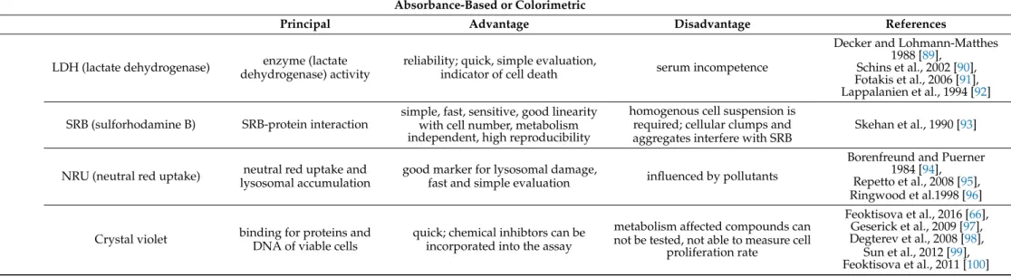
LDH (lactate dehydrogenase) enzyme (lactate dehydrogenase) activity
reliability; quick, simple evaluation,
indicator of cell death serum incompetence
Decker and Lohmann-Matthes 1988 [89],
Schins et al., 2002 [90], Fotakis et al., 2006 [91], Lappalanien et al., 1994 [92]
SRB (sulforhodamine B) SRB-protein interaction
simple, fast, sensitive, good linearity with cell number, metabolism independent, high reproducibility
homogenous cell suspension is required; cellular clumps and aggregates interfere with SRB
Skehan et al., 1990 [93]
NRU (neutral red uptake) neutral red uptake and lysosomal accumulation
good marker for lysosomal damage,
fast and simple evaluation influenced by pollutants
Borenfreund and Puerner 1984 [94], Repetto et al., 2008 [95], Ringwood et al.1998 [96]
Crystal violet binding for proteins and DNA of viable cells
quick; chemical inhibtors can be incorporated into the assay
metabolism affected compounds can not be tested, not able to measure cell
proliferation rate
Feoktisova et al., 2016 [66], Geserick et al., 2009 [97], Degterev et al., 2008 [98],
Sun et al., 2012 [99], Feoktisova et al., 2011 [100]
1Trypan Blue: Staining with trypan blue is one of the oldest viability assays. In a viable cell, the intact membrane will prevent trypan blue from entering cells. In dead or dying cells, trypan blue will enter the cell, staining it blue. This method was traditionally quantified manually using microscopes and hemocytometers, making it very labor-intensive. However, the recent availability of affordable automated cell counters makes this assay less time consuming and more accurate. MTT (3-(4,5-dimethylthiazole-2-yl)-2,5-diphenyl-tetrazolium bromide) is a tetrazolium salt that gets reduced by both mitochondrial and extra-mitochondrial dehydrogenases to form insoluble blue formazan crystals, meaning a solubilization step is required before the assay can be read. MTS/XTT: MTS (3-(4,5-dimethylthiazol-2-yl)-5-(3- carboxymethoxyphenyl)-2-(4-sulphophenyl)-2H-tetrazolium) and XTT (2,3-bis-(2-methoxy-4-nitro-5-sulfophenyl)-2H-tetrazolium-5-carboxanilide) substrates are similar to MTT. However, one advantage is that the reactions are carried out intracellularly in the presence of the intermediate electron acceptor phenazine methosulfate (PMS), which enhances their sensitivity. In addition, the reduced formazan product is soluble and gets released to the culture media, removing the need for the extra solubility step that is required with MTT. However, phenol red in cell culture media, fatty acids, and serum albumin have all been reported to distort data obtained from MTS, XTT, and WST assays over prolonged incubation periods. WST: Water-soluble tetrazolium salts (WSTs) are cell-impermeable tetrazolium dyes that get reduced extracellularly via plasma membrane electron transport, and combined with the electron acceptor PMS to generate water-soluble formazan dyes. LDH assay: Lactate dehydrogenase is a ubiquitous, stable cytoplasmic enzyme that converts lactate to pyruvate. If the cell membrane has been damaged, LDH, and therefore, its enzymatic activity is released from cells and can be detected in cell culture media.
Table 5.
Fluorometric cell viability and cytotoxicity assays
1.
Fluorometric AssayPrincipal Advantage Disadvantage References
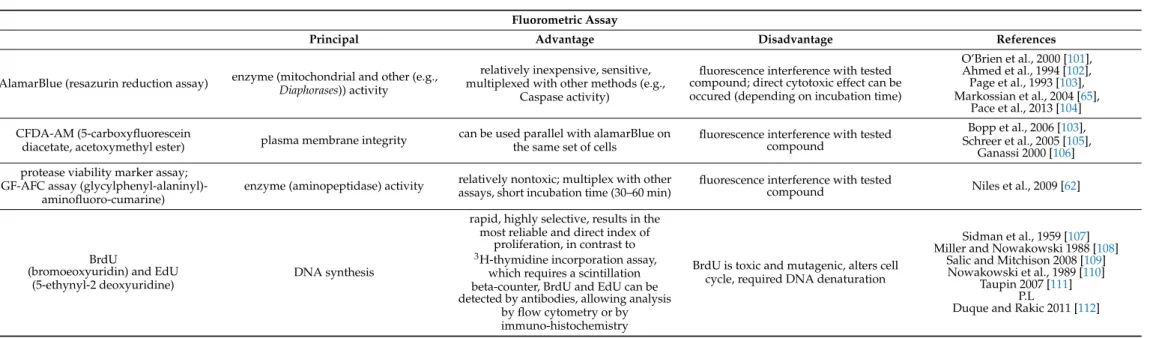
AlamarBlue (resazurin reduction assay) enzyme (mitochondrial and other (e.g., Diaphorases)) activity
relatively inexpensive, sensitive, multiplexed with other methods (e.g.,
Caspase activity)
fluorescence interference with tested compound; direct cytotoxic effect can be occured (depending on incubation time)
O’Brien et al., 2000 [101], Ahmed et al., 1994 [102], Page et al., 1993 [103], Markossian et al., 2004 [65],
Pace et al., 2013 [104]
CFDA-AM (5-carboxyfluorescein
diacetate, acetoxymethyl ester) plasma membrane integrity can be used parallel with alamarBlue on the same set of cells
fluorescence interference with tested compound
Bopp et al., 2006 [103], Schreer et al., 2005 [105],
Ganassi 2000 [106]
protease viability marker assay;
GF-AFC assay (glycylphenyl-alaninyl)- aminofluoro-cumarine)
enzyme (aminopeptidase) activity relatively nontoxic; multiplex with other assays, short incubation time (30–60 min)
fluorescence interference with tested
compound Niles et al., 2009 [62]
BrdU
(bromoeoxyuridin) and EdU (5-ethynyl-2 deoxyuridine)
DNA synthesis
rapid, highly selective, results in the most reliable and direct index of
proliferation, in contrast to
3H-thymidine incorporation assay, which requires a scintillation beta-counter, BrdU and EdU can be detected by antibodies, allowing analysis
by flow cytometry or by immuno-histochemistry
BrdU is toxic and mutagenic, alters cell cycle, required DNA denaturation
Sidman et al., 1959 [107]
Miller and Nowakowski 1988 [108]
Salic and Mitchison 2008 [109]
Nowakowski et al., 1989 [110]
Taupin 2007 [111]
P.L
Duque and Rakic 2011 [112]
1Alamar Blue: is a resazurin compound that gets reduced to resorufin and dihydroresorufin in viable cells. It can enter live cells so does not require cell lysis, and is stable in culture media. This assay has the added advantage that it can be measured in both fluorimetric and colorimetric plate readers. Pace et al., reported incubation time dependent cytotoxic effect [104]. Calcein-AM: Calcein-acetoxymethylester is a non-fluorescent dye that is used in both cell viability and apoptosis assays and its lipophilic, allowing easy passage through the cell membrane. Once inside the viable cell, intracellular esterases cleave the ester bonds of the acetomethoxy group, resulting in the formation of a fluorescent anionic and hydrophilic calcein dye. Non-viable cells do not contain active esterases. Also need to consider that: Cu2+, Co2+, Fe3+, Mn2+, and Ni2+quench the fluorescent signal from calcein at physiological pH, which means care must be taken to select the appropriate cell culture media.
Table 6.
Luminometric cell viability and cell cytotoxicity assays
1. Luminometric Assay
Principal Advantage Disadvantage References
ATP assay membrane integrity
the fastest and the most sensitive assay to use; no artefacts; no
plate handling step
sensitivity depends on reproducibility of pipetting
Maehara et al., 1987 [113], García et al., 2003 [114],
Andreotti et al., [115], Markossian et al., 2004 [65]
Real-time viability assay metabolic activity real-time measurement;
multiplex
incubation time is cell type and seeding density dependent
Duellman et al., 2015 [116], Markossian et al., 2004 [65]
1ATP content: there are numerously available assays that measure ATP levels as an output, when cells begin to undergo cell death process (apoptosis) or lose membrane integrity, ATP stocks become depleted through the activity of ATPases that concurrently prevent any new ATP synthesis. This leads to a rapid depletion of intracellular ATP levels. Luminescent ATP assays function by lysing cells to release ATP stores, while concurrently inhibiting ATPases. Luciferase catalyzes the oxidation of luciferin to oxyluciferin in the presence of magnesium and ATP, resulting in a luminescent signal that directly correlates with the intracellular ATP concentration.
Table 7.
Cell viability assays for flow cytometry and microscopic imaging
1. Cell Viability Assays for Flow Cytometry and Microscopic Imaging
λ ex Advantage Disadvantage References
Nucleic acid dies
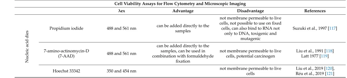
Propidium iodide 488 and 561 nm can be added directly to the samples
not membrane permeable to live cells, not possible to use on fixed cells, can also bind to RNA not
only to DNA, toxigenic and mutagenic
Suzuki et al., 1997 [117]
7-amino-actinomycin-D
(7-AAD) 488 and 561 nm
can be added directly to the samples, can be used in combination with formaldehyde
fixation
not membrane permeable to live cells, potential carcinogen
Liu et al., 1991 [118]
Latt 1977 [119]
Hoechst 33342 350 and 454 nm not membrane permeable to live
cells
Liu et al., 2019 [120],
Réu et al., 2019 [121]
Table 7.
Cont.
Cell Viability Assays for Flow Cytometry and Microscopic Imaging
λex Advantage Disadvantage References
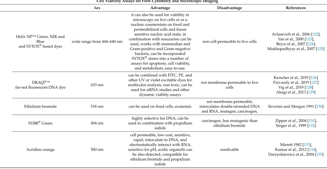
Helix NP™ Green, NIR and Blue
and SYTOX
®based dyes
wide range from 444–640 nm
it can also be used for viability in microscopy on live cells or as a nuclear counterstain on fixed and
permeabilized cells and tissue sensitive nucleic acid stain, in combination with resuzarine can be
used, works with mammalian and Gram-positive and Gram-negative
bacteria, can be incorporated SYTOX
®stains into a number of assays for apoptosis, cell viability,
and metabolism, easy-to-use.
non cell-permeable to live cells
Avlasevich et al., 2006 [122], Yan et al., 2000 [123], Bryce et al., 2007 [124], Mukhopadhyay et al., 2007 [125]
DRAQ7™
far-red fluorescent DNA dye 633 nm
can be combined with FITC, PE, and other UV or violet excitable dyes for multicolor analysis, non toxic, can be used for siRNA studies and other
dynamic viablity assays
not membrane-permeable to live cells
Kerscher et al., 2019 [126]
Paivandy et al., 2019 [127]
Vig et al., 2019 [128]
Akagi et al., 2013 [129]
Ethidium bromide 518 nm can be used on fixed cells, economic
not membrane-permeable, intercalates double-stranded DNA
and RNA, mutagen, carcinogen,
Severini and Morgan 1991 [130]
SYBR
®Green 494 nm
highly selective for DNA, can be used in combination with propidium
iodide
carcinogen, less mutagenic than ethidium bromide
Zipper et al., 2004 [131], Singer et al., 1999 [132]
Acridine orange 500 nm
cell permeable, low-cost, sensitive, rapid, intercalate to DNA, and electrostatically interact with RNA, sensitive for pH, acidic organells can
be also detected, compatible for ethidium bromide and propidium
iodide
nonfixable
Mirrett 1982 [133],
Kumar et al., 2012 [134],
Darzynkiewicz et al., 2004 [135]
Table 7.
Cont.
Cell Viability Assays for Flow Cytometry and Microscopic Imaging
λex Advantage Disadvantage References
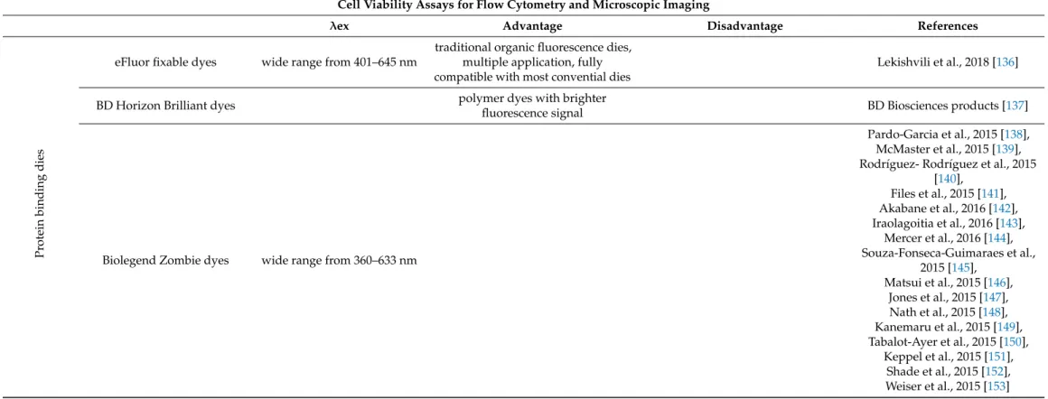
Pr otein binding dies
eFluor fixable dyes wide range from 401–645 nm
traditional organic fluorescence dies, multiple application, fully compatible with most convential dies
Lekishvili et al., 2018 [136]
BD Horizon Brilliant dyes polymer dyes with brighter
fluorescence signal BD Biosciences products [137]
Biolegend Zombie dyes wide range from 360–633 nm
Pardo-Garcia et al., 2015 [138], McMaster et al., 2015 [139], Rodríguez- Rodríguez et al., 2015
[140], Files et al., 2015 [141], Akabane et al., 2016 [142], Iraolagoitia et al., 2016 [143],
Mercer et al., 2016 [144], Souza-Fonseca-Guimaraes et al.,
2015 [145], Matsui et al., 2015 [146],
Jones et al., 2015 [147], Nath et al., 2015 [148], Kanemaru et al., 2015 [149], Tabalot-Ayer et al., 2015 [150],
Keppel et al., 2015 [151],
Shade et al., 2015 [152],
Weiser et al., 2015 [153]
Table 7.
Cont.
Cell Viability Assays for Flow Cytometry and Microscopic Imaging
λex Advantage Disadvantage References
Calcein AM 496
cell permeant, indicator of lipid vesicle leakage, neutral substrate for
MDR efflux transporters, selective for live cells; suitable for proliferating and non-proliferating
cells; ideal for suspension and adherent cells, rapid, ideal for high-throughput assays; commonly used for cell tracing and in studies of endocytosis, cell migration, and gap junctions; adaptable to a wide variety of techniques, including: microplate assays, immunocytochemistry, flow cytometry, and in vivo cell tracking
Allen and Cleland 1980 [154], Patel et al., 2009 [155], Glavinas et al., 2004 [156]
1Calcein-AM: Calcein-acetoxymethylester is a non-fluorescent dye that is used in both cell viability and apoptosis assays and is lipophilic, allowing easy passage through the cell membrane. Once inside the viable cell, intracellular esterases cleave the ester bonds of the acetomethoxy group, resulting in the formation of a fluorescent anionic and hydrophilic calcein dye. Non-viable cells do not contain active esterases. Also need to consider that: Cu2+, Co2+, Fe3+, Mn2+,and Ni2+quench the fluorescent signal from calcein at physiological pH, which means care must be taken to select the appropriate cell culture media.
Propidium Iodide/7-AAD: These intercalating agents are frequently used to study the cell cycle and they are membrane-impermeable, they are excluded from viable cells. This means that the fluorescence signal emitted by PI or 7-AAD in non-viable cells can be measured either by fluorescence microscopy or FACS analysis. Cell-impermeable DNA-binding dyes such (DRAQ7 from Abcam, Cambridge, UK or SYTOX from Thermo Fisher, Waltham, MA, USA) enter cells through compromised cell membranes and display strong fluorescence upon binding with DNA.
![Table 1. Ligands and functions of different integrins of human leukocytes (RGD, Arg-Gly-Asp sequence; VCAM-1, vascular cellular adhesion molecule-1; ICAM-1, Intercellular adhesion molecule-1) [10,30–33].](https://thumb-eu.123doks.com/thumbv2/9dokorg/768074.34007/7.892.248.840.209.356/ligands-functions-different-integrins-leukocytes-sequence-vascular-intercellular.webp)