23
Accepted for publication March 25, 2014, Published online May 30, 2014 Correspondence to Wolf Dieter Grimm
SpecialistTM of Periodontology of German Association of Periodontology and Specialist of Periodontology of European Association of Periodontology, Ruhrstr. 76, 58452 Witten, Germany Tel: +490230252971, Fax: +490230252972
E-mail: prof_wolf.grimm@yahoo.de
This is an open-access article distributed under the terms of the Creative Commons Attribution Non-Commercial License (http://creativecommons.org/
licenses/by-nc/3.0/), which permits unrestricted non-commercial use, dis- tribution, and reproduction in any medium, provided the original work is properly cited.
Translational Research: Palatal-derived Ecto-mesenchymal Stem Cells from Human Palate: A New Hope for Alveolar Bone
and Cranio-Facial Bone Reconstruction
Wolf Dieter Grimm1, Aous Dannan2, Bernd Giesenhagen3, Ingmar Schau4, Gabor Varga5, Mark Alexander Vukovic6, Sergey Vladimirovich Sirak7
1Periodontology, University of Witten, Germany, 2Periodontology, Syrian Private University - Damascus, Syrian Arab Republic,
3Implantology, Implantogy Center Kassel, Germany, 4Implantology, Implantology Center Detmold, Germany, 5Cell Biology, SE University Budapest, Hungary, 6Implantology, Praxisteam Hasslinghausen, Germany,
7Stomatology, Stavropol State Medical University, Stavropol, Russia
The management of facial defects has rapidly changed in the last decade. Functional and esthetic requirements have steadily increased along with the refinements of surgery. In the case of advanced atrophy or jaw defects, extensive horizontal and vertical bone augmentation is often unavoidable to enable patients to be fitted with implants. Loss of vertical alveolar bone height is the most common cause for a non primary stability of dental implants in adults.
At present, there is no ideal therapeutic approach to cure loss of vertical alveolar bone height and achieve optimal pre-implantological bone regeneration before dental implant placement. Recently, it has been found that specific pop- ulations of stem cells and/or progenitor cells could be isolated from different dental resources, namely the dental fol- licle, the dental pulp and the periodontal ligament. Our research group has cultured palatal-derived stem cells (paldSCs) as dentospheres and further differentiated into various cells of the neuronal and osteogenic lineage, thereby demonstrat- ing their stem cell state. In this publication will be shown whether paldSCs could be differentiated into the osteogenic lineage and, if so, whether these cells are able to regenerate alveolar bone tissue in vivo in an athymic rat model.
Furthermore, using these data we have started a proof of principle clinical- and histological controlled study using stem cell-rich palatal tissues for improving the vertical alveolar bone augmentation in critical size defects. The initial results of the study demonstrate the feasibility of using stem cell-mediated tissue engineering to treat alveolar bone defects in humans.
Keywords: Human palatal-derived stem cells (paldSCs), Osteogenic differentiation, Athymic immunodeficient rats, Stem-cell enriched palatal-derived tissues, Proof of principle clinical-and histologically-con, Osteoplastic surgical methods
Introduction
The management of facial defects has rapidly changed in the last decade. Functional and esthetic requirements have steadily increased along with the refinements of surgery. Facial defects include nearly all aspects of the face, i.e. the facial soft tissues, skull, and teeth. Disfigur- ement and extension of a defect go inparallel; seen in clefts, Noma, which is a devastating gangreneous disease that leads to severe tissue destruction of the face and
long-lasting defects after trauma and tumour surgery.
The fact that speaking, mastication, deglutition, and ef- fortless respiration are localized in the face, dictates the goal for any treatment: the most lifelike restoration possi- ble to restore physiological function. To date the most fre- quently used technique for reconstruction of extended de- fects is the transfer of vascularized osseous respectively os- seo(-myo-)cutaneous free flaps in particular as part of on- cologic surgery of the head and neck region. Fibula, scap- ula, and iliac crest were the preferred donor sites (Vinzenz and C. Schaudy, 2011).
In the case of advanced atrophy or jaw defects, ex- tensive horizontal and vertical bone augmentation is often unavoidable to enable patients to be fitted with implants.
These implantological procedures are usually two-stage.
Bone block transplantation is a good model to evaluate bone regeneration. It is common to combine autologous bone blocks with alloplastic material. Drawback of the procedure is that, to obtain autologous bone, an additional surgical step is needed, causing donor-site morbidity (Grimm et al. 2012, 2013). If alloplastic materials are used without addition, normally longer healing times are chos- en to compensate for the initial lack of regeneration. This explains why there are minor reports about healing peri- ods of less than 6 months when pure allogenic material is used. The report of our research group (Plöger and Schau, 2010) has showed in the first time the healing peri- ods of more than three years when pure allogenic material is used.
The regeneration of bone requires an adequate scaffold as well as cells that are capable to attach to the scaffold, proliferate, and differentiate. The dynamics of bone heal- ing are controlled by growth factors, such as bone morpho- genetic proteins. To promote these requirements, several strategies for bone and tissue regeneration have been developed.
The discovery of stem cells and recent advances in cel- lular and molecular biology has led to the development of novel therapeutic strategies that aim at the regeneration of many tissues that were injured by disease. Tissue en- gineering is a multidisciplinary field that combines biol- ogy, engineering, and clinical science with the goal of gen- erating new tissues and organs. It is a science based on fundamental principles that involves the identification of appropriate cells, the development of scaffolds and mor- phogenic signals required to induce cells to regenerate a tissue or organ.
Neural crest cells could be termed a dormant stem cell population in the adult, as they are pluripotent at first, but with migration they become restricted during develop-
ment (Cheng et al. 2012). Neural crest cells that form the vertebrate head skeleton migrate and interact with sur- rounding tissues to shape the skull. In common with most organs, teeth develop from interactions between epithelial cells (oral epithelium) and mesenchymal cells (cranial neural-crest-derived ectomesenchyme).
From a biological perspective, current and future pros- pects for improved regeneration of bone in the facial skull and upper and lower jaw are dependent on the ability to facilitate the repopulation of the wound by cells capable of promoting regeneration.
Several recent studies showed that neural crest-related stem cells can be isolated from mammalian craniofacial tissues such as periodontal ligament, dental pulp, and palate. These cells have in common that they form neuro- sphere-like clusters and proliferate in serum-free culture in the presence of fibroblast growth factor-2 (FGF-2) and epidermal growth factor (EGF).
From this perspective, the human palate has been shown to be of critical importance in the regenerative process. Although it is clear that cells residing in the sub- epithelial soft tissue of the palate can achieve bone re- generation, this population is heterogenous (Harris, 1999), but it is not known which subpopulations are capable of achieving regeneration. Indeed, cells derived from palate were found to have specific properties, such as increased proliferation rates, and representative of a regenerative phenotype as shown by research group (Greiner et al.
2011, Widera et al. 2009).
In order to understand the cellular origins of Neural Crest Stem Cells from Dental and Periodontal Tissues, transplanted tooth buds were used to show that the mesen- chymal-derived dental follicle surrounding the developing tooth root is the source of progenitor cells for cementum, alveolar bone and periodontal ligament (Heinemann and Götz 2013). Further evidence, that cells from the dental follicle are precursors to cementoblasts and to osteoblasts, has been shown by direct cell migration in the rat molars.
More recently, the dental follicle associated with third mo- lars has been shown to contain precursor cells which are clonogenic and have the ability to differentiate under in vitro conditions to a membrane-like structure containing calcified nodules (Morsczeck et al. 2009).
The source of post-natal progenitor cells which may be capable of regenerating the alveolar bone has been inves- tigated for a number of years. Cell kinetic experiments in mice and rats have shown that osteoblast populations of the subepithelial palatal soft tissue represent a steady-state renewal system, with the number of new cells generated by mitosis equal to the number of cells lost through apop-
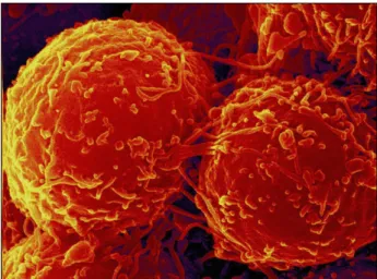
Fig. 1. SEM: Cultured palatal-derived stem cells (paldSC) form spheres.
tosis and migration. This capacity for self-renewal, which is further evidenced by the rapid turnover of these stem cells, supports the notion of progenitor/stem cell popu- lations. Furthermore, a significant number of pala- tal-derived stem cells do not enter the cell cycle, suggest- ing that these cells may act in a similar manner to quies- cent, self-renewable and multipotent stem cells.
The relationship between progenitor cells in regenerat- ing tissues and normally functioning (steady-state) tissues has been investigated in studies performed in normal mouse alveolar bone, wounded mouse alveolar bone de- fects, normal rat gingiva and inflamed monkey gingiva (see overview by Mao et al.). These studies have identified a common paravascular location for osteo- and fibroblast progenitors. These cells exhibit some of the classical cyto- logical features of stem cells, including small size, re- sponsiveness to stimulating factors, and slow cycle time (Schomann et al. 2013). Furthermore, these paravascular cells exhibit spatial clustering which suggests a possible clonal distribution of progenitors and their progeny (Vertelov et al. 2013).
Other possible sources of osteoblast and cementoblast precursors are the endosteal spaces of alveolar bone from which cells have been observed to adopt a paravascular lo- cation in the bone of mice (Marx and Tursun 2011).
Through a combination of transplanted biomaterials containing appropriately selected and primed stem cells, together with an appropriate mix of regulatory factors and extracellular matrix components to allow growth and spe- cialization of the cells, the research of new therapies with significant clinical potential in alveolar bone surgery and facial skull surgery are emerging (Bartold et al. 2000).
Neural Crest Stem Cells from Dental and Periodontal Tissues
Innovative therapeutic concept
Our innovation describes a new and previously over- looked way to prepare the endogenous repairing stem cells on the basis of their ecto-mesenchymal like phenotype (Molthera; PCT/EP2006/066221). Our new cell therapeutic product is already developed on a research laboratory scale. The patient-specific adult stem cells can be easily isolated and propagated without human serum (serum- free medium) from the oral cavity.
Thus neural crest cells might be the endogenous repair troop, which is also in the right differentiation stage to regenerate multi-lineage tissues. Generally, most organs including teeth develop from interactions between epi- thelial cells (oral epithelium) and mesenchymal cells
(cranial neural-crest-derived ectomesenchyme).
Several recent studies showed that neural crest-related stem cells can be isolated from mammalian craniofacial tissues (Fig. 1) such as periodontal ligament and from hu- man palate as shown by our research group (Widera et al. 2007, 2009, Molnar et al. 2008) and by Techawattan- awisal et al. (2007) or dental pulp as shown by our inter- national research group (Kiraly et al. 2009).
These cells have in common that they form neuro- sphere-like clusters and proliferate in serum-free culture in the presence of fibroblast growth factor-2 (FGF-2) and epidermal growth factor (EGF). In light of these findings, the reason for this consortium is the identification of stem cells within the human palate. Palate is a highly re- generative and richly innervated craniofacial tissue. It is well described that wounds within oral mucosa heal rapidly. This capability for rapid regeneration may be ex- plained either by the presence of growth factors, such as basic fibroblast growth factor in the saliva, or by the poten- tial presence of at least one stem cell type within the tissue.
To decipher the developmental make-up of the alveolar and facial bone region, we will follow the path of the mi- gratory neural crest, the neural crest lineage segregation as a blueprint for bone regeneration since it gives rise to bone progenitor tissues, which in turn are subjected to the influence of diverse bone extracellular matrices and pep- tide growth factors. We propose that the developmental history of a tissue is a highly instructive design template for the discovery of novel bioengineering tools and ap- proaches (Kharazi et al. 2013). Further developments will include a formulation for clinical applications. This for- mulation will be based on scaffolds or delivery systems, which will be developed during this application. This will
lead to the therapy of previously untreatable cases of facial defects and of advanced alveolar bone loss.
Purpose
Several tissue engineering approaches have been tried to stimulate bone formation. The addition of in vitro cul- tured mesenchymal stem cells (MSCs) has proven to stim- ulate osteogenesis. Preclinical and clinical studies have demonstrated the ability of bone marrow-derived stem and progenitor cells to regenerate various tissues, including bone (Marx et al. 2012). To bypass the problem of se- lection, multiplication, and differentiation, most of the mentioned studies selected MSCs, multiplied them, and then differentiated them to the osteogenic lineage. As this is unreasonable to do in daily practice, in this “proof of principle” clinical-controlled study approach was chosen in which allogenic bone blocks will be combined with pala- tal-derived stem cells (paldSCs, see purpose).
Based on the hypothesis that the number of paldSCs is secondary, the individual potential of paldSCs to survive high stress and chemotactically attract osteogenic progeni- tor cells will dominate. As the augmentation material is far from local blood supply, it can be assumed that a natu- ral selection procedure will be applied to the cells. As blood and oxygen supply are reestablished, the paldSCs could then unfold their full potential.
During the physiological bone healing, osteoprogenitor cells migrate into the defect, differentiate into osteoblasts, and produce calcified matrix. These osteoprogenitor cells derive from multipotent ecto-mesenchymal palatal-derived stem cells (paldSCs) that are recruited from palate.
PaldSCs can tolerate low oxygen concentrations and have the potential for osteogenic, chondrogenic, and adipogenic differentiation, depending on the external stimulus by cytokines. To stimulate bone healing, various grafting techniques have been developed to supply additionally ei- ther cytokines, progenitor cells, suitable scaffolds, or any combination of them. In our study design we propose add- ing EMDOGAIN®. EMDOGAIN® can increase bone for- mation dynamics.
The purpose of this study is to evaluate the potential of Adult Palatinum as a Novel source of stem cells in com- bination with transplantation of OsteoGraft Spongiosa/
Corticalis Bonering Blocks (maxgraft® bonering, botiss bi- omaterials, Germany) for one-stage bone augmentation and immediatly implant placement. These combination of methods for vertical augmentation in difficult anatomical sites will be tested in in a multicentric, “proof of principle”
randomized, controlled, clinical, radiological and, histo-
logical trial.
Our group gained clinical experience with the bone block technique and isolation, cultivation, and character- ization of palatinal-derived stem cells (paldSCs). Based on this key discovery we aim to develop a regenerative cell therapy.
Characterization of paldSCs by RT-PCR and flow cy- tometry revealed expression of several stem cell markers, such as nestin and Sox2 as well as STRO-1 and CD146.
In 2007 our research group first describe the character- ization and neuronal differentiation capacity of stem cells derived from inflamed periodontal tissues (Widera et al., 2007a). These so called periodontal-derived stem cells (pdSCs) were isolated by minimally invasive periodontal surgery and cultured as so-called dentaspheres under se- rum-free conditions in the presence of FGF-2 and EGF.
Differentiation studies revealed that paldSCs exhibit an osteogenic differentiation capacity. The osteogenic differ- entiation capacity of paldSCs was markedly developed suggesting that these cells might be a suitable cellular tool for bone regeneration purposes (Widera et al. 2007b).
Cultivation of paldSCs in neuronal differentiation me- dia indicated the high neuronal differentiation capacity of these stem cells from periodontal tissue. Cells adopted a neuronal morphology and expressed a variety of neuronal markers, such as β-III-tubulin, Map2, GAD67, neurofila- ment-L (NF-L), neurofilament-M (NF-M), neurofilament-H (NF-H), and synaptophysion (results not shown).
Thus, we looked for a novel source of stem cells from the oral cavity being higly suited for regenerative purposes. In 2009 Widera et al. described for the first time the presence of nestin-positive neural crest-related stem cells within Meissner corpuscles and Merkel cell-neurite complexes located in the hard palate of adult Wistar rats.
These cells were characterized by expression of nestin, the transcription factor Sox2, and additional neural crest markers such as Slug or Snail.
To prove for the putative bone regeneration capacity of paldSCs in an in vivo (Grimm et al. 2011) experimental setting an athymic rat model was conducted (Fig. 2). The current study demonstrates that isolating paldSCs from patients undergoing open flap minimally invasive perio- dontal surgery seems to be a simple and reliable method.
These cells can be easily propagated ex vivo, thereby still preserving their stem cell state. As demonstrated pre- viously and shown here, paldSCs are capable to differ- entiate both into the neuronal lineage as well as the osteo- genic lineage. In vivo studies clearly showed that human adult pdSCs being transplanted into an athymic rat model were able to regenerate periodontal tissue elements at dif-
Fig. 2. Experimental setting on an athymic rat model.
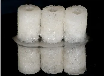
Fig. 3. Allogen human bone rings (maxgraftⓇ bonering, botiss bio- materials, Germany) with preserved biomechanical properties:
Sterile without antigenic effects, Storable at room temperature for 5 years, Osteoconductive properties supporting natural and con- trolled tissue remodeling.
ferent levels.
Case presentation (
proof of principle
study) and Protocol specificationsThe clinical study has been approved by the ethics com- mittees of the Zahnärztekammer Westfalen/Lippe and the University of Münster (Germany).
The study will be conducted in accordance with the Declaration of Helsinki (Fifth Revision, 2004). Patients older than 18 years with need of dental implant placement will be eligible if they had a maximum of 4 mm residual alveolar height. They had to be able to comply with study-related procedures, such as returning for follow- up examinations, exercising good oral hygiene, and being able to understand the nature of the proposed surgery.
Written informed consent will be obtained prior to any study-related procedures. Exclusion criteria will be smok- ing, history, signs of malignancy, radiotherapy or chemo- therapy, pregnancy or nursing, general contraindications for dental or surgical treatment, medications, treatments or diseases, which may have an effect on bone remodeling, bone or connective tissue metabolism, or an allergy to collagen.
The patients included in the study will be divided into three groups:
ㆍpatients belonging to only test or comparitive groups and
ㆍpatients with a split-mouth (cross-over) treatment Primary parameter of the study will be new bone for- mation (NBF) including the histological assessment.
Secondary parameters will be volume of the augmentate and bone height, see details.
Patient population
Thirty patients will be assessed for the study: Each pa- tient will be randomly assigned to either comparative or test arm. Because of the surgical intervention, neither sur- geon nor patient can be effectively blinded.
Step by step surgical procedures
For experienced oral surgeons, allograft bone blocks for block augmentation are the only real alternative to har- vesting patients own autogenic bone blocks, preventing well known risks as donor-site morbidity, infection risk, pain and loss in bone stability.
As a carrier of our stem cell containing tissues we are using a sterile, high-safety (donor selection, virus testing, chemical cleaning, processing and sterilization) allograft product, derived of human donor bone, processed by the Cell+Tissue Bank Austria (CTBA). The high biologic re- generation capability of allogenic bone blocks results in a predictable clinical outcome.
Properties (Fig. 3)
ㆍPreserved biomechanical properties ㆍSterile without antigenic effects
ㆍStorable at room temperature for 5 years
ㆍOsteoconductive properties supporting natural and controlled tissue remodeling.
In the case presented here one allogen bonering was used. Until the lateral access to the maxillary sinus is pre- pared, the allogen bonering is steeped in native blood
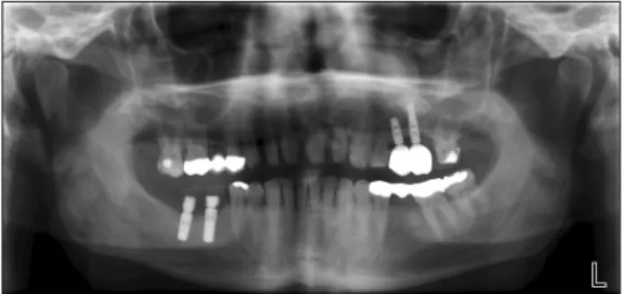
Fig. 4. X-ray situation after loading and finally clinical result with placed implants and superconstruction.
mixed. The bonering was introduced vertically into the maxillary sinus. It stabilizes the implant by buttressing them. The cortical portion of the rings was aligned cra- nially since this makes adaptation of the cancellous por- tion of the ring to the concave-shaped sinus floor much easier. The still existing cavity is filled with bone re- generation material. By screwing in a membrane screw, the bone ring is stabilized within the sinus and the im- plant is anchored in a primary stable position. This proce- dure enables good results with long-term effects. For har- vesting the stem cell-containing subepithelial connective tissue graft from the palate a horizontal incision to the bone will be made 5 mm from the palatal gingival margin and the micro-blade will be subsequently placed parallel to the long axis of the roots. Another horizontal incision will be made 2 mm coronal to the first incision and the periosteum will be dissected before removing the wedge of soft tissue. An approximately 10×6 mm subepithelial connective tissue graft (SCTG) has been harvested from the palate in the second premolar to second molar region as the source of ecto-mesenchymal stem cells. The SCTG trimmed precisely to adapt to the recipient site. X-ray shows the situation after loading (Fig. 4) and finally clin- ical result with placed implants and superconstruction.
In summary this case report confirms that bone re- generation is possible with PALATAL-DERIVED STEM CELLS.
References
1. Vinzenz K, Schaudy C. Osteoplastic surgery of the face - state of the art and future aspects. Eur Surg 2011;43:1-14 2. Grimm WD, Dittmar T, Varga G, Giesenhagen B. Use of
stem-cell induced allogenic bonerings for pre-implanto- logical vertical augmentations of alveolar bone defects in humans- a clinical controlled study. Proceedings, BIT’s 5h Annual World Congress of Regenerative Medicine & Stem Cells 2012, December 2-4, 2012, Guangzhou, China.
3. Grimm WD, Varga G, Dittmar T. Palate-derived ecto-mes- enchymal stem cells in Periodontology and Implant Dentistry. Does The Chronically Inflamed Periodontium Harbour Cancer Stem Cells? Proceedings, BIT’ 5th Annual World Congress of Regenerative Medicine & Stem Cell 2012, December 2-4, 2012, Guangzhou, China
4. Cheng I, Mayle RE, Cox CA, Park DY, Smith RL, Corcoran-Schwartz I, Ponnusamy KE, Oshtory R, Smuck MW, Mitra R, Kharazi AI, Carragee EJ. Functional assess- ment of the acute local and distal transplantation of human neural stem cells after spinal cord injury. Spine J 2012;12:1040-1044
5. Plöger, Mathias, I. Schau. Kieferkammrekonstruktion mit CAD-CAM-gefertigten allogenen Knochenblöcken. Quintes- senz Publishing Company 2010
6. Harris RJ. Successful root coverage: a human histologic evaluation of a case. Int J Periodontics Restorative Dent 1999;19:439-447
7. Greiner JF, Hauser S, Widera D, Müller J, Qunneis F, Zander C, Martin I, Mallah J, Schuetzmann D, Prante C, Schwarze H, Prohaska W, Beyer A, Rott K, Hütten A, Gölzhäuser A, Sudhoff H, Kaltschmidt C, Kaltschmidt B.
Efficient animal-serum free 3D cultivation method for adult human neural crest-derived stem cell therapeutics. Eur Cell Mater 2011;22:403-419
8. Widera D, Zander C, Heidbreder M, Kasperek Y, Noll T, Seitz O, Saldamli B, Sudhoff H, Sader R, Kaltschmidt C, Kaltschmidt B. Adult palatum as a novel source of neural crest-related stem cells. Stem Cells 2009;27:1899-1910 9. Heinemann F, Götz W. Oral tissue engineering and
implants. Ann Anat 2012;194:501
10. Morsczeck C, Petersen J, Völlner F, Driemel O, Reichert T, Beck HC. Proteomic analysis of osteogenic differ- entiation of dental follicle precursor cells. Electrophoresis 2009;30:1175-1184
11. Mao JJ, Giannobile WV, Helms JA, Hollister SJ, Krebsbach PH, Longaker MT, Shi S. Craniofacial tissue engineering by stem cells. J Dent Res 2006;85:966-979
12. Schomann T, Qunneis F, Widera D, Kaltschmidt C, Kaltschmidt B. Improved method for ex ovo-cultivation of developing chicken embryos for human stem cell xenografts.
Stem Cells In. 2013;2013:960958
13. Marx RE, Tursun R. A qualitative and quantitative analysis of autologous human multipotent adult stem cells derived from three anatomic areas by marrow aspiration: tibia, ante- rior ilium, and posterior ilium. Oral Craniofac Tissue Eng 2011;1:98-102
14. Arnold WH, Becher S, Dannan A, Widera D, Dittmar T, Jacob M, Mannherz HG, Dittmar T, Kaltschmidt B, Kaltschmidt C, Grimm WD. Morphological character- ization of periodontium-derived human stem cells. Ann Anat 2010;192:215-219
15. Bartold PM, McCulloch CA, Narayanan AS, Pitaru S.
Tissue engineering: a new paradigm for periodontal re- generation based on molecular and cell biology. Periodontol 2000 2000;24:253-269
16. Widera D, Grimm WD, Moebius JM, Mikenberg I, Piechaczek C, Gassmann G, Wolff NA, Thévenod F, Kaltschmidt C, Kaltschmidt B. Highly efficient neural dif- ferentiation of human somatic stem cells, isolated by mini- mally invasive periodontal surgery. Stem Cells Dev 2007;16:447-460
17. Widera D, Grimm WD, Möbius JM, Dannan A, Becher S, Mikenberg I, Piechaczek C, Gassmann G, Kaltschmidt B.
Human periodontium-derived neural stem cells: chance or risk? Regenerative Medicine 2007;2:3-61
18. Molnár B, Kádár K, Király M, Porcsalmy B, Somogyi E, Hermann P, Grimm WD, Gera I, Varga G. Isolation, culti- vation and characterisation of stem cells in human perio- dontal ligament. Fogorv Sz 2008;101:155-161
19. Techawattanawisal W, Nakahama K, Komaki M, Abe M, Takagi Y, Morita I. Isolation of multipotent stem cells from adult rat periodontal ligament by neurosphere-forming cul- ture system. Biochem Biophys Res Commun 2007;357:
917-923
20. Király M, Porcsalmy B, Pataki A, Kádár K, Jelitai M, Molnár B, Hermann P, Gera I, Grimm WD, Ganss B, Zsembery A, Varga G. Simultaneous PKC and cAMP activa- tion induces differentiation of human dental pulp stem cells
into functionally active neurons. Neurochem Int 2009;55:
323-332
21. Kharazi A, Levy ML, Visperas MC, Lin CM. Chicken em- bryonic brain: an in vivo model for verifying neural stem cell potency. J Neurosurg 2013;119:512-519
22. Vertelov G, Kharazi L, Muralidhar MG, Sanati G, Tankovich T, Kharazi A. High targeted migration of human mesenchymal stem cells grown in hypoxia is associated with enhanced activation of RhoA. Stem Cell Res Ther 2013;4:5 23. Marx RE, Harrell DB. Translational Research: The CD34+
cell is crucial for large-volume bone regeneration from the milieu of bone marrow progenitor cells in craniomandibular reconstruction. Int J Oral Maxillofac Implants 2014;
29:e201-e209
24. Grimm WD, Dannan A, Becher S, Gassmann G, Arnold W, Varga G, Dittmar T. The ability of human periodontium- derived stem cells to regenerate periodontal tissues: a pre- liminary in vivo investigation. Int J Periodontics Restorative Dent 2011;31:e94-e101
25. Keeve PL, Dittmar T, Gassmann G, Grimm WD, Niggemann B, Friedmann A. Characterization and analysis of migration patterns of dentospheres derived from periodontal tissue and the palate. J Periodontal Res 2013;48:276-285