Enteric neural crest-derived cells promote their migration by modifying their microenvironment through tenascin-C
production
Sophia E. Akbareiana,*, Nandor Nagyb,*, Casey E. Steigera, John D. Mablyc, Sarah A.
Millera, Ryo Hottaa, David Molnarb, and Allan M. Goldsteina,#
aDepartment of Pediatric Surgery, Massachusetts General Hospital, Harvard Medical School, Boston, MA
bDepartment of Human Morphology and Developmental Biology, Faculty of Medicine, Semmelweis University, Budapest-1094, Hungary
cDepartments of Pediatrics and Genetics, Children’s Hospital Boston, Harvard Medical School, Boston, MA
Abstract
The enteric nervous system (ENS) is derived from vagal and sacral neural crest cells that migrate, proliferate, and differentiate into enteric neurons and glia within the gut wall. The mechanisms regulating enteric neural crest-derived cell (ENCC) migration are poorly characterized despite the importance of this process in gut formation and function. Characterization of genes involved in ENCC migration is essential to understanding ENS development and could provide targets for treatment of human ENS disorders. We identified the extracellular matrix glycoprotein tenascin-C (TNC) as an important regulator of ENCC development. We find TNC dynamically expressed during avian gut development. It is absent from the cecal region just prior to ENCC arrival, but becomes strongly expressed around ENCCs as they enter the ceca and hindgut. In aganglionic hindguts, TNC expression is strong throughout the outer mesenchyme, but is absent from the submucosal region, supporting the presence of both ENCC-dependent and independent expression within the gut wall. Using rat-chick coelomic grafts, neural tube cultures, and gut explants, we show that ENCCs produce TNC and that this ECM protein promotes their migration. Interestingly, only vagal neural crest-derived ENCCs express TNC, whereas sacral neural crest-derived cells do not. These results demonstrate that vagal crest-derived ENCCs actively modify their
microenvironment through TNC expression and thereby help to regulate their own migration.
Keywords
Enteric nervous system; extracellular matrix; neural crest cells; tenascin-C; Hirschsprung disease
© 2013 Elsevier Inc. All rights reserved.
#Corresponding Author: Allan M. Goldstein, Massachusetts General Hospital, Warren 1153, Boston, MA 02114, Tel: 617-726-0270, Fax: 617-726-2167, agoldstein@partners.org.
*These authors contributed equally to this work.
Publisher's Disclaimer: This is a PDF file of an unedited manuscript that has been accepted for publication. As a service to our customers we are providing this early version of the manuscript. The manuscript will undergo copy editing, typesetting, and review of
NIH Public Access
Author Manuscript
Dev Biol. Author manuscript; available in PMC 2014 October 15.
Published in final edited form as:
Dev Biol. 2013 October 15; 382(2): 446–456. doi:10.1016/j.ydbio.2013.08.006.
NIH-PA Author Manuscript NIH-PA Author Manuscript NIH-PA Author Manuscript
Introduction
Enteric neural crest-derived cells (ENCCs) arise predominantly from the vagal region of the neural tube, adjacent to somites 1–7 (Yntema and Hammond, 1954) and migrate rostro- caudally along the gastrointestinal tract to form two ganglionated plexuses of neurons and glial cells that comprise the enteric nervous system (ENS). A smaller proportion of ENCCs come from the sacral neural crest, caudal to somite 28 (Burns and Douarin, 1998), and migrate caudo-rostrally to colonize the distal gut. An intact ENS is critically important for regulating essential functions of the intestine, including peristalsis. Therefore, understanding the factors that control ENCC migration, proliferation, and differentiation during
embryogenesis is of fundamental importance, particularly for understanding the
pathogenesis of Hirschsprung disease, a congenital disorder in which ganglion cells fail to colonize the distal colorectum(Gershon, 2010; Goldstein et al., 2013).
Interactions between ENCCs and the extracellular matrix (ECM) play a critical role during ENS formation. Disruption of this interaction via conditional deletion of β1 integrin from ENCCs leads to their impaired migration, abnormal aggregation, and colonic aganglionosis (Breau et al., 2006). The essential role of β1 integrin signalling has also been shown in avians, where it mediates ENCC migration along the vascular ECM (Nagy et al., 2009).
Specific ECM proteins have been implicated in ENS development, including laminin, which promotes enteric neurogenesis (Chalazonitis et al., 1997). Colorectal aganglionosis in endothelin-3 (ET3) mutant mice has been attributed to an accumulation of laminin in the aganglionic mesenchyme (Payette et al., 1988). Loss of ET3 signalling increases expression of laminin (Wu et al., 1999), leading to premature neuronal differentiation and thus arrested migration and distal aganglionosis (Gershon et al., 1993). The distal aganglionosis in endothelin receptor B (EDNRB) mutant mice has been attributed to delayed ENCC migration, which results in their arrival in a non-permissive colonic environment due to increased laminin expression within the gut wall (Druckenbrod and Epstein, 2009).
In characterizing ECM expression in the avian gut, we identified a dynamic pattern for tenascin-C (TNC) in the cecal region that was temporally related to ENCC arrival, suggesting a potential role in colorectal ENS development. TNC, previously referred to as cytotactin (Grumet et al., 1985), myotendinous antigen (Chiquet and Fambrough, 1984), hexabrachion(Erickson and Taylor, 1987), and J1 (Kruse et al., 1984), is a multifunctional ECM glycoprotein with important roles during neural crest cell (NCC) migration (Bronner- Fraser, 1988; Epperlein et al., 1988; Jones and Jones, 2000; Mackie et al., 1988; Tucker, 2001), where it decreases cell adhesion to the ECM substrate and thereby promotes crest cell migration (Halfter et al., 1989). Inhibition of tenascin adjacent to the neural tube confirmed its essential role for cranial neural crest migration in avian embryos (Bronner-Fraser, 1988).
We have characterized the role of TNC during ENCC colonization of the distal intestine and find that TNC is produced not only by the gut environment but also by migrating vagal, but not sacral, crest-derived ENCCs. We further show that TNC promotes the migration of ENCCs. These results reveal a novel mechanism whereby ENCCs modify their microenvironment and thereby promote their own migration.
Materials & Methods
Animals
Fertilized White Leghorn chicken eggs were obtained from commercial breeders and maintained at 37°C in a humidified incubator. Embryos were staged according to Hamburger and Hamilton (HH) stage (Hamburger and Hamilton, 1992) or number of embryonic days (E).
NIH-PA Author Manuscript NIH-PA Author Manuscript NIH-PA Author Manuscript
Immunohistochemistry
Cryosections, 10–12µm thick, were collected on Probe-on poly-L-lysine-coated slides (Fisher Scientific, Pittsburgh, PA) and immunohistochemistry performed as described (Nagy and Goldstein, 2006a) using the antibodies listed in Table 1. For non-fluorescent
immunohistochemistry, sections were incubated with primary antibody then with biotinylated goat anti-mouse IgG or IgM (Vector Labs, Burlingame, CA) for 45 minutes each. Endogenous peroxidase activity was quenched with 3% hydrogen peroxide (Sigma, St.
Louis, MO) for 10 minutes and slides incubated in avidin-biotinylated peroxidase complex (Vectastain Elite ABC kit, Vector Labs) for 30 minutes. Binding sites were visualized by 4- chloro-1-napthol (Sigma). For fluorescent immunohistochemistry, secondary antibodies included Alexa Fluor 488 and 594-conjugated anti-mouse IgG, anti-mouse IgM, and anti- rabbit IgG (Invitrogen, Grand Island, NY) were used. Cell nuclei were visualized with DAPI (4’, 6-diamidine-2-phenylidole-dihydrochloride; Vector Labs). Samples were examined under a Nikon Eclipse 80i microscope and photographed with a Spot camera. Images were compiled with Adobe Photoshop.
Immunoblotting
Embryonic gut was digested in lysis buffer (1.21% 76.5mM Tris, 2% SDS, 10% glycerol) and homogenized with a 19 gauge needle and Sonic Dismembrator Model 500 (Fisher Scientific). Protein concentration was determined using a Pierce BCA Protein Assay Kit (Thermo Scientific, Billerica, MA) and a Victor2 1420 Multilabel counter (Perkin Elmer Life Sciences, Waltham, MA). Lysates were heated at 95°C for 5 minutes, loaded onto 7.5%
or 10% Mini protean TGX precast gels (Biorad, Waltham, MA) and transferred to PVDF membrane (Biorad) using a buffer consisting of 0.02M glycine, 0.25M Tris in 20% MeOH.
Membranes were probed with the antibodies in Table 1 for TNC (1:125), laminin (1:125), and fibronectin (1:1000), followed by goat anti-mouse HRP-conjugated secondary antibody (Biorad; 1:5000), and Immun-Star HRP luminol/enhancer with HRP peroxide buffer (Biorad). Immunoblotting for TNC and laminin was done in non-reducing conditions.
Chorioallantoic membrane (CAM) grafts
For experiments shown in Fig. 4, E5 chick intestine was harvested and the cloaca removed.
Ganglionic grafts, termed “+ceca,” included the hindgut and ceca, which has ENCCs at this stage. Aganglionic grafts, termed “−ceca,” include only the post-cecal intestine and therefore have no ENCCs. CAM grafts were performed as previously described (Nagy and Goldstein, 2006a; Newgreen et al., 1980). Briefly, host E9 chick embryos were prepared by gently abrading the CAM and placing the graft on that region. Grafts were collected 8 days later and processed for immunohistochemistry. For Fig. 4F, E5 hindgut (preganglionic) was harvested without ceca, but with cloaca and nerve of Remak (NoR) attached to serve as a source of sacral neural crest-derived cells. This hindgut was grafted onto the CAM of an E9 host embryo for 6 days in order to generate a hindgut containing only sacral (and not vagal) neural crest-derived cells.
Vagal neural tube ablations
The neural tube adjacent to somites 3–6 was removed from HH9-10 chick embryos using a tungsten wire and microscalpel (Kind gift of Dr. Alan Burns, ICH, UCL, London, UK).
Sham ablations consisted of opening the vitelline membrane. Embryos were harvested at the desired stages and the intestines collected for immunohistochemistry or RNA extraction.
Flow Cytometry
Postumbilical intestine was harvested from E7 chicks and the cloaca and NoR removed.
Intestine was dissociated to single cells in dispase (lmg/ml; StemCell Technologies,
NIH-PA Author Manuscript NIH-PA Author Manuscript NIH-PA Author Manuscript
Vancouver, Canada) for 5 minutes, pelleted, and resuspended with N-cadherin (NCAD) antibody (DSHB) conjugated to Alex Fluor 488 using the Zenon Antibody Labeling Kit (Invitrogen). NCAD+ and NCAD− cells were sorted on a Becton Dickinson 5-laser FACS DiVa as described (Preffer and Dombkowski, 2009).
Quantitative PCR (qPCR)
Total RNA was extracted using RNeasy (Qiagen, Valencia, CA) and cDNA generated with Superscript III First-Strand Synthesis System for RT-PCR (Invitrogen). qPCR primers are listed in Table 2. Primers for Tnc isoforms were designed to cross the exon 10/14 boundary for the short isoform (Tnc-S), exon 12/14 boundary for the medium isoform (Tnc-M), and exon 13/14 boundary for the long isoform (Tnc-L). qPCR was performed on a CFX96 Real- Time PCR Detection System. The expression level of each gene was normalized to Gapdh.
Statistical comparisons were made between groups for each isoform separately using Student’s t-test.
Rat-chick coelomic explants
Aneural colon from Sprague-Dawley rats (Charles River Labs) was prepared by dissecting the distal, pre-ganglionic colon from E13.5 rat embryo, just prior to the arrival of ENCCs (Newgreen and Hartley, 1995). The isolated colon was transplanted into the coelomic cavity of E3 chicks (n=11) for 7 days as previously described (Nagy and Goldstein, 2006b) and then harvested for immunohistochemistry.
In vitro ENCC migration assays
ENCC migration was analyzed as previously described (Nagy et al., 2009). E6 chick intestine without cloaca was cultured onto plastic tissue culture dishes coated with chick- derived tenascin protein (1µg/ml; Millipore, Billerica, MA) with or without 10µg/ml fibronectin (Biomedical Technologies Inc, Stoughton, MA). Culture media containing DMEM with glutamine, 10% FBS, and pen/strep was added and the cultures incubated for 48 hours. Cultures were fixed in 2% paraformaldehyde and immunohistochemistry
performed. For cell migration, approximately 10–15 measurements were performed in each of 3–4 guts per experimental group. Statistical significance was calculated using Student’s t- test.
Neural tube cultures
Neural tube cultures were performed as described (Bronner-Fraser, 1996). Briefly, chick vagal neural tube adjacent to somites 1–7 was microsurgically excised from HH10-12 embryos, while sacral neural tube caudal to somite 28 was removed from HH16 embryos.
Dissection was facilitated by addition of dispase (1mg/ml) for 20 minutes at 37°C. Neural tubes were cultured onto dishes coated with fibronectin (10µg/ml; Sigma). After 24 hours, cultures were fixed and processed for immunohistochemistry.
Results
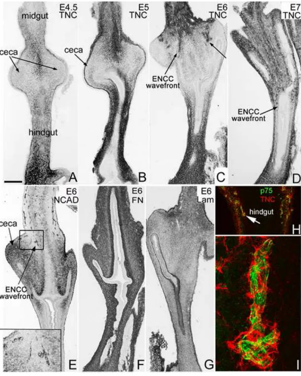
Tenascin-C expression in the gut is dynamic and colocalizes with migrating ENCCs TNC expression during ENS development in the post-umbilical intestine was assessed by immunohistochemistry. At E4.5-E5, when ENCCs are migrating in the distal midgut, TNC is present in the gut mesenchyme proximal and distal to the ceca, in the midgut and hindgut, respectively, but absent from the cecal region itself (Fig. 1A,B). As the ENCC wavefront enters the ceca at E6 and the proximal colon at E7, TNC continues to be expressed in the gut mesenchyme proximal and distal to the cecal region. Interestingly, we also noted TNC immunoreactivity in a small cluster of cells in the proximal ceca at E6 that are found in the
NIH-PA Author Manuscript NIH-PA Author Manuscript NIH-PA Author Manuscript
same region as invading ENCCs (Fig. 1C, arrows). N-cadherin expression at E6 highlights the ENCC wavefront at this stage (Fig. 1E, boxed area). Note that N-cadherin transiently stains the cecal mesenchyme at E6, similar to HNK-1 and p75, as previously described (Nagy et al., 2012). In contrast to the dynamic expression of TNC, fibronectin and laminin are uniformly expressed in all segments of the post-umbilical intestine from E5 through E8 (Fig. 1F,G). Given the spatiotemporal concordance between TNC immunoreactivity and the migratory ENCC wavefront (Fig. 1C–E), particularly evident in the cecal region, we performed double-label immunofluorescence with antibodies to TNC and p75 to determine if TNC protein colocalizes with migrating ENCCs. We find that at the stages when ENCCs are colonizing the cecum and proximal hindgut, TNC expression is strong surrounding the migrating ENCCs (Fig. 1H,I).
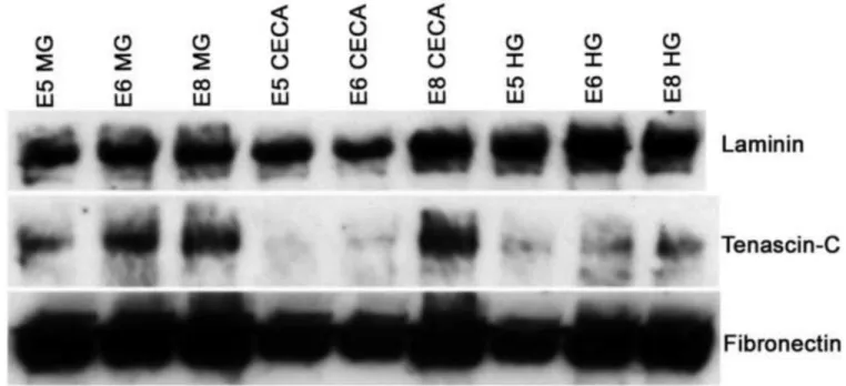
The expression of several ECM proteins was examined at multiple stages and segments of the postumbilical intestine by immunoblotting. As shown in Fig. 2, laminin and fibronectin are both strongly expressed in the midgut, ceca, and hindgut at E5, E6, and E8. In contrast, TNC is absent from the ceca at E5 and expressed at very low levels at E6 (Fig. 2), consistent with the immunohistochemistry results. In all segments of the post-umbilical intestine examined, TNC expression increases as ENS development proceeds. Notably, TNC is also expressed in the hindgut at E5 and E6, prior to ENCC arrival.
Vagal, but not sacral, neural crest-derived cells express TNC
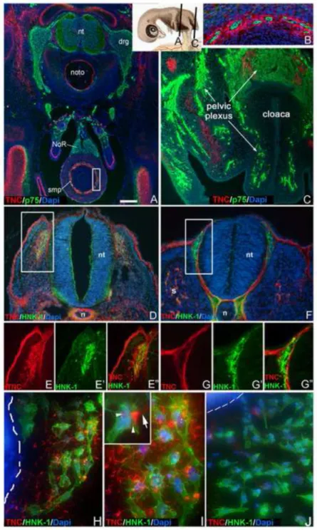
The pattern of TNC immunoreactivity and its relationship with NCCs was examined in cross-sections of early E8 chick embryos. Vagal crest-derived structures, like the
submucosal plexus of the colon, are surrounded by TNC immunostaining (Fig. 3A,B). In the midgut (not shown), both submucosal and myenteric plexuses show strong TNC
immunoreactivity. In contrast, sacral crest-derived structures, including the nerve of Remak and pelvic plexus, do not (Fig. 3A,C). To further explore this vagal-sacral difference, cross- sections through a HH17 embryo were taken at the vagal (3rd somite) and sacral (30th somite) levels. Although there is conflicting data about the precise stages when vagal and sacral NCCs migrate away from the neural tube, HH17 was chosen as ventral migration of vagal NCCs occurs between HH13 and HH21 (Kuo and Erickson, 2011), while sacral NCCs emerge from the neural tube between HH17 and HH19 (Serbedzija et al., 1991). Early migrating NCCs arising from the vagal neural tube (NT) are surrounded by TNC immunoreactivity (Figs. 3D,E), whereas migrating sacral NCCs are not (Figs. 3F,G). NT explants were also performed using HH10-12 vagal NT and HH16 sacral NT cultured onto fibronectin-coated dishes. After 24 hours, HNK-1+ cells migrate from the vagal NT explant and these cells are surrounded by fibrillary extracellular TNC (Fig. 3H,I). Vagal NCCs can also be seen expressing TNC protein intracellularly (Fig. 3I, inset). In contrast, using sacral NT explants that contain only sacral crest-derived ENCCs (Fig. 3J), HNK-1+ cells migrate onto the culture dish, but no TNC immunoreactivity is present within or around these cells.
Appearance of TNC in submucosa requires the presence of ENCCs
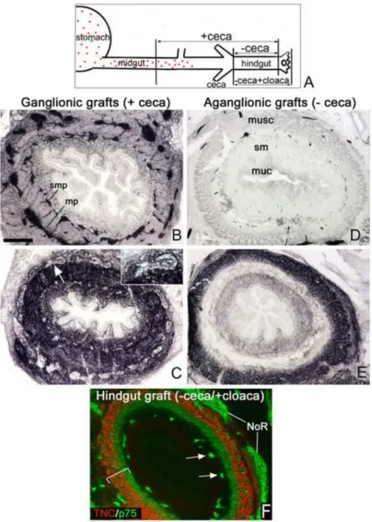
Based on the presence of TNC immunoreactivity surrounding vagal ENCCs, we hypothesized that ENCCs are required for TNC expression. To test this hypothesis, we generated aganglionic colon using two different methodologies. In the first method, we harvested intestine at E5, a stage at which ENCCs are located just beyond the level of the umbilicus (Fig. 4A). Grafts that included midgut (designated “+ceca” grafts) and those that did not (“−ceca” grafts) were cultured on the chorioallantoic membrane (CAM) of E9 host embryos for 8 days. The +ceca grafts became fully colonized by Tuj1+ enteric ganglia (Fig.
4B), and showed strong TNC expression throughout the gut mesenchyme (Fig. 4C). In contrast, -ceca grafts remained aganglionic (Fig. 4D), and TNC expression in these grafts was significantly altered, with no TNC present in the submucosa or lamina propria (Fig. 4E).
NIH-PA Author Manuscript NIH-PA Author Manuscript NIH-PA Author Manuscript
The CAM graft technique also allowed us to test whether sacral ENCCs fail to express TNC.
While NoR and pelvic plexus are not TNC-immunoreactive (Figs. 3A,C), these structures are not part of the ENS proper. We therefore harvested E5 chick hindgut without the ceca, but with the cloaca and NoR attached to serve as sources of sacral crest-derived cells. After 6 days on a host chick CAM, the resulting graft contains only sacral-derived ENCCs (arrows in Fig. 4F). These p75-immunoreactive ENCCs have no surrounding TNC expression (Fig.
4F).
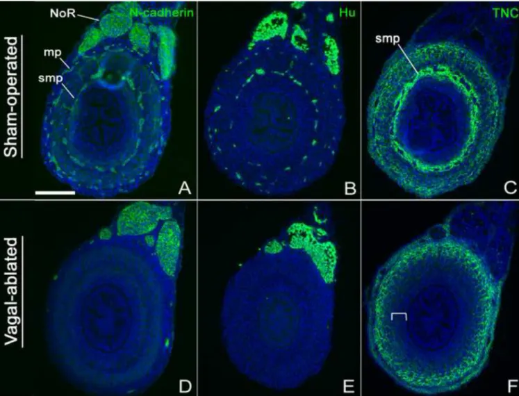
As a complementary method to verify the role of the ENS for TNC expression, aganglionic hindguts were generated in vivo by performing vagal neural tube ablations in HH9-10 chick embryos and allowing them to develop until E9. Hindguts from sham-operated embryos had normal expression of N-cadherin (Fig. 5A), Hu (Fig. 5B), and TNC (Fig. 5C). Note the strong expression of TNC in the area of the submucosal plexus (Fig. 5C). Successful ablation of the vagal neural tube was confirmed by the presence of aganglionosis in the E9 hindgut, with no expression of N-cadherin (Fig. 5D) or Hu (Fig. 5E), except in the nerve of Remak, which is sacral neural crest-derived. In these aganglionic hindguts, TNC expression is again absent from the submucosa and lamina propria (Fig. 5F), similar to the results obtained with the CAM grafts (Fig. 4E). Interestingly, TNC expression in these aganglionic colons is very similar to that observed in the hindgut+cloaca graft (Fig. 4F), which contains submucosal ENCCs of sacral neural crest origin only. Together, these results demonstrate that TNC expression in the inner, submucosal layers of the colon wall is dependent on the presence of vagal crest-derived ENCCs, whereas expression of TNC in the outer, muscular layer is ENCC-independent.
ENCCs produce TNC during ENS development
The colocalization of ENCC markers and TNC, as well as the loss of submucosal TNC expression in aganglionic hindguts, suggests that ENCCs either produce TNC themselves or induce its expression locally. To distinguish between these possibilities, we used rat-chick intestinal coelomic explants. Aneural colon from E13.5 embryonic rat was harvested and transplanted into the coelomic cavity of E3 chick embryos for 7 days. We have previously shown that these coelomic grafts generate chimeric guts by becoming colonized by host (i.e.
chick) vagal neural crest-derived cells (Nagy and Goldstein, 2006b). The graft undergoes significant growth during this coelomic culturing period (Fig 6A). After 7 days, the myenteric plexus is colonized by chick-derived ENCCs, as confirmed by co-labelling using 8F3, an antibody specific for cells of chick origin, and the NCC marker, p75 (Fig. 6B). An inner layer of p75-negative chick-derived cells is present and labels with a chick-specific endothelial marker, MEP21 (Fig. 6C), demonstrating that both NCCs and endothelial cells from the chick host colonize the graft. CN, an antibody specific for differentiated chick neurons, confirms the avian origin of the myenteric plexus (Fig. 6D) in the rat grafts.
Staining the graft with a chick-specific TNC antibody that does not stain rat TNC (Fig. 6G) shows TNC expression surrounding the p75-immunoreactive cells and confirms that chick- derived ENCCs are the source of TNC protein in these grafts (Fig. 6E,F).
These results confirm that ENCCs express TNC. However, the gut mesenchyme also produces TNC, as evidenced by the presence of TNC protein prior to ENCC colonization of the colon (Figs. 1,2) and its presence in the outer mesenchyme in the setting of
experimentally-induced aganglionosis (Figs. 4,5). We hypothesized that ENCCs and the gut mesenchyme may express different isoforms of the protein. TNC has three alternative splice variants in the chick which differ in the number of fibronectin type III repeats. The long, medium, and short variants refer to a 230 kD TNC with 11 fibronectin type III repeats, a 200 kD TNC with 9 repeats, and a 190 kD TNC with 8 repeats, respectively (Tucker, 1993).
These different variants appear to have distinct roles during development, with the long variant promoting cell motility and the smaller ones associated with cell proliferation and
NIH-PA Author Manuscript NIH-PA Author Manuscript NIH-PA Author Manuscript
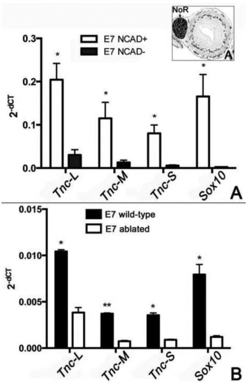
differentiation (Tucker, 1993). To test our hypothesis, we isolated wild-type E7 post- umbilical intestine, removed the nerve of Remak, and performed flow cytometry using N- cadherin to separate ENCCs (NCAD+) from non-ENCC cells (NCAD−). In the E7 hindgut, N-cadherin is specific for ENCCs, with no mesenchymal immunoreactivity (Fig. 7A’).
Quantitative PCR was performed using Tnc isoform-specific primers. Successful sorting of ENCCs from the remainder of the gut was confirmed by the strong expression of Sox10 by the NCAD+ population, with no expression in the NCAD− population (Fig. 7A). All three isoforms of Tnc were expressed by both NCAD+ and NCAD− cells, and all isoforms were expressed at much higher levels by the NCAD+ population. This result refutes our hypothesis that ENCCs and the gut mesenchyme have a distinct profile of isoform expression.
The qPCR described above compared Tnc isoform expression in ENCCs and the E7 gut environment after the gut had already been colonized. We also compared isoform expression in E7 wild-type colon and E7 aganglionic colon generated by vagal neural tube ablation. As expected, Sox10 expression is much lower in the aganglionic colon (Fig. 7B). The presence of a low level of Sox10 is likely due to inclusion of some nerve of Remak in the
preparations. However, this should not impact the results as nerve of Remak is a sacral neural crest-derived structure that does not express TNC (Fig. 3). Consistent with the results shown in Fig. 7A, the aganglionic colon expresses much lower levels of all three Tnc isoforms as compared to wild-type, with the relative expression of each isoform similar in both tissues.
TNC promotes migration of enteric neural crest-derived cells
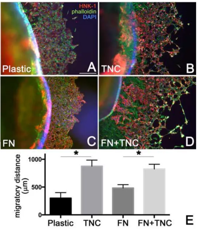
The effect of TNC on ENCC migration was tested by culturing E6 chick intestine onto plastic, fibronectin, and TNC substrates. Both HNK-1+ and HNK-1− cells migrate away from the explant, the latter representing smooth muscle cells and fibroblasts (Nagy et al., 2009). The distance ENCCs (HNK-1+ cells) migrated away from the gut after 48 hours was measured. Measurements were made at the cecal region, since this is the location of the migratory wavefront at E6. Measurements were made in 3–4 guts per group. As shown in Fig. 8E, ENCCs migrated further on a TNC-coated surface (875 + 222 µm) as compared to plastic alone (300 + 173 µm), and this difference was statistically significant. Similarly, when comparing ENCC migration onto a fibronectin (483 + 104 µm) versus fibronectin +TNC surface (825 + 171 µm), ENCCs again migrated further when TNC was present (Fig.
8C–E).
To determine whether the enhanced migration observed on TNC-coated surfaces was due to increased ENCC proliferation, we repeated the migration assay and stained the cultures for HNK-1 and phosphohistone H3, a marker of mitosis. The rate of ENCC proliferation on plastic and TNC-coated surfaces was 3.2 ± 1.2% and 3.0 ± 1.3 %, respectively (p=NS), suggesting that the promigratory effect of TNC is not due to enhanced cell proliferation.
Discussion
During ENS development, the cecal area represents a specialized region where ENCC migration is normally delayed (Druckenbrod and Epstein, 2005) and where ENCCs separate from their strands and advance into the colon as isolated cells (Druckenbrod and Epstein, 2005). The cecal region is also where Gdnf and Et3 levels are most strongly expressed (Barlow et al., 2003; Leibl et al., 1999; Nagy and Goldstein, 2006a) and where specific factors, such as β1 integrin (Breau et al., 2009), exert their influence. The unique features of this region are consistent with its potential role as a staging area to prepare ENCCs for colonization of the hindgut. In examining the developing gut for cecal-specific factors that may contribute to its special role during ENS development, we examined ECM protein
NIH-PA Author Manuscript NIH-PA Author Manuscript NIH-PA Author Manuscript
expression and found TNC specifically absent from the cecal region prior to the arrival of the migratory wavefront. As the advancing ENCCs entered the cecal area, TNC surrounded the arriving cells. This dynamic expression pattern led us to hypothesize that migrating ENCCs influence their extracellular environment as they invade the distal gut. We tested this by generating aganglionic hindguts and found that TNC immunostaining, specifically in the submucosal region, was absent, suggesting either that ENCCs produce TNC or that they induce its expression in the local microenvironment. Using rat-chick intestinal coelomic explants, we found that avian ENCCs colonize the aneural rat graft and produce chick- derived TNC, thus confirming that ENCCs produce TNC. This finding was further
supported by quantitative PCR showing that N-cadherin-immunoselected cells from the gut express TNC, and by vagal neural tube explants demonstrating TNC expression by vagal- derived NCCs.
Published studies examining whether NCCs produce tenascin have generated conflicting results. During early NCC patterning, Tan et al (Tan et al., 1987; Tan et al., 1991) found tenascin exclusively expressed by the somite in a pattern independent of the presence of NCCs. This lack of tenascin expression by NCCs was supported by Mackie et al (Mackie et al., 1988). In contrast, results by Stern et al (Stern et al., 1989) suggested that NCCs express tenascin as they migrate through the somites, a conclusion supported by several others (Tongiorgi et al., 1995; Tucker, 2001; Tucker and McKay, 1991). Our results are consistent with the latter studies and, furthermore, represent the first observation that TNC, or any other ECM protein, is expressed by ENCCs.
Interestingly, the pattern of TNC expression observed in the avian embryo is quite different from what has been described in rat (Newgreen and Hartley, 1995) and mouse (Breau et al., 2009), where tenascin is expressed diffusely in the outer mesenchyme with no obvious increased expression surrounding ENCCs, although this was not specifically examined in those studies. We find the strongest colocalization of TNC with ENCCs in the submucosal plexus. Interestingly, it is also the submucosal expression of TNC that is specifically absent in our experimentally generated aganglionic hindguts. Unlike in avians, the submucosal plexus of the rodent colon does not develop until postnatally (McKeown et al., 2001). In fact, in avians the submucosa of the hindgut region is the first to be colonized by the
migrating ENCC wavefront (Conner et al., 2003). Whether the difference in TNC expression between avians and rodents contributes to these species-specific differences in hindgut colonization merits further investigation.
Prior studies have shown that TNC promotes the migration and invasiveness of NCCs (Halfter et al., 1989; Tan et al., 1987; Tucker, 1993, 2001). Using an in vitro migration assay, we find that TNC promotes ENCC migration on both plastic and fibronectin-coated surfaces. These results are consistent with TNC’s known anti-adhesive role (Fischer et al., 1997; Tucker, 2001) and its ability to inhibit integrin-mediated cell adhesion to fibronectin (Chiquet-Ehrismann et al., 1988; Lotz et al., 1989; Probstmeier and Pesheva, 1999), an ECM protein strongly expressed in the gut mesenchyme. This anti-adhesive effect may allow faster cell detachment from the ECM substratum and thereby facilitate NCC migration (Halfter et al., 1989). Breau et al (Breau et al., 2009) recently proposed that TNC is inhibitory to ENCC migration, in contrast to our findings. This difference may result from species differences (i.e. chick versus mouse) or from several technical factors. First, the concentration of TNC was much higher in their study (20µg/ml) than ours (1µg/ml). Higher levels of TNC may inhibit cell migration (Melkonian et al., 2004). Second, they tested the effect of TNC on ENCC migration on a vitronectin surface, while we used plastic and fibronectin. Finally, while we focused on migration of ENCCs at the level of the cecum, the location of the wavefront in our E6 gut cultures, their study used E12.5 mouse midgut.
NIH-PA Author Manuscript NIH-PA Author Manuscript NIH-PA Author Manuscript
Given the highly dynamic nature of ENS development, this seemingly subtle difference may be profoundly important.
The expression of TNC by ENCCs emphasizes the close relationship between ENCCs and their microenvironment during ENS development. While colorectal aganglionosis is commonly thought to be due to defects either in ENCCs or in the gut environment, our findings demonstrate that ENCCs can modulate their microenvironment and therefore one cannot easily separate these two essential regulatory components of ENS development. To evaluate the impact of ENCCs on their mesenchymal environment, we generated
aganglionic hindguts both in vivo by vagal neural tube ablation and ex vivo using CAM grafts. In both models, the aganglionic hindgut exhibited a marked difference in ECM expression, with loss of TNC specifically from the submucosal region. This suggests that while the outer gut wall is able to produce its own TNC, possibly from the smooth muscle cells (Grumet et al., 1985), TNC expression by the inner mesenchyme is dependent on the presence of ENCCs in the submucosal region. Thus, ENCCs not only respond to their microenvironment, they also modify it by modulating ECM expression. This is reminiscent of a prior study showing that HNK-1-expressing mesenchymal cells are present in the gut prior to ENCC arrival, but absent once the gut is colonized (Luider et al., 1992). The loss of TNC expression in the aganglionic colonic mesenchyme may contribute to the decreased permissiveness of the aganglionic gut to late-arriving ENCCs (Druckenbrod and Epstein, 2009; Meijers et al., 1987) or to transplanted ENCCs (Rothman et al., 1993). This
observation also emphasizes that aganglionosis, as occurs in human Hirschsprung disease, may have additional adverse effects on the structure and function of the gut beyond the absence of enteric ganglia. Understanding the microenvironmental changes caused by aganglionosis is critical for developing effective strategies for cell-based therapies in Hirschsprung and other neurointestinal diseases (Hotta et al., 2013; Metzger et al., 2009).
A particularly intriguing finding is the dramatic difference between vagal and sacral-derived ENCCs with respect to TNC expression. We found that only vagal neural crest-derived cells express TNC. The only previously identified molecular difference between ENCCs of vagal and sacral origin was the finding that vagal NCCs express much higher levels of Ret than sacral crest cells (Delalande et al., 2008), which was felt to explain, at least partially, their increased invasiveness. The absence of TNC expression by sacral-derived ENCCs may further contribute to the decreased invasiveness of these cells and may also provide a clue to account for how these two cell populations migrate in opposing directions during
colonization of the distal gut. Identifying additional differences between vagal and sacral crest-derived ENCCs will contribute to our understanding of ENS development in general and of the etiology of Hirschsprung disease.
Acknowledgments
AMG is supported by NIH-NIDDK R01DK080914. NN is supported by a Bolyai Fellowship and Tamop-4.2.2/
B-10/1-2010-2013. RH is supported by a grant from the Uehara Memorial Foundation. Antibodies for TNC, Fibronectin, Laminin, N-cadherin, and 8F3 were obtained from the Developmental Studies Hybridoma Bank, developed under the auspices of the NICHD and maintained by The Univ. of Iowa.
References
Barlow A, de Graaff E, Pachnis V. Enteric nervous system progenitors are coordinately controlled by the G protein-coupled receptor EDNRB and the receptor tyrosine kinase RET. Neuron. 2003;
40:905–916. [PubMed: 14659090]
Breau MA, Dahmani A, Broders-Bondon F, Thiery JP, Dufour S. Beta1 integrins are required for the invasion of the caecum and proximal hindgut by enteric neural crest cells. Development. 2009;
136:2791–2801. [PubMed: 19633172]
NIH-PA Author Manuscript NIH-PA Author Manuscript NIH-PA Author Manuscript
Breau MA, Pietri T, Eder O, Blanche M, Brakebusch C, Fassler R, Thiery JP, Dufour S. Lack of beta1 integrins in enteric neural crest cells leads to a Hirschsprung-like phenotype. Development. 2006;
133:1725–1734. [PubMed: 16571628]
Bronner-Fraser M. Distribution and function of tenascin during cranial neural crest development in the chick. J Neurosci Res. 1988; 21:135–147. [PubMed: 2464073]
Bronner-Fraser, M. Methods in Avian Embryology. Inc, London: Academic Press; 1996.
Burns AJ, Douarin NM. The sacral neural crest contributes neurons and glia to the post-umbilical gut:
spatiotemporal analysis of the development of the enteric nervous system. Development. 1998;
125:4335–4347. [PubMed: 9753687]
Chalazonitis A, Tennyson VM, Kibbey MC, Rothman TP, Gershon MD. The alpha1 subunit of laminin-1 promotes the development of neurons by interacting with LBP110 expressed by neural crest-derived cells immunoselected from the fetal mouse gut. J Neurobiol. 1997; 33:118–138.
[PubMed: 9240369]
Chiquet M, Fambrough DM. Chick myotendinous antigen. II. A novel extracellular glycoprotein complex consisting of large disulfide-linked subunits. J Cell Biol. 1984; 98:1937–1946. [PubMed:
6202699]
Chiquet-Ehrismann R, Kalla P, Pearson CA, Beck K, Chiquet M. Tenascin interferes with fibronectin action. Cell. 1988; 53:383–390. [PubMed: 2452695]
Conner PJ, Focke PJ, Noden DM, Epstein ML. Appearance of neurons and glia with respect to the wavefront during colonization of the avian gut by neural crest cells. Dev Dyn. 2003; 226:91–98.
[PubMed: 12508228]
Delalande JM, Barlow AJ, Thomas AJ, Wallace AS, Thapar N, Erickson CA, Burns AJ. The receptor tyrosine kinase RET regulates hindgut colonization by sacral neural crest cells. Dev Biol. 2008;
313:279–292. [PubMed: 18031721]
Druckenbrod NR, Epstein ML. The pattern of neural crest advance in the cecum and colon. Dev Biol.
2005; 287:125–133. [PubMed: 16197939]
Druckenbrod NR, Epstein ML. Age-dependent changes in the gut environment restrict the invasion of the hindgut by enteric neural progenitors. Development. 2009; 136:3195–3203. [PubMed:
19700623]
Epperlein HH, Halfter W, Tucker RP. The distribution of fibronectin and tenascin along migratory pathways of the neural crest in the trunk of amphibian embryos. Development. 1988; 103:743–
756. [PubMed: 2470571]
Erickson HP, Taylor HC. Hexabrachion proteins in embryonic chicken tissues and human tumors. J Cell Biol. 1987; 105:1387–1394. [PubMed: 3654758]
Fischer D, Brown-Ludi M, Schulthess T, Chiquet-Ehrismann R. Concerted action of tenascin-C domains in cell adhesion, anti-adhesion and promotion of neurite outgrowth. J Cell Sci. 1997;
110(Pt 13):1513–1522. [PubMed: 9224768]
Gershon MD. Developmental determinants of the independence and complexity of the enteric nervous system. Trends Neurosci. 2010; 33:446–456. [PubMed: 20633936]
Gershon MD, Chalazonitis A, Rothman TP. From neural crest to bowel: development of the enteric nervous system. J Neurobiol. 1993; 24:199–214. [PubMed: 8445388]
Goldstein AM, Hofstra RM, Burns AJ. Building a brain in the gut: development of the enteric nervous system. Clin Genet. 2013; 83:307–316. [PubMed: 23167617]
Grumet M, Hoffman S, Crossin KL, Edelman GM. Cytotactin, an extracellular matrix protein of neural and non-neural tissues that mediates glia-neuron interaction. Proc Natl Acad Sci U S A. 1985;
82:8075–8079. [PubMed: 2415980]
Halfter W, Chiquet-Ehrismann R, Tucker RP. The effect of tenascin and embryonic basal lamina on the behavior and morphology of neural crest cells in vitro. Dev Biol. 1989; 132:14–25. [PubMed:
2465193]
Hamburger V, Hamilton HL. A series of normal stages in the development of the chick embryo. 1951.
Dev Dyn. 1992; 195:231–272. [PubMed: 1304821]
Hotta R, Stamp LA, Foong JP, McConnell SN, Bergner AJ, Anderson RB, Enomoto H, Newgreen DF, Obermayr F, Furness JB, Young HM. Transplanted progenitors generate functional enteric neurons in the postnatal colon. J Clin Invest. 2013; 123:1182–1191. [PubMed: 23454768]
NIH-PA Author Manuscript NIH-PA Author Manuscript NIH-PA Author Manuscript
Jones FS, Jones PL. The tenascin family of ECM glycoproteins: structure, function, and regulation during embryonic development and tissue remodeling. Dev Dyn. 2000; 218:235–259. [PubMed:
10842355]
Kruse J, Mailhammer R, Wernecke H, Faissner A, Sommer I, Goridis C, Schachner M. Neural cell adhesion molecules and myelin-associated glycoprotein share a common carbohydrate moiety recognized by monoclonal antibodies L2 and HNK-1. Nature. 1984; 311:153–155. [PubMed:
6206400]
Kuo BR, Erickson CA. Vagal neural crest cell migratory behavior: a transition between the cranial and trunk crest. Dev Dyn. 2011; 240:2084–2100. [PubMed: 22016183]
Leibl MA, Ota T, Woodward MN, Kenny SE, Lloyd DA, Vaillant CR, Edgar DH. Expression of endothelin 3 by mesenchymal cells of embryonic mouse caecum. Gut. 1999; 44:246–252.
[PubMed: 9895385]
Lotz MM, Burdsal CA, Erickson HP, McClay DR. Cell adhesion to fibronectin and tenascin:
quantitative measurements of initial binding and subsequent strengthening response. J Cell Biol.
1989; 109:1795–1805. [PubMed: 2477381]
Luider TM, Peters-van der Sanden MJ, Molenaar JC, Tibboel D, van der Kamp AW, Meijers C.
Characterization of HNK-1 antigens during the formation of the avian enteric nervous system.
Development. 1992; 115:561–572. [PubMed: 1385063]
Mackie EJ, Tucker RP, Halfter W, Chiquet-Ehrismann R, Epperlein HH. The distribution of tenascin coincides with pathways of neural crest cell migration. Development. 1988; 102:237–250.
[PubMed: 2458221]
McKeown SJ, Chow CW, Young HM. Development of the submucous plexus in the large intestine of the mouse. Cell Tissue Res. 2001; 303:301–305. [PubMed: 11291776]
McNagny KM, Pettersson I, Rossi F, Flamme I, Shevchenko A, Mann M, Graf T. Thrombomucin, a novel cell surface protein that defines thrombocytes and multipotent hematopoietic progenitors. J Cell Biol. 1997; 138:1395–1407. [PubMed: 9298993]
Meijers JH, Tibboel D, van der Kamp AW, Van Haperen-Heuts IC, Kluck P, Molenaar JC. The influence of the stage of differentiation of the gut on the migration of neural cells: an experimental study of Hirschsprung's disease. Pediatr Res. 1987; 21:466–470. [PubMed: 3588084]
Melkonian G, Wang JL, Chung J, Munoz N, Talbot P. CD44 and tenascin play critical roles in growth and vascular development of the chick chorioallantoic membrane and are targets of cigarette smoke. Anat Embryol (Berl). 2004; 208:109–120. [PubMed: 15052477]
Metzger M, Caldwell C, Barlow AJ, Burns AJ, Thapar N. Enteric nervous system stem cells derived from human gut mucosa for the treatment of aganglionic gut disorders. Gastroenterology. 2009;
136:2214–2225. e2211–e2213. [PubMed: 19505425]
Nagy N, Burns AJ, Goldstein AM. Immunophenotypic characterization of enteric neural crest cells in the developing avian colorectum. Dev Dyn. 2012; 241:842–851. [PubMed: 22411589]
Nagy N, Goldstein AM. Endothelin-3 regulates neural crest cell proliferation and differentiation in the hindgut enteric nervous system. Dev Biol. 2006a; 293:203–217. [PubMed: 16519884]
Nagy N, Goldstein AM. Intestinal coelomic transplants: a novel method for studying enteric nervous system development. Cell Tissue Res. 2006b; 326:43–55. [PubMed: 16736197]
Nagy N, Mwizerwa O, Yaniv K, Carmel L, Pieretti-Vanmarcke R, Weinstein BM, Goldstein AM.
Endothelial cells promote migration and proliferation of enteric neural crest cells via beta1 integrin signaling. Dev Biol. 2009; 330:263–272. [PubMed: 19345201]
Newgreen DF, Hartley L. Extracellular matrix and adhesive molecules in the early development of the gut and its innervation in normal and spotting lethal rat embryos. Acta Anat (Basel). 1995;
154:243–260. [PubMed: 8773711]
Newgreen DF, Jahnke I, Allan IJ, Gibbins IL. Differentiation of sympathetic and enteric neurons of the fowl embryo in grafts to the chorio-allantoic membrane. Cell Tissue Res. 1980; 208:1–19.
[PubMed: 7388924]
Payette RF, Tennyson VM, Pomeranz HD, Pham TD, Rothman TP, Gershon MD. Accumulation of components of basal laminae: association with the failure of neural crest cells to colonize the presumptive aganglionic bowel of ls/ls mutant mice. Dev Biol. 1988; 125:341–360. [PubMed:
3338619]
NIH-PA Author Manuscript NIH-PA Author Manuscript NIH-PA Author Manuscript
Preffer F, Dombkowski D. Advances in complex multiparameter flow cytometry technology:
Applications in stem cell research. Cytometry B Clin Cytom. 2009; 76:295–314. [PubMed:
19492350]
Probstmeier R, Pesheva P. Tenascin-C inhibits beta1 integrin-dependent cell adhesion and neurite outgrowth on fibronectin by a disialoganglioside-mediated signaling mechanism. Glycobiology.
1999; 9:101–114. [PubMed: 9949188]
Rothman TP, Goldowitz D, Gershon MD. Inhibition of migration of neural crest-derived cells by the abnormal mesenchyme of the presumptive aganglionic bowel of ls/ls mice: analysis with aggregation and interspecies chimeras. Dev Biol. 1993; 159:559–573. [PubMed: 8405679]
Serbedzija GN, Burgan S, Fraser SE, Bronner-Fraser M. Vital dye labelling demonstrates a sacral neural crest contribution to the enteric nervous system of chick and mouse embryos. Development.
1991; 111:857–866. [PubMed: 1879357]
Stern CD, Norris WE, Bronner-Fraser M, Carlson GJ, Faissner A, Keynes RJ, Schachner M. J1/
tenascin-related molecules are not responsible for the segmented pattern of neural crest cells or motor axons in the chick embryo. Development. 1989; 107:309–319. [PubMed: 2483682]
Tan SS, Crossin KL, Hoffman S, Edelman GM. A symmetric expression in somites of cytotactin and its proteoglycan ligand is correlated with neural crest cell distribution. Proc Natl Acad Sci U S A.
1987; 84:7977–7981. [PubMed: 2446315]
Tan SS, Prieto AL, Newgreen DF, Crossin KL, Edelman GM. Cytotactin expression in somites after dorsal neural tube and neural crest ablation in chicken embryos. Proc Natl Acad Sci U S A. 1991;
88:6398–6402. [PubMed: 1713677]
Tanaka H, Kinutani M, Agata A, Takashima Y, Obata K. Pathfinding during spinal tract formation in the chick-quail chimera analysed by species-specific monoclonal antibodies. Development. 1990;
110:565–571. [PubMed: 1723947]
Tongiorgi E, Bernhardt RR, Zinn K, Schachner M. Tenascin-C mRNA is expressed in cranial neural crest cells, in some placodal derivatives, and in discrete domains of the embryonic zebrafish brain.
J Neurobiol. 1995; 28:391–407. [PubMed: 8568519]
Tucker RP. The in situ localization of tenascin splice variants and thrombospondin 2 mRNA in the avian embryo. Development. 1993; 117:347–358. [PubMed: 7693413]
Tucker RP. Abnormal neural crest cell migration after the in vivo knockdown of tenascin-C expression with morpholino antisense oligonucleotides. Dev Dyn. 2001; 222:115–119. [PubMed: 11507773]
Tucker RP, McKay SE. The expression of tenascin by neural crest cells and glia. Development. 1991;
112:1031–1039. [PubMed: 1718677]
Weskamp G, Reichardt LF. Evidence that biological activity of NGF is mediated through a novel subclass of high affinity receptors. Neuron. 1991; 6:649–663. [PubMed: 1849725]
Wu JJ, Chen JX, Rothman TP, Gershon MD. Inhibition of in vitro enteric neuronal development by endothelin-3: mediation by endothelin B receptors. Development. 1999; 126:1161–1173.
[PubMed: 10021336]
Yntema CL, Hammond WS. The origin of intrinsic ganglia of trunk viscera from vagal neural crest in the chick embryo. J Comp Neurol. 1954; 101:515–541. [PubMed: 13221667]
NIH-PA Author Manuscript NIH-PA Author Manuscript NIH-PA Author Manuscript
Highlights
• In the embryonic gut, enteric neural crest-derived cells (ENCCs)produce tenascin-C (TNC).
• TNC is produced by vagal, but not sacral, ENCCs in the intestine.
• Expression of TNC is absent in the submucosa of aganglionic hindgut.
• TNC promotes the migration of ENCCs.
• ENCCs modify their microenvironment to promote their own migration.
NIH-PA Author Manuscript NIH-PA Author Manuscript NIH-PA Author Manuscript
Figure 1. TNC expression colocalizes with the ENCC migratory wavefront in the cecal region The dynamic pattern of TNC expression is shown by immunohistochemistry on longitudinal sections of postumbilical intestine at E4.5 (A), E5 (B), E6 (C) and E7 (D). TNC is present in the mesenchyme proximal and distal to the ceca, but is notably absent from the cecal region from E4.5-E5 (arrows in A,B) and appears with the advancing ENCC wavefront at E6 (C, arrows) and E7 (D, arrow). N-cadherin immunostaining of longitudinal E6 gut shows the ENCC wavefront in the cecal region (E, boxed area magnified in inset). Fibronectin (F) and laminin (G) are both expressed throughout the gut at E6. A confocal image from
longitudinal section of E7 proximal hindgut shows strong TNC labelling (red) around p75+
ENCCs (green) at the migrating wavefront (H, arrow points to area magnified in I).
NIH-PA Author Manuscript NIH-PA Author Manuscript NIH-PA Author Manuscript
Scale bar in A=380µm (A,B), 440µm (C-G), 130µm (H), 140µm (inset in E), 15µm (I)
NIH-PA Author Manuscript NIH-PA Author Manuscript NIH-PA Author Manuscript
Figure 2. Dynamic expression of TNC protein during distal gut development
Immunoblot analysis was performed to detect the expression levels of laminin, TNC, and fibronectin in tissue lysates prepared from the post-umbilical midgut, cecal region, and hindgut of E5, E6, and E8 avian intestine.
MG, midgut; HG, hindgut
NIH-PA Author Manuscript NIH-PA Author Manuscript NIH-PA Author Manuscript
Figure 3. Vagal, but not sacral, neural crest-derived cells produce TNC
Transverse sections through an E8 embryo at the levels shown in the schematic inset were stained with antibodies to p75 and TNC (A-C). Vagal crest-derived ENCCs in the hindgut submucosal plexus are surrounded by TNC (A; boxed area in A is magnified in B), while sacral crest-derived structures, including NoR and pelvic plexus, are not (A,C). Cross sections of HH17 embryos through the vagal (D; 3rd somite) and sacral (F, 30th somite) levels were stained for HNK-1 and TNC. Magnified views of the boxed areas in (D) and (F) are shown in E-E” and G-G”, respectively, demonstrating that vagal, but not sacral, NCCs are surrounded by TNC immunoreactivity. Vagal and sacral neural tube explants were cultured for 24 hours on fibronectin-coated plates. Vagal crest-derived NCCs (HNK-1+)
NIH-PA Author Manuscript NIH-PA Author Manuscript NIH-PA Author Manuscript
migrate away from the neural tube and are surrounded by TNC (H,I), while sacral NCCs, which also migrate onto the dish, are not (J). Inset in Fig. 3I shows a vagal NCC with both intracellular (arrowheads) and extracellular (arrow) TNC protein. The edge of the neural tube explants is marked with a hatched line in H and J.
drg, dorsal root ganglion; NoR, nerve of Remak; n, notochord; nt, neural tube; s, somite;
smp, submucosal plexus. Scale bar in A=220µm (A), 60µm (B), 130µm (C), 200µm (D,F), 25µm (H,J), 10µm (I)
NIH-PA Author Manuscript NIH-PA Author Manuscript NIH-PA Author Manuscript
Figure 4. Submucosal expression of TNC is absent in aganglionic hindgut grafts
Diagram of gut at E5 indicates position of ENCCs, which have migrated just past the umbilicus (A). As illustrated in A, segments of distal gut including (+ceca) or excluding (−ceca) the ENCC wavefront were cultured on the CAM for 8 days to generate ganglionic (+ceca) or aganglionic (−ceca) hindguts. Transverse sections of ganglionated grafts show normal Tuj1 (B) and TNC (C) expression.
TNC is expressed both in and around the enteric ganglia (arrow depicts the myenteric ganglion magnified in the inset, C). Aganglionic grafts lack enteric neurons (Tuj1, D) and have no TNC expression in the submucosal region (E). Hindgut containing only sacral- derived ENCCs (F, arrows) was generated by grafting E5 (preganglionic) hindgut with
NIH-PA Author Manuscript NIH-PA Author Manuscript NIH-PA Author Manuscript
attached cloaca and nerve of Remak onto an E9 CAM for 6 days, then stained for p75 (green) and TNC (red). The bracket in (F) denotes the smooth muscle layer, which is TNC- and p75-immunoreactive.
mp, myenteric plexus; muc, mucosa; muse, muscular layer; NoR, nerve of Remak; sm, submucosa; smp, submucosal plexus. Scale bar in A=260µm in all panels.
NIH-PA Author Manuscript NIH-PA Author Manuscript NIH-PA Author Manuscript
Figure 5. ENCCs are required for normal TNC expression in the submucosal region of the hindgut
Vagal neural tube ablations were performed to generate aganglionic guts in ovo. Transverse sections through the hindgut of sham-operated E9 embryos have normal ENCCs (N- cadherin, A), enteric neurons (Hu, B), and TNC expression (C). In contrast, vagal-ablated embryos are aganglionic (D,E) and lack submucosal TNC (F, bracket marks submucosa). A- C, sham-operated control embryos; D-F, vagal-ablated embryos.
mp, myenteric plexus; NoR, nerve of Remak; smp, submucosal plexus. Scale bar in A=200µm in all panels.
NIH-PA Author Manuscript NIH-PA Author Manuscript NIH-PA Author Manuscript
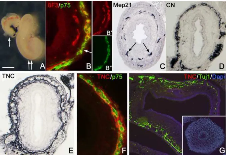
Figure 6. Rat-chick coelomic explants confirm that ENCCs produce TNC
Aneural rat hindgut was isolated at E13.5 and transplanted into the coelomic cavity of an E3 chick embryo for 7 days. The graft grows significantly after 2 days (A, arrow) and 7 days (A, double arrow) in the coelom. Double immunofluorescence with 8F3 and p75 antibodies reveals chick-derived ENCCs in the myenteric plexus of the rat gut (B, arrow). Staining with 8F3 and p75 are shown separately in B’ and B”, respectively. Unlike the outer layer of 8F3+
cells, the inner layer does not co-express p75, but does express the chick-specific endothelial cell marker, MEP-21 (C, arrows). CN-immunoreactivity in the graft confirms the presence of chick-derived neurons (D). Staining with a chick-specific anti-TNC antibody shows TNC expression around the myenteric ganglia of the rat hindgut graft (E). Double
immunofluorescence with chick-specific antibodies confirms that both ENCCs and TNC are of chicken origin (F). Rat E14.5 stomach (G; Tuj1, green) and preganglionic colon (G, inset) do not stain with chick-specific TNC antibody (red).
Scale bar in A=400µm (A), 50µm (B,F), 100µm (C,D,G), 80µm (E)
NIH-PA Author Manuscript NIH-PA Author Manuscript NIH-PA Author Manuscript
Figure 7. ENCCs preferentially express the long isoform of TNC
(A) The relative expression of Tnc isoforms was determined by qPCR analysis of NCAD+
and NCAD− cells sorted by flow cytometry from wild-type E7 post-umbilical gut, excluding NoR and cloaca (n=6). 2−dCT indicates the standard relative expression level of each gene normalized to GAPDH. The inset (A’) shows N-cadherin immunohistochemistry of a cross- section of E7 hindgut. (B) Expression of TNC isoforms was also compared between wild- type and vagal neural tube-ablated embryos (n=3 for each group). Statistical significance was calculated using Student’s t-test, comparing NCAD+ to NCAD− (A) or wild-type to ablated (B) for each gene. *p<0.05;**p<0.001
NIH-PA Author Manuscript NIH-PA Author Manuscript NIH-PA Author Manuscript
NCAD, N-cadherin; NoR, nerve of Remak; Tnc-S, Tnc short isoform; Tnc-M, Tnc medium isoform; Tnc-L, Tnc long isoform
NIH-PA Author Manuscript NIH-PA Author Manuscript NIH-PA Author Manuscript
Figure 8. TNC promotes ENCC migration
To determine the effect of TNC on ENCC migration, E6 chicken midgut and hindgut (excluding nerve of Remak) was explanted onto plastic (A), TNC (1 µg/ml) (B), fibronectin (10 µg/ml) (C), or fibronectin + TNC (D) coated surfaces. After 48 hours, ENCC migration was examined near the cecal region by staining for HNK-1 (red), phalloidin (green), and DAPI (blue). The distance of ENCC migration on each surface was calculated (E). *p<0.05 FN, fibronectin. Scale bar in A=200µm in all panels
NIH-PA Author Manuscript NIH-PA Author Manuscript NIH-PA Author Manuscript
NIH-PA Author Manuscript NIH-PA Author Manuscript NIH-PA Author Manuscript
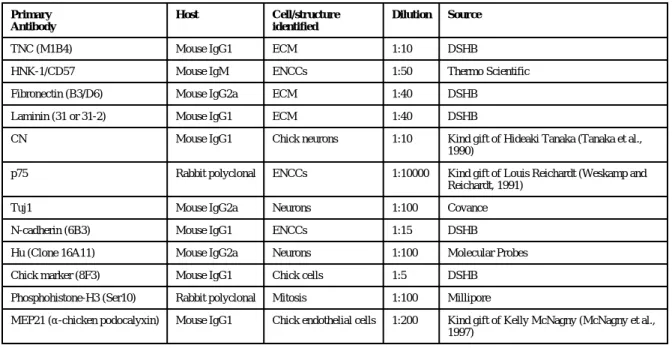
Table 1 Primary antibodies used for immunohistochemistry.
Primary Antibody
Host Cell/structure
identified
Dilution Source
TNC (M1B4) Mouse IgG1 ECM 1:10 DSHB
HNK-1/CD57 Mouse IgM ENCCs 1:50 Thermo Scientific
Fibronectin (B3/D6) Mouse IgG2a ECM 1:40 DSHB
Laminin (31 or 31-2) Mouse IgG1 ECM 1:40 DSHB
CN Mouse IgG1 Chick neurons 1:10 Kind gift of Hideaki Tanaka (Tanaka et al., 1990)
p75 Rabbit polyclonal ENCCs 1:10000 Kind gift of Louis Reichardt (Weskamp and Reichardt, 1991)
Tuj1 Mouse IgG2a Neurons 1:100 Covance
N-cadherin (6B3) Mouse IgG1 ENCCs 1:15 DSHB
Hu (Clone 16A11) Mouse IgG2a Neurons 1:100 Molecular Probes
Chick marker (8F3) Mouse IgG1 Chick cells 1:5 DSHB
Phosphohistone-H3 (Ser10) Rabbit polyclonal Mitosis 1:100 Millipore
MEP21 (α-chicken podocalyxin) Mouse IgG1 Chick endothelial cells 1:200 Kind gift of Kelly McNagny (McNagny et al., 1997)
NIH-PA Author Manuscript NIH-PA Author Manuscript NIH-PA Author Manuscript
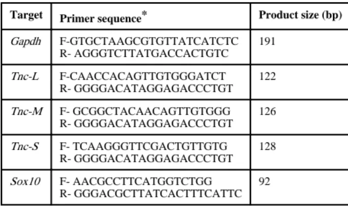
Table 2 Primers for qPCR.
Target Primer sequence* Product size (bp) Gapdh F-GTGCTAAGCGTGTTATCATCTC
R- AGGGTCTTATGACCACTGTC 191
Tnc-L F-CAACCACAGTTGTGGGATCT R- GGGGACATAGGAGACCCTGT
122
Tnc-M F- GCGGCTACAACAGTTGTGGG R- GGGGACATAGGAGACCCTGT
126
Tnc-S F- TCAAGGGTTCGACTGTTGTG R- GGGGACATAGGAGACCCTGT
128
Sox10 F- AACGCCTTCATGGTCTGG R- GGGACGCTTATCACTTTCATTC
92
*F, forward primer; R, reverse primer