Characterising human pluripotent stem cells-derived endothelial cells
PhD thesis
Dr. Edit Gara
Doctoral School of Basic Medical Sciences Semmelweis University
Supervisor: Dr. Gábor Földes PhD
Official reviewers: Dr. Melinda Pirity PhD Dr. János Réthelyi PhD
Head of the Final Examination Committee:
Dr. Péter Ferdinandy DSc
Members of the Final Examination Committee:
Dr. Lívia Jánoskuti PhD Dr. Ágota Apáti PhD
Budapest, 2015
2 INTRODUCTION
Definite therapy to improve reduced myocardial function or replace injured cells after ischemic or degenerative injuries of the cardiovascular system is lacking. Many cell types have been considered as future sources for cardiovascular cell therapy. Earlier studies focused on bone marrow-derived mononuclear cells, endothelial progenitor cells, and mesenchymal stem cells. First-in-man and early clinical trials with these cells had only moderate or non-beneficial outcomes.
Furthermore, many of these trials published conflicting data. The ideal cell type for regenerative purposes should be easily available, easy to be collected and expanded, should have a stable karyotype and phenotype, should be efficiently and feasibly differentiated to the desired cell type, may not be immunogenic and must provide desired functional activity in vivo. Human embryonic stem cells (hESC) are derived from the inner cell mass of blastocyst stage embryos; human induced pluripotent stem cells (hiPSC) are developed by genetic reprogramming from somatic cells. Both cell types are able to differentiate into all cell types of the three germ lines. Human pluripotent stem cells are therefore now considered as an alternative source for cardiovascular regeneration. We believe that hiPSC may be one of the most promising cell types for cardiovascular regeneration. Human iPSC may overcome ethical issues associated with hESC and may lack immunogenic complications.
However, challenges with genetic and epigenetic changes during reprogramming need further considerations.
My project focuses on endothelial derivatives of human pluripotent stem cells (hESC-EC and hiPSC-EC). Endothelial cells play a major role in the physiology of the cardiovascular system, e.g. regulating vascular tone and inflammation as well as platelet activation and clotting. Their efficient function is crucial both in vascular tone and microcirculatory organisation. Indeed, endothelial cells provide a semipermeable membrane between blood and vessel wall. Furthermore,
3
cells release vasoactive factors to regulate vessel tone and vascular resistance. It is therefore not surprising that endothelial dysfunction plays a key role in development of cardiovascular diseases, including hypertension, atherosclerosis and diabetes mellitus.
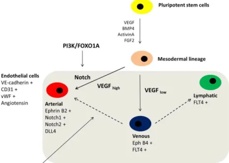
Arterial and venous subpopulations of endothelial cells differ not only in their localisation but also in their functional properties. Arterial endothelial cells regulate vessel tone and vascular resistance by defining the tone of the pre-capillary arteries. Post-capillary venules and veins are the site of inflammatory reaction where white blood cell rolling and extravasation occur. The vasoactive role may be less important for venous endothelial cells. It is well-known that environmental cues (paracrine signals, effects of wall shear stress) may alter arterial and venous endothelial fate; e.g. coronary grafts derived from saphenous veins function properly after coronary artery bypass graft surgery.
Recent studies have shown that apart from the plasticity of arterial and venous fate, developmental potential may be regulated at transcription levels. The major regulatory pathways for arterial development are controlled by VEGF and Notch. Notch1, Notch2, EphrinB2 and DLL4 are known to be regulators for arterial development. EphB4 is responsible for venous development, as well as FLT4; the latter is more likely to be a lymphatic marker for the endothelial phenotype.
A recent study has shown that the transcription factor FOXO1 (downstream member of the phosphatidylinositol 3-kinase (PI3K) signalling pathway) leads to the development of arterial endothelial cells via upregulation of Notch1. PI3K is a lipid kinase, and has been shown to play an important role in regulating cell proliferation, adhesion, DNA repair, senescence and stemness. PI3K is well suited for pharmacological interventions, which makes the pathway one of the most attractive therapeutic targets in cancer and diabetes. Members of FOXO (Forkhead box O transcription factor) family play a crucial role during embryonic
4
development of the cardiovascular system, as FOXO1A-deficient embryos die around embryonic day 11 because of their inefficient vascular and cardiac systems. FOXO1A is also important in regulation of endothelial cell fate. Indeed, it has been found that mouse embryonic stem cells-derived endothelial cells lacking FOXO1A are unable to respond to vascular endothelial growth factor (VEGF), an important angiogenic growth factor for normal vascular development. A better understanding of PI3K-FOXO1A-related signalling control of specification and function of human endothelial cells may provide a therapeutic advantage in endothelial cell transplantation and tissue engineering.
Figure 1. Endothelial differentiation from human pluripotent stem cells
5 AIMS
The aim of this study was to differentiate and characterise endothelial cells from hESC and hiPSC. Gaining detailed information on endothelial differentiation and phenotype in vitro and in vivo increases our understanding of cardiovascular diseases. Development of new cell products enhances utilisation in preclinical studies.
The specific aims of this project were:
1. To optimise endothelial differentiation protocols and characterise endothelial fate, particularly arterial and venous subpopulations and angiogenic properties.
2. To investigate the role of the PI3K/FOXO1A signalling pathway in endothelial differentiation, proliferation, viability and angiogenesis- related properties.
3. To develop three-dimensional endothelial cell culture systems for tissue engineering.
4. To define anticlotting characteristics of 3D endothelial cell culture.
5. To modulate endothelial maturation in vivo.
6 METHODS
I. Cell culture
All cell culturing were performed under sterile conditions, using a biosafety cabinet; cells were kept in 37°C, 5% CO2 and 21% O2 conditions. Human embryonic stem cells used in these studies were commercially available. All ethical approvals had been acquired for research purposes. Experiments were carried out on WA007 (H7) female hESC line (WiCell Research Institute Bank, Madison, USA).
Human iPSC were available from Osaka University, Japan (ReproCell, Kanagawa, Japan) or purchased from WiCell (IMR-90-4). Human iPSC were also generated from human adult fibroblasts via overexpression of pluripotency genes (episomal plasmids from Addgene, Cambridge, USA), although these hiPSC were not used in further experiments. All pluripotent stem cells were cultured in feeder-free conditions in mTeSR1 medium. Combined (mechanical and enzymatic) passaging was performed every 4-10 days.
Human ESC-EC, hiPSC-EC and control endothelial cells (HUVEC:
human umbilical vein, HCAEC: human coronary artery, HAEC: human aorta, BOEC: blood outgrowth) were all maintained in EGM2 endothelial medium. Endothelial cells were used for experiments between passages 2 and 6.
For 3D endothelial cell culture endothelial cells were seeded on commercially available porcine intestinal extracellular matrix, or decellularised human aortic pieces. Aortic decellularisation procedure was carried out in detergent solution. The small bioreactor system composed of scale-up spinner flask, endothelial cells and extracellular matrices for endothelial growth (50000 cells/ 0.5cm2 biomatrix).
Endothelial cell alignment under shear stress was determined using a sheer stress model in vitro. Cells were grown to confluence and placed on an orbital shaker. The wave of media resulted in a complex pattern
7
of shear stress with directional (laminar) shear towards the edge of the well, and non-directional (turbulent) shear at the centre. Alignment of cells was visualised by light microscopy.
II. Endothelial differentiation
Protocols involved embryoid body formation (EB) or monolayer methods. To force differentiation into mesodermal lineage differentiating colonies were treated with extra growth factors and morphogenes (Activin A, FGF-2, VEGF165 and BMP4). Differentiating cells were maintained in Stemline II medium. EBs were developed by forced aggregation from suspension. EBs were cultured for four days on ultra-low attachment plates either in normoxic or hypoxic conditions and EBs were seeded onto gelatine in EGM2 medium after four days.
At day 13 days, CD31 positive endothelial cells were sorted from differentiating culture by fluorescence activated cell sorting method (FACS).
III. Cell treatments
Endothelial cells were treated with PI3K inhibitor/FOXO1A activator LY294002 (10 μM, 24 h) during and after endothelial differentiation.
For FOXO1A knockdown on differentiating hESC and purified hESC- EC cultures, FOXO1A siRNA (10 nM, 6 hours and 24 hours) was used.
Scrambled non-targeting (NT) siRNA (10 nM) was used as negative control.
To overexpress FOXO1A, transfection of hPSC-EC with plasmids encoding FOXO1A-eGFP was carried out by electroporation.
H2O2 was used as a stable reactive oxygen species for induction of cell death in three different concentrations (high dose: 900 μM, medium dose: 600 μM, low dose: 300 μM) for 12 hours.
8 IV. Endothelial characteristics
Matrigel tube formation assay
Control, PI3K inhibitor LY294002, scrambled (NT) or FOXO1A siRNA-pretreated hESC-EC (50000cells/well) were plated onto Matrigel. Cultures were imaged and quantitated after 24 hours. Cell nuclei were stained for Hoechst-33342. Colony formation and the number of nuclei per colony were assessed by Colony Formation BioApplication on automated Cellomics ArrayScanner platform.
Immunocytochemistry and immunohistochemistry
Endothelial cells were assessed by immunocytochemistry. Cells were labelled with anti-human CD31, anti-human vWF, anti-human DLL4 and anti-human FOXO1A antibodies. As secondary antibody, Alexa Fluor 546 was used. Cell nuclei were stained with Hoechst-33342. For live imaging in order to measure of cell death parameters, cells were stained with vital dyes TOTO-1 or TO-PRO-3.
3D vascular constructs were embedded in paraffin to perform histochemical analysis. Histological sections were analysed with 3D- HisTech software, which enables three dimensional analysis of whole slides in section, tissue segmentation and morphometric analysis.
Human endothelial cells were stained with anti-human CD31 antibody.
Gene expression analyses
For quantitative, real-time PCR, RNA was extracted from cellular samples and complimentary-DNA (cDNA) was synthesized by reverse transcriptase. Messenger RNA levels of Notch1, Notch2, EphrinB2, EphB4, FLT4, ve-Cadherin, Angiopoietin-2, Tie-2, CD31 and NOS-3 were quantified. As housekeeping gene control human glyceraldehyde 3-phosphate dehydrogenase (GAPDH) endogenous control was used.
Relative expression was determined by the ΔΔCt method in which fold increase = 2 -ΔΔCt.
9 Proteomics and ELISA
To identify protein production of angiogenesis and hematopoietic soluble receptors, proteome profiling measurement was performed.
Both cell surface supernatants and cell lysates were used in these studies. Pixel densities on assay membranes were analysed by Image J software. Human IL-6, IL-8 and ET-1 ELISA assays were performed.
Rantes ELISA assay was used to study anticlotting effect of hESC-EC and hiPSC-EC. Platelet rich plasma was added to 3D vascular constructs; increase in Rantes levels refers to platelet activation.
V. In vivo experiments
Human ESC-EC, hiPSC-EC and control HUVEC were implanted into athymic nude rats (Crl:NIH-Foxn1rnu, Charles River), to investigate survival, engraftment and angiogenic activity in vivo. Animals were housed in microbe-free conditions, care and handling fulfilled all regulations. Before transplantation animals received general anaesthesia with Ketamine-Xylazine cocktail (80-100 mg/kg and 5-10 mg/kg).
Endothelial cells (106/implanted plug) were transplanted in Matrigel (250µl), heparin (64U/ml), recombinant murine basic FGF (80ng/ml) and EGM2 (70µl) containing plugs. Cells were transplanted subcutaneously at four sites of the abdominal region of each rat. Two weeks after the implantation cells were re-isolated via enzymatic digestion and CD31-positive human endothelial cells were sorted and cultured. Further gene expression analyses were performed.
VI. Statistics
Statistical analysis was carried out using GraphPad Prism 5 software.
Data are presented as the mean ± standard error of mean (SEM). All experiments were carried out in least three biological replicates. Data were analysed by Student’s t-test, paired t-test or using one-way ANOVA following Tukey post test. In all cases significance was taken as * p <0.05, ** p < 0.01, *** p < 0.001.
10 RESULTS
I. Endothelial characteristics
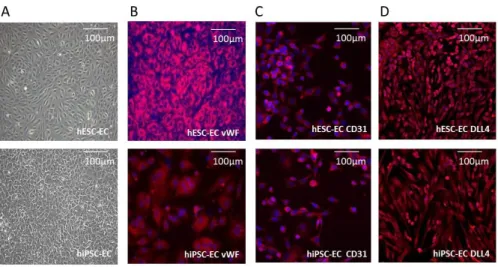
Both hESC-EC and hiPSC-EC cultures showed cobblestone pattern in vitro (Figure 2. A). Immunocytochemistry analysis showed that hESC- EC and hiPSC-EC are stained positive for von Willebrand factor and CD31 as well as arterial endothelial marker DLL4 (Figure 2. B-D).
Control endothelial cells (HAEC and BOEC) were cobblestone when grown under static culture conditions in vitro (Figure 3. A). BOEC and HAEC changed morphology in response to shear stress. Both HAEC and BOEC elongated and aligned when exposed to directional shear stress but remained cobblestoned in morphology when exposed to non- directional, turbulent shear stress (Figure 3. B). Human ESC-EC did not appear to respond to shear stress (Figure 3. B).
Figure 2. In vitro morphology and immunocytochemistry of hESC-EC and hiPSC-EC
11
Figure 3. Responses of endothelial cells to shear stress
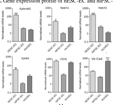
As assessed by quantitative RT-PCR expression of endothelial and angiogenesis-related genes was abundant in hESC-EC and hiPSC-EC.
Significant increase was found both in arterial (EphrinB2, Notch1, Notch2) and venous (EphB4) marker genes during endothelial differentiation. No significant difference was found between the mRNA levels of arterial and venous endothelial markers. Angiogenesis marker genes, CD31 and ve-cadherin, showed increased expressions in hESC- EC and hiPSC-EC as compared to those in undifferentiated pluripotent stem cells, normalised to GAPDH housekeeping control (Figure 4.).
Figure 4. Gene expression profile of hESC-EC and hiPSC-EC
hESC-EC hiPS
C-EC HUVEC 0.1
1 10 100
1000 EphrinB2
***
Normalised mRNA levels
hESC-EC hiP
SC-EC HUVEC 0.1
1 10 100
1000 Notch1
*
Normalised mRNA levels
hESC-EC hiPS
C-EC HUVEC 0.1
1 10 100
1000 Notch2
***
Normalised mRNA levels
hES C-EC
hiPSC-EC HUV
EC 0.1
1 10 100
1000 EphB4
**
Normalised mRNA levels
hESC -EC
hiPSC -EC
HUVEC 0.1
1 10 100
1000 CD31
Normalised mRNA levels
hES C-EC
hiPSC-EC HUV
EC 0.1
1 10 100 1000
Ve-Cad
***
Normalised mRNA levels
Ve-Cad
12
II. Role of PI3K/FOXO1A on endothelial subpopulations, angiogenesis, proliferation and survival
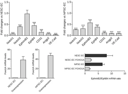
FOXO1A overexpression downregulates expression of all endothelial genes investigated. Figure 5. shows that CD31, angiopoietin-2 and ve- cadherin levels were decreased in response to high levels of FOXO1A.
Arterial and venous marker genes were parallel decreased. Ratio of EphrinB2/EphB4 was markedly decreased after FOXO1A overexpression in hESC-EC and hiPSC-EC, suggesting that FOXO1A regulates arterial phenotype.
Figure 5. FOXO1A overexpression affects endothelial gene expression
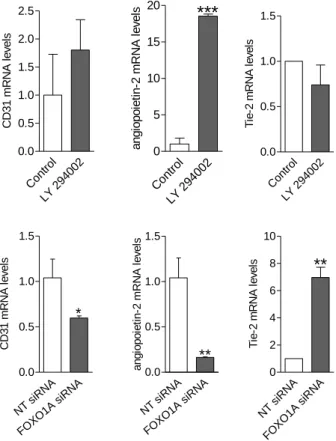
The mRNA levels of CD31 and angiopoietin-2 were increased in response to PI3K inhibitor/FOXO1A activator LY294002; on the other hand, FOXO1A siRNA resulted in lower mRNA levels of angiopoietin- 2 (Figure 6.). The mRNA levels of the angiopoietin-2 receptor, Tie-2 showed inverse regulation as angiopoietin-2. LY294002 decreased the mRNA levels of Tie-2, whilst treatment with FOXO1A siRNA resulted in increased Tie-2 mRNA levels (Figure 6.).
Notch1 Notch2
EphrinB2 EphB4
CD31 Angp2
VE-Cad 0.0
0.5 1.0 1.5
***
*** ***
***
***
***
Fold changes vs hESC-EC
Notch1 Notch2
EphrinB2 EphB4
CD31 Angp2
VE-Cad 0.0
0.5 1.0 1.5
***
***
***
***
*** *** ***
Fold changes vs hiPSC-EC
hESC -EC
Overexpressio n 0 20 40 60
Foxo1A mRNA levels
hiPS C-EC
Overexpressio n 0 20 40 60
Foxo1A mRNA levels
0 5 10 15
hiPSC-EC FOXO1A hiPSC-EC hESC-EC FOXO1A hESC-EC
EphrinB2/EphB4 mRNA ratio
13
Figure 6. PI3K/FOXO1A signalling modulates angiogenesis-related gene expression in hESC-derived endothelial cells
NT siRNA FOXO1A siR
NA 0.0 0.5 1.0 1.5
*
CD31 mRNA levels
NT siRN A
FOX O1A
siRNA 0.0 0.5 1.0 1.5
**
angiopoietin-2 mRNA levels
NT siRNA FO
XO1A siR NA 0 2 4 6 8 10
**
Tie-2 mRNA levels
Control LY 29
4002 0.0 0.5 1.0 1.5
Tie-2 mRNA levels
Cont rol LY
294002 0.0 0.5 1.0 1.5 2.0 2.5
CD31 mRNA levels
Contro l
LY 29400
2 0
5 10 15
20 ***
angiopoietin-2 mRNA levels
In vitro Matrigel tube formation assay revealed that LY294002 inhibits hESC-EC migration and formation of tubes. Figure 7. shows representative Matrigel tube formation images of control hESC-EC, pre-treated with LY294002 or FOXO1A siRNA. Silencing of FOXO1A by siRNA markedly increased in vitro tube formation activity of hESC- EC as shown by total tube length and node count (Figure 8.).
Figure 7. Matrigel tube formation in control, LY294002 or FOXO1A si- RNA pre-treatment
Control LY290042 FOXO1A
14
Figure 8. PI3K/FOXO1A pathway modulates in vitro tube formation of hESC-EC
NT siRNA FO
XO 1A
siRNA 0.0 0.5 1.0 1.5 2.0
average tube length (fold changes vs NT siRNA) control
LY294002 0.0 0.2 0.4 0.6 0.8 1.0 1.2
*
total node count (fold changes vs control)
control LY294002 0.0
0.2 0.4 0.6 0.8 1.0 1.2
*
average tube length (fold changes vs control)
contro l LY2
94002 0.0
0.2 0.4 0.6 0.8 1.0 1.2 1.4
total tube length (fold changes vs control)
NT siRN A
FO XO1A siRN
A 0
1 2 3 4 5
***
total node count (fold changes vs NT siRNA)
NT siRN A
FO XO1A siRN
A 0
2 4 6
***
total tube length (fold changes vs NT siRNA)
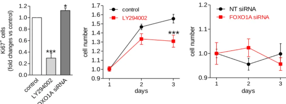
Proliferative activity of sorted hESC-EC cultures were assessed by Ki67 marker and colony formation. Percentage of Ki67+ cells and colony formation activity after 3 days were significantly decreased in response to LY294002 (Figure 9.); in contrast, further downregulation of FOXO1A expression by siRNA treatment resulted in a modest increase in the ratio of Ki67+ cells with no effect on cell number (Figure 9.).
15
Figure 9. PI3K/FOXO1A pathway modulates proliferative activity of hESC-EC
cont rol LY294
002
FOXO 1A siRNA 0.0
0.2 0.4 0.6 0.8 1.0 1.2
***
*
Ki67+ cells (fold changes vs control)
1 2 3
0.9 1.0 1.1
1.2 NT siRNA FOXO1A siRNA
days
cell number
1 2 3
0.9 1.0 1.1 1.2 1.3 1.4 1.5 1.6
1.7 control
LY294002
days
***
cell number
Next, the role of PI3K/FOXO1A pathway on viability and cell survival of hESC-EC was tested. Using H2O2 as a danger signal caused necrosis and nuclear remodelling in hESC-EC in a dose-dependent manner (Figure 10.). Silencing of FOXO1A had no effect on levels of necrosis marker TOTO1 or nuclear remodelling in hESC-EC (Figure 10.). Pre- treatment with LY294002 increased the pro-necrotic effects of H2O2 (at a dose of 600 µM).
Figure 10. Cell death of hESC-derived endothelial cells is modulated by PI3K/FOXO1A signalling pathways
cont rol LY294
002
FOXO1A siRNA H2O
2 (300 )
H2O 2 (600
)
H2O 2 (900
)
H2O 2 (300
)
H2O 2 (600
)
H2O 2 (900
)
H2O 2 (300
)
H2O 2 (600
)
H2O 2 (900 0 )
5 10 15 20 25
***
***
*** ***
***
LY294002 FOXO1A siRNA TOTO-1 intensity (fold changes vs control)
control LY294002 FO
XO1A siRNA H2O2 (300)
H2O2 (600) H2O2 (900)
H2O2 (300) H2O2 (600)
H2O2 (900) H2O2 (300)
H2O2 (600) H2O2 (900) 0.0
0.5 1.0 1.5
***
***
*** ***
LY294002 FOXO1A siRNA
***
nuclear size (fold changes vs control)
16 III. Anticlotting activity in 3D culture
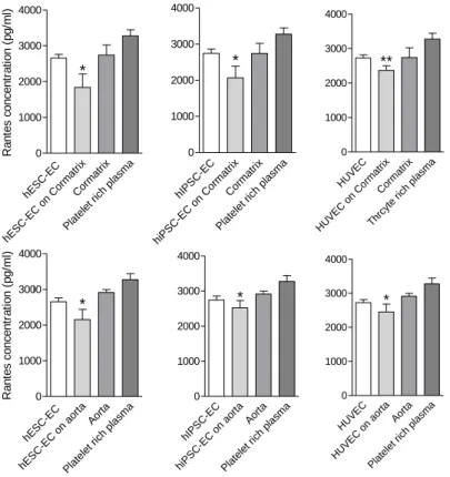
Figure 12. Anticlotting effects of endothelial cells in 3D culture
hESC-EC
hESC-EC on
Corm atrix
Corm atrix
Plat elet rich pla
sma 0
1000 2000 3000 4000
*
Rantes concentration (pg/ml)
hIPSC-EC
hiPSC-EC on Co
rmatrix Cor
matrix
Platelet r ich pla
sma 0
1000 2000 3000 4000
*
HUVEC
HUVEC on Co rma
trix Corma
trix
Thr cyte rich pl
asma 0
1000 2000 3000 4000
**
hESC-EC hESC-EC on aorta
Aorta
Platelet rich p lasma 0
1000 2000 3000 4000
*
Rantes concentration (pg/ml)
hIPSC-EC hiPSC-EC
on aorta Aorta
Platelet rich plasma 0
1000 2000 3000 4000
*
HUVEC HUVEC on aort
a Aort
a
Platelet rich plasm
a 0
1000 2000 3000 4000
*
Endothelial cells remained viable in 3D culture. Results of anticlotting assay showed similar results in control and matrix with plasma samples, suggesting that the matrix itself was not thrombogenic. Endothelial cells decreased secretion of Rantes, suggesting lower platelet activation. Anticlotting effects of endothelial cells were stronger when they were cultured in 3D conditions (Figure 12.).
Figure 11.
Anticlotting assay
1.Cells+plasma
2.Biomatrix+cells+plasma 3.Biomatrix+plasma 4.Plasma (control)
17
IV. In vivo modulation of endothelial maturation
Human ESC-EC, hiPSC-EC and control endothelial HUVEC showed subcutaneous engraftment upon in vivo transplantation. After 21 days conditioning of cells, all general endothelial genes and arterial and venous marker genes were increased. mRNA levels of CD31, NOS-3 as well as arterial (EphrinB2, Notch1 and Notch2) and venous (EphB4) endothelial markers were increased significantly in in all endothelial cells (hESC-EC, hiPSC-EC and HUVEC) upon engraftment (Figure 13.).
Figure 13. In vivo conditioning of hESC-EC, hiPSC-EC and HUVEC
hES C-EC hES
C-EC plu
g hiPS
-EC
hiPS C-EC p
lug 0
10 20 30 40 400
Normalised mRNA levels
EphrinB2
*
hES C-EC hES
C-EC plu
g hiPS
-EC
hiPS C-EC p
lug 0
40 100 200 300
400 Notch1
*
hES C-EC hES
C-EC plu
g hiPS
-EC
hiPS C-EC p
lug 0
10 20 30 40
400 Notch2
*
hES C-EC hES
C-EC plu
g hiPS
-EC
hiPS C-EC p
lug 0
10 20 50000 100000
150000 EphB4
hESC-EC hESC-EC
plug hiPS-EC hiPSC
-EC p lug 1
10 100 1000
10000 CD31
Normalised mRNA levels
hESC -EC
hESC -EC
plug hiPS-EC hiPSC
-EC plug 1
10 100 1000
10000 NOS3
**
Normalised mRNA levels
18 CONCLUSIONS
Endothelial cells were successfully differentiated from human pluripotent stem cells. Generated hESC-EC and hiPSC-EC showed similar characteristics in vitro to mature endothelial cells. Indeed, we found that their phenotype such as immunocytochemical properties and gene expression profile was comparable to those in control HUVEC.
Analysis of the marker genes showed high expression of general endothelial marker genes as well as specific arterial and venous genes.
Functional assays revealed that hESC-EC and hiPSC-EC can share anticlotting function when grown as 3D vascular constructs.
PI3K/FOXO1A is one of the particular key signalling pathways for function and survival of hESC-EC. Results suggest that PI3K/FOXO1A acts as an underlying regulator in endothelial differentiation (Figure 14.). Activated FOXO1A is also a central regulator of hESC-EC function. On the one hand, it mediates endothelial generation; on the other, it at least partly inhibits proliferative and angiogenic activities of the cells, and plays a role in the response to oxidative stress. To test whether modulating FOXO to release inhibitory feedback may be advantageous in cardiovascular cell therapy still needs to be determined.
Figure 14. Endothelial differentiation and specification
19
Publications directly related to the PhD dissertation
1. Merkely B*, Gara E*, Lendvai Z, Skopál J, Leja T, Zhou W, Kosztin A, Várady G, Mioulane M, Bagyura Z, Németh T, Harding SE, Földes G. (2015) Signalling via PI3K/FOXO1A Pathway Modulates Formation and Survival of Human Embryonic Stem Cell- Derived Endothelial Cells.
Stem Cells Dev 24: 869-878.
IF: 3.727 *Equal contribution
2. Reed DM, Földes G, Kirkby NS, Ahmetaj-Shala B, Mataragka S, Mohamed NA, Francis C, Gara E, Harding SE, Mitchell JA. (2014) Morphology and vasoactive hormone profiles from endothelial cells derived from stem cells of different sources.
Biochem. Biophys. Res. Commun. 455: 172-177.
IF: 2.300
3. Kosztin A*, Gara E*, Harding SE, Földes G. (2015) Stem Cell Therapy to Treat Heart Failure
Reference Module in Biomedical Sciences, London, Elsevier IF: N/A *Equal contribution
4. Gara E, Merkely B, Földes G. (2013) Őssejtek és érrendszeri megbetegedések. Hogyan készítsünk ereket?
Élet és Tudomány 68: 470-471.
IF: N/A
20
Publications not related to the PhD dissertation
1. Kovács A, Tapolyai M, Celeng C, Gara E, Faludi M, Berta K, Apor A, Nagy A, Tislér A, Merkely B. (2014) Impact of hemodialysis, left ventricular mass and FGF-23 on myocardial mechanics in end-stage renal disease: a three-dimensional speckle tracking study.
International Journal of Cardiovascular Imaging 30: 1331-1337.
IF: 2.539
2. Debreczeni B, Gara E, Veresh Z, Márki A, Rácz A, Matics R, Hamar J, Koller Á. (2013) Hydrogen peroxide via thromboxane A2 receptors mediates myogenic response of small skeletal muscle veins in rats. Clin Hemorheol Microcirc 54: 393-407.
IF: 2.242
3. Gara E, Gesztes É, Doroszlai R, Zacher G. (2014) Sürgősségi császármetszés szén-monoxid-mérgezésben.
Orvosi Hetilap 155: 871-875.
IF: N/A