DEVELOPMENT OF BIOCONJUGATES AND THEIR MODUL CONSTRUCTS FOR TARGETED THERAPY
OF CANCERS WITH HIGH MORTALITY
Excerption from the results obtained in frame of the grant NVKP_16-1-2016-0036
supported by the National Research, Development and Innovation Office
Budapest, 2020
DEVELOPMENT OF BIOCONJUGATES AND THEIR MODUL CONSTRUCTS FOR TARGETED THERAPY
OF CANCERS WITH HIGH MORTALITY
Excerption from the results obtained in frame of the grant NVKP_16-1-2016-0036
supported by the National Research, Development and Innovation Office
Budapest, 2020
ISBN 978-963-489-286-1
50
Peptide-based delivery vehicles for tumor tissue targeting
Zsuzsa Baranyai1, Katalin Uray1, Beáta Biri-Kovács1,2, Lilla Horváth1,2, Szilvia Bősze1
1MTA-ELTE Research Group of Peptide Chemistry, Hungarian Academy of Sciences, Eötvös L.
University, Budapest, Hungary
2Eötvös Loránd University (ELTE), Faculty of Science, Institute of Chemistry, Budapest, Hungary
Introduction
Conjugating antitumor compounds with peptide-based delivery vehicles, particularly cell-penetrating or cell surface protein (receptor/adhesion protein etc.) specific peptides could enhance their cellular internalization rate and efficacy. Cell membranes and other tissue barriers hamper drug candidates’ distribution and cellular uptake; therefore, most of the active compounds are of limited therapeutic value. Targeted tumor therapy is based on anticancer drugs being delivered to tumor cells by specific carrier molecules, with the result of lowering side effects of the chemotherapeutic agent. Different peptide-based delivery vehicles can be applied. (1) One of these approaches is using cell-penetrating peptides such as SynB3 or others conjugated not only to the drug molecule but to a targeting moiety. (2) Another approach is the use of peptides targeting cell surface proteins, which can be (a) ligands of cell surface receptors expressed exclusively or in highly elevated level on cancer cells, (b) or other cell surface structures, e.g. adhesion molecules such as nectin-1, characteristic for certain cell types (Figure 1).
SynB3 peptide (RRLSYSRRRF) is a cell-penetrating peptide that is reported to be able to cross the blood-brain barrier with high efficiency.1-3 It is a derivative of a natural antimicrobial peptide called protegrin-1. The transport mechanism of SynB3 has been identified as temperature and energy-dependent adsorptive-mediated transcytosis. It is also suggested that SynB3 is sequestered within endocytotic vesicles and might be degraded within lysosomal compartments.4
Tuftsin is a naturally occurring tetrapeptide produced by enzymatic cleavage from immunoglobulin G. Tuftsin derivatives have immunostimulatory effect and antitumor activity through the activation of immunologic effector cells. Moreover, tuftsin can bind to the neuropilin-1 receptor (NRP-1) and can be transported by the CendR pathway, which is an endocytotic/exocytotic transport.5 NRP-1 is upregulated in angiogenic tumor blood vessels and in tumor cells and its ligands often have CendR motif (R/KXXR/K, C-end rule) with the ability of tumor and tissue penetration through the CendR pathway.6,7 A tuftsin analogue with the sequence of TKPPR possesses the CendR motif and have a high affinity to NRP-1.
51
Figure 1. Targeting tumor tissue with antitumor conjugates containing peptide-based carriers bearing different features as (1) cell-penetrating, (2) tumor cell receptor-specific and (3) adhesion protein-
specific peptides.
Herpes simplex viruses (HSV-1 and HSV-2 of Alphaherpesvirinae) show unique entry mechanism into the host cells using their gD envelop glycoprotein as well as gB, gC and gH/gL, as it is usual in case of other herpes viruses. gD glycoprotein selectively binds the nectin-1 adhesion protein on the surface of the host cells, then the glycoprotein undergoes major conformational changes resulting in the triggering of the other viral proteins to effect the fusion.8 According to the known 3D structure of the HSV-1 gD – nectin-1 three regions are in close contact with each other.9,10 We hypothesized that parts of these regions may internalize into nectin-1 bearing cells and can also be used as delivery vehicles.
Results
We have designed and synthesized different SynB3 and tuftsin derivatives and a combined peptide composed of SynB3 and tuftsin. The SynB3 peptide was decanoylated in order to modify the lipophilicity, penetration ability and membrane interaction of the original peptide. The intracellular localization of selected, fluorescently labeled peptides was investigated on HUVECs (human umbilical vein endothelial cells), these cells were used to model tumor-related vascular endothelial cells that have NRP-1 on their surface (Figure 2).
The intracellular localization was also studied on U87 human glioblastoma cell line as a model cell for gliomas (Figure 2). The localization patterns of the fluorescently labeled
52
peptides were similar in case of the two different cell types. The SynB3 peptide (Cf-S) can be found in the cytosol, in the nucleus and in a small amount of lysosomal localization was also detected. In contrast, the tuftsin analogue Cf-T showed high lysosomal localization but no co- localization with the nucleus. The combined peptide (Cf-ST) had the intracellular distribution characteristics of the parent Cf-S and Cf-T peptides. It could be detected in both the lysosomes and in the nucleus. The decanoyl side chain containing peptide (Cf-SD) was cytotoxic on the cells, probably it has a membrane damaging effect, it can be seen in the cytosol and in the nucleus with a highly homogeneous distribution. These findings suggest that the uptake of the cell-penetrating Cf-S peptide is a complex process; probably direct penetration and vesicular transport are also present. As expected, the tuftsin analogue mainly can enter the cells by receptor-mediated endocytosis. The combined peptide follows both internalization mechanisms of its parent peptides. Modification of a cationic cell-penetrating peptide with a hydrophobic fatty acid chain leads to higher cellular uptake and at the same time it has cytotoxic effect probably due to its highly amphiphilic characteristic.
Figure 2. Localization of Cf-peptides in HUVEC and U87 cells by confocal microscopy. Nuclei were stained with Hoechst 33342 (blue), lysosomes were stained with LysoTracker Deep Red (red), cells were incubated with Cf-peptides (green, 25 µM, 30 min except for Cf-SD where 12.5 µM was used).
Scale bar represents 10 µm.
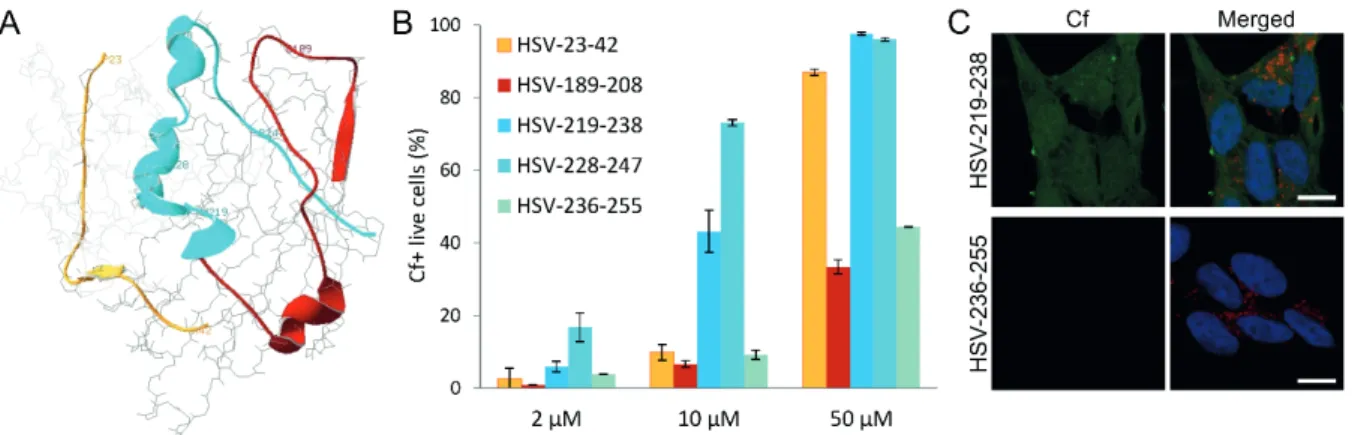
Based on the known 3D structure of the HSV1 gD – nectin-1 complex (PDB ID:
3U82)9,10, we have selected three regions of gD making contact with nectin-1 (Figure 3A), and carboxyfluorescein labeled overlapping peptides were designed and synthesized representing these regions. The cellular internalization of these peptides into SH-SY5Y
53
neuroblastoma cells (as a model of neuroblastomas) has been determined by flow cytometry and is depicted in Figure 3B for representative peptides. Region II peptides showed negligible internalization. Some Region I peptides showed cellular entry at higher concentrations, but Cf-HSV 228-247 (228QRTVAVYSLKIAGWHGKPAP247) peptide showed the most efficient cellular entry, with Cf-HSV-219-238 (219NleLPRFIPENQRTVAVYSLKI238 where Nle represents norleucine substituting the native methionine) also showing significant entry in lower (10 μM) concentration. Certain C- and N-terminal truncation of 228-247 and the substitution of Lys237Arg and Trp241Phe were well tolerated.11 The propensity of the peptides to adopt helical conformation in the lipomimetic solvent trifluoroethanol showed a strong correlation with their ability to internalize into the neuroblastoma cells.11
Parallel with flow cytometry measurements, to assess qualitative information regarding the subcellular localization, internalized peptides were imaged by confocal laser scanning microscopy. Peptides with high ability to internalize (as positive and negative examples, see Cf-HSV-219-238 and Cf-HSV-236-255, respectively, in Figure 3C) could be imaged in the cytosol and the nucleus, but there is no direct co-localization with lysosomal staining. This suggests that there is no vesicular transport involved in the uptake of the Cf- peptide.
Figure 3. (A) Structure of the HSV1 gD (dark grey) – nectin-1 (light grey) complex (PDB ID: 3U82), the selected regions of the HSV1 gD (region I: yellow, region II: red, region III: cyan), (B) internalization of Cf-HSV1 gD peptides into SH-SY5Y cells at c = 2, 10 and 50 μM, percentage of Cf-
positive live cells, (C) internalization of Cf-HSV1 gD peptides visualized by confocal microscopy (green), nuclei and lysosomes are stained with Hoechst 33342 (blue) and LysoTracker (red),
respectively, scale bar represents 10 µm.
Conclusion
In the framework of grant NVKP_16-1-2016-0036 we have identified potential new carrier peptides of different origin as delivery vehicles. They can improve the cellular uptake
54
of different cargoes (i.e., drugs or drug candidates). These peptide carriers possess favorable internalization properties and they have different intracellular routes. The localization of the cell-penetrating type peptide was mainly cytosolic, while that of the receptor-specific tuftsin carrier was rather lysosomal. The viral carriers after internalization displayed ubiquitous distribution in the cytosol and in the nucleus as well.
Acknowledgements
These studies were also supported by ELTE Thematic Excellence Programme 2020 (National Research, Development and Innovation Office: TKP2020-IKA-05, NKFIH-1157- 8/2019-DT) and by grant VEKOP-2.3.3-15-2017-00020 from the European Union and the State of Hungary, co-financed by the European Regional Development Fund.
References
1. Rousselle C, Clair P, Lefauconnier JM, Kaczorek M, Scherrmann JM, Temsamani J. Mol Pharmacol 57: 679-686 (2000)
2. Stalmans S, Bracke N, Wynendaele E, Gevaert B, Peremans K, Burvenich C, Polis I, De Spiegeleer B. PLoS One 10: e0139652 (2015)
3. Oller-Salvia B, Sanchez-Navarro M, Giralt E, Teixido M. Chem Soc Rev 45: 4690-4707 (2016) 4. Drin G, Rousselle C, Scherrmann JM, Rees AR, Temsamani J. AAPS PharmSci 4: E26 (2002) 5. Nissen JC, Selwood DL, Tsirka SE. J Neurochem 127: 394-402 (2013)
6. Leng Q, Woodle, MC, Mixson AJ. Drugs Future 42: 95-104 (2017) 7. Ruoslahti E. Adv Drug Deliv Rev 110-111: 3-12 (2017)
8. Lazear E, Whitbeck JC, Zuo Y, Carfí A, Cohen GH, Eisenberg RJ, Krummenacher C. Virology 448: 185-195 (2014)
9. Zhang N, Yan J, Lu G, Guo Z, Fan Z, Wang J, Shi Y, Qi J, Gao GF. Nat Commun 2: 577 (2011) 10. Di Giovine P, Settembre EC, Bhargava AK, Luftig MA, Lou H, Cohen GH, Eisenberg RJ,
Krummenacher C, Carfi A. PLoS Pathog 7: e1002277 (2011)
11. Bősze Sz, Zsila F, Biri-Kovács B, Szeder B, Majer Zs, Hudecz F, Uray K. Biomolecules 10: 721 (2020)