DEVELOPMENT OF BIOCONJUGATES AND THEIR MODUL CONSTRUCTS FOR TARGETED THERAPY
OF CANCERS WITH HIGH MORTALITY
Excerption from the results obtained in frame of the grant NVKP_16-1-2016-0036
supported by the National Research, Development and Innovation Office
Budapest, 2020
DEVELOPMENT OF BIOCONJUGATES AND THEIR MODUL CONSTRUCTS FOR TARGETED THERAPY
OF CANCERS WITH HIGH MORTALITY
Excerption from the results obtained in frame of the grant NVKP_16-1-2016-0036
supported by the National Research, Development and Innovation Office
Budapest, 2020
ISBN 978-963-489-286-1
34
Design, synthesis and characterization of Pancreatic Ductal Adenocarcinoma (PDAC) targeting antitumor daunomycin-peptide
conjugates
Levente E. Dókus1,2, Eszter Lajkó3, Orsolya Láng3, Diána Mező3, Zsófia Szász3, Angéla Takács3, László Kőhidai3, Gitta Schlosser2, Ivan Ranđelović4, József Tóvári4, Gábor
Mező1,2
1MTA-ELTE Research Group of Peptide Chemistry, Hungarian Academy of Sciences, Eötvös L.
University, Budapest, Hungary
2Institute of Chemistry, Eötvös L. University, Budapest, Hungary
3Department of Genetics, Cell- and Immunobiology, Semmelweis University, Budapest, Hungary
4Department of Experimental Pharmacology, National Institute of Oncology, Budapest, Hungary
Introduction
Pancreatic Ductal Adenocarcinoma (PDAC) is one of the most aggressive and dangerous cancerous diseases with a high mortality rate.1 The average 5-year survival rate is less than 5%.2 The early diagnosis of PDAC is still difficult, and most patients have already progressed to not operable and incurable statuses at the recognition of the disease.3 In addition, the chemotherapy applied to treat pancreatic cancers is usually ineffective due to the fast development of resistance. Furthermore, chemotherapy causes many side effects because of the low selectivity of the currently used drugs.4 Therefore, the design of efficient anticancer agents against PDAC is one of the most challenging tasks for scientists working on cancer research.5 Targeted tumor therapy could be a promising strategy to overcome these drawbacks in pancreatic cancer treatment – similarly to other types of cancers.6 Targeted tumor therapy is based on targeting tumor-specific or overexpressed receptors or other cell surface compartments on tumor cells that can be recognized selectively by antibodies or small molecules such as folic acid or peptides.7,8 Several homing peptides have been described in the literature that recognize pancreatic cancer cells and could be used for drug targeting directly or as part of nanoparticles.9-12
Here we report peptide–drug conjugates in which daunomycin (Dau) as an anticancer agent is linked via oxime bond to different types of homing peptides. For this study, two homing peptides were selected. The first one is based on a neurotensin fragment (6PRRPYIL13) which binds to neurotensin receptors that are overexpressed in numerous tumor types.13 The other one is KTLLPTP heptapeptide, which is able to recognize plectin, a protein
35
that is overexpressed in PDAC-cells.14 Six conjugates were developed with GFLG spacer between the homing peptide and the aminooxy moiety which can be cleaved by the lysosomal enzyme Cathepsin B,15 therefore, they can be suitable to enhance the intracellular degradation and the release of the active metabolite (Dau=Aoa-Gly-OH).
Results
Fourteen daunomycin−peptide conjugates were synthesized by solid-phase peptide synthesis (Table 1). The homing peptides were prepared either on Wang resin that provides free carboxyl group at the C-terminus (neurotensin derivatives) or on Rink-Amide MBHA resin for peptides with carboxamide C-terminus (plectin recognizing derivatives) using Fmoc/tBu strategy. Prior to the cleavage of the peptides from resins, isopropylidene protected aminooxyacetic acid (>=Aoa-OH) was attached to the amino function(s) of peptides. The isopropylidene protecting group was cleaved from the purified peptide derivatives. Purified peptide derivatives were linked via oxime bond to Dau. The synthesis route of a selected conjugate is presented in Scheme 1.
Scheme 1. Development of conjugate 1 as a representative synthesis route
The antitumor effect of conjugates was investigated in vitro on PANC-1, a human PDAC cell line by xCELLigence-system which is an impedimetric technique. Conjugate 1, based on the 8-13 part (8RRPYIL13) of the neurotensin hormone peptide, showed only a moderate antitumor effect. When an additional Lys was incorporated into the N-terminus of the sequence, the antitumor effect of the conjugate increased significantly (conjugate 3).
However, the sequence elongation either with the native sequence elements or with GFLG
O O
C O
H3 OH
OH OH
CH3
N O O
NH2
CH3 OH N H2 O O
CH3
N O O
C
H3 methoxyamine (1.5 M)
0.2 M NH4OAc buffer (pH= 5) RT; 2h
daunomycin (2 equiv.) 0.2 M NH4OAc buffer (pH= 5) RT; 24h
Arg-Arg-Pro-Tyr-Ile-Leu-OH Arg-Arg-Pro-Tyr-Ile-Leu-OH Arg-Arg-Pro-Tyr-Ile-Leu-OH
36
spacer did not provide a positive effect on the antitumor activity (conjugate 2, 5 and 6). In addition, the elimination of the Proin position 10 caused the full loss of the antitumor effect (4).
In the case of plectin targeting (KTLLPTP) peptide, which was identified by phage- display, a similar effect was detected. Conjugate 7 proved to be one of the most efficient antitumor agents but the GFLG containing analogue (8) did not show any activity.
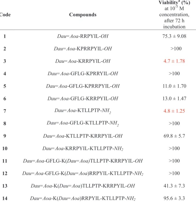
Table 1. Antitumor effect of the conjugates on PANC-1 cell –line
Code Compounds
Viabilitya (%) at 10-5 M concentration,
after 72 h incubation
1 Dau=Aoa-RRPYIL-OH 75.3 ± 9.08
2 Dau=Aoa-KPRRPYIL-OH >100
3 Dau=Aoa-KRRPYIL-OH 4.7 ± 1.78
4 Dau=Aoa-GFLG-KPRRYIL-OH >100 5 Dau=Aoa-GFLG-KPRRPYIL-OH 11.0 ± 1.70 6 Dau=Aoa-GFLG-KRRPYIL-OH 13.0 ± 1.47
7 Dau=Aoa-KTLLPTP-NH2 4.8 ± 1.25
8 Dau=Aoa-GFLG-KTLLPTP-NH2 >100
9 Dau=Aoa-KTLLPTP-KRRPYIL-OH 69.8 ± 5.7
10 Dau=Aoa-KRRPYIL-KTLLPTP-NH2 >100 11 Dau=Aoa-GFLG-K(Dau=Aoa)TLLPTP-KRRPYIL-OH >100 12 Dau=Aoa-GFLG-K(Dau=Aoa)RRPYIL-KTLLPTP-NH2 >100 13 Dau=Aoa-K(Dau=Aoa)TLLPTP-KRRPYIL-OH 41.3 ± 7.3 14 Dau=Aoa-K(Dau=Aoa)RRPYIL-KTLLPTP-NH2 95.6 ± 3.3
a Cell index (CI) values of the treated cells are normalized to the CI values of the control wells and expressed as percentages. Data are given as mean values ± standard deviation (SD), (n=3).
37
Further derivatives were developed by the combination of the most effective homing sequences: KRRPYIL and KTLLPTP. These constructions were synthesized using the successful linear (9, 10) or branched structure (11-14) which may lead to the design of efficient antitumor agents.16 Unfortunately, most of these conjugates lost the antitumor effect completely except conjugates 9 and 13 that showed moderate activity.
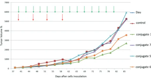
The four most efficient conjugates (3, 5, 6, 7) identified in the in vitro studies were applied in an in vivo experiment using s.c. developed PANC-1 tumor-bearing SCID mice.
Results were compared with free drug administration. It can be observed that the antitumor effect of conjugates is different considering the tumor volume and weight.
Green arrows indicate the days of administration with conjugates Red arrows indicate the days of administration with daunomycin
Figure 1. In vivo tumor growth inhibition on PANC-1 tumor-bearing mice measured by tumor volume
After the termination of animals (on day 85), it was indicated that the tumor volume decreased by 49% for conjugate 3, 1.2% for conjugate 7 and 32.6% in case of conjugate 6, respectively (Figure 1), but conjugate 5 did not present any effect. In the case of tumor weight measurement, 5 also did not inhibit tumor growth, while conjugates 6 and 3 showed very modest, non-significant tumor growth inhibition (3.3% and 4.6%, respectively). In this case, conjugate 7 induced 15.9% decrease of tumor weight compared to the control animals (Figure 2). Conjugate 3 showed the highest antitumor activity followed by conjugates 6 and 7 (considering the tumor volume). Conjugate 5 showed the lowest antitumor activity. It is worth mentioning that on day 59 when the Dau treated group had to be terminated because of toxic side effects, all conjugates showed a bit lower activity compared with the free Dau. In summary, the results indicated that the two most efficient agents were conjugate 3 that
38
inhibited better the growth of tumor volume and conjugate 7 that has a higher inhibition effect on the growth of tumor weight.
Figure 2. In vivo tumor growth inhibition on PANC-1 tumor bearing mice measured by tumor weight
References
1. Vincent A, Herman J, Schulick R, Hruban, RH, Goggins M. Lancet 378: 607-620 (2011) 2. Hidalgo M. N Engl J Med. 362: 1605-1617 (2010)
3. Zhang L, Sanagapalli S, Stoita A, World J. Gastroenterol 24: 2047-2060 (2018)
4. Diab M, Azmi A, Mohammad R, Philip PA. Expert Opin Pharmacother 20: 535-546 (2019) 5. Xie D, Xie K. Genes Dis. 2: 133-143 (2015)
6. Szepeshazi K, Schally AV, Block NL, Halmos G, Nadji M, Szalontay L, Vidaurre I, Abi-Chaker A, Rick FG. Oncotarget 4: 751-760 (2013)
7. Vrettos EI, Mező G, Tzakos AG. Beilstein J Org Chem 14: 930-954 (2018)
8. Kutova OM, Guriyev EL, Sokolova EA, Alzeibak R, Balalaeva IV. Cancers 11: 68 (2019) 9. Matters GL, Harms J. Biomedicines 6: E65. (2018)
10. Sanna V, Nurra S, Pala N, Marceddu S, Pathania D, Neamati N, Sechi M. J Med Chem 59: 5209- 5220 (2016)
11. Liu X, Jiang J, Ji Y, Lu J, Chan R, Meng H. Mol Syst Des Eng 2: 370-379 (2017)
12. Nishimoto T, Yamamoto Y, Yoshida K, Goto N, Ohnami S, Aoki K. PLoS One 7: e45550 (2012) 13. Leeman SE, Carraway RE. Ann N Y Acad Sci 400: 1-16. (1982)
14. Kelly KA, Bardeesy N, Anbazhagan R, Gurumurthy S, Berger J, Alencar H, Depinho RA, Mahmood U, Weissleder R. PloSMed 5: e85 (2008)
15. Peterson JJ, Meares CF. Bioconjugate Chem 9: 618-626 (1998)
16. Dókus LE, Lajkó E, Ranđelović I, Mező D, Schlosser G, Kőhidai L, Tóvári J, Mező G.
Pharmaceutics 12: 576 (2020)