DEVELOPMENT OF BIOCONJUGATES AND THEIR MODUL CONSTRUCTS FOR TARGETED THERAPY
OF CANCERS WITH HIGH MORTALITY
Excerption from the results obtained in frame of the grant NVKP_16-1-2016-0036
supported by the National Research, Development and Innovation Office
Budapest, 2020
DEVELOPMENT OF BIOCONJUGATES AND THEIR MODUL CONSTRUCTS FOR TARGETED THERAPY
OF CANCERS WITH HIGH MORTALITY
Excerption from the results obtained in frame of the grant NVKP_16-1-2016-0036
supported by the National Research, Development and Innovation Office
Budapest, 2020
ISBN 978-963-489-286-1
23
Structural and binding characteristics of HER2 receptor targeting peptides
Beáta Biri-Kovács1,2, Afrodité Adorján1,2, Ildikó Szabó2, Bálint Szeder3, Szilvia Bősze2, Gábor Mező1,2
1Institute of Chemistry, Eötvös L. University, Budapest, Hungary
2MTA-ELTE Research Group of Peptide Chemistry, Hungarian Academy of Sciences, Eötvös L.
University,Budapest, Hungary
3Institute of Enzymology, Research Centre for Natural Sciences, Hungarian Academy of Sciences, Budapest, Hungary
Introduction
HER2 (ErbB2) is overexpressed in 15-30% of breast cancers and is also amplified in several other cancers (gastric, ovarian, prostate), its increased expression prognoses high mortality rate and the development of metastases. HER2 is a transmembrane glycoprotein that belongs to the epidermal growth factor receptor tyrosine kinase protein family and plays role in mitogenic signaling. HER2 is the only receptor in the protein family that does not have a known ligand, its activation occurs through the formation of homo- or heterodimers (formed with other members of the protein kinase family). Its increased level results in uncontrollable tumor growth due to the excessive formation of HER2-containing dimers leading to enhanced cell proliferation and survival.1,2
As HER2 is an important target in breast cancer research, several therapies have been developed during the last decades that include the use of monoclonal antibodies, small molecule tyrosine kinase inhibitors and antibody-drug conjugates. Despite successful anti- HER2 agents that achieved improvement in tumor therapy, some patients suffer from serious side effects or have developed resistance, thus, new approaches in HER2-targeting are needed.3 Moreover, for selection of the appropriate therapeutical agent, new, fast, reliable and quantitative diagnostical tools are also required.
Tumor targeting peptides have become an important field in targeted tumor therapy due to their low molecular weight, easy synthesis, high receptor recognition rate and better tissue penetration characteristics. With the use of peptide bacteriophage display technology, Karasseva et al. identified a hexapeptide (KCCYSL) that bound with high affinity and specificity to HER2-expressing cells.4 Hence, we chose this peptide as a starting point of our study to design new, modified peptides with increased HER2 binding affinity.
Geng et al. used the method of molecular dynamics simulation to identify peptides involved in the dimerization at the extracellular domain of HER2. These peptides were used
24
to build a „One bead one compound” (OBOC) library and were screened for specificity and affinity resulting in 17-mer peptides binding with high affinity to HER2 receptor.5 Parts of the peptides with the strongest binding ability show similarities with the KCCYSL peptide. Thus, we decided to synthesize combined peptides where the modified KCCYSL sequence precedes GYYNPN (taken from the OBOC library) and compare their binding to HER2 expressing cells.6
Results
A set of hexa- and 12-mer peptide analogues were synthesized by solid-phase peptide synthesis on Rink Amide MBHA resin using Fmoc/tBu protocol. In the last step, 5(6)- carboxyfluorescein (CF) as fluorescent dye was attached to the N-terminus of the peptidyl resins. The compounds were analyzed by analytical RP-HPLC, their molecular weight was identified by ESI-MS (Table 1).
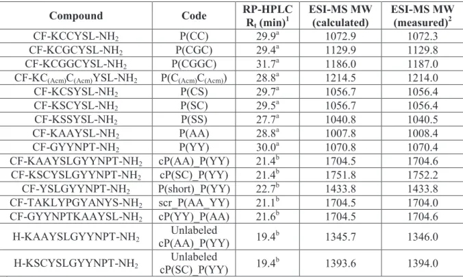
Table 1. List of HER2 binding compounds, their codes and chemical characteristics.
Compound Code RP-HPLC
Rt (min)1 ESI-MS MW
(calculated) ESI-MS MW (measured)2
CF-KCCYSL-NH2 P(CC) 29.9a 1072.9 1072.3
CF-KCGCYSL-NH2 P(CGC) 29.4a 1129.9 1129.8
CF-KCGGCYSL-NH2 P(CGGC) 31.7a 1186.0 1187.0
CF-KC(Acm)C(Acm)YSL-NH2 P(C(Acm)C(Acm)) 28.8a 1214.5 1214.0
CF-KCSYSL-NH2 P(CS) 29.7a 1056.7 1056.4
CF-KSCYSL-NH2 P(SC) 29.5a 1056.7 1056.4
CF-KSSYSL-NH2 P(SS) 27.7a 1040.8 1040.5
CF-KAAYSL-NH2 P(AA) 28.8a 1007.8 1008.4
CF-GYYNPT-NH2 P(YY) 30.0a 1070.8 1070.4
CF-KAAYSLGYYNPT-NH2 cP(AA)_P(YY) 21.4b 1704.5 1704.6 CF-KSCYSLGYYNPT-NH2 cP(SC)_P(YY) 21.4b 1751.8 1752.2 CF-YSLGYYNPT-NH2 P(short)_P(YY) 22.7b 1433.8 1433.8 CF-TAKLYPGYANYS-NH2 scr_P(AA_YY) 21.1b 1704.5 1704.0 CF-GYYNPTKAAYSL-NH2 cP(YY)_P(AA) 21.6b 1704.5 1704.6
H-KAAYSLGYYNPT-NH2 Unlabeled
cP(AA)_P(YY) 19.4b 1345.7 1346.0
H-KSCYSLGYYNPT-NH2 Unlabeled
cP(SC)_P(YY) 19.4b 1393.6 1394.0
1 RP-HPLC: a: column: Phenomenex Aeris Peptide XB-C18 column (250 x 4.6 mm) with 3.6 μm silica; eluents:
0.1% TFA in water (A) and 0.1% TFA in acetonitrile-water (80:20, v/v) (B); gradient: 0 min 0% B, 5 min 0% B, 50 min 90% B; flow rate: 1 mL/min; detection: λ= 220 nm, b: column: Macherey–Nagel Nucleosil C18 column (250 x 4.6 mm) with 5 µm silica (100 Å pore size); eluents: 0.1% TFA in water (A) and 0.1% TFA/ acetonitrile
−water (80:20 v/v) (B); gradient 0 min 2% B, 5 min 2% B, 30 min 90% B; flow rate 1 mL/min; detection: 220 nm 2 ESI-MS: Esquire 3000+ ion trap mass spectrometer (Bruker Daltonics)
25
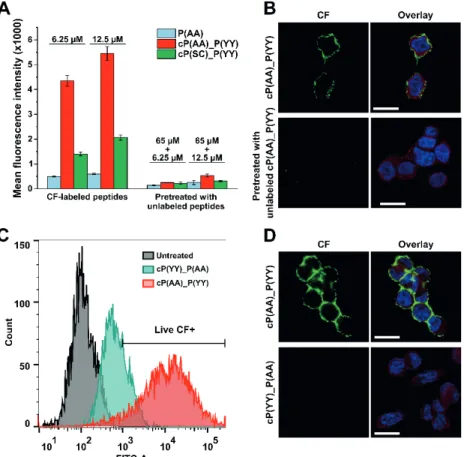
Cellular uptake was measured by flow cytometry using HER2-overexpressing breast cancer cells. HER2 expression was detected by Western blot (Figure 1A), as a result, MDA- MB-453 breast adenocarcinoma cell line was chosen for further studies. First, variations of the KCCYSL hexapeptide (role of cysteine residues, distance between them) were studied based on the measured fluorescence intensity after incubation of the cells with CF-labeled peptides for 3 h. Consequently, the two most promising analogues (P(AA) and P(SC)) were chosen for further experiments and the design of combined peptides. The next cellular uptake study demonstrated that combined peptide cP(AA)_P(YY) shows ten times higher fluorescence intensity values compared to the P(AA) hexapeptide (Figure 1B).
Cellular localization was detected by confocal microscopy, as expected, peptides could be detected at the membrane of MDA-MB-453 cells suggesting their binding to the extracellular domain of HER2 (Figure 1C).
Figure 1. (A) Western blot analysis of breast cancer cells for HER2 expression. (B) Cellular uptake profile of CF-labeled peptides measured by flow cytometry on MDA-MB-453 cells. (C) Confocal microscopy imaging of MDA-MB-453 cells incubated with cP(AA)_P(YY) peptide (green), nuclei were stained with DAPI (blue), HER2 was visualized by immunofluorescence staining (red), scale bar
represents 20 µm.
To monitor specificity, cells were pre-incubated with unlabeled peptides before addition of CF-labeled derivatives. Cellular uptake and microscopic measurements demonstrated that fluorescent signal was decreased supporting that peptides bind to a specific
26
receptor on the surface of cells (Figure 2A and 2B). The most promising combined peptide (cP(AA)_(P(YY)) was also studied for specificity by designing and analysing a reversed version of it (cP(YY)_(P(AA)). Flow cytometric and confocal microscopic analysis confirms the importance of the order of the two hexapeptides as the reversed peptide bound to cells at a much lower extent (Figure 2C and 2D).
Figure 2. (A, B) Cellular uptake and confocal microscopic imaging of CF-labeled peptides using MDA-MB-453 cells pre-incubated with corresponding unlabeled peptide. (C, D) Study of cellular
uptake of the reversed combined peptide by flow cytometry and confocal microscopy (green: CF, blue: nuclei, red: HER2, scale bars: 20 µm).
Finally, the secondary structure of the peptides was predicted using the PEP-FOLD algorithm.7 The predicted helix formation is quite low for the negative control scrambled peptide, however, it increases in case of peptides binding with a higher rate to HER2- expressing cells and the highest is in case of the most promising combined peptide (Figure 3).
27
Figure 3. The estimated α-helical content of HER2-binding peptides is proportional to the fluorescence intensity detected on the surface of MDA-MB-453 cells incubated with CF-labeled
peptides.
In conclusion, we developed targeting peptides against the extracellular domain of HER2 receptor. We generated new analogues by modifying and combining two sets of known HER2-binding peptides that resulted in the revealing of a 12-mer peptide that binds to HER2 overexpressing cells with high affinity and specificity. Microscopy experiments validated extracellular localization; specificity was verified by preincubation of cells with unlabeled peptides. Secondary structure prediction revealed the importance of the α-helical structure of the conjugates. This combined homing peptide with high affinity and specificity for HER2 extracellular domain can be applied in further studies for tumor diagnostics and drug targeting.6
References
1. Lv Q, Meng Z, Yu Y, Jiang F, Guan D, Liang C, Zhou J, Lu A, Zhang G. Int J Mol Sci 17: 2095 (2016)
2. Hsu JL, Hung MC. Cancer Metastasis Rev 35: 575-588 (2016)
3. Escriva-de-Romani S, Arumi M, Bellet M, Saura C. Breast. 39: 80-88 (2018)
4. Karasseva NG, Glinsky VV, Chen NX., Komatireddy R, Quinn TP. J Prot Chem 21: 287-96.
(2002)
5. Geng L, Wang Z, Jia X, Han Q, Xiang Z, Li D, Yang X, Zhang D, Bu X, Wang W, Hu Z, Fang Q. Theranostics. 6: 1261-1273 (2016)
6. Biri-Kovács, B, Adorján A, Szabó I, Szeder B, Bősze S,. Mező G. Biomolecules 10: 183 (2020) 7. Shen Y, Maupetit J, Derreumaux P, Tuffery P. J Chem Theory Comput 10: 4745-4758 (2014)