Role of preoperative imaging in the diagnosis and planning of surgical radicality of endometrial and cervical cancer
Ph.D. Thesis DOROTTYA BÚS M.D.
Supervisor:
György Vajda M.D., Ph.D., Department of Obstetrics and Gynecology Zala County Saint Rafael Hospital, Zalaegerszeg, Hungary
Director of Doctoral School of Clinical Medicine:
Prof. Lajos Kemény, M.D., Ph.D., D.Sc., member of the Hungarian Academy of Science
Head of Reproductive Health Subprogram:
Prof. emeritus György Bártfai M.D., D.Sc.
Szeged, 2020
LIST OF PUBLICATIONS related to the subject of the dissertation
I. Bús D, Nagy G, Póka R and Vajda G. Clinical Impact of Preoperative Magnetic Resonance Imaging in the Evaluation of Myometrial Infiltration and Lymph-Node Metastases in Stage I Endometrial Cancer. Pathology and Oncology Research 2021, 27:1-8. IF: 2,826.
II. Bús D, Nagy G, Vajda G. A preoperatív mágnesesrezonanciavizsgálat klinikai jelentősége az I.
stádiumú endometriumcarcinoma myometrialis infiltrációjának és nyirokcsomó-státuszának megítélésében. Magyar Nőorvosok Lapja. 2021, 84:37-41.
LIST OF PUBLICATIONS not related to the subject of the dissertation
III. Bús D, Buzogány M, Nagy G, Vajda G. Rare virilizing granulosa cell tumor in an adolescent.
Molecular and Clinical Oncology. 2017; 6:88-90.
IV. Husz V, Bús D, Vajda Gy. Extremely large epithelial ovarian cancer associated with pregnancy: A case report. Molecular and Clinical Oncology. 2018, 8:103-106 V. Bús D, Buzogány M, Nagy Gy, Vajda Gy. Menarchét követő, amenorrheát okozó ritka virilizáló granulosasejtes tumor (diagnosztikus és terápiás konzekvenciák). Interdiszciplináris Magyar Egészségügy. 2017, 7: 41-43. 3.
VI. Husz V, Bús D, Vajda Gy. Subpubicus képlet diagnózisának és terápiájának határterületi kérdései. Interdiszciplináris Magyar Egészségügy. 2017, 7: 47-50. 4.
VII. Goldfinger J, Bús D, Husz V, Nagy Gy, Tóth Z, Vernarelli F, Vajda Gy. Terhességet komplikáló kiterjedt petefészektumor. Interdiszciplináris Magyar Egészségügy. 2017, 7: 44-46. 5.
Table of Contents
1. Abbreviations 4
2. Summary of the thesis 5
3. Introduction 6
3.1 Endometrial cancer 6
3.2 Cervical cancer 8
3.3 Therapy and treatment 9
3.3.1 Endometrial cancer 9
3.3.2 Cervical cancer 10
4. Material and methods 11
4.1 Design 11
4.2 Diagnostic algorithm 12
4.3 Data collection 12
4.3.1 Surgical diagnostic methods 13
4.3.2 Radiology findings 13
4.3.3 Surgical treatment 16
4.3.4 Histopathological evaluation 16
4.3.5 Postoperative treatment and follow-up 16
4.4 Statistical analysis 17
5. Results 18
5.1 Endometrial cancer 18
5.2 Cervical cancer 24
6. Discussion 30
6.1 Endometrial cancer 30
6.1.1 Accuracy of pre-and postoperative staging in endometrial cancer 31
6.2 Cervical cancer 34
6.2.1 Accuracy of pre-and postoperative staging in cervical cancer 34
7. Conclusion 37
8. Acknowledgements 38
9. References 39
1. Abbrevations
18F-FDG 2-deoxy-2-[fluorine-18]fluoro- D-glucose
BMI Body mass index
CEA Carcinoembryonal antigene CIN Cervical Intraepithelial Neoplasia
CT Computer tomography
DNA Deoxyribonucleic acid DWI Diffusion weighted imaging
EIC Endometrial intraepithelial carcinoma ESMO European Society for Medical Oncology ESUR European Society of Urogenital Radiology
FIGO International Federation of Gynecology and Obstetrics HPV Human papillomavirus
IARC International Agency for Research on Cancer ICC Intraclass correlation coefficient
LEEP Loop Electrosurgical Excision Procedure MRI Magnetic resonance imaging
NCCN National Comprehensive Cancer Network NPV Negative predictive value
PET-CT Positron emission tomography PPV Positive predictive value
SEER Surveillance, Epidemiology and End Results Program TNM Tumor, nodes and metastasis System
TRUFISP True fast imaging with steady state precession US
USA
Ultrasound
United States of America
2. Summary of the thesis
Gynecological cancers are the most common tumors among women in the developed world. Due to a more excessive screening, diagnosis is available at an early stage. Furthermore, advanced diagnostic imaging methods allow appropriate preoperative staging, therefore the radicality of surgery and adjuvant therapy can be more tailored to the patient and the tumor-spread.
In this study we analyzed the reliability of preoperative MRI findings in the staging of endometrial and cervical cancer, as well as the clinical characteristics of patients underwent radical hysterectomy and the histopathologic evaluation of their tumor.
The results of the preoperative radiology staging were compared with the final histological analysis of the surgical specimen. The sensitivity, specificity, positive- and negative predictive values of the preoperative assessment were calculated for each endpoint. The percentage of the underdiagnosed or overdiagnosed cases and accuracy rate in terms of stage, local invasion and lymph-node metastases were also evaluated.
In order to analyze the role of the rater’s expertise assessing the MRI findings, we divided the specialists into two groups: a radiologist specialized in imaging of gynecological tumors and a subgroup of non-specialized evaluators. Inter-rater agreement was calculated to determine the conformity of pre- and postoperative staging.
Based on our results, we report similar findings as found in international literature. MRI is the method of choice in the terms of evaluating overall staging, as well as myometrial and stromal invasion, as its specificity and negative predictive value is rather high. Our studies showed that the diagnosis of lymph node metastases is difficult with MRI modality, since hyperplastic and metastatic nodes cannot be easily differentiated leading to high percentage of false positive results, therefore other imaging modalities can be used for more accurate evaluation.
We have also found that more experienced raters in gynecological imaging provide more consistent evaluations of staging, local invasion and finding lymph node metastases, however, the difference between inter-rater agreements was not significant between the two groups.
3. Introduction
Gynecological cancers are the most common malignancies among women in industrialized countries.1 According to the National Cancer Institute SEER2 and IARC databases, the incidence and mortality of endometrial cancer is slightly increasing in the developing countries. It is estimated to be 3.5% of all cancer cases and 2.0% of all cancer-related deaths in 2019 in the USA.
Due to a more excessive screening of cervical cancer, diagnosis is available at an early stage leading to decreased mortality, which is estimated to be 0.7% both in all cancer cases and all cancer-related deaths in 2019 in the USA.
Advanced diagnostic methods and imaging modalities, such as the worldwide available MRI and CT technology, allow staging and treatment to be based on histopathological and magnetic resonance findings. Therefore, the specificity and sensitivity of these diagnostic methods is of great importance in making an appropriate diagnostic choice and in determining the radicality of the treatment.3
3.1 Endometrial cancer
Endometrial cancer is the 9th most common cancer among women in developing countries.
According to the National Cancer Institute SEER2 and IARC databases the number of new cases of uterine cancer was 27.5 per 100,000 women per year, and the number of related deaths was 4.7 per 100,000 women per year in the USA – based on 2012-2016 statistics, with the prevalence of an estimated 61,880 new cases in 2019. The incidence is also increasing in Hungary, with an increase of 8% between 2010 and 2014, according to the Hungarian National Cancer Database.
Despite the increase of incidence and mortality, the 5-year relative survival rate in the USA is 81.2% overall, with a 95% survival rate in localized tumors – based on data from SEER 18, 2009- 2015.
Most cases of endometrial cancer are diagnosed in women aged between 45 and 74. Risk factors include extended hyperestrogenism (due to anovulation, nulliparity, polycystic ovarian syndrome or tamoxifen therapy), obesity (BMI>30), diabetes and hypertension. In rare cases (5% of endometrial cancers), tumors are associated with Lynch-syndrome type II. Symptoms of the cancer are postmenopausal bleeding and premenopausal menstrual disorder.4
Based on histopathological characteristics, two types of endometrial cancers have been recognized.
Type 1: estrogen-dependent endometrioid adenocarcinomas represent 80% of cases, often strongly associated with endometrial hyperplasia. Type 2: estrogen-independent non-endometrioid forms, generally presenting as serous carcinomas, are often associated with a form of endometrial intraepithelial carcinoma (EIC) as precursor lesion.5
Diagnosis is based on dilatation and curettage, minimally invasive methods (endometrial biopsy, hysteroscopy) and vaginal ultrasound scan. MR imaging is the modality of choice for staging, with CT having relatively low specificity (especially for myometrial invasion).6
Endometrial cancer is generally staged according to the International Federation of Gynecology and Obstetrics (FIGO)6 and TNM7 system which is based on histopathological characteristics, tumor grade, rate of myometrial invasion, the presence or absence of lymph-node and distant metastases. (Table 1.)
TNM FIGO Description
T0 Stage 0 Carcinoma in situ
T1 Stage I Cancer limited to the body of the uterus (or endocervical glandular involvement only)
T1a Ia No invasion or tumor invades less than one-half (≤ 50%) of the myometrium T1b Ib Tumor invades one-half (≥ 50%) or more of the myometrium
T2 Stage II Cervical stromal involvement T3 and/
or N1
Stage III Local or regional spread of the tumor
T3a IIIa Tumor invades the serosa of the body of the uterus and/or adnexa T3b IIIb Vaginal or parametrial involvement
T3c IIIc Pelvic or para-aortic lymphadenopathy T3c1 IIIc1 Positive pelvic nodes
T3c2 IIIc2 Positive para-aortic nodes with or without pelvic nodes
T4 Stage IV Involvement of rectum and or bladder mucosa and or distant metastasis T4a IVa Bladder or rectal mucosal involvement
M1 IVb Distant metastases, malignant ascites, peritoneal involvement Table 1. Based on Revised 2009 FIGO staging and TNM Classification of malignant tumours. 6,7
3.2 Cervical cancer
Cervical cancer is still the 2nd most frequently diagnosed cancer, and the 3rd cause of cancer- related mortality among women in developing countries.8 However, due to specific screening methods and their increasing availability, a decreasing rate of incidence and mortality is seen in both developing and developed countries. Cervical cancer represents 0.7% of all newly diagnosed cancer cases and of cancer-related deaths in the USA, with a prevalence of estimated 13,170 new cases and 4250 deaths in 2019, according to the to the National Cancer Institute SEER2 and IARC databases. The incidence is also decreasing in Hungary, with a decrease of 28% between 2010 and 2016, according to the Hungarian National Cancer Database. The 5-year relative survival rate in the USA is 65.8% overall, with a 91.8% survival rate in localized tumors – based on data from SEER 18, 2009-2015.
Cervical cancer is usually diagnosed in women aged between 35 and 74, with a median age of 50.
Its main risk factor is human papilloma virus (HPV) infection, with an increased prevalence in groups with certain epidemiologic risk factors, including first intercourse at a young age, sexually transmitted diseases, promiscuity, smoking, multiparity and chronic immunosuppression. The cancer is often asymptomatic in the early stages. The leading symptoms of an advanced cervical cancer are menstrual disorders, abnormal vaginal bleeding or discharge, postcoital contact bleeding or pain, abdominal pain and uraemia.9,10
Based on histopathological characteristics, squamous cell carcinoma represents 85% of all cases, while adenocarcinomas are less common, about 10%, and 5% of cases constitute other types, such as neuroendocrine tumors (small cell carcinoma) or other epithelial tumors. Cervical carcinomas usually originate in precursor lesions in the squamous or glandular epithelium, and are highly associated with HPV-derived lesions.10
Diagnosis is based on cervical cytology, colposcopy-guided cervical biopsy and HPV DNA testing.11
The clinical staging of cervical cancer is determined by tumor size, vaginal/parametrial involvement, or distant metastasis (based on physical examination, histopathology, colposcopy, cystoscopy and MR imaging results of the uterus, kidneys, lung and skeleton. Interpretation is based on FIGO6 or TNM staging7. (Table 2.)9
TNM FIGO Description T0 Stage 0 Carcinoma in situ
T1 Stage I Limited to the cervix (or endocervical glandular involvement only).
T1a IA Invasive cancer identified only microscopically. Invasion is limited to measured stromal invasion with a maximum depth of 5 mm and no wider than 7 mm.
T1a1 IA1 Measured invasion of the stroma no greater than 3 mm in depth and no wider than 7 mm diameter.
T1a2 IA2 Measured invasion of stroma greater than 3 mm but no greater than 5 mm in depth and no wider than 7 mm in diameter.
T1b IB Clinical lesions confined to the cervix or preclinical lesions greater than Stage IA. (All gross lesions even with superficial invasion)
T1b1 IB1 Clinical lesions no greater than 4 cm in size.
T1b2 IB2 Clinical lesions greater than 4 cm in size.
T2 Stage II Carcinoma that extends beyond the cervix but does not extend into the pelvic wall. The carcinoma involves the vagina but not as far as the lower third.
T2a IIA No obvious parametrial involvement. Involvement of up to the upper two-thirds of the vagina.
T2b IIB Obvious parametrial involvement, but not into the pelvic sidewall.
T3 and/
or N1
Stage III Carcinoma that has extended into the pelvic sidewall. On rectal examination, there is no cancer-free space between the tumor and the pelvic sidewall. The tumor involves the lower third of the vagina. (All cases with hydronephrosis or a non-functioning kidney)
T3a IIIA No extension into the pelvic sidewall but involvement of the lower third of the vagina.
T3b and/
or M1
IIIB Extension into the pelvic sidewall or hydronephrosis or non-functioning kidney.
T4 Stage IV Carcinoma that has extended beyond the true pelvis or has clinically involved the mucosa of the bladder and/or rectum.
T4a IVA Spread of the tumor into adjacent pelvic organs.
M1 IVB Spread to distant organs.
Table 2. Based on revised FIGO 2009 staging and TNM Classification of malignant tumours.6,7
3.3 Therapy and treatment 3.3.1. Endometrial cancer
Surgery is the primary treatment of endometrial cancer.
With a tumor confined to the uterus, total abdominal hysterectomy and bilateral salpingo- oophorectomy are performed. In stage II disease, radical hysterectomy is the treatment of choice.12 An alternative therapeutic solution for stage II uterine cancer is regular hysterectomy followed with radiation treatment.13
In the case of serous-papillary and clear-cell tumors, omentectomy and lymph node extirpation are always performed.
Endometrioid tumors in stage IB with grade 3 are considered high risk with a recurrence rate of about 25%. Adjuvant irradiation to the pelvis reduces the frequency of local recurrences, but do not increase survival.
Serous papillary as well as clear cell tumors have a high risk for recurrence both in stage IA and in stage IB. Adjuvant irradiation to the pelvis reduces the frequency of local recurrences, but do not increase survival.14
In the case of patients with grade 3 tumor or more than 50% of myometrial invasion, furthermore for those diagnosed with carcinosarcoma or lymph node metastases, postoperative chemotherapy of carboplatin and paclitaxel should be considered.14
In stage II tumors, radical hysterectomy with bilateral salpingo-oophorectomy and pelvic and paraaortic lymph node staging are recommended with postoperative chemotherapy of carboplatin and paclitaxel.15
In stage III or IV tumors, radical hysterectomy with bilateral salpingo-oophorectomy and pelvic and paraaortic lymphadenectomy is the treatment of choice after neoadjuvant chemotherapy, and with supplemental postoperative radiotherapy or chemoradiotherapy.16
3.3.2. Cervical cancer
The choice of treatment depends on the stage of the disease.
Conisation is the treatment of choice in stage IA1 of the disease, CIN 2-3 lesions and cases with microinvasion with the lack of visible cancer. If fertility is not desired, a total or modified radical hysterectomy may be performed.
The traditional treatment for stages IA2 and IB2 is radical hysterectomy with pelvic lymph node dissection.17,18 In women desiring future fertility, a radical trachelectomy with pelvic lymph node dissection is also considered to be an adequate treatment. In women with stage IB1, where the tumor is less than 2 cm in diameter, fertility sparing surgery can be considered as well.
Patients with positive lymph nodes, positive margins or parametrial involvement require postoperative treatment with radiation therapy to the pelvis to reduce the risk of recurrence.
Radiation therapy is also recommended for patients with negative lymph nodes but with certain pathologic risk factors, including deep cervical stromal invasion, large tumor size or lymphovascular space invasion.19
Primary radiation therapy may also be given in cases where surgery is not advisable due to advanced age or other medical comorbidities.
In stages IIB to IVA of the disease, full radiation therapy with neoadjuvant chemotherapy is advisable. Cisplatin and a combination of external radiation therapy and brachytherapy is the method of choice.
In case of lymph node metastases in the common iliac or paraaortal area, radiation is applied to the paraaortal field as well.
In stage IVB cancer, palliative radio- or chemotherapy is considerable.20 4. Material and methods
4.1 Design
In the period between January 01, 2010 and December 31, 2019, 254 radical hysterectomies and lymphadenectomies were performed at the Department of Obstetrics and Gynecology of Zala County Saint Rafael Hospital due to endometrial or cervical cancer of the uterus.
Eligibility criteria included: 1. evidence of endometrial or cervical cancer of any type, grade or stage, confirmed by a preoperative endometrial or cervical sample, 2. available documentation on preoperative and postoperative cancer treatment stored in the Medical Network System database of the hospital, 3. achievable information of the preoperative clinical and imaging findings, 4.
histological evaluation of endometrial or cervical cancer, 5. intraoperative findings, 6.
postoperative histopathological staging and 7. data upon follow-up and treatment until the end of 2019.
Six patients were excluded from data collection due to the lack of preoperative MRI or CT findings, and 12 patients due to their missing data on follow-up.
Patients that underwent radical hysterectomy with a final histopathologic diagnosis of ovarian cancer were excluded from the study due to the low number of cases.
Data collection was approved by the Ethics Committee of the Hospital.
4.2 Diagnostic algorithm
On all patients with abnormal vaginal bleeding or positive Papanicolau-smear dilatation and curettage, cervical biopsy or conisation was performed. Based on a positive histopathological result of endometrial or cervical cancer, a preoperative radiological investigation was performed for local and distant cancer staging. Modalities involved magnetic resonance imaging, chest X-ray, transvaginal ultrasound and computer tomography, when necessary.
Based on radiologic staging, a multidisciplinary tumor board, consisting of an oncologist, a pathologist, a radiologist and a gynecologist, decided on the necessity of neoadjuvant oncological therapy and the radicality of hysterectomy with the possibility of involving a multidisciplinary surgical team in case local intestinal or urological metastases were found.
Staging was re-evaluated by the tumor board postoperatively, based on intraoperative and histopathological findings. A postoperative oncological therapy was designed individually for each patient.
Follow-up visits, conducted in the Department of Oncology and the Department of Gynecology, included routine physical and radiological check-ups.
4.3 Data collection
The following data were obtained from the Medical Network System database of the Hospital:
The age of the patient at the time of surgery, the surgical diagnostic methods (conisation, curettage, biopsy), preoperative MRI or CT findings and staging, the surgical description of the radical hysterectomy and lymphadenectomy, the histopathological type and stage of the tumor, postoperative treatment (irradiation, chemotherapy), recurrence of tumor or metastases in the follow-up period, 1- and 5-year mortality.
4.3.1. Surgical diagnostic methods
Preliminary histological evaluation and grading of the tumors were based on conisation, curettage or cervical biopsy performed at the Hospital.
LEEP (Loop Electrosurgical Excision Procedure) conisation and curettage of the residual endocervical canal were performed on patients with positive cytological screening test, severe cervical dysplasia or cervical cancer.
Cervical punch biopsy was performed in macroscopically visible cervical cancer cases where conisation was contraindicated due to the risk of bleeding.
Dilatation and curettage were performed in patients with abnormal or postmenopausal vaginal bleeding.
4.3.2 Radiology findings
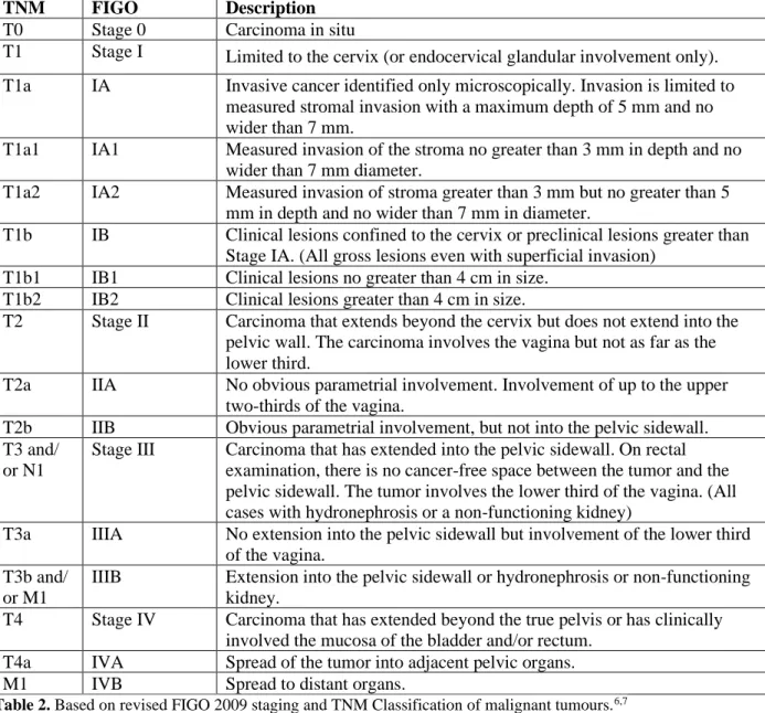
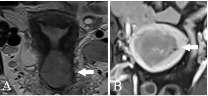
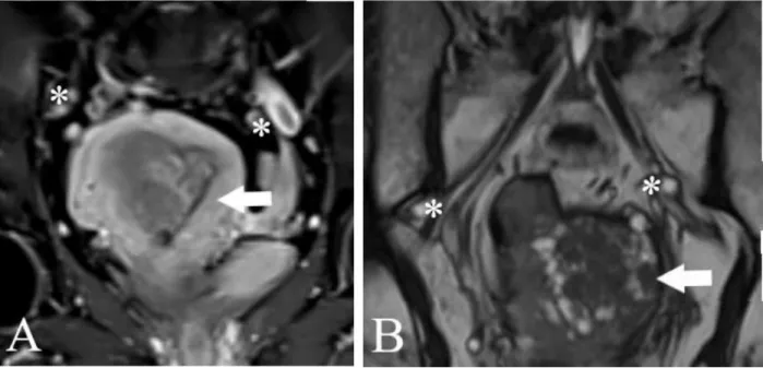
The preoperative MRI was performed using a Siemens Magnetom Area 1.5 Tesla device. Data required for FIGO 2009 staging were collected: the degree of myometrial, cervical, serosal, adnexal and parametrial invasion (Figure 1 and 2.), as well as metastases of pelvic or para-aortic lymph nodes (Figure 3.)
Figure 1. MR imaging. A. Cervical cancer, stromal invasion (T2 sequence) B. Endometrial cancer, myometrial invasion >50% (T1 sequence)
Images of the retroperitoneal, parailiacal, pelvic and cervical area were taken with axial oblique fat-saturated T1, sagittal and axial oblique fast spin-echo T2, T2 TRUFISP and DWI sequences, with gadolinium-based contrast medium.
Figure 2 . MR imaging A. Cervical cancer, tumour spread to upper third of vagina and rectum (T2 sequence, sagittal view)
B: Endometrial cancer, mass is close to the serosa of the uterus. (T2 sequence, coronal view)
Figure 3 MR imaging. Evaluation of lymph-node metastases. A.: Endometrial cancer, * enlarged lymph-nodes (T2 sequence, phase 3 of gadolinium enhancement). B.: Endometrial cancer with leiomyomas, * parailacal enlarged lymph nodes (T2 TRUFI sequence)
Preoperative CT was performed in patients with contraindications for MRI or with extreme obesity using a Siemens Somatom Definition Edge device, with iobitridol as contrast medium.
Osseal and pulmonary metastases were evaluated preoperatively for staging of distant tumor- spread.
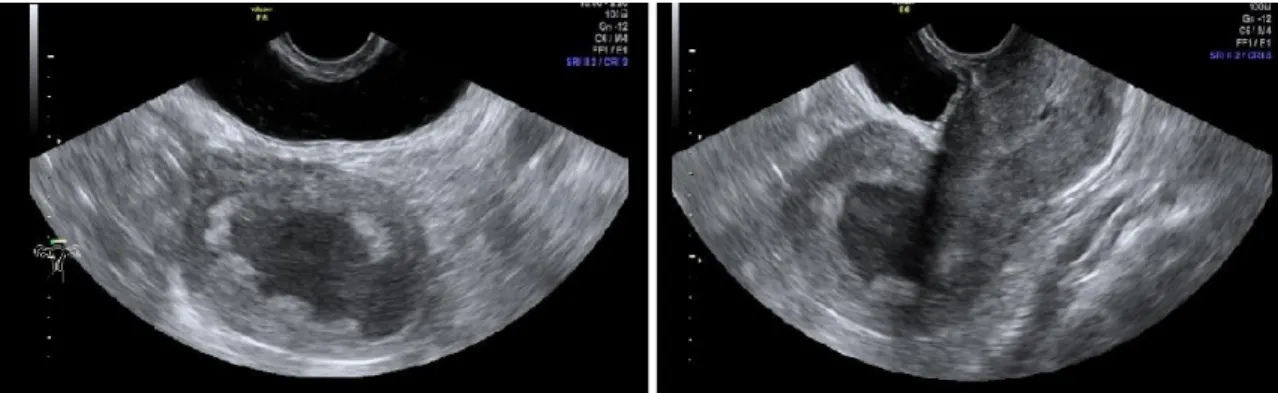
Transvaginal ultrasound was performed using a GE Voluson E6 device with a multifrequency endovaginal probe (5-8 MHz); it had a role in evaluating cervical stroma infiltration, endometrial thickness and structure, adnexal and parametrial invasion, endometrial color-flow index and vascularization of the masses. (Figure 4 and 5.) Two-dimensional coronal and sagittal planes were used to assess myometrial invasion of the uterine corners, and 3D sampling frame was employed for more accurate evaluation.
Figure 4. Transvaginal ultrasound. Enlarged uterine cavity with necrotic mass. 2D imaging.
Figure 5. Transvaginal ultrasound. Cervical cancer, 2D imaging and color-flow.
4.3.3 Surgical treatment
Indication for surgery was based upon preoperative histological and radiological findings, indicated by the Multidisciplinary Tumor Board of the institution, which involves specialists of radiology, oncology, pathology and gynecology.
Abdominal, laterally extended radical hysterectomy was in all cases performed by a gynecologist and a urologist specialist trained in oncosurgery. Initial steps included catheterization of the ureter, followed by parailiacal and pelvic/obturator lymphadenectomy in the pelvic retroperitoneal space.
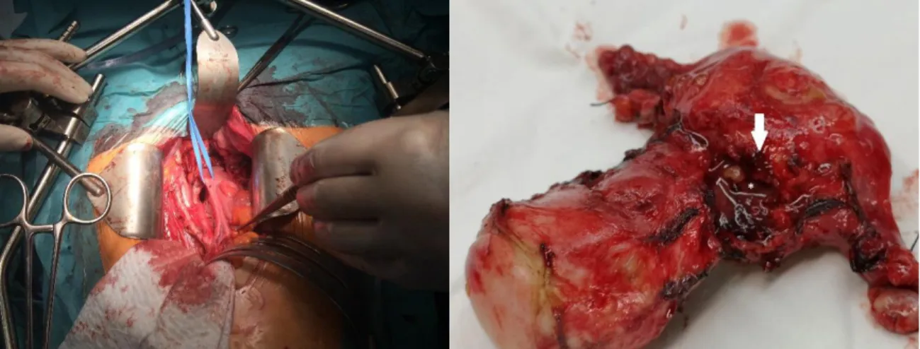
Radical hysterectomy included the removal of the cervix and uterus, Fallopian tubes and ovaries, together with the parametries and upper third of vagina. (Figure 6.)
Figure 6. Intraoperative Picture with preparation and surgical sample of the uterus (arrow: cancer spread beyond the uterinal wall)
4.3.4 Histopathological evaluation
Macroscopic and microscopic histopathological evaluation of the surgical specimen were performed. Findings included the description of the type of tumor, differentiation, TNM and FIGO staging based on the extent of infiltration, and the presence or absence of parametrian, cervical, adnexal, rectal and urinary bladder involvement, lymph-node and pelvic wall invasion.
4.3.5 Postoperative treatment and follow-up
Method of adjuvant treatment was based on final histopathological staging, and was decided by the institutional Oncology Team as required by ESMO guidelines.
Postoperative irradiation in the form of external beam radiotherapy or brachytherapy was recommended in most cases. In patients with a high-risk of recurrence or with confirmed metastases, chemo- or radiochemotherapy was performed using cisplatin–5-fluorouracil, paclitaxel or carboplatin dublets.
Follow-up visits included a complete physical and pelvic examination performed by an oncologist and a gynecologist specialist, with regular radiology follow-ups to exclude the recurrence of cancer.
4.4 Statistical analysis
The age at the time of surgery, prevalence of different tumor histological subtypes, tumor grades and differentiation were taken into account at analysis. The 1- and 5-year survival rates were calculated accounting for deaths occurring before December 31, 2019.
The results of the preoperative radiology staging were compared with the final histological analysis of the surgical specimen, using χ2 or Fisher’s exact test to compare variables. P-values of < 0.05 were considered to be significant. The sensitivity, specificity, positive- and negative predictive values of the preoperative assessment were calculated for each endpoint, together with 95 % confidence intervals (95 % CI). The percentage of the underdiagnosed or overdiagnosed cases and accuracy rate in terms of stage, local invasion and lymph-node metastases were also calculated.
All data were collected in an Excel database, and they were analyzed using SPSS statistical software. The intraclass correlation coefficient (ICC) and Cohen’s kappa value were used to determine the inter-rater agreement of the overall results concerning myometrial invasion and lymph-node metastases, and to evaluate the role of experience between radiologist specialists. ICC and Cohen’s kappa below 0.50 were considered as poor, between 0.50 and 0.75 as moderate, between 0.75 and 0.90 as good, respectively, and above 0.90 ICC it was considered as excellent inter-rater reliability. SPSS software (version 25; SPSS Inc., Chicago, IL, USA) was used for the statistical analyses, and p-values < 0.05 were considered to be significant.
5. Results
5.1 Endometrial cancer
A total of 148 patients with a final histopathological diagnosis of endometrial cancer were enrolled in this study. All patients underwent dilatation and curettage for endometrial sampling, those with a positive result were followed by MR staging of the tumor. Based on the decision of a multidisciplinary tumor board, radical hysterectomy with paraaortic and pelvic lymph-node dissection was performed. Pathologists used macroscopic and microscopic evaluations on hysterectomy specimen with a staining of hematoxylin and eosin, vimentin, estrogen, CEA and p16. Tumor histologic subtype, grade, depth of myometrial invasion and lymph-node positivity were examined. The tumor board evaluated the postoperative results and final stages, according to the FIGO 2009 classification to determine the optimal treatment for the patient.
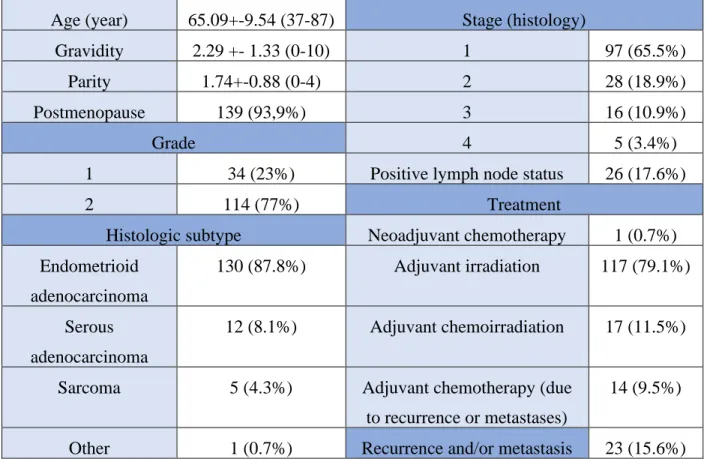
The clinical and histopathological characteristics of the patients are seen in Table 3.
Age of the patients ranged from 37 to 87 years with a mean of 65.09+-9.54 years. of the majority of patients were postmenopausal (93.9%) (Figure 6) and overweight (body mass index > 25 kg/m2;
74.2%).
According to the decision of the tumor board, 1 patient had neoadjuvant chemotherapy (0.7%), 117 patients (79.1%) received adjuvant irradiation and 17 of them (11.5%) had the treatment of chemoirradiation. Fourteen patients (9.1%) were diagnosed with metastases (lung, osseal and lymph-node) and local recurrence occurred in 9 patients (6.1%) during the follow-up period, 96.4% of these patients received chemotherapy.
In the follow-up period, overall 16 patients died, 8 of the deaths were endometrial cancer-related.
The overall 1-year mortality rate was 2.7% (0.7% cancer-related), 5-year mortality rate was 10.8%
(5.4% cancer-related).
Postoperative histologic assessment revealed histopathologic subtype of endometrioid adenocarcinoma in 87.8 %, serous adenocarcinoma in 8.1 %, sarcoma in 3.4 % and other subtypes in 0.7 % of the cases, respectively. Based on FIGO 2009 classifications, the specimens were evaluated as stadium of IA in 35.1%, IB in 30.4%, II in 18.9%, IIIA in 4.1%, IIIB in 6.1%, IIIC in
0.7%, IVA in 2.0% and IVB in 1.4%, respectively. Seventy seven percent of the cases were diagnosed as high-grade, 23 % as low-grade tumors. (Figure 7.)
Figure 7 A. Age of patients with endometrial cancer (years) B. Final stages of endometrial cancer (FIGO 2009 classification)
Table 3. Characteristics. Age, gravidity, parity: mean, SD, range. Postmenopausal state, grade, Histopathologic subtype, Stage, Treatment, Recurrence and metastasis: frequency (%)
Age (year) 65.09+-9.54 (37-87) Stage (histology)
Gravidity 2.29 +- 1.33 (0-10) 1 97 (65.5%)
Parity 1.74+-0.88 (0-4) 2 28 (18.9%)
Postmenopause 139 (93,9%) 3 16 (10.9%)
Grade 4 5 (3.4%)
1 34 (23%) Positive lymph node status 26 (17.6%)
2 114 (77%) Treatment
Histologic subtype Neoadjuvant chemotherapy 1 (0.7%) Endometrioid
adenocarcinoma
130 (87.8%) Adjuvant irradiation 117 (79.1%)
Serous adenocarcinoma
12 (8.1%) Adjuvant chemoirradiation 17 (11.5%)
Sarcoma 5 (4.3%) Adjuvant chemotherapy (due
to recurrence or metastases)
14 (9.5%)
Other 1 (0.7%) Recurrence and/or metastasis 23 (15.6%)
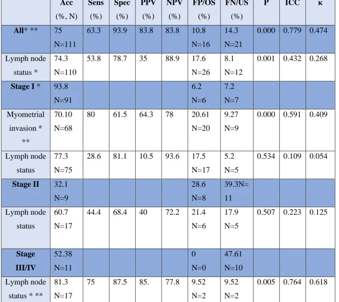
The overall accuracy of MRI evaluating the correct staging was 75%, with the intraclass correlation coefficient of 0.779 ( 0.695-0.840) and κ-coefficient of 0.474 (both with 95% confidence intervals [CI]; p <0,001). Rates of overdiagnosis were 10.8% and underdiagnosis 14.3% of the cases. (Table 4.)
The accuracy of MRI for the detection of lymph-node in all stages status was 74.3%, and its sensitivity, specificity, PPV, and NPV were 53.8%, 78.7%, 35%, and 88.9%, respectively, with the intraclass correlation coefficient of 0.425 (0.214-0.589) and κ-coefficient of 0.268 (both with 95% confidence intervals [CI]; p <0,001).. False negative results were 8.1%, false positive were 17.6% of the cases.
In stage I cancers, the accuracy of MRI regarding stage was 93.8%, with an underestimation in 6.2% of the cases.
The accuracy of MRI for the detection of myometrial invasion in stage I cancers was 70.1%, and its sensitivity, specificity, PPV, and NPV were 80.0%, 61.5%, 64.3%, and 78%, respectively, with the intraclass correlation coefficient of 0.591 (0.388-0.726) and κ-coefficient of 0.409 (both with 95% confidence intervals [CI]; p <0,001). The rates of overdiagnosis were 20.61%, and that of underdiagnosis were 9.27%.
The accuracy of MRI for the detection of lymph-node in stage I cases was 77.3%, and its sensitivity, specificity, PPV, and NPV were 28.6%, 81.1%, 10.5%, and 93.6%, respectively, with the intraclass correlation coefficient of 0.109 (-0.332-0.404) and κ-coefficient of 0.054 (both with 95% confidence intervals [CI]; p=0.054). False negative results were in 5.2% of cases, false positives in 17.5%, respectively.
In stage II cancers, the accuracy of MRI regarding stage was 32.1%. The MRI staging was underestimated in 11 and overestimated in 8 of the cases.
The accuracy of MRI for the detection of lymph-node in stage II cases was 60.7%, and its sensitivity, specificity, PPV, and NPV were 44.4%, 68.4%, 40%, and 72.2%, respectively, with the intraclass correlation coefficient of 0.223 (-0.680-0,640) and κ-coefficient of 0.125 (both with 95% confidence intervals [CI]; p=0.507). There were 6 false positive and 5 false negativecases with respect to the lymph-node status.
In advanced, stage III and IV cancers, the accuracy of MRI regarding stage was 52.38. The MRI staging was underestimated in 47,61% of the cases.
The accuracy of MRI for the detection of lymph-node in advanced stages cases was 80.95%, and its sensitivity, specificity, PPV, and NPV were 80%, 81.8%, 80%, and 81.8%, respectively, with the intraclass correlation coefficient of 0.764 (0.418-0.904) and κ-coefficient of 0.618 (both with 95% confidence intervals [CI]; p =0.005). There were 2 false positive and false negative cases as well.
Acc (%, N)
Sens (%)
Spec (%)
PPV (%)
NPV (%)
FP/OS (%)
FN/US (%)
P ICC κ
All* ** 75 N=111
63.3 93.9 83.8 83.8 10.8 N=16
14.3 N=21
0.000 0.779 0.474
Lymph node status *
74.3 N=110
53.8 78.7 35 88.9 17.6 N=26
8.1 N=12
0.001 0.432 0.268
Stage I * 93.8 N=91
6.2 N=6
7.2 N=7 Myometrial
invasion *
**
70.10 N=68
80 61.5 64.3 78 20.61 N=20
9.27 N=9
0.000 0.591 0.409
Lymph node status
77.3 N=75
28.6 81.1 10.5 93.6 17.5 N=17
5.2 N=5
0.534 0.109 0.054
Stage II 32.1 N=9
28.6 N=8
39.3N=
11 Lymph node
status
60.7 N=17
44.4 68.4 40 72.2 21.4 N=6
17.9 N=5
0.507 0.223 0.125
Stage III/IV
52.38 N=11
0 N=0
47.61 N=10 Lymph node
status * **
81.3 N=17
75 87.5 85. 77.8 9.52 N=2
9.52 N=2
0.005 0.764 0.618
Table 4. Results. Frequency (N) and Percentage (%). Sens: Sensitivity; Spec: Specificity; PPV: Positive predictive value; NPV: Negative predistive value; FP: False positive; OS: Overstaging; FN: False negative; US:
understaging. P: significance value, ICC: intraclass correalation coefficient, κ: Cohen’s kappa value
*p<0.05 **moderate to excellent inter-rater reliability (ICC >0.500)
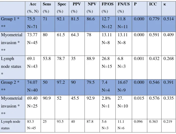
In order to analyze the role of the rater evaluating the MRI findings, we divided the specialists into two groups: a radiologist specialized in imaging of gynecological tumors and a subgroup of non-specialized evaluators. (Table 5.)
A radiologist specialized in the evaluation of gynecological imaging staged 94 cases. The accuracy of overall staging was 75.5%, with the intraclass correlation coefficient of 0.779 (0.790-0.908) and κ-coefficient of 0.514 (both with 95% confidence intervals [CI]; p <0,001), with understaging in 11.8% and overstaging in 12.7%. Sensitivity, specificity, positive predictive value and negative predictive value were 71%, 92.1%, 81.50% and 86.60%, respectively.
The accuracy of the assessment of myometrial invasion was 73.77%, with a sensitivity, specificity, positive predictive value and negative predictive value of 80%, 61.5%, 64.3% and 78%, respectively, with the intraclass correlation coefficient of 0.591 (0.388-0.726) and κ-
coefficient of 0.409 (both with 95% confidence intervals [CI]; p <0,001). The rate of myometrial invasion was understaged in 23.5% and overstaged 29.6% of the cases.
The accuracy of MRI for the detection of lymph-node with experienced rater was 69.1%, and its sensitivity, specificity, PPV, and NPV were 53.8%, 78.7%, 35%, and 88.9%, respectively, with the intraclass correlation coefficient of 0.432 (0.214-0.585) and κ-coefficient of 0.268 (both with 95% confidence intervals [CI]; p =0,001). False positive and negative rates were 26.8% and 6.8%, respectively.
In the second rater group, four radiologists of average experience in the evaluation of
gynecological imaging staged 54 cases. The accuracy of overall staging was 74.07%, with the intraclass correlation coefficient of 0.546 (0.217-0.736) and κ-coefficient of 0.391 (both with 95% confidence intervals [CI]; p <0,001) with understaging in 9 and overstaging in 4 cases.
Sensitivity, specificity, positive predictive value and negative predictive value were 50%, 97.2%, 90% and 79.5%, respectively.
The accuracy of the assessment of myometrial invasion was 69.40%, with a sensitivity, specificity, positive predictive value and negative predictive value of 90.9%, 52%, 45.4% and 90.9%, respectively, with the intraclass correlation coefficient of 0.576 (0.169-0.785) and κ- coefficient of 0.334 (both with 95% confidence intervals [CI]; p =0,015). The rate of myometrial invasion was overstaged in 1 and understaged in 10cases.
The accuracy of MRI for the detection of pathologic lymph-nodes for moderate-experienced raters was 83.3%, and its sensitivity, specificity, PPV, and NPV were 25%, 93.5%, 40% and 87.8%, respectively, with the intraclass correlation coefficient of 0.363 (0.098-0.630) and κ- coefficient of 0.219 (both with 95% confidence intervals [CI]; p=0,096). There were 3 false positive and 6 false negative cases.
Acc (%, N)
Sens (%)
Spec (%)
PPV (%)
NPV (%)
FP/OS (%)
FN/US (%)
P ICC κ
Group 1 *
**
75.5 N=71
71 92.1 81.5 86.6 12.7 N=12
11.8 N=11
0.000 0.779 0.514
Myometrial invasion *
**
73.77 N=45
80 61.5 64.3 78 13.11 N=8
13.11 N=8
0.000 0.591 0.409
Lymph node status
*
69.1 N=43
53.8 78.7 35 88.9 26.8 N=15
6.8 N=3
0.001 0.432 0.268
Group 2 *
**
74.07 N=40
50 97.2 90 79.5 7.4 N=4
16.67 N=9
0.000 0.546 0.391
Myometrial invasion *
**
69.40 N=25
90.9 52 45.5 92.9 2.8%
N=1
27.
N=10
0.015 0.576 0.335
Lymph node status
83.3 N=45
25 93.5 40 87.8 5.6
N=3
11.1 N=6
0.096 0.363 0.219
Table 5. Rater-related results. Frequency (N) and Percentage (%). Sens: Sensitivity; Spec: Specificity; PPV:
Positive predictive value; NPV: Negative predictive value; FP: False positive; OS: Overstaging; FN: False negative; US: understaging.
*p<0.05 **moderate to excellent inter-rater reliability (ICC > 0.500)
5.2 Cervical cancer
A total of 88 patients with a final histopathological diagnosis of cervical cancer were enrolled in this study. Sixty-one patients (69.3%) underwent conisation due to abnormal citology result, or were diagnosed by dilatation and curettage (27 patients, 30.7%). The positive result was
followed by MRI staging of the tumor. Based on the decision of a multidisciplinary tumor board, radical hysterectomy with paraaortic and pelvic lymph-node dissection was performed.
Pathologists used macroscopic and microscopic evaluations on hysterectomy specimen with a staining of hematoxylin and eosin, p16, direct HPV, Ki-67 and p53 tests. Tumor histologic subtype, grade, depth of stromal and parametrian invasion and lymph-node positivity were examined. The tumor board evaluated thepostoperative results and final stages according to the FIGO 2009 classification were determine for optimal treatment of each patient.
The clinical and histopathological characteristics of the patients are seen in Table 6.
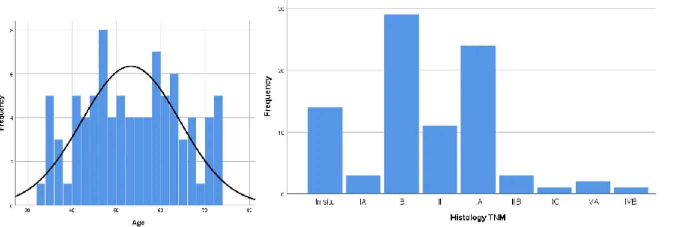
Age of the patients ranged from 33 to 73 years with a mean of 53,26+11,06 years. (Figure 8.) Fifty-three patients were in postmenopause (60.2%) and body mass index was between 19–53.2, with only 23% overweight patients (body mass index > 25 kg/m2)
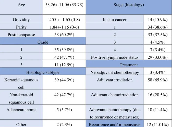
Figure 8 A. Age of patients with cervical cancer (years) B. Final stages of cervical cancer (FIGO 2009 classification)
According to the decision of the tumor board, 3 patients had neoadjuvant chemotherapy (3,4%), 58 patients (68,9%) received adjuvant irradiation and 18 of them (20,5%) had the treatment of chemoirradiation. Eleven patients (12,6%) were diagnosed with metastases (lung, brain, osseal and lymph-node) and local recurrence occurred in 6 patients (6,8%) during the follow-up period, 98,2% of these patients received chemotherapy.
In the follow-up period, overall 12 patients died, 5 of the deaths were cervical cancer-related.
The overall 1-year mortality rate was 5,7% (4,5% cancer related), 5-year mortality rate was 12,5% (5,7% cancer-related).
Postoperative histologic assessment revealed histopathologic subtype of keratoid squamous-cell cancer in 44,3 %, nonkeratoid squamous-cell cancer in 47,7 %, adenocarcinoma in 5,7 % and other subtypes (adenosquamous and small-cell carcinoma) in 2,3 % of the cases. Based on FIGO 2009 classifications, the specimens classified as stadium of IA in 3,4%, IB in 33,0%, IIA in 12,5%, IIB in 27,3%, IIIA in 3,4%, IIIB in 1,1%, IVA in 2,3% and IVB in 1,1%, respectively.
Thirty-nine point eight percent of the cases were diagnosed as well-differentiated, 47,7 % as medially-differentiated and 12,5% as poorly-differentiated tumors. (Figure 8.)
Table 6. Characteristics. Age, gravidity, parity: mean, SD, range. Postmenopausal state, grade, Histopathologic subtype, Stage, Treatment, Recurrence and metastasis: frequency (%)
Age 53.26+-11.06 (33-73) Stage (histology)
Gravidity 2.55 +- 1.65 (0-8) In situ cancer 14 (15.9%)
Parity 1.84+-1.15 (0-6) 1 34 (38.6%)
Postmenopause 53 (60.2%) 2 33 (37.5%)
Grade 3 4 (4.5%)
1 35 (39.8%) 4 3 (3.4%)
2 42 (47.7%) Positive lymph node status 29 (33.0%)
3 11 (12.5%) Treatment
Histologic subtype Neoadjuvant chemotherapy 3 (3.4%) Keratoid squamous
cell
39 (44.3%) Adjuvant irradiation 58 (65.9%)
Non-keratoid squamous cell
42 (47.7%) Adjuvant chemoirradiation 16 (20.5%)
Adenocarcinoma 5 (5.7%) Adjuvant chemotherapy (due to recurrence or metastases)
10 (11.4%)
Other 2 (2.3%) Recurrence and/or metastasis 12 (11.01%)
The overall accuracy of MRI evaluating the correct staging was 61.4%, with the rates of overdiagnosis in 31.8% and underdiagnosis in 6.8% of the cases, with the intraclass correlation coefficient of 0.824 (0.731-0.885) and κ-coefficient of 0.425 (both with 95% confidence intervals [CI]; p <0,001). Sensitivity, specificity, PPV, and NPV were 87.5%, 68.6%, 70% and 86.8%, respectively. (Table 7.)
The accuracy of MRI for the detection of lymph-node status was 67%, and its sensitivity, specificity, PPV, and NPV were 58,6%, 71,2%, 50%, and 77.8%, respectively, with the intraclass correlation coefficient of 0.447 (0.155-0.638) and κ-coefficient of 0.286 (both with 95% confidence interval [CI]; p =0,007). False negative and positive rates were 13.6% and 19.3%, respectively.
In stage I cancers, the accuracy of MRI regarding stage was 61.8%, with an underestimation in 13 cases (38.2%).
The accuracy of MRI for the detection of stromal invasion was 79.4%, and its sensitivity, specificity, PPV, and NPV were 80%, 75%, 96%, and 33.3%, respectively, with the intraclass correlation coefficient of 0.553 (0.106-0.777) and κ-coefficient of 0.357 (both with 95%
confidence intervals [CI]; p =0,019). The rate of overdiagnosis was 2.9%, and that of underdiagnosis was 17.6%.
The accuracy of MRI for the detection of pathologic lymph-nodes in stage I cases was 70.6%, and its sensitivity, specificity, PPV, and NPV were 66.7%, 71.4%, 33.3%, and 90.9%,
respectively, with the intraclass correlation coefficient of 0.457 (0.087-0.729) and κ-coefficient of 0.274 (both with 95% confidence intervals [CI]; p =0.076). False negative results were 5.9%, false positives 23.5%, respectively.
In stage II cancers, the accuracy of MRI regarding stage was 72.7%. MRI staging was underestimated in 15.1% and overestimated in 12.2% of the cases.
The accuracy of MRI for the detection of abnormal lymph-nodes in stage II cases was 54.5%, and its sensitivity, specificity, PPV, and NPV were 58.8%, 50%, 55.6%, and 53.3%, respectively, with the intraclass correlation coefficient of 0.163 (0.069-0.586) and κ-coefficient of 0.088 (both with 95% confidence intervals [CI]; p=0.611). False negative results were detected in 21.2%, false positives in 24.2% of the cases.
In advanced, stage III and IV cancers, the accuracy of MRI regarding stage was 85.7%. The MRI staging was underestimated in 14.3% of the cases.
The accuracy of MRI for the detection of lymph-node in advanced stages was 50%, and its sensitivity, specificity, PPV, and NPV were 60%, 50%, 75%, and 33.3%, respectively, with the intraclass correlation coefficient of 0.167 (0.038-0.857) and κ-coefficient of 0.0.87 (both with 95% confidence intervals [CI]; p =0,809). There were 2 false negative and 1 false positive lymph-node detection.
Acc (%, N)
Sens (%)
Spec (%)
PPV (%)
NPV (%)
FP/OS (%)
FN/US (%)
P ICC κ
All* ** 61.4 N=54
87.5 68.8 70 86.8 31.8 N=28
6.8 N=6
0.000 0.824 0.425
Lymph node status *
67 N=59
58.6 71.2 50 77.8 19.3 N=17
13.6 N=12
0.007 0.447 0.286
Stage I 61.8 N=21
38.2 N=13 Stromal
invasion *
**
79.4 N=27
80 75 96 33.3 2.9
N=1
17.6 N=6
0.019 0.553 0.357
Lymph node status *
70.6 N=24
66.7 71.4 33.3 90.9 23.5 N=8
5.9 N=2
0.076 0.457 0.274
Stage II *
**
72.7 N=24
12.2 N=4
15.1 N=5 Lymph node
status
54.5 N=18
58.8 50 55.6 53.3 24.2 N=8
21.2 N=7
0.611 0.163 0.088
Stage III/IV
* **
85.7 N=6
0 N=0
14.3 N=1 Lymph node
status
50 N=4
60 50 75 33.3 12.5
N=1 25 N=2
0.809 0.167 0.087
Table 7 Results. Frequency (N) and Percentage (%). Sens: Sensitivity; Spec: Specificity; PPV: Positive predictive value; NPV: Negative predictive value; FP: False positive; OS: Overstaging; FN: False negative; US: understaging P: significance based on Chi-square or Fisher’s exact test, ICC: intraclass correlation coefficient, κ: Cohen’s kappa value
*p<0.05 **moderate to excellent inter-rater reliability (ICC >0.500)
In order to analyze the role of the rater evaluating the MRI findings, we divided the specialists into two groups: a radiologist specialized in imaging of gynecological tumors and a subgroup of non-specialized evaluators. (Table 8.)
A radiologist specialized in the evaluation of gynecological imaging staged 55 cases (62.5%).
The accuracy of overall staging was 60%, with the intraclass correlation coefficient of 0.806 (0.668-0.887) and κ-coefficient of 0.406 (both with 95% confidence intervals [CI]; p <0,001).
Understaging rates were 7.3% and overstaging 32.7% of the cases. Sensitivity, specificity, positive and negative predictive values were 87.5%, 68.8%, 70% and 86.8%, respectively.
The accuracy of the assessment of stromal invasion in stage 1 was 82.6%, with a sensitivity, specificity, positive predictive value and negative predictive value of 81.8%, 100%, 100% and 20%, respectively, with the intraclass correlation coefficient of 0.486 (0.211-0.782) and κ-
coefficient of 0.281 (both with 95% confidence intervals [CI]; p =0.052). The rate of myometrial invasion was understaged in 4 cases and there were no overstaging.
The accuracy of MRI for the detection of pathologic lymph-nodes by an experienced rater was 61.8%, and its sensitivity, specificity, PPV, and NPV were 66.7%, 64.7%, 40% and 84.6%, respectively, with the intraclass correlation coefficient of 0.432 (0.338-0.759) and κ-coefficient of 0.258 (both with 95% confidence interval [CI]; p =0.183). Fourteen cases were false positive (25.5%) and 7 cases false negative (12.7%).
In the second rater group, four radiologists of average experience in the evaluation of
gynecological imaging staged 33 cases. The accuracy of overall staging was 63.6%, with the intraclass correlation coefficient of 0.849 (0.694-0.925) and κ-coefficient of 0.457 (both with 95% confidence intervals [CI]; p <0,001). Understaging was found in 2 and overstaging in 10 cases. Sensitivity, specificity, PPV, and NPV were 86.7%, 72.2%, 72.2% and 86.7%,
respectively.
The accuracy of the assessment of stromal invasion in stage 1 tumors was 72.7%, with a sensitivity, specificity, positive predictive value and negative predictive value of 75%, 66.7%, 85.7% and 50%, respectively, with the intraclass correlation coefficient of 0.556 (0.140-0.880) and κ-coefficient of 0.377 (both with 95% confidence intervals [CI]; p =0,201). Myometrial invasion was overstaged in 1 and understaged in 2 cases.
The accuracy of MRI for the detection of pathologic lymph-nodes by moderate-experienced raters was 75.8%, and its sensitivity, specificity, PPV, and NPV were 44.4%, 87.5%, 57.1%, and 80.8%, respectively, with the intraclass correlation coefficient of 0.515 (0.018-0.760) and κ- coefficient of 0.343 (both with 95% confidence intervals [CI]; p=0,046). There were 3 false positive and 5 false negative cases with regard to the lymph-node status.
Acc (%, N)
Sens (%)
Spec (%)
PPV (%)
NPV (%)
FP/OS (%)
FN/US (%)
P ICC κ
Group 1
* **
60.0 N=33
87.5 68.8 70 86.8 32.7 N=18
7.3 N=4
0.000 0.806 0.406
Stromal invasion
82.6 N=19
81.8 100 100 20 0 18.2
N=4
0.052 0.486 0.281
Lymph node status
61.8 N=34
66.7 64.7 40 84.6 25.5 N=14
12.7 N=7
0.183 0.432 0.258
Group 2
* **
63.6 N=21
86.7 72.2 72.2 86.7 30.3 N=10
6.1 N=2
0.000 0.849 0.457
Stromal invasion
**
72.7 N=8
75 66.7 85.7 50 9.1 N=1
18.2 N=2
0.201 0.556 0.377
Lymph node status *
**
75.8 N=25
44.4 87.5 57.1 80.8 9.1 N=3
15.2 N=5
0.046 0.515 0.343
Table 8. Rater-related results. Frequency (N) and Percentage (%). Sens: Sensitivity; Spec: Specificity; PPV:
Positive predictive value; NPV: Negative predictive value; FP: False positive; OS: Overstaging; FN: False negative; US: understaging
P: significance based on Chi-square or Fisher’s exact test, ICC: intraclass correlation coefficient, κ: Cohen’s kappa value
*p<0.05 **moderate to excellent inter-rater reliability (ICC >0.500)
6. Discussion
The aim of our study was to evaluate the role and reliability of preoperative imaging and staging in the planning process, regarding the radicality of surgery in gynecological cancers.
According to the 2018 FIGO staging revision, gynecological and pathological examinations, as well as imaging assessments are incorporated in the staging of gynecological cancers. Countries with a developed healthcare system have already been using various imaging modalities at pretreatment and staging, leading to an increased diagnostic accuracy, and therefore to a more tailored surgical and oncological treatment. 21
The National Cancer Comprehensive Network (NCCN) guidelines for cervical and endometrial cancer recommend pelvic MRI, chest radiography or CT, or whole body PET-CT for primary diagnostics in the evaluation of local tumor extent and metastases.
MRI has been considered the imaging method of choice for the assessment of tumor size, localization, parametrial-, myometrial- and stromal infiltration and lymph-node status.22 With at least two T2-weighted sequences in sagittal, axial oblique or coronal oblique orientation, local tumor extension and parametrial invasion can be evaluated. An axial T1-weighted sequence is used for the detection of enlarged pelvic or abdominal lymph nodes; however, CT and MRI tend to have low sensitivity for metastatic lymph-node status: since enlarged metastatic and hyperplastic nodes are hard to differentiate, the criteria based on the size often lead to overlooking smaller metastases.23
6.1 Endometrial cancer
In the developed world, endometrial cancer is one of the most common malignant gynecological cancer types. Due to the currently available diagnostic modalities and patient education, the early detection of the tumor leads to high overall survival. Prognostic factors include histopathologic subtypes, grade, myometrial invasion and lymph node metastases.
In the detection and staging of endometrial cancer, patients with abnormal vaginal bleeding are candidates for endometrial thickness evaluation by transvaginal ultrasound technique; however, a definitive diagnosis requires endometrial sampling by biopsy or dilatation and curettage for histopathological evaluation.24
In the preoperative staging, MRI is considered as the best method of choice to assess myometrial invasion depth, cervical involvement and tumor grade, as well as raise the probability of lymph node metastases. NCCN and ESUR advise MR imaging in type II endometrial cancers, suspected cervical invasions and to identify patients with stage Ia disease.25 As patients with type I endometrial cancer with myometrial invasion of more than 50%, or with type II endometrial cancer are considered to be intermediate to high-risk of lymph node metastasis, preoperative staging is essential for a tailored pre- and postoperative treatment, and for planning the radicality of the surgical treatment.26
6.1.1 Accuracy of pre-and postoperative staging in endometrial cancer
According to our findings, the overall accuracy of MRI in regards of staging and identifying lymph node metastases was 75% and 74.3% with high specificity, high negative predictive value and low sensitivity. (Figure 9.) As the majority of the tumors are detected at an early stage, the high sensitivity of MRI for myometrial invasion, its moderate specificity and high negative predictive value for lymph node metastases in stage I diseases means that this modality plays an essential role in planning the radicality of hysterectomy and lymphadenectomy in localized tumors.
However, due to its low sensitivity and low positive predictive value for lymph node detection, a considerable number, 17.5%, of surgeries at an early stage were complemented with lymphadenectomy due to false positive MRI results.27 In literature reviews of the past few years’
studies similar results were reported, showing satisfactory specificity but low sensitivity in regards to lymph node detection due to similar radiological findings of hyperplastic and metastatic lymph- node enlargement.28
At advanced stages (stage II-IV) the overall accuracy of MRI was moderate, with relatively high negative predictive value. In terms of lymph-node status, 60.7-80.95% of the metastases were evaluated correctly; however, due to the low number of advanced cases, the results were not considered statistically significant. As the postoperative treatment of endometrial cancer in advanced stages depends on further imaging and pathological findings, such as tumor grade, histopathological subtype and presence or absence of distant metastases, our detection rate of lymph node metastases with MRI modality is satisfactory in order to make the most tailored
decision for post-staging treatment, which leads to only 5.4% cancer-related 5-year mortality at our hospital.
Figure 9. Accuracy between MRI and histology evaluation.
Another main objective of our study was to analyze inter-rater agreement. Due to a high BMI in 74.2% of our patients and to the high percentage of stage I disease, which presents difficulty in lymph-node evaluation, we studied, whether there is a significant difference between the findings of a radiologist specialized in gynecological tumors and of a non-specialized radiologist, in endometrial cancer cases.
Overall MRI staging, in comparison with histopathological staging had good inter-rater agreement with ICC of 0.742-0.865, with good to excellent agreement with specialized radiologists and moderate to good agreement with non-specialized radiologists. In terms of accuracy, there was no significant difference between the two groups. In group 1, sensitivity, specificity, positive and negative predictive values were more consistent and satisfactory than in group 2. Findings of radiologists with less gynecological experience showed higher sensitivity for myometrial invasion and higher specificity for lymph node evaluation. Both groups had moderate agreement with
histopathologic staging, when assessing the rate of myometrial invasion and poor to moderate agreement regarding the detection of lymph node metastases, compared to final pathologic results.
(Figure 10.)
Figure 10. Inter-rater agreement between MRI and histopathologic staging based on intraclass correlation.
*p<0.05
Compared to CT and MRI studies recommend 18F-FDG PET-CT, which allows more accurate staging and detection of lymph node metastases, as MRI can only find lymph node metastases when the nodes are swollen, with a diameter of >10mm.29
Unfortunately, PET-CT for preoperative investigation is still not routinely supported by health insurance in Hungary, and only special cases are evaluated with this imaging technique, such as ovarian cancer cases and investigations of remote metastases.
A 2D and 3D transvaginal ultrasound examination is (routinely) used in preoperative evaluation of staging in middle-income countries. Some studies have shown, that its accuracy and positive predictive value is equal to that of an MRI finding, assuming we can rely on an experienced investigator.30 In our study, we started using the ultrasound modality only with patients presenting after June of 2017 to improve preoperative diagnostics ; therefore, we have a low number of ultrasound findings. Where transvaginal ultrasound was used for preoperative imaging, the
0 0.1 0.2 0.3 0.4 0.5 0.6 0.7 0.8 0.9
Staging (Rater 1) Staging (Rater 2) Infiltration rate (Rater 1)
Infiltration rate (Rater 1)
Lymph-node status (Rater 1)
Lymph-node status (Rater 2)
Inter-rater agreement
*
* *
*
*