Differentiations of the Ground Cytoplasm and Their Significance for the Generation of the Motive Force of Ameboid Movement
1Κ. E . W O H L F A R T H - B O T T E R M A N N Zentral-Lab Oratorium für angewandte Übermikroskopie am Zoologischen Institut
der Universität Bonn, Bonn, Germany
Throughout the history of the investigation of ameboid movement the relevant theories have often been closely tied to current views of cytoplasmic structure (cf. De Bruyn, 1957; Noland, 1957). T h e mor- phologist, dealing with protoplasmic streaming, has primarily to an- swer the question of which cytoplasmic structures furnish the motive force for this interesting and important phenomenon of living matter.
Furthermore, it is a problem of morphological interest to see how these structures might be ordered to give a functional pattern, i.e., a mode of action resulting in protoplasmic movement. Such knowledge would be a highly desirable basis for physiological and biochemical studies.
The purpose of this paper is to show that today, for the first time, modern cell morphology seems able to demonstrate by means of the phase-contrast and electron microscopes differentiations of the ground- plasm or ground cytoplasm which may be responsible for the genera- tion of the motive force. We studied the classic objects for this kind of research, namely, amebae and slime molds.
Thus far electron microscopy has not been very successful in reveal- ing the structural organization of the groundplasm. In the past decade there has emerged an image of the typical cell structure, which is so generally well understood today that it is unnecessary to give details here. T h e spatial relationships of the newly discovered membrane sys- tems are surely a major contribution to animal and plant cytology.
These membranes provide an enormous expanse of inner surface and achieve a separation of different spaces within the cytoplasm (cf. Palade,
1956; Danielli, 1958; Ruska, 1962). In most cells, the groundplasm is the most significant of these spaces as far as the volume is concerned.
It is the groundplasm in which the membrane structures of the cell are embedded, and therefore it has been called the cytoplasmic matrix:
ι Support from the Deutsche Forschungsgemeinschaft for the experimental work is gratefully acknowledged.
79
80 Κ. Ε. WOHLFARTH-BOTTERMANN
(cf. Wohlfarth-Bottermann, 1960, 1961a,b,c). It is reasonable to hold groundplasm responsible for the generation of the motive force. How- ever, two uncertainties must be considered in connection with this as- sumption:
1. It is not clear in all cases, whether protoplasmic streaming is set in motion by contraction phenomena (cf. Stewart and Stewart, 1959a;
Allen, 1961b; Abé, 1962; Kavanau, 1962a,b).
2. Because of the artifact problem, it is more difficult to obtain reli- able morphological information about the groundplasm than about membranes.
If the cytoplasm is thought of as the source of motive force resulting from contraction phenomena (cf. De Bary, 1864; Schulze, 1875; Mast, 1926, 1931), then the morphologist immediately wants to know whether there is any evidence of threadlike or reticular structures. Such struc- tures have been postulated since the beginning of protoplasmic investi- gation (cf. Zeiger, 1943; Haas, 1955; Oberling, 1959). In most electron microscope studies it has not been possible to distinguish clearly the ultrastructure of the cytoplasmic ground substance: a survey of the existing literature reveals mostly negative findings and so the general opinion has been that the groundplasm, even after the best fixation, appears structureless under the electron microscope.
A promising approach to the study of groundplasm has seemed to us to be to work with cells in which large and defined areas contain only this material. The ameba Hyalodiscus simplex (Wohlfarth-Bottermann,
1960), the normal form of which always shows a clear separation of hyaline ectoplasm and granular endoplasm, has proved particularly suitable for this purpose. The large front lobe consists of pure ground- plasm, and contains no membrane structures such as endoplasmic reticulum or vacuoles and is intimately connected with the motive mechanism of this cell. T h e electron microscope has revealed, inde- pendently of the fixation and electron-staining methods used, three structural aspects: the groundplasm is either structureless, or it is com- posed either of globular or threadlike structures (Wohlfarth-Botter- mann, 1960, 1961a). We found pleomorphism of the groundplasm (even
in single cells), which may be correlated with different physiological states.
Though threadlike structures have been postulated for a long time as components of a "contractile gel reticulum" we must ask to what degree they represent real, vital components of the cytoplasmic matrix.
In living amebae, fibrils have been described several times in the light microscope (Wittmann, 1950, 1951; Goldacre, 1961; Käppner, 1961).
Also by means of the electron microscope similar fibrils are detected in
Differentiations of the Ground Cytoplasm 81 Hyalodiscus: they represent bundles of filamentous structures of the groundplasm (Schneider and Wohlfarth-Bottermann, 1959, p. 379, Fig. 1;
cf. Wohlfarth-Bottermann, 1963a); we can thus assume that they are not artifacts. The evidence for this will now be given in connection with another "classic" object for studying protoplasmic streaming, i.e., the slime mold, Physarum polycephalum.
This organism is particularly suitable for morphological investiga- tions, and the physiology of this type of protoplasmic streaming is well known (cf. Kamiya, 1959, 1960a,b). T h e observation of living micro- plasmodia of Physarum suggests strongly that contraction mechanisms are involved. We know from many studies some of the chemical proc- esses which underlie the generation of the motive force in Physarum, but until now no one has demonstrated a physical basis for this type of streaming.
I need not cite here the numerous physiological studies that have been made; I only want to recall that they seem to suggest a pressure- flow mechanism. Similarly, biochemical methods in general (Loewy, 1952; Ts'o et al, 1956, 1957; Nakajima, 1956, 1957, 1960) demonstrate that it is possible to prepare an adenosine triphosphate (ATP)-sensitive,
"contractile protein" from the plasmodium. These proteins behave much like actomyosin and myosin Β of muscle and were, therefore, designated "myxomyosin" and "plasmodial myosin B . " T h e myxomyosin molecules are threadlike structures of 70 A in width.
Kamiya et al. (1957) were able to show that the motive force of this streaming (which alternates its direction rhythmically) is significantly increased by the addition of ATP. All these results indicate a mechanism for the conversion of chemical to mechanical energy, which is tied to an ATP-sensitive protein, and which thus may be related to the mechanism of muscle contraction. Unfortunately, until the present, morphologists have been unable to detect relevant filamentous structures in the cyto- plasm of Physarum (Stewart and Stewart, 1959b), i.e., a substrate which might be structurally capable of contracting and whose functional dis- position might speak for a pressure-flow mechanism indicated by the physiological results cited previously.
The plasmodial channels in which the endoplasm flows back and forth have an outer ectoplasmic gel layer. The electron microscope has revealed that this ectoplasmic gel layer is substantially groundplasm (Wohlfarth-Bottermann, 1962) containing threadlike structures mor- phologically identical with those found in the groundplasm of amebae.
We shall henceforth designate these filamentous structures as "plasma filaments" (Wohlfarth-Bottermann, 1962), and we believe that they rep-
82 Κ. Ε. WOHLf ARTH-BOTTERMANN
FIG. 1. Highly enlarged electron micrograph (ultrathin section) of a part of a protoplasmic fibril from the outer region of a protoplasmic thread. Normal plasmodium of Physarum polycephalum. The fibril (f) is formed by very narrow, longitudinally
Differentiations of the Ground Cytoplasm 83 resent an interesting and important differentiation of the cytoplasmic matrix. Let us see whether we can gain more information about their function.
Individual plasma filaments of the Physarum groundplasm (Fig. 1) can be seen electron optically with approximately the same fidelity as the thin myofilaments of smooth muscle cells. In the course of our electron microscopic studies on slime mold plasmodia we very soon found that the plasma filaments can form compact fibrils (Figs. 1 to 3), in part by means of parallel arrangement (Wohlfarth-Bottermann, 1962).
This finding supports the similar observation on amebae, where the groundplasm may likewise contain plasma filaments (Wohlfarth-Botter- mann, 1961a), which can orientate themselves to form fibrils (Schneider and Wohlfarth-Bottermann, 1959, Part 1; cf. Wohlfarth-Bottermann,
1963a). In contrast with amebae, one finds the fibrils in plasmodia in great numbers and a thorough electron microscopic analysis has re- vealed that the fibrils are often intricately interconnected at "nodal points" (Fig. 3). In the cytoplasm of Physarum plasmodia there is a more or less coherent network of fibrils, and these fibrils are simply a conspicuous differentiation of the groundplasm. It must be pointed out that not all fibrils possess the same fine structure. Many of them are built up by structural units showing no parallel arrangement (Fig. 2).
It is likely that the variations in the fine structure can be interpreted as functional differences, but until now it has not been possible to show a clear correlation, say, for instance, with respect to their dynamic be- havior.
The electron microscope pictures of sections through fibrils show that the dimension of many fibrils reaches a size visible in the light microscope (Fig. 3). For the study of the functional arrangement of the fibrillar network within the larger plasmodia, the electron microscope is for several reasons quite unsuitable: thin-sectioning techniques pro- viding preparations of 200-600 A thickness and of limited area do not allow a quick and quantitative survey. For this purpose we had to find a light microscopic method, which enabled us to cut plasmodial strands and protoplasmic drops in serial sections according to conditions of fix- ation and embedding that make it possible to see the fibrils in the light microscope. We succeeded in developing a quite simple procedure (Wohlfarth-Bottermann, 1963b): after fixation as used for electron mi- croscopy (Wohlfarth-Bottermann, 1957), we embedded the plasmodia in
arranged plasma filaments (arrows) of the groundplasm. On the right side of the picture
—normal cytoplasm (cy) with ribosomes and membrane structures. Magnification:
χ 107,000. (Wohlfarth-Bottermann, 1962.)
FIG. 2. Highly enlarged part of a protoplasmic fibril from the outer region of a protoplasmic drop (10 min old) (Physarum polycephalum). T h e fibril (f) is formed by plasma filaments showing no parallel arrangement. On the right side of the picture
—normal cytoplasm (cy) with ribosomes. Magnification: χ 107,000 (unpublished).
84
Differentiations of the Ground Cytoplasm 85
FIG. 3. Protoplasmic fibrils (arrows) of Physarum polycephalum. Note that the fibril branches out at different "ramification points." Electron micrograph of an ultra- thin section. Magnification: χ 15,000. (Wohlfarth-Bottermann, 1963c.)
86 Κ. Ε. WOHLFARTH-BOTTERMANN
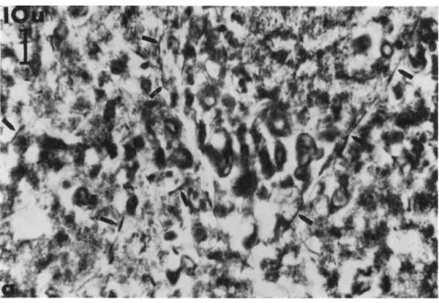
FIG. 4 . Electron and phase-contrast micrographs of the protoplasmic fibrils of Physarum polycephalum for comparison, (a) Low-power electron micrograph; ultra- thin section; magnification: χ 4 0 0 0 . (b) Phase-contrast microscopic picture; paraffin wax section 3-5 μ thick; magnification: χ 725. Note the attachment of fibrils to vacuoles in both cases and the deformation of the vacuoles (unpublished).
Differentiations of the Ground Cytoplasm 87 paraffin wax ("Tissuemat") and cut serial sections of 3-5 μ thickness on the L K B ultramicrotome (Schneider, 1962). Without any staining, phase- contrast microscopic examination of the sections is possible after the embedding medium has been removed. In Fig. 4 one can compare the electron and phase-contrast microscopic images. It is absolutely clear that these microscopes reveal identical structures. There are many reasons for this statement, one of them being that the filamentous struc-
FIG. 5. Protoplasmic fibril of Physarum polycephalum. Osmium-chromium fixation;
paraffin wax embedding; section of 3-5 μ thickness. Top: phase-contrast micrograph.
Bottom: polarizing micrograph, revealing the weak birefringence of the fibril. Mag- nification: χ 1250. Photograph, W. Cebulla, Ultraphot Carl Zeiss, Oberkochen (un- published).
88 Κ. Ε. WOHLFARTH-BOTTERMANN
tures seen in the phase-contrast microscope are biréfringent in the polarizing microscope (Fig. 5). This finding is in accordance with the fine structure of certain fibrils revealed by the electron microscope, namely, their composition of longitudinally arranged plasma filaments.
After a suitable method had been developed for examining the fibrils in the light microscope, we found it was possible to isolate them by means of quite simple procedures developed by muscle physiologists:
the protoplasmic fibrils survive for many days after glycerine extraction of the plasmodia and protoplasmic drops. Figure 6 shows fibrils after glycerine extraction, comparing results achieved by phase contrast (Fig. 6a) and electron microscopy (Fig. 6b). This means that they could be used as "models" in the sense of the muscle physiologists. Indeed we have tried to test their contractility by the methods worked out by Hoffmann-Berling (1958), but until now we have not been able to find clear evidence of shortening. This negative finding, however, is not evidence against their being contractile, because our control experi- ments with amebae and muscles only gave positive results on muscles
FIG. 6. Protoplasmic fibrils (arrows) of Physarum polycephalum after glycerine extraction (72 hr) of a protoplasmic drop, followed by osmium-chromium fixation, (a) Paraffin wax embedding; section of 3-5 μ thickness; phase-contrast microscope;
magnification: χ 800 (unpublished), (b) Vestopal-W embedding; ultrathin section;
electron microscope; magnification: χ 25,000 (unpublished). Note the clear representa- tion of the plasma filaments (b) after glycerine extraction.
'erentiations of the Ground Cytoplasm
(b)
90 Κ. Ε. W Ο Η L F A RT Η - Β OTT Ε R Μ Α Ν Ν
and never on amebae (Amoeba proteus), whereas Hoffmann-Berling described contraction phenomena on models in both cases. Beyond this it must be considered [though the plasma filaments after glycerine ex- traction are revealed particularly well (Fig. 6b)] that the electron micro- scopic pictures point to disintegration processes of the fibrils, possibly preventing a contraction of the model. Yet the extraction experiments with glycerine show that the isolation of the fibrils for biochemical and further physiological studies is possible.
The occurrence and the functional arrangement of the fibrils in pro- toplasmic drops from the plasmodial strands of Physarnm may give some further information about their function. When a plasmodial channel is punctured with a glass needle, the endoplasm flows out form- ing a drop which is reabsorbed after a certain time by the plasmodial network. We can assume that this is a process that occurs quite often in nature, too, when the plasmodium, living in the soil gets injured.
Before the reabsorption of the endoplasmic drop by the plasmodial channel can take place, the protoplasm must in any case be differentiated into an "outer gel layer" and an inner, "more fluid portion." It is well known that this process of differentiation is an important one for ame- boid movement. It can be studied quite well in protoplasmic drops which are from 0 to 10 min old. In this connection it is only of interest that the occurrence of fibrils in drops of different ages and the func- tional arrangement of the fibrils give us further information about their function. The protoplasmic drops are objects yielding easily ob- tainable and readily reproducible results.
Drops fixed at the moment of formation ("0 min drops") contain no fibrils visible in the phase-contrast or electron microscope. Several min- utes later, the formation of the fibrils and a differentiation of the pro- toplasm into an outer layer and an inner core begins. Drops 10 min old contain in their outer plasmic gel layer many large and interconnected fibrils (Wohlfarth-Bottermann, 1962), whose thickness and configura- tion are characterized in Figs. 12b and 13B. Paraffin sections 3-5 μ thick, examined under the phase-contrast microscope have thus far revealed maximum lengths of 1 / 3 mm (!) and maximum widths of 8 μ (Wohlfarth-Bottermann, 1963b). It is a curious fact that these structures have not been seen earlier. They are often interconnected by ramifica- tions and thus form a coherent network. In interpreting their function it is important to remember that the fibrils are not present in "0 min drops," but appear for the first time some minutes later, that is, precisely when the drop begins to be reabsorbed by the plasmodial channel. A simple explanation for this morphological finding would be the as- sumption that these fibrils, which simply represent a big threadlike
Differentiations of the Ground Cytoplasm 91 differentiation of the groundplasm, supply the motive force needed to bring the drop of protoplasm back into the plasmodial channels, even against a pressure within the strands.
Let us see whether we can find further support for this view. We have already cited the myxomyosin molecules as threadlike structures 70 A in diameter according to Ts'o et al. (1957). We found that the plasma filaments have the same size. If the hypothesis is correct that myxomyosin and plasma fdaments are identical structures, then one could make the prediction that our fibrils should show ATPase activity, because myxomyosin is an ATP-sensitive, contractile protein as found by biochemical methods (Loewy, 1952; Ts'o et al., 1956, 1957; Nakajima, 1956, 1957, 1960).
Indeed, ATPase activity in the protoplasmic fibrils can be demon- strated cy to chemically both in the light and in the electron microscope (Wohlfarth-Bottermann and Komnick, see Wohlfarth-Bottermann,
1963a). We used frozen sections of unfixed, 10-min-old drops of Physarum protoplasm. The control preparations for these experiments were the usual ones: incubation without ATP, as well as normal incuba- tion after thermal inactivation of the enzymes. Neither control showed any positive reaction. The greatest amount of ATPase activity is re- stricted to the outer region of the drops (cf. O R Fig. 13B), whereas the inner region (cf. I R Fig. 13B) shows no important reaction. The fibrils in the outer region of the drop show a heavy positive reaction. T o be sure, this ATPase activity is not restricted to the fibrils but is also found, though to a lesser extent, in the whole cytoplasm of the drop's outer region. But this finding is in accordance with our view that the fibrils are only a specialized form of the groundplasm and that, of course, this matrix exists in a less differentiated form all over the cytoplasm.
The demonstration by light microscope of ATPase activity could, with suitable modifications of method, be supplemented by relevant electron microscopic investigations (cf. Wohlfarth-Bottermann, 1963a).
There is an amassing of the reaction product from the cytochemical ATPase reaction in the protoplasmic fibrils, and higher magnification revealed the reaction product adhering to the plasma filaments, i.e., the units of the fibrils. One should point out here, however, that demon- strations of enzyme activity by means of the electron microscope are still problematical with respect to certainty and reproducibility. The significance of such findings depends on the extent to which they can be corroborated by other methods. With regard to this finding, how- ever, we should therefore mention that the electron microscopic result is confirmed by the classic light microscopic technique which also reveals
92 Κ. Ε. WOHLFARTH-BOTTERMANN
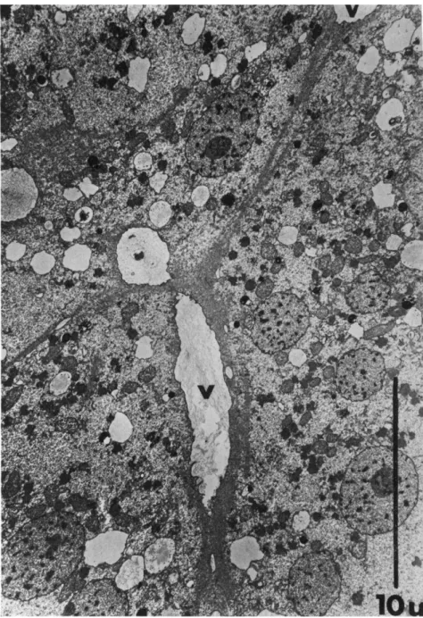
FIG. 7. Low-power electron micrograph revealing the characteristic deformation of vacuoles (V) with fibril attachments; (Physarum polycephalum). Note the deforma- tion of the vacuole in the longitudinal direction of the fibril. Magnification: χ 5500 (unpublished).
Differentiations of the Ground Cytoplasm 93 the ATPase activity of the fibrils. This cytochemical result supports our assumption that myxomyosin and plasma filaments are identical.
After this excursion into the field of cytochemistry let us return to the solid basis of morphological facts. Both light microscopic and electron microscopic investigation of drops, strands, or compact layers of Physarum plasmodia prove that many of the fibrils found have con- tact with vacuoles of different sizes. Such vacuoles occur abundantly in the Physarum protoplasm (a further observation that is very easy
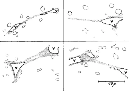
FIG. 8. Drawings of electron micrographs of four fibrils deforming vacuoles (v) in the direction of the longitudinal axis of the fibrils (unpublished).
to make is the frequent attachment of fibrils on the plasmalemma of drops, channel walls, and the limiting membrane of plasmodial proto- plasm). Figure 7 shows this finding in a low-power electron microscopic survey picture. It is nearly impossible to find vacuoles with fibrils with- out the vacuoles seeming to be considerably deformed in a characteristic way. Figure 8 presents four drawings from electron micrographs, which illustrate that the vacuoles with attached fibrils are always deformed in the longitudinal direction of the fibrils. The morphologist can scarcely interpret this phenomenon in any other way than that the fibrils im- pose a tension on the vacuole membrane to which they are attached.
94 Κ. Ε. WOHLFARTH-BOTTERMANN
We planned and carried out the following experiments with the hypothesis in mind that the protoplasmic fibrils of Physarum are rather ephemeral structures. It is not appropriate to give extensive support for this assumption here. It will be enough to mention that the very rapid formation of the fibrils in protoplasmic drops and general and special experiences with other cellular ultrastructures as well (Wohlfarth-Bot- termann, 1959a, 1963a,c) demonstrated a remarkably dynamic capacity of the cytoplasmic building elements. Our idea was that, provided the fibrils were indeed short-lived, we could find differences in their quan- titative occurrence in small plasmodial threads with defined proto- plasmic streaming activity. For this purpose, we used the following experi- mental arrangement (Fig. 9): protoplasmic strands of convenient thickness (0.1-0.5 mm) and length (15 mm) were cut from the plasmodium of Physa- rum grown on filter paper (Camp, 1936). In a moist chamber, which per- mitted observation by the aid of a stereomicroscope, we mounted the strands in two different ways. One position was to hang them up, so that the longitudinal axis of the strand was oriented vertically2) the other was to lay the strands on glass slides so that the threads were orientated horizontally. It is well known that a short time after isolation the pro- toplasmic streaming reappears under such conditions and is quite normal.
We now watched by means of a stereomicroscope the direction of the moving protoplasm in the plasmodial capillary and the rhythmic re- versal of the flow. Sometimes it was possible to make the plasmic flow directly visible (this is the case with very thin strands lying on glass), but we often had to depend on measuring the lengthening or shorten- ing of the ends of the strands as a sign of influx or efflux, respectively, of protoplasm. The objects were fixed just at the moment when either the influx or the efflux of protoplasm had reached its maximum speed.
After fixation we cut off the ends we had observed, embedded them in paraffin wax, and divided them in serial sections of 3 μ thickness. In this manner we examined 80 plasmodial strands with different positions and directions of plasmic flow. Figures 10 and 11, which show the results of this investigation, are drawings of unstained sections evaluated in the phase-contrast microscope. For each drawing, the position of the strand, the plane of the section, and the direction of the protoplasmic flow at the moment of fixation are recorded in a diagram.
Transverse sections through the front of horizontally lying proto-
2 In a further group of experiments the "twisting movement'' of free hanging threads (Kamiya and Seifriz, 1954) was avoided by adhering the strands to glass slides in the same vertical orientation.
Differentiations of the Ground Cytoplasm 95 plasmic strands often reveal no very distinct differences between threads with different streaming directions. Counting the fibrils in terms of in- flux and efflux, however, proved that those endpieces, which squeezed out the protoplasm, contained on an average nearly twice as many fibrils as those threads with an influx of protoplasm. Longitudinal sections, es- pecially in the tangential region of the threads (Fig. 10A and B), show this finding more clearly.
In another group of experiments (cf. Fig. 9 ) we hung up threads, with the idea in mind that here in a vertically oriented thread, the efflux of
/ \ /
W g g / ^ ^ ^
•
I
1 1
\ Ù
••• \ 1
\V V
Ά ϋ
. .1
F i
\ - - -
1 0 m m
/ - - - -- ~ - " Ι - Ô U T
/
LFIG. 9. Schematic representation of the experimental arrangement using proto- plasmic threads of Physarum with defined direction of protoplasmic flow. Drawing, W. P . Fischer (unpublished).
protoplasm had to be effected against the force of gravity, and, in con- trast to this, the influx should be assisted by gravitation. Provided that our view of the function of the fibrils was correct and, also, provided that they are rather ephemeral structures, the quantitative differences in the number of the fibrils should be clearer in this experimental arrangement.
If the fibrils have a direct function for the generation of the motive force we should expect to see the fibrils in large numbers in those threads pumping the protoplasm against gravity. This prediction proved to be
96 Κ. Ε. WOHLFARTH-BOTTERMANN
s ä
Differentiations of the Ground Cytoplasm 97
FIG. 10. Functional arrangement of fibrils. Drawings of phase-contrast microscopic preparations (paraffin wax sections of 3-5 μ thickness) of horizontally lying protoplasmic strands of Physarum, fixed while streaming in a defined direction. Orientation of section and direction of streaming is shown by arrow. Drawing, B. Koeppen-Lesche. (A) Fixation at the moment of influx of protoplasm; (B) fixation at the moment of efflux of protoplasm (unpublished).
98 Κ. Ε. WOHLE ARTH-BOTTERM ANN
FIG. 11. Functional arrangement of fibrils. Drawings of phase-contrast microscopic preparations of vertically suspended protoplasmic strands of Physarum. Drawing, B.
Differentiations of the Ground Cytoplasm
Koeppen-Lesche. (A) Influx of protoplasm; (B) eiïhix of protoplasm.
100 Κ. Ε. WOHLFARTH-BOTTERMANN
FIG. 1 1 . (C) Median section through the thread presented in Β (unpublished).
Differentiations of the Ground Cytoplasm 101 correct. In both transverse and longitudinal sections, there was an ac- cumulation of fibrils in those specimens where the motive force had to work against gravity3 (Fig. 11A-C). Indeed the threads coming down also contain fibrils, but at first sight they seem far fewer. There seems to be a direct relationship between the amount of motive force needed and the number of fibrils. T h e fibrils show many interconnections by branching (Fig. I I B and C), so we can be sure they frequently form a cohesive net- work. This network obviously represents a highly dynamic organization.
Figure 11C shows a median section of the thread in Fig. I I B . This il- lustration (Fig. 11C) demonstrates the occurrence of the fibrils exclusively in an outer zone (probably equivalent to the channel wall) of the thread whereas they cannot be found in the inner core.
Equally interesting and convincing results can be obtained by a com- parative investigation of protoplasmic drops fixed at different times after their formation. Drops fixed immediately after formation (0 min drops) have a very homogeneous cytoplasm without fibrils (Fig. 12a); 10 min afterward, when the reabsorption of the drop is in full progress, the fibril- lar network is present (Fig. 12b).
Figure 13 shows drawings of sections through drops of different ages.
The diagrams explain the ages of the drops and the direction of the preponderant protoplasmic flow. T h e morphologist, without knowing the physiological and biochemical findings that support a pressure-flow mechanism by contractility phenomena tends to interpret the results illustrated in Fig. 13A and Β in the sense cited previously. We believe that the fibrillar network revealed is one site of the force causing stream- ing.
Admittedly, the mode of action of these fibrillar elements remains to be analyzed. One should remember, however, that there is no direct proof of contractility in the myofilaments of smooth muscle cells; nevertheless, nobody doubts their capacity for contraction. Even so we shall shortly review and discuss the facts presented to relate them to the physiological and biochemical results and to some current theoretical interpretations.
In both amebae and slime molds, the groundplasm shows filamentous structures, the plasma filaments, which can form compact fibrils in part
3 It seems probable that a certain number of the fibrils are involved in delivering the motive force for the "twisting movement" observable on free hanging threads (Kamiya and Seifriz, 1954). W e avoided as far as possible fixing threads which showed greater activity in this respect. In any case this does not put in doubt the validity of the result because, as demonstrated by Kamiya and Seifriz (1954), this twisting motion is in- dependent of the direction of protoplasmic streaming. But if this were not so, the abundant occurrence of fibrils in twisting threads would speak for the dynamic function of the fibrillar network.
1 0 2 Κ. Ε. WOHLFARTH-BOTTERMANN
FIG. 1 2 . Phase-contrast micrographs of 3 - 5 μ paraffin wax sections (median section through protoplasmic drops of different ages), (a) " 0 min drop" (fixed as soon as it had emerged from the plasmodial tube network). T h e homogeneous protoplasm of the outer region (OR) contains no fibrillar structures visible in the light microscope. IR
Differentiations of the Ground Cytoplasm 103
= inner region of the drop. Magnification: χ 620 (unpublished), (b) "10 min drop"
(fixed 10 min after it had emerged from the plasmodial tube network). Note that the outer region (OR) of the drop now contains large fibrils joined into a network through ramification points. I R = inner region of the drop. Magnification: χ 420 (un- published).
104 Κ. Ε. WOHLFARTH-BOTTERMANN
FIG. 13. Functional arrangement of fibrils in protoplasmic drops of different ages (Physarum polycephalum). Drawings of phase-contrast microscopic preparations (median paraffin wax sections of 3-5 μ thickness). Osmium-chromium fixation, no staining.
Drawing, B . Koeppen-Lesche. (A) Drop fixed just after it had emerged from the strand (S). T h e arrow in the diagram represents the direction of the main streaming tendency
Differentiations of the Ground Cytoplasm 105 by means of parallel arrangement.4 The fibrils possess ATPase activity, and have a functional disposition in protoplasmic threads of Physarum justifying the assumption that the cohesive network built up by them in the plasmagel wall of the capillary tubes represents the physical struc- ture responsible for the generation of the motive force of the streaming.
This thesis is in accordance with physiological and biochemical results indicating that the enzymatic and mechanochemical system of proto- plasmic streaming in Physarum has much in common with the basic mechanisms of muscle contraction (cf. Kamiya, 1959; Hasselbach, 1962).
The possibility of making this long postulated "gel-reticulum" visible perhaps helps us to throw further light on the dynamic organization producing the motive force.
These conspicuous structures now revealed, which probably produce contraction of a cortical gel layer, seem to corroborate from the mor- phological view the biochemical and physiological results of Kamiya and his group (cf. 1959, 1962) according to which the streaming in Physarum (at least in part) "is caused passively by a difference in internal pressure"
(cf. De Bary, 1864). However, it should be mentioned that not all move- ment phenomena of this organism can be explained by a "simple" pres- sure-flow theory alone, because the facts are more complicated (Stewart and Stewart, 1957, 1959a). Though the microscopic observation of living microplasmodia seems to reveal active contraction phenomena and a squeezing out of protoplasm in the sense of a pressure flow at the moment of the efflux of protoplasm, it is unlikely also with Physarum that such "a simple mechanism" alone deals with all aspects. Here, however, we should bear in mind that the fibrillar network now demonstrated is nothing but a particularly high differentiation of the cytoplasmic matrix. This "un- differentiated" matrix, existing everywhere in both a stiff protoplasmic gel and the flowing endoplasm, should be capable of producing motive force at any point where it might be necessary. In view of this, both localized and limited counterstreaming and a movement of individual
4 Last year, De Pétris et al. (1962) described in normal mononuclear phagocytes filamentous structures with a diameter of 40-60 A, often forming bundles. T h e authors discuss the hypothesis "that these filaments are contractile in nature and are related in some way to the dynamic activity of the cells, either to the movements of the cell as a whole, or to internal shifts of organelles." Joyon and Chawet (1962) found filaments of unknown nature in the cytoplasm of the ameba Hyalosphenia papilio.
of the protoplasm at the moment of fixation. I R = inner region of the drop. (B) Drop fixed 10 min after it had emerged from the strand (S). Note the amassment of fibrils in the outer region (OR) of the drop. T h e arrow in the diagram represents the direction of the main streaming tendency of the protoplasm at the moment of fixation. I R — inner region of the drop (unpublished).
106 Κ. Ε. WOHLE ARTH-BOTTERM ANN
particles against the main stream (Stewart and Stewart, 1959a) are not fully unintelligible.5
In view of our results both in amebae and in slime molds contractility phenomena (which are perhaps comparable to a certain extent with muscle contraction) remain the most satisfactory explanations regarding the source of motive force.
At the present time there seem to be no compelling reasons to abandon the contractility theories in favor of surface tension theories (Kavanau, 1962a,b) or other modern explanations (Jahn and Rinaldi, 1959; Stewart and Stewart, 1959a; Bingley and Thompson, 1962). The front contraction theory of Allen (1961a,b), postulated for amebae contractility of the endoplasm, can also exert a fruitful influence on further morphological research with Physarum.
Though modern cell morphology is now able to reveal the gel reticu- lum of the protoplasm, the morphologist today does not yet seem to be in a position to decide between the different modern contraction theories—
of which one should also mention the interesting concepts of Goldacre (1961) and Yagi (1961)—but perhaps this is only a question of time and effort.
It remains to be tested whether the ideas of "sol-gel transformation"
are not intimately correlated with the conceptions of contractility (cf.
Wohlfarth-Bottermann and Schneider, 1958; Abé, 1961, 1962; Wohlfarth- Bottermann, 1963a). Undoubtedly, the streaming in a plasmodial tube of Physarum requires that an outer, gel-like zone should surround the endo- plasm flowing within. Furthermore, it should be noted that sol-gel trans- formation theories cannot be sharply differentiated from contraction theories, since essential elements of the sol-gel transformation theory are contained in the various contractility theories, or to state it more accu- rately, sol-gel transformation theories have nearly always assumed a
5 The enigmatic counterstreaming in Foraminifera rhizopodia observable in the light microscope cannot be definitely demonstrated with this instrument: the electron microscope clearly revealed that a small rhizopod, appearing in the light microscope as one thread, really consists of a conglomeration of many minute strands of sub- microscopic size (Wohlfarth-Bottermann, 1961b). This finding makes it conceivable that within a single strand there is a one-way streaming, but within different, closely neighboring strands the protoplasm streams in opposite directions, thus simulating a counterstreaming in the light microscope, whose resolution power is unable to visualize the individual threads composing a rhizopod. T h e lack of proof of a counter- streaming in Foraminifera, the fact that each minute thread is limited individually by a definite membrane, and the morphological finding that one rhizopod is simply an open bundle of many individual threads make it difficult at present to explain this type of streaming with various kinds of interfacial forces in the sense of Jahn and Rinaldi (1959) and of Kavanau (1962b).
Differentiations of the Ground Cytoplasm 107 contractility of the ectoplasm or "plasmagel" (cf. Landau et al., 1954;
Marsland, 1956a,b; Hirshfield et al, 1958; Landau, 1959; Yagi, 1961;
Landau and Thibodeau, 1962).
The purpose of this paper was to point out that modern cell mor- phology is able today to demonstrate the long-postulated cytoplasmic gel reticulum and its interesting functional pattern in Physarum. Therefore, morphology is in a better position than ever before to cooperate with cell physiology and promises to make significant contributions toward the goal of elucidating the secret of protoplasmic streaming.
REFERENCES Abé, T. H. (1961). Cytologia 2 6 , 378.
Abé, T . H. (1962). Cytologia 2 7 , 111.
Allen, R. D. (1961a). Exptl. Cell Res. Suppl. 8 , 17.
Allen, R. D. (1961b). In "The Cell" (J. Brächet and A. E. Mirsky, eds.), Vol. II, pp. 135-216. Academic Press, New York.
Bingley, M. S., and Thompson, C M. (1962). J. Theoret. Biol. 2 , 16.
Camp, W. G. (1936). Bull. Torrey Botan. Club 6 3 , 205.
Danielli, J . F. (1951). In "Cytology and Cell Physiology" (G. H. Bourne, ed.), pp. 150- 182. Clarendon Press, Oxford, England.
Danielli, J . F. (1958). In "Surface Phenomena in Chemistry and Biology," pp. 246- 265. Pergamon Press, New York.
Davson, H., and Danielli, J . F. (1943). In "The Permeability of Natural Membranes."
Cambridge Univ. Press, Cambridge, England.
De Bary, E. (1864). "Die Mycetozoen (Schleimpilze). Ein Beitrag zur Kenntnis der niedersten Organismen," 2nd ed. Engelmann, Leipzig, Germany.
De Bruyn, P. P. H. (1957). Quart. Rev. Biol. 2 2 , 1.
De Pétris, S., Karlsbad, G., and Pernis, Β. (1962). / . Ultrastruct. Res. 7, 39.
Goldacre, R. J . (1961). Exptl. Cell Res. Suppl. 8 , 1.
Haas, J . (1955). "Physiologie der Zelle." Borntraeger, Berlin.
Hasselbach, W. (1962). Fortschr. Zool. 1 5 , 1.
Hirshfield, H. I., Zimmerman, A. M., and Marsland, D. (1958). / . Cellular Comp.
Physiol. 5 2 , 269.
Hoffmann-Berling, H. (1958). Fortschr. Zool. 1 1 , 142.
Jahn, T . L., and Rinaldi, R. (1959). Biol. Bull. 117, 100.
Joyon, L., and Chawet, R. (1962). Compt. Rend. Acad. Sei. 2 5 5 , 2661.
Käppner, W. (1961). Protoplasma 5 3 , 504.
Kamiya, Ν. (1959). Protoplasmatologia 8 , 3a.
Kamiya, N. (1960a). Ann. Rev. Plant Physiol. 1 1 , 323.
Kamiya, N. (1960b). Ann. Rep. Sei. Works, Fac. Sei. Osaka Univ. 8 , 13.
Kamiya, N. (1962). In "Handbuch der Pflanzenphysiologie" (W. Ruhland, ed.), Vol.
XVII, part 2, pp. 979-1035. Springer, Berlin.
Kamiya, N., and Seifriz, W. (1954). Exptl. Cell Res. 6 , 1.
Kamiya, N., Nakajima, H., and Abé, S. (1957). Protoplasma 4 8 , 94.
Kavanau, J . L. (1962a). Life Sciences 5 , 177.
Kavanau, J . L. (1962b). Exptl. Cell Res. 2 7 , 595.
Landau, J . V. (1959). Ann. N. Y. Acad. Sei. 7 8 , 487.
Landau, J . V., and Thibodeau, L . (1962). Exptl. Cell Res. 2 7 , 591.
108 Κ. Ε. WOHLFARTH-BOTTERMANN
Landau, J . V., Zimmerman, A. M., and Marsland, D . A. (1954). / . Cellular Comp.
Physiol. 44, 211.
Loewy, A. G. (1952). / . Cellular Comp. Physiol. 4 0 , 127.
Marsland, D . A. (1956a). Pubbl. Staz. Zool. Napoli 3 3 , 182.
Marsland, D . A. (1956b). Intern. Rev. Cytol. 5 , 199.
Mast, S. O. (1926). / . Morphol. 4 1 , 347.
Mast, S. O. (1931). Protoplasma 1 4 , 321.
Nakajima, H. (1956). Seita No Kagaku 7, 256.
Nakajima, H. (1957). 22nd. Ann. Meeting Botan. Soc. Japan.
Nakajima, H . (1960). Protoplasma 5 2 , 413.
Noland, L. E. (1957). / . Protozool. 4, 1.
Obeiiing, Ch. (1959). Intern. Rev. Cytol. 8 , 1.
Palade, G. E. (1956). / . Biophys. Biochem. Cytol. 2 , 85.
Robertson, J . D . (1960a). Progr. Biophys. Biophys. Chem. 1 0 , 343.
Robertson, J . D . (1960b). Proc. Intern. Conf. Electron Microscopy, 4th, Berlin, 1958, 2, 159-171.
Ruska, H. (1962). Proc. Intern. Congr. Neuropathol., 4th. 2 , 42-49.
Schneider, L. (1962). Sei. Tools 9 , 16.
Schneider, L., and Wohlfarth-Bottermann, Κ. E. (1959). Protoplasjna 5 1 , 377.
Schulze, F. Ε. (1875). Arch. Mikroskop. Anat. Entwicklungsmech. 1 1 , 329.
Stewart, P. Α., and Stewart, Β. T. (1957). Federation Proc. 1 6 , 125.
Stewart, P. Α., and Stewart, Β. T. (1959a). Exptl. Cell Res. 1 7 , 44.
Stewart, P. Α., and Stewart, Β. T. (1959b). Exptl. Cell Res. 1 8 , 374.
Ts'o, P. O. P., Eggman, L., and Vinograd, J . (1956). / . Gen. Physiol. 3 9 , 801.
Ts'o, P. O. P., Eggman, L., and Vinograd, J . (1957). Biochim. Biophys. Acta 2 5 , 532.
Wittmann, Η. (1950). Protoplasma 3 9 , 450.
Wittmann, Η . (1951), Protoplasma 4 0 , 23.
Wohlfarth-Bottermann, Κ. Ε. (1957). Naturivissenschaften 44, 287.
Wohlfarth-Bottermann, Κ. Ε. (1959a). Zool. Anz. 2 3 , Suppl., 393.
Wohlfarth-Bottermann, Κ. E. (1959b). Z. Zellforsch. Mikroskop. Anat. 5 0 , 1.
Wohlfarth-Bottermann, Κ. E. (1960). Protoplasma 5 2 , 58.
Wohlfarth-Bottermann, Κ. E. (1961a). Protoplasma 5 3 , 259.
Wohlfarth-Bottermann, Κ. E. (1961b). Protoplasma 5 4 , 1.
Wohlfarth-Bottermann, Κ. E. (1961c). Protoplasma 5 4 , 307.
Wohlfarth-Bottermann, Κ. E. (1962). Protoplasma 5 4 , 514.
Wohlfarth-Bottermann, Κ. E. (1963a). Intern. Rev. Cytol. 1 6 , pp. 61-132.
Wohlfarth-Bottermann, Κ. E. (1963b). Protoplasma 5 4 , 514.
Wohlfarth-Bottermann, Κ. E. (1963c). Naturwissenschaften, 5 0 , 237.
Wohlfarth-Bottermann, Κ. E. (1963c). Naturwissenschaften 5 0 , 237.
Yagi, K. (1961). Comp. Biochem. Physiol. 3 , 73.
Zeiger, Κ. (1943). Klin. Wochschr. 2 2 , 201.
DISCUSSION
CHAIRMAN THIMANN: Would you care to guess the fraction of the slime mold material which is occupied by the fibers?
DR. WOHLFARTH-BOTTERMANN: Yes, I think the number of fibrils depends on the physiological state and is very different in different areas of the plasmodium. If you have a very thin microplasmodium, then there are not many fibers; but if you ask for the total mass of fibril-forming material, then the whole mass of groundplasm has to be considered. In that case, the value would be rather high.
Differentiations of the Ground Cytoplasm 109
DR. BOUCK: What is the nature of the endoplasmic reticulum?
DR. WOHLFARTH-BOTTERMANN: I think mostly one doesn't find endoplasmic reticu- lum but rather vacuoles. Most people, I think, will call them regular endoplasmic reticulum; but I would not. I never understood why a vacuole should be called a reticulum.
DR. STEWART: Have you ever seen the fibers in a living plasmodium?
DR. WOHLFARTH-BOTTERMANN: Yes. We have occasionally seen them in living micro- plasmodia with the phase-contrast microscope. Also, I saw polarization microscopic pictures made by Dr. Nakajima which were very convincing.6
DR. GOLDACRE: I demonstrated fibrils that can be seen in the light microscope by the technique of allowing an ameba to lose half its water.
DR. WOHLFARTH-BOTTERMAN: I know. W e intended to try using your technique before we saw fibers in our ameba after mere fixation. However, I have referred to your paper.
6 Note added in proof: Using the freeze-substitution techniques, G . S. Omenn saw
"strand-like material which might possibly resemble endoplasmic reticulum" in plas- modia of Physarum in the electron microscope without recognizing the importance of these pictures (Plates 8 and 9, G . S. Omenn: T h e myxomycete Physarum poly- cephalum. Cultivation, Ultrastructure, Molecular Model for Contractility. Unpublished Senior Thesis carried out under the direction of L. I. Rebhun, Department of Biology, Princeton University, 1961). There is no doubt that the "strands" of Omenn are identi- cal with the fibrils discussed here. T h a t means that these structures can also be demon- strated by freeze-substitution techniques.