IN CULTURES OF AGGREGATES OF DISSOCIATED EMBRYONIC CHICK OTOCYSTS
MARY FAITH ORR
Department of Anatomy Northwestern University Medical and Dental Schools
Chicago, Illinois
I. INTRODUCTION
Several studies have shown that the embryonic chick otocyst developed sensory epithelia of the inner ear in organ culture that were similar to the sensory epithelia that developed in vivo
(Fell, 1928; Friedmann, 1956, 1965, 1968; Orr, 1968). Basilar papillae and cristae were frequently observed. These structures developed morphologically although there was no anatomical devel- opment in culture. The sensory epithelia were composed of fully differentiated sensory cells and supporting cells. In electron microscopic studies Friedmann (1959, 1968, 1969) and Friedmann and Bird (1961) demonstrated that sensory cells in the sensory epithelium of the basilar papillae have a surface structure (hair) composed of sterocilia; ocassionally a basal body was observed.
363
It was also shown by Friedmann that nerve fibers had formed synap- tic contacts with the sensory cells. The sensory cells in the epithelium covering a ridge-shaped crista have a surface structure composed of a kinocilium and numbers of sterocilia.
A light microscopic study of organ cultures of aggregates of dissociated otocysts and associated tissues revealed that areas of reconstructed epithelium differentiated into sensory cells and supporting cells. The epithelium in these areas was innervated by nerve fibers. There were other areas of reconstructed epithelium in which a few poorly differentiated sensory cells were observed;
no nerve fibers were demonstrated in these epithelia (Orr, 1968).
This is a report of the preliminary electron microscopic ob- servations of the surface structures of sensory cells that have differentiated in reconstructed epithelium in organ cultures of aggregates of dissociated embryonic chick otocysts.
II. MATERIALS AND METHODS
A. Dissociation and Organ Culture Methods
1. Dissociation
Embryonic chick otocysts along with the surrounding mesen- chyme and acoustic ganglion were dissected out of the embryo on the 4th day of incubation and dissociated according to the method of Moscona (1961). The otocysts were incubated for 15 minutes in Calcium- and Magnesium- free Hanks' balanced salt solution (Ca- and Mg- free HBSS) and then for 45 minutes in 1% trypsin (Difco 1:250) dissolved in Ca- and Mg- free HBSS. The tissue was washed three times in Ca- and Mg- free HBSS and then placed in one ml of nutrient medium or regular HBSS. The otocysts were dis- sociated by flushing through a fine pipette and the cellular sus- pension was allowed to aggregate for one hour in a test tube that
was turned by hand at frequent intervals or by rotation in a rol- ler drum (revolving 10 times per hour).
2 . Organ Culture
Aggregates were placed in organ culture chambers according to the method previously described (Orr, 1968, 1975b). Control oto- cysts were cultivated in the same manner. Two nutrients were used with equal results: (1) The supernatant from clotted chicken plasma and embryo extract (equal parts), or (2) equal parts fetal
calf serum and embryo extract. The pH was adjusted to 7.4-7.6.
The medium was changed at 2 or 3 day intervals for 12 days.
B. Preparation of Histologic and Electron Microscopic Specimens
1. Histologic Preparations
Otocysts and cultures of otocysts and aggregates were fixed, processed, embedded in paraffin and serially sectioned according to the methods described in previous reports (Orr, 1968, 1975b).
Some of the sections were stained with periodic acid-Schiff tech- nique (PAS) followed by hematoxylin, while others were impreg- nated with silver according to Bodian1 s protargol method followed by the periodic acid-Schiff technique. Details of these staining methods were described by Orr (1975b).
2. Preparation of Electron Microscopic Specimens
Cultures of aggregates and intact otocysts were fixed for electron microscopy in 1% osmium tetroxide in Veronal acetate buffer (Palade, 1952) adjusted to pH 7.2-7.4. Specimens were also fixed in 1% glutaraldehyde in HBSS followed by post-fixation in 1% osmium in HBSS. Tissues were processed in graded ethanols, cleared in propylene oxide, infiltrated in equal parts propylene oxide and Araldite 502, and embedded in Araldite 502 (Orr, 1975 a and b ) . Light microscopic sections (0.5 to 1.0 micron in thick-
ness) were stained with, a mixture (equal volumes) of 1% methylene blue in 1% borax solution and 1% azure II (Richardson et al., 1960). Ultrathin sections were cut on a Porter-Blum MT I ultra- microtome, stained with saturated uranyl acetate followed by lead citrate (Reynolds, 1963) and examined with a Hitachi 11 F electron microscope.
III. RESULTS
As noted in the introduction certain observations have been reported that were made on serial sections of histologic prepara- tions of organ cultures of aggregates and intact otocysts (Orr, 1968). In order to make a clear presentation of the results of this study it was considered necessary to illustrate similar ob- servations in this report. The photomicrographs shown here have not appeared in any other publications.
A. Embryonic Chick Otocyst
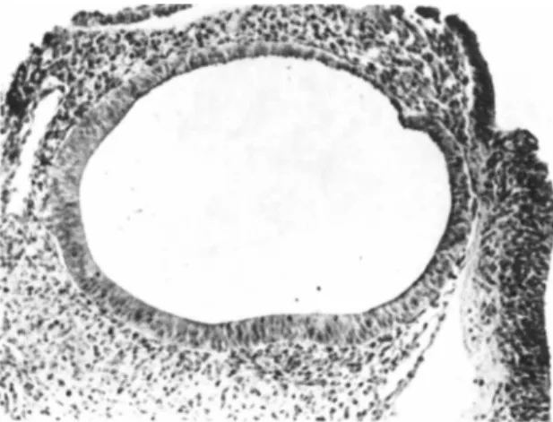
Figure 1 is a photomicrograph of an embryonic chick otocyst on the 4th day of incubation. The pseudostratified epithelium in the ventral portion of this sac-like structure merged with a cu- boidal epithelium on the dorsal surface. The epithelium contained cells undergoing cell division and cells with a cilium. Under- lying the epithelium was a thin basement membrane and the acoustic ganglion was adjacent to the pseudostratified epithelium (Orr, 1968, 1975a).
B. Sensory Epithelia in Twelve-day-old Cultures
A basilar papilla that developed and differentiated in a twelve-day-old culture is shown in figure 2. The basilar papil- lae were composed of rows of sensory cells; each sensory cell was surrounded by supporting cells. The presence of hair structures on the surface of sensory cells was considered one characteristic
FIGURE 1 This is a photomicrograph of a histologic section of an embryonic chick otocyst on the 4th day of incubation. The acoustic ganglion is not shown in this section. PAS and hema- toxylin. X75.
FIGURE 2 This is a section of a basilar papilla in a 12-day- old culture. Note the hairs extending from the surface of the hair cells towards the tectorial membrane (TU) . Nerve fibers (NF, arrow) can be observed innervating the sensory epithelium. Bodian and PAS. X650.
FIGURE 3 Ά sensory cell in a basilar papilla that developed in a 12-day-old culture is shown in this electron micrograph.
Note the sterocilia on the surface; the gradation from the tallest to the shortest sterocilia can be seen in this section. Xll,750.
that indicated complete cytodifferentiation of the sensory cells.
Hairs extended from the surface of sensory cells towards the tec- torial membrane. Nerve fibers impregnated with silver were ob- served innervating the sensory epithelium. An electron micro- scopic examination of a basilar papilla revealed that there were numbers of sterocilia on the surface of the sensory cells (Figure 3 ) . Basal bodies of kinocilia were also observed in some cells.
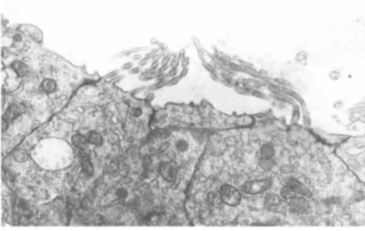
The sensory cells in the sensory epithelium that covered a crista were 'tear-drop' shaped; two sensory cells are shown in Figure 4. An electron microscopic examination of a cross section of the surface structure (hair) of a similar sensory cell demon- strated that the sterocilia were arranged in orderly rows with a kinocilium located in a constant position (Figure 5 ) .
FIGURE 4 Two sensory cells in the sensory epithelium of a crista can be seen in this electron micrograph. XI1,750.
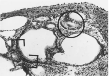
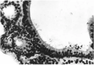
C. Sensory Epithelia in Twelve-day-old Cultures of Aggregates Cellular suspensions (that contained undissociated groups of cells) of embryonic otocysts reaggregated into clumps of cells within an hour. After three days in culture reconstructed epi- thelium had formed in organ cultures of aggregated clumps of cells. Although morphologic development did not occur, areas of the reconstructed epithelium differentiated into sensory cells and supporting cells (Figure 6 ) . Figure 7 is an example of a sensory epithelium in which sensory cells with demonstrable hairs are shown in a histologic section. An examination of histologic preparations impregnated with silver revealed that nerve fibers were present in these sensory areas of epithelium (Orr, 1968).
Furthermore, with electron microscopy it was shown that the
FIGURE 5 An electron micrograph of a cross section of the surface structure of a sensory cell similar to those depicted in Figure 4. The orderly arrangement of the sterocilia and the lo- cation of the kinocilium are demonstrated in this micrograph.
X7,050.
FIGURE 6 This is a photomicrograph of a histologic section of a 12-day-old culture of aggregates of dissociated otocysts.
One area of sensory epithelium is within brackets. Another sen- sory epithelium is circled; PAS positive material is present in the lumen of this structure. Bodian and PAS. X250.
FIGURE 7 The sensory epithelium within brackets in Figure 6 is shown here at higher magnification. Note the well-aligned row of sensory cells with hair structures extending into the lumen.
The hairs have been distorted by the preparation procedure.
Bodian and PAS. X400.
sterocilia of the sensory cells had developed and were comparable with the sterocilia of sensory cells that had developed in cul- tures of intact otocysts (Figure 8 ) .
There were other areas of reconstructed epithelium that con- tained a few sensory cells that were poorly differentiated (Fig- ure 9 ) . Nerve fibers were not observed innervating these areas of epithelium. The electron microscopic examination showed that there were several abnormal configurations of the sterocilia on the surface of the sensory cells. In some electron micrographs of longitudinally cut sensory cells the sterocilia were disorga- nized and some appeared enlarged (Figure 1 0 ) . Sterocilia that were abnormally bent were observed in other sections (Figure 1 1 ) . In Figure 12 there is one sensory cell cut in longitudinal sec- tion with a few sterocilia that appeared normal. A cross section of a surface structure in this same electron micrograph revealed a kinocilium and numbers of sterocilia, however, the arrangement
FIGURE 8 An electron micrograph of the sterocilia of a sen- sory cell that differentiated in a 12-day-old culture of aggre- gates. These sterocilia are comparable with those observed on the surface of sensory cells that differentiated in cultures of intact otocysts. X7,050.
FIGURE 9 This photomicrograph of a histologic section of a 12-day-old culture of aggregates reveals a sensory epithelium
(within brackets) in which only two poorly differentiated sensory hair cells can be observed. Bodian and PAS. X250.
FIGURE 10 The sterocilia shown in this electron micrograph are abnormal in development and distribution. Note that one sterocilium in this electron micrograph is enlarged (arrow).
X7,000.
FIGURE 11 In this electron micrograph the sterocilia of the sensory cell on the right are abnormally bent. X7,000.
of the sterocilia was disorganized. Compare this cross section with the cross section in Figure 5.
FIGURE 12 In this electron micrograph there is one cell cut in longitudinal section with a few sterocilia that appear to be normal; the tip of a kinocilium can be seen. Ά kinocilium (cir- cled) and numbers of disorganized sterocilia can be observed in this tangential section across a hair structure in the same micrograph. X7,000.
IV. DISCUSSION
This electron microscopic study has shown that there were well-developed sterocilia on the surface of sensory cells that had developed in organ cultures of aggregates of dissociated em- bryonic chick otocysts. It has been shown in a study of histo- logic preparations of cultures of aggregates that nerve fibers innervated areas of epithelium that had differentiated into sen- sory hair cells and supporting cells (Orr, 1968). Synaptic con-
tacts between nerve fibers and sensory cells in reconstructed epithelia have not been explored with the electron microscope.
The findings in this study have also confirmed that the cyto- differentiation of the sensory cells in some reconstructed epi- thelia was affected by the lack of some factor. The sterocilia of some of the sensory cells were revealed to be abnormal in distribution, development and physical structure; andf there was a definite indication that the presence of a kinocilium did not prevent disorganization of the sterocilia. Based on light micro- scopic studies of histologic preparations it was suggested that complete cytodifferentiation of sensory cells was dependent on in- nervation of epithelium (Orr, 1968). Friedmann (1968) had also noted that "the inclusion of certain neurogenic elements plays an essential role in the differentiation of the sensory areas" of the inner ear of the embryonic chick in culture. Sher (1971) re- ported that innervation of the sensory areas of the inner ear of the mouse preceded differentiation of the epithelium.
The most likely explanation for the poor differentiation of the surface structures of the sensory cells was the lack of in- nervation of the sensory epithelium. However, it should be noted that there may be other factors involved in some instances. Some of the undissociated groups of cells in the cellular suspension were probably responsible for some of the sensory epithelium in the reconstructed epithelium in cultures of aggregates. It has been shown that there were groups of cells in the embryonic chick otocyst that have cohesive apical junctions that were not dis- rupted by trypsin although the desmosomes were affected (Orr, 1975a). The poor differentiation of some of the sensory cells may be due to incompletely reformed intercellular connections.
This study was restricted to the formation of sterocilia on the surface of the sensory hair cells since this was considered one characteristic of cytodifferentiation that would be most re- vealing. The influence of the innervating nerve fibers has been discussed. However, it should be noted that a previous study
also indicated that histodifferentiation of the epithelium into sensory epithelia required the proximity of mesenchymal and epi- thelial tissue (Orr, 1968). This requirement would be particu- larly important in the early stages of localization and develop- ment of the sensory epithelia.
V. CONCLUSION
This study with electron microscopy has shown that the stero- cilia were well-formed on the surface of sensory cells in some of the reconstructed epithelium in cultures of aggregates of dis- sociated embryonic chick otocysts. The sterocilia of other sen- sory cells were abnormal in distribution, development and physi- cal structure. The most probable cause was considered to be the lack of innervation of these epithelia.
ACKNOWLEDGMENTS
The author wishes to thank Mrs. Ethel Golliday for technical assistance and Ms. Sue Decker for assisting in preparing the prints. This investigation was supported by a United States Pub- lic Health Service Grant NS 08569.
REFERENCES
Fell, H. B. (1928). Arch. Exptl. Zellforsch. 7, 69-81.
Friedmann, I. (1956). Ann. Otol. Rhinol. Laryngol. 65, 98-109.
Friedmann, I. (1959). J. Biophys. Biochem. Cytol. 5, 263-268.
Friedmann, I. (1965). In "Cells and Tissues in Culture" (Ε. N.
Willmer, e d . ) . Vol. II, pp. 521-548. Academic Press, New York.
Friedmann, I. (1968). J. Laryng. and Otol. 82, 185-201.
Friedmann, I. (1969). Acta Oto-laryngologica 67, 224-238.
Friedmann, I,, and Bird, E, S. C1961), J. Ultrstruct. Res. 20, 356-365.
Moscona, A. A. (1961). Exptl. Cell Res. 22, 455-475.
Orr, M. F. (1968). Devel. Biol. 17, 39-54.
Orr, M. F. (1975a). Devel. Biol. 47, 325-340.
Orr, M. F. (1975b). in "Handbook of Auditory and Vestibular Re- search Methods" (C. A. Smith and J. A. Vernon, eds.), Chapt. 5, pp. 127-174. C. C. Thomas, Springfield, Illinois.
Palade, G. E. (1952). J. Exptl. Med. 95, 285-298.
Reynolds, E. S. (1963). J. Cell Biol. 17, 208-212.
Richardson, K. C., Jarell, L., and Fincke, Ε. H. (1960). Stain Technology 35, 313-323.