III./7.2. Clinical characteristics and symptoms
In this chapter, the symptoms of intracranial tumors and certain related concepts are discussed. Anatomical and physiological knowledge needs to be first reviewed in order to understand the information provided in this chapter.
Introduction – location of tumors
Elements
Some tumors originate from the nervous system itself (peripheral nerves, brain or spinal cord), while others from the surrounding organs and tissues (e.g. skull, meninges, spinal column) causing secondary damage to neural elements. Neurosurgeons prefer to use the intraaxial-extraaxial distinction:
tumors located within the brain and spinal cord are called intraaxial, while tumors located outside the nervous system, but within the bony boundaries of the central nervous system are called extraaxial.
What is the
difference between an intracranial and brain tumor?
Tumors growing inside the skull are often referred to as “brain tumors”, but it is more appropriate to talk about intracranial tumors in general, because only some of them originate from the brain or are located in the brain. Many tumors originate inside the skull from other, non-neural tissues (e.g.
meningiomas from the meninges), not to mention metastases, which comprise half of intracranial tumors in adults.
Monroe-Kellie principle
Intracranial pressure gradients
The symptoms of intracranial (IC) tumors derive partly from the Monroe- Kellie principle, which states that the total volume of the three components (brain tissue, blood, cerebrospinal fluid) of intracranial space is constant, because the volume inside the cranium is fixed. An increase of volume of one of the three components can only happen on the expense of another. Because intracranial space has limited possibilities of compensation, a rise of the intracranial pressure can occur. The normal value of IC pressure is 100-170 mm of water.
What is a mass lesion?
Intracranial tumors act as “mass lesions” or “space-occupying lesions”, which terms are used in clinical practice as synonyms for tumors.
It is important to emphasize that increased intracranial pressure is not necessarily a sign of a space-occupying lesion. It may be caused by many reasons, which are divided into three groups. However, some pathological processes may belong to 2 or 3 groups (e.g. sinus thrombosis).
Classification of causes of raised intracranial pressure
Which diseases cause raised
1. Space-occupying processes Tumors and tumor-like lesions
Vascular disease (brain hemorrhage or large infarct) Infections (brain abscess, herpes encephalitis, etc.)
intracranial
pressure? Traumatic disorders (contusion, epi-and subdural hematoma) Brain edema from any cause
2. Abnormalities of CSF
Disorders of CSF circulation and absorption, hydrocephalus Changes in the composition of CSF (meningitis, subarachnoid hemorrhage, high protein content)
3. Circulatory causes
Intracranial venous (sinus) thrombosis Hypertensive crisis
Causes and types of cerebral edema
Cerebral edema is a reaction of brain tissue to a variety of effects. Based on the mechanism of development, three types of brain edema are differentiated:
1. Cytotoxic brain edema (intracellular)
Mechanism of action: in the absence of energy supply, sodium- potassium pumps fail and water accumulates inside the cells.
Causes: ischemia, hypoxia, metabolic diseases, and intoxications 2. Vasogenic brain edema (extracellular)
Mechanism of action: due to the damage of the blood-brain barrier, water exits into the space between cells.
Causes: tumors, infections, trauma, hypertensive crisis 3. Interstitial brain edema
Mechanism of action: in hydrocephalus, CSF enters the
periventricular brain tissue through micro-lesions of the ependyma.
Importance and forms of herniation
The space within the skull is divided into different compartments, occupied by specific parts of the brain. If a space-occupying lesion develops in one compartment, brain tissue is displaced into the adjacent compartment. This is called herniation. As a result of this process, not only certain brain centers and pathways may be affected, but also blood vessels, nerves or CSF circulation pathways, causing serious secondary complications. As the only natural exit of the skull is the foramen magnum, most of the large space- occupying processes eventually converge in the direction of the foramen magnum causing tonsillar herniation, which is fatal due to the compression of the medulla oblongata.
Fig. 1: Signs of tonsillar herniation in a post-mortem specimen
Fig. 2: Lateral (subfalcial) herniation:
the cingulate gyrus is displaced and compressed under the falx cerebri towards the opposite side, caused by a
glioblastoma
Fig. 3: Transtentorial (uncus) herniation with the compression of the mesencephalon, caused by a space-occupying glioblastoma
The symptoms of nervous system tumors are determined by their location and size, rather than by their histological type.
IC tumors usually show a slow and gradual progression in symptoms and signs. However, it is not uncommon that severe neurological symptoms develop suddenly in asymptomatic patients, suggesting first a cerebrovascular disease, but a tumor is shown by imaging studies. This sudden deterioration may be caused by a hemorrhage within the tumor, herniation, cerebral infarction due to the compression of a blood vessel by the tumor, and epileptic seizure.
Hogyan alakulnak a daganatok tünetei?
As the IC tumor grows, it damages different areas of the brain, causing focal signs. These may be symptoms functional deficit (paresis, hypesthesia, speech disorder) or partial and secondary generalized epileptic seizures.
Areas adjacent to the tumor (cortical centers, tracts) may also come under compression, leading to even more symptoms. Even small tumors may cause the obstruction of cerebrospinal fluid circulation pathways, which leads to occlusive hydrocephalus and rapid deterioration of clinical symptoms.
The intracranial hypertension syndrome
After reaching a certain size, all intracranial tumors - regardless of their initial location and histological type –lead to the syndrome of elevated IC pressure mentioned above. These symptoms are nonspecific, but their co-occurrence is characteristic for raised IC pressure. Perifocal cerebral
edema has a major role in the development of the space-occupying effect of a tumor. Gliomas with an infiltrative growth are often associated with a space- occupying effect, which is less pronounced in comparison to other tumors.
This is explained by the fact that tumor tissue is located in between normal cerebral fibers and cells.
Fig. 4: 53-year-old female patient. Slowly-growing glioma in the left hemisphere, discovered 25 years ago (histology: A2). Despite the large size of the tumor, the space occupying effect is mild.
The patient has only mild motor aphasia. MRI images, FLAIR (Fluid Attenuated Inversion Recovery) sequences.
Disorders of consciousness
Headache is one of the most typical symptoms of elevated IC pressure. It usually has a diffuse, overwhelming, compressive character, exacerbated in the morning or in lying position. The intensity of headache increases over time: progressive headache is very typical for IC tumors. The location of the headache is usually not related to the location of the tumor.
Nausea and vomiting are common accompanying symptoms. Severe
vomiting without nausea is seen in childhood in posterior fossa tumors, but it is not typical in adulthood.
Organic
psychosyndromes
In an advanced stage of increased IC pressure, the level of consciousness gradually decreases. The patient first becomes lethargic, disinterested and drowsy, which is followed by stages of somnolence, stupor, and coma.
Cushing’s phenomenon is a significant increase of systolic blood pressure, with a decrease of diastolic blood pressure, heart rate and respiratory rate.
Bilateral abducens nerve palsy with double vision may develop due to the compression of the abducens nerve on the skull base, where the nerve runs freely over a long segment.
If raised IC pressure is persistent, congestive signs in the optic fundus develop due to the obstruction of venous flow, and bilateral edema of the optic nerve head (papilloedema) is seen. Papilloedema does not cause deterioration of vision for a long time, but eventually concentric narrowing of the visual field, and gradual loss of vision may occur. Papilloedema is
detected by the ophthalmoscopic examination of the optic fundus, and its severity is given in diopters.
The phenomenon of obscuration may occur as a non-specific symptom, which is caused by bilateral posterior ischemia presenting as a bilateral transient loss of vision, especially when bending forward.
Nuchal rigidity and a head tilt resembling torticollis occurs mainly in conditions close to herniation, in posterior fossa tumors, primarily in childhood.
Tumors damaging the association areas of the cerebral cortex cause changes
in higher cognitive functions, rather than focal signs. Depending on the location of the tumor, various syndromes may occur, such as a prefrontal or parietal lobe syndrome. Tumors growing in the prefrontal region may reach a large size before being recognized, because only symptoms suggesting a mental disorder are present for a long period of time, and focal neurological signs appear quite late.
Endocrine symptoms
Endocrine symptoms in most cases are caused by pituitary or other sellar tumors. Hormonally active tumors are discovered earlier because of the typical hyper-functional endocrine syndromes. Tumors that do not produce hormones may cause pituitary underfunction (panhypopituitarism) after reaching a certain size by damaging the normal gland.
Paraneoplastic syndromes
Although the nervous system is relatively often affected by paraneoplastic disorders caused by tumors of other organs, paraneoplastic syndromes are not typically associated with primary CNS tumors. The rare opsoclous- myoclonus-cerebellar ataxia is one exception, which is considered to be a distant effect of neuroblastoma occurring in childhood, and caused by the cross-reaction of the antibodies produced against the tumor.
The special oncological aspects of the CNS tumors deserve a separate discussion.
Because of their special location and certain characteristics, general
oncological principles have only a limited value in central nervous systems tumors. This is especially important in determining the type of therapy.
In brain and spinal cord tumors, histological benignancy or malignancy is often not closely related with the actual biological behavior of the tumor. For example, a benign tumor growing in a critical location that is poorly
accessible surgically or by other means may cause life-threatening complications by compressing vital centers or leading to increased IC pressure.
Grading
The behavior of IC tumors may be characterized by a grading system ranging from I to IV: grade I-II is considered benign, and grade III-IV malignant.
However, tumors are often not homogeneous, different areas of a given tumor may have different grades.
With time, the grade can increase, which may also be inhomogeneous within a tumor.
Infiltrative tumors
Some tumors are well defined with macroscopically and microscopically sharp borders, located within a capsule, and they show a non-infiltrative, expansive growth. These are usually extraaxial tumors (e.g. meningioma, vestibular schwannoma). However this does not apply to most of the cerebral gliomas (and other intraaxial tumors); they have no sharp borders within brain tissue, tumor cells can be detected very far from the borders seen macroscopically or by imaging methods, even up to 9 cm away in brain tissue of normal appearance. This explains the high rate of recurrence.
Eloquent areas
In addition to the points mentioned above, in brain and spinal cord tumors the proximity of vital brain centers or tracts may be a limiting factor in the radical surgical removal of the tumor or it’s in toto removal with excisions in normal area. These are called eloquent areas, which include for example the
speech centers, or the primary motor field. During surgery, eloquent areas should be spared at all times, or surgery may be contraindicated if this is technically impossible. Accordingly, the operability of brain tumors is significantly influenced by the location of the tumor.
The limitations of surgical treatment
Because of the difficulties mentioned above, the removal of brain tumors is incomplete in many cases, and the reduction of tumor size and the temporary reduction of space-occupying effect is often the only realistic goal. In this case, relapse is inevitable.
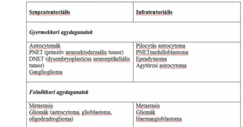
Relationship between age, tumor location and histological type Some nervous system tumors may occur at any age, but certain types of tumors typically appear at a particular age. In childhood, brain tumors are the second most common tumors after hematologic malignancies. The incidence of primary brain tumors shows a peak in children and young adults, and in the elderly, whereas metastatic tumors are more frequent in the elderly. Age and location of tumors are also related, as childhood tumors are commonly infratentorial, while tumors in adulthood are mainly supratentorial.
Fig. 5: Primary intraaxial tumors grouped according to age and location
Metastases in the nervous system
Most of CNS tumors may occur in any location within the brain and the spinal cord, but certain types of tumors occur exclusively in specific locations. For example, medulloblastoma originates only from the cerebellum, and vestibular schwannoma from the cerebellopontine angle.
However, other tumors (e.g. meningioma) may also occur in the cerebellopontine angle.
Nervous system malignant tumors - in contrast to other malignancies - usually do not give metastases to extraneural organs or tissues. Neither is it typical for metastases to form within the nervous system, with the exception of a few tumors where tumor cells spread mainly via the CSF. The latter are called "drop metastases", which may occur in medulloblastomas, malignant gliomas, ependymomas, and chorioideus plexus tumors. For this reason, a special treatment, the irradiation of the entire spinal cord is necessary in medulloblastoma.
Causes
IC metastatic tumors, especially if located on the surface, tend to spread in the CSF space and form infiltrates along the surface, which is called meningeal carcinomatosis.
Another specific form of tumor propagation is along white matter tracts, which occurs mainly in glioblastoma. The tumor may grow via the corpus callosum into the contralateral hemisphere ("butterfly-tumor").
In rare cases, CNS tumors may have a multifocal primary origin, i.e. multiple
tumors appear simultaneously in distant brain areas.
The cause of nervous system tumors
The causes of primary CNS tumors are mostly unknown. In some types of tumors, the role exogenous factors (such as X-ray radiation in meningioma) or inherited genetic factors (e.g. in hereditary phacomatoses) is confirmed.
There is no significant difference in the average incidence of tumors between genders, however, a significant difference may exist in particular tumor types.
For example, the incidence of meningioma is twice as common in females as in males.
Infections rarely play a role in tumor genesis. Primary central nervous system B-cell non-Hodgkin's lymphoma is an exception, which is common in AIDS and in other immunodeficiency syndromes.
Since the general risk factors of nervous system tumors are unknown and the nervous system cannot be tested with simple methods, there are no routinely available screening methods for detecting brain and spinal tumors at an early stage.
For further reading
The symptoms of the intracranial tumors are presented in detail on the following page:
http://www.virtualtrials.com/symptoms.cfm