Experimental therapeutic approaches against hyperglycemia-induced mitochondrial injury
in endothelial cells
PhD thesis
Domokos Ger ő M.D.
Doctoral School of Basic and Translational Medicine Semmelweis University
Consultant: Miklós Mózes, M.D., Ph.D.
Official reviewers: János Nacsa, M.D., Ph.D.
Ákos Zsembery, M.D., Ph.D.
Head of the Complex Exam Committee:
Anikó Somogyi, M.D., D.Sc.
Members of the Complex Exam Committee:
György Jermendy, M.D., D.Sc.
György Nádasy, M.D., Ph.D.
Éva Szőke, D.Sc.
Budapest
2018
TABLE OF CONTENTS
LIST OF ABBREVIATIONS ... 4
1. SCIENTIFIC BACKGROUND ... 8
1.1. Introduction ... 8
1.2. Characteristics of the damage ... 8
1.2.1. Glucose and oxidative stress in diabetic vascular damage ... 8
1.2.2. Target cells of hyperglycemia ... 10
1.2.3. Time course of hyperglycemic injury ... 11
1.3. Triggers of endothelial dysfunction and damage ... 12
1.3.1 Hyperglycemia and ‘glucose memory’ ... 12
1.3.2. Downstream molecules responsible for the injury ... 15
1.4. Mechanisms of ROS production in hyperglycemia ... 15
1.4.1. Glucose-induced oxidative stress pathways ... 15
1.4.2. Unifying hypothesis: the role of mitochondrial oxidants ... 19
1.4.3. The mechanism of glucose-induced mitochondrial superoxide generation .. 20
1.5. Mechanism of damage: Cell damaging responses to ROS production in hyperglycemia ... 26
2. AIMS ... 30
3. MATERIALS AND METHODS ... 32
3.1. Compound libraries and commercially available drugs ... 32
3.2. Synthesis of mitochondrial H2S donors (10-(4-Carbamothioylphenoxy)-10- oxodecyl) triphenylphosphonium bromide (AP123) and (10-Oxo-10-(4-(3-thioxo- 3H-1,2-dithiol-5-yl)phenoxy) decyl)triphenylphosphonium bromide (AP39) . ... 32
3.3. H2S release detection in solution and in situ in endothelial cells ... 34
3.4. Cell culture and cell-based screening for inhibitors of hyperglycemia
induced mitochondrial ROS production ... 35
3.5. Measurement of cytoplasmic ROS generation ... 36
3.6. In situ detection of ROS generation ... 37
3.7. Mitochondrial ROS measurement in isolated mitochondria ... 37
3.8. Xanthine-oxidase assays ... 38
3.9. Viability assays: MTT and LDH assays, ATP measurement ... 38
3.10. Mitochondrial membrane potential measurement ... 39
3.11. Gene expression array ... 40
3.12. siRNA mediated gene silencing and real-time PCR measurements ... 40
3.13. Mitochondria isolation and western blotting ... 41
3.14. Detection of oxidative nucleic acid and protein damage ... 42
3.15. Detection of oxidative damage of the mitochondrial DNA (mtDNA) ... 43
3.16. Respiratory complex II/ III activity assay ... 43
3.17. Extracellular Flux Analysis ... 44
3.18. Vascular studies of in vitro hyperglycemia ... 45
3.19. Vascular studies of streptozotocin-induced diabetes ... 45
3.20. Statistical analysis ... 45
4. RESULTS ... 46
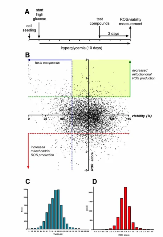
4.1. Characterization of the hyperglycemic endothelial cell injury model ... 46
4.2. Cell-based screening for inhibitors of hyperglycemia-induced mitochondrial ROS production in endothelial cells ... 49
4.3. Characterization of the mode of action of hit compounds ... 53
4.3.1. The mechanism of action of paroxetine: mitochondrial superoxide
scavenging in hyperglycemic endothelial cells ... 53
4.3.2. Paroxetine protects against oxidative damage: prevents the hyperglycemia- and diabetes-induced endothelial dysfunction in vascular rings ... 60
4.3.3. Glucocorticoids inhibit the mitochondrial ROS production in microvascular endothelial cells ... 63
4.3.4. The mode of action of glucocorticoids: restoration of the mitochondrial potential via UCP2 induction ... 66
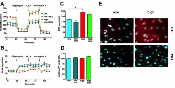
4.3.5. The mode of action of mitochondria-targeted H2S-donor compounds against mitochondrial ROS production in hyperglycemic endothelial cells ... 75
5. DISCUSSION ... 84
5.1. Cell-based screening identifies currently approved drugs with repurposing potential and novel chemotypes as inhibitors of hyperglycemic endothelial ROS production ... 84
5.2. The mode of action of select hit compounds ... 89
5.2.1. Paroxetine acts as mitochondrial superoxide scavenger ... 89
5.2.2. Glucocorticoids block the mitochondrial ROS production via UCP2 induction in microvascular endothelial cells ... 91
5.2.3. H2S donors act as electron donors to the respiratory chain in endothelial cells ... 93
6. CONCLUSIONS ... 98
7. SUMMARY ... 99
8. REFERENCES ... 101
9. BIBLIOGRAPHY OF THE CANDIDATE’S PUBLICATIONS ... 132
10. ACKNOWLEDGEMENTS ... 140
11. SUPPLEMENTARY MATERIAL ... 141
List of abbreviations
ADP adenosine diphosphate
ADT-OH desmethyl anethole dithiolethione AGEs advanced glycation end products AMP adenosine monophosphate
AMPK AMP-activated protein kinase ANOVA one-way analysis of variance
AP123 (10-(4-Carbamothioylphenoxy)-10-oxodecyl) triphenylphosphonium bromide AP39 (10-Oxo-10-(4-(3-thioxo-3H-1,2-dithiol-5-yl)phenoxy)
decyl)triphenylphosphonium bromide ATP adenosine triphosphate
ATP5A1 ATP synthase subunit alpha AzMc 7-azido-4-methylcoumarin BCA bicinchoninic acid assay BCAA branched chain amino acid BER base excision repair
BSA bovine serum albumin CCD charge-coupled device cDNA complementary DNA
cGMP cyclic guanosine monophosphate
CM-H2DCFDA 5-(and-6)-chloromethyl-2',7'-dichlorodihydrofluorescein diacetate CN-POBS N-cyclohexyl-4-(4-nitrophenoxy)benzenesulfonamide
CoQ coenzyme Q, ubiquinone COX3 cytochrome c oxidase III CTL control
Cyt C cytochrome C DADS diallyldisulfide
DAMP damage-associated molecular patterns DATS diallyltrisulfide
DC protein assay detergent compatible protein assay DETAPAC diethylenetriaminepentaacetic acid DHAP dihydroxyacetone phosphate
DMEM Dulbecco’s modified Eagle’s medium DMSO dimethyl-sulfoxide
DNA deoxyribonucleic acid
ECAR extracellular acidification rate EDTA ethylenediaminetetraacetic acid eNOS endothelial nitric oxide synthase EXOG exo/endonuclease G
Ex/Em excitation/emission F6P fructose-6-phosphate
FAD/FADH2 flavin adenine dinucleotide FBS fetal bovine serum
FCCP carbonyl cyanide p-trifluoromethoxyphenylhydrazone FOXO1 forkhead-box O1 protein 1
GAPDH glyceraldehyde-3-phosphate dehydrogenase G6PDH glucose-6-phosphate dehydrogenase
GLUT1 glucose transporter 1 GR glucocorticoid receptor
GRE glucocorticoid response element GSH/GSSG reduced/oxidized glutathione GTP guanosine triphosphate
H2O2 hydrogen peroxide H2S hydrogen sulfide
HbA1c glycated hemoglobin
HEPES (4-(2-hydroxyethyl)-1-piperazineethanesulfonic acid) HMGB1 high-mobility group box 1
hnRNP-K heterogeneous nuclear ribonucleoprotein-K hOGG1 8-oxoguanine DNA glycosylase
HTB 4-hydroxythiobenzamide
INT 2-(4-Iodophenyl)-3-(4-nitrophenyl)-5-phenyl-2H-tetrazolium chloride
Keap1 Kelch-like erythroid cell-derived protein with Cap 'n' collar (CNC) homology (ECH)-associated protein 1
LDH lactate dehydrogenase
LKB1 tumor suppressor liver kinase B1 miRNA microRNA
MnSOD manganese-dependent superoxide dismutase mtDNA mitochondrial DNA
MAO monoamine oxidase
mitoKATP mitochondrial ATP-sensitive potassium channel MitoQ mitochondria-targeted ubiquinol
mitoTEMPO mitochondria-targeted piperidine nitroxide TEMPO MTCO1 Cytochrome c oxidase subunit I
mTOT mitochondrial target of TZDs MTT thiazolyl blue tetrazolium bromide
NAD+/NADHnicotinamide adenine dinucleotide
NADP+/NADPH nicotinamide adenine dinucleotide phosphate NamPRT nicotinamide phosphoribosyltransferase
NaSH sodium hydrosulfide Na2S sodium sulfide
NDUFB8 NADH dehydrogenase (Ubiquinone) 1 beta subcomplex 8 NF-κB nuclear factor kappa B
NO• nitric oxide
NOD mice non obese diabetic mice Nrf2 nuclear factor E2-related factor 2
NSAIDs non-steroidal anti-inflammatory drugs O2•- superoxide
OCR oxygen consumption rate OD optical density
ONOO•- peroxynitrite
OXPHOS oxidative phosphorylation
p66SHC 66-kDa Src homology 2 domain-containing protein PARP poly(ADP-ribose) polymerase
PBS phosphate-buffered saline PCR polymerase chain reaction PDE5 cGMP phosphodiesterase PGC-1 PPAR-γ coactivator 1-α PKC protein kinase C
PMS N-methylphenazonium methyl sulfate
Polγ DNA polymerase gamma
PPAR-γ peroxisome proliferation activating receptor-γ PPR proton production rate
RAGE receptor of AGEs RET reverse electron transfer
RIPA buffer radio immunoprecipitation assay buffer ROS reactive oxygen species
RNA ribonucleic acid
S3QELs (“sequels”) selective suppressors of site IIIOQ electron leak SDHB Succinate dehydrogenase [ubiquinone] iron-sulfur subunit sGC soluble guanylate cyclase
siRNA silencing RNA SIRT1 sirtuin 1
SOD superoxide dismutase SOD2 superoxide dismutase 2 SQR sulfide:quinone oxidoreductase SSRI selective serotonin reuptake inhibitor STZ streptozotocin
TC50 toxic concentration for the 50% of the cells TCA cycle tricarboxylic acid cycle
TFAM mitochondrial transcription factor A Tris tris(hydroxymethyl)aminomethane TPP+ triphenylphosphonium
TZD thiazolidinedione UCP1 uncoupling protein 1 UCP2 uncoupling protein 2 UCP3 uncoupling protein 3
UKPDS United Kingdom Prospective Diabetes Study UV ultraviolet
VEGF vascular endothelial growth factor Vmax maximum velocity
WHO World Health Organization
1. Scientific background
1.1. Introduction
The significance of hyperglycemia-induced endothelial damage is underlined by its pathogenic role in diabetes complications and the associated costs of diabetes management. The global prevalence of diabetes among adults over 18 years of age has risen from 4.7% in 1980 to 8.5% in 2014 with a steep increase over the age of 50, reaching the peak prevalence of 25% above 80 years of age [1, 2]. The (direct and indirect) medical costs for patients with diabetes are double the amount compared to expenses for non-diabetic individuals and three times higher in case of cardiovascular diseases such as myocardial infarction or stroke [3]. Currently, diabetes-related healthcare expenditure accounts for 10% of the total healthcare costs and it is estimated to increase by 70% over the next 25 years leading to a serious societal and economic burden [4]. Diabetes complications are responsible for the majority of the associated costs and excess costs gradually increase with the duration of the disease leading to substantially higher expenses after 8-10 years [5, 3]. Hyperglycemia- induced endothelial dysfunction is the major contributor to the development of vascular disease in diabetes mellitus [6]. While insulin resistance may be present in patients with no increase in plasma glucose level and it may contribute to endothelial dysfunction, the major pathway that is responsible for endothelial damage is glucose- induced oxidative stress in diabetes [6, 7].
1.2. Characteristics of the damage
1.2.1. Glucose and oxidative stress in diabetic vascular damage
Endothelial dysfunction is a pathological state of the endothelium and can be defined as an aberration of the normal endothelial function of vascular relaxation, blood clotting and immune function. In general, it means impaired endothelium-dependent vasodilation as a result of imbalance between vasodilating and vasoconstricting substances produced by (or acting on) the endothelium. Endothelial dysfunction can be a significant predictor of coronary artery disease and atherosclerosis and it increases the risk of stroke and heart attack [8]. In basic science and in clinical research, endothelial function is commonly assessed by the use of the acetylcholine-
mediated vasodilatation test or by flow-mediated vasodilation, and this methodology is considered the ‘gold standard’ at this moment [9, 8]. Endothelial dysfunction is primarily responsible for the impaired vasorelaxation in diabetes but it is closely followed by the development of vascular smooth muscle cell dysfunction [10, 11].
Impaired relaxation may be caused by diminished production or increased destruction of vasodilating factors or impaired response to them in diabetes. Oxidative stress is considered as one of the major underlying mechanisms that leads to endothelial dysfunction in hyperglycemia, since the therapeutic supplementation of antioxidants or antioxidant enzymes can restore the endothelium-dependent vasodilation in experimental models of diabetes [10].
Glucose-induced damage is apparently controversial: glucose is a major source of energy and a small increase in blood glucose, that have no obvious ill effect on the short term, can cause serious long-term complications in diabetes. Glucose uptake is non-insulin dependent in endothelial cells and it occurs via GLUT1 (glucose transporter 1), thus high blood glucose level results in similarly high intracellular glucose concentration in endothelial cells [12, 13]. Endothelial cells have few mitochondria and primarily use glycolysis to produce ATP molecules, which suggests low oxygen consumption and relatively low level of oxidant production [14].
Furthermore, higher glucose concentration would allow even higher rate of anaerobic metabolism to produce the necessary amount of ATP and limit aerobic metabolism, oxygen consumption and reactive oxygen species (ROS) production in the cells. Still, hyperglycemia is associated with the activation of various ROS producing pathways and increased oxidant production in endothelial cells [15, 16]. Oxidants play a significant role in the destruction of nitric oxide and other signaling molecules and result in impaired vasoreactivity [17, 10, 18]. Inflammatory pathways may be implicated in the early stages of the injury and they are typically involved in the later stages of the disease and contribute to oxidant production and inflammatory cytokine secretion, which can also change the vascular function [19]. Oxidative stress also induces DNA damage that triggers endothelial cell senescence that might have an impact on vascular function in the later stages of the injury [20]. There are approximately 2-10 trillion (2-10 x 1012) endothelial cells in the human body and they form the endothelial surface of 500 m2 of blood vessels and require constant renewal [21-23]. Mostly, the resident stem cells (located in the vessel wall) take part in the
repair processes but also circulating progenitor cells that arise from the bone marrow are involved in the process [22]. In diabetes, endothelial cell turnover is impaired and it might be a consequence of accelerated aging or reduced renewal of cells [24, 25].
While ROS-mediated injury dominates in the earlier stages of hyperglycemia-induced damage, cell senescence and impairment of endothelial cell turnover may play the leading part in the later stages.
1.2.2. Target cells of hyperglycemia
Hyperglycemia induces damage in a select cell population in the body, including mainly the mesangial cells in the kidney, neurons and Schwann cells in peripheral nerves and a subset of endothelial cells: only the microvascular and the arterial endothelial cells show impairment [26]. Interestingly, this dichotomy in the vulnerability is often preserved in in vitro experiments: microvascular endothelial cells are more susceptible to glucose-induced injury, whereas venous endothelial cells show reduced oxidant production and damage. This suggests that differences in the pressure, blood flow or vessel function in various parts of the circulation may not be accounted for the susceptibility. It is rather an inherent difference between the cells that explain the vulnerability of the microvasculature [27]. There are differences in the protein and RNA expression patterns, including the miRNA expression profiles, and the different responses of micro- and macrovascular endothelial cells to various metabolic stimuli may be attributed to this difference [28].
Differences in glucose uptake may be partially responsible for the susceptibility: most cells tightly regulate the glucose transport rate and prevent the unrestricted uptake, but endothelial and mesangial cells are unable to decrease the transport rate [29, 30].
Glucose overload induces a gradual increase in the mitochondrial membrane potential and the elevated protonic potential increases the superoxide generation by the respiratory chain [31]. The mitochondrial membrane potential is regulated by uncoupling proteins in the cells: these channels release excess protons from the intermembrane space to the matrix and protect against mitochondrial hyperpolarization. Endothelial cells express uncoupling protein 2 (UCP2) and its transport capacity is controlled by oxidative stress: high levels of oxidants open the channel, while the absence of oxidants closes the channel [32, 33]. In venous endothelial cells, hyperglycemia upregulates the expression of UCP2 and it
effectively protects against mitochondrial hyperpolarization and ROS production [34, 35]. This process does not work in microvascular endothelial cells: there is no change in UCP2 expression in response to elevated glucose concentration resulting in mitochondrial hyperpolarization with a simultaneous rise in mitochondrial superoxide generation [35]. In many cases, endothelial cells were found to produce excess levels of mitochondrial oxidants in response to hyperglycemia only in the presence of pro- inflammatory cytokines suggesting further mechanisms to be involved in the hyperglycemia-induced cell-damaging processes but the potential implication of inflammatory pathways has not been clarified [36].
1.2.3. Time course of hyperglycemic injury
At cellular level hyperglycemic damage occurs within a few days and induce compensatory and repair mechanisms that may have consequences in the cell population. Vascular endothelium covers a huge surface in the body and possesses a huge capacity to compensate for any damage that occurs over longer periods, thus changes in vascular function may occur with a delay.
In experimental models glucose levels are often above 20-30 mmol/L and vascular dysfunction develops over weeks or within a few months [37]. The development of hyperglycemia-induced endothelial cell damage is neither instantaneous in vitro, it usually takes a few days of exposure to high glucose levels to induce a significant increase in the mitochondrial membrane potential and oxidant production [38, 35].
Hyperglycemia-induced ROS production induce RNA and DNA damage that may be responsible for the reduced proliferation rate observed in endothelial cells [39].
Reduced proliferation and senescence occur after more than 10 doublings of endothelial cells exposed to 25 mmol/L glucose in vitro [25].
On the other hand, diabetic vascular complications occur after years of hyperglycemic exposure and poor glycemic control accelerates the development of the disease [40, 41]. Although, complications usually first appear some years after clinical diagnosis, retinopathy and nephropathy were often present (in 10-37% of patients) at the time of clinical diagnosis or within the first year after diagnosis [42]. Glucose levels that induce endothelial damage are moderately elevated in most patients due to improved diabetes care and diabetes self-management education and support (DSME/S) [43, 44].
Endothelial cell senescence and reduced proliferation are the dominant features in diabetes, still pathological proliferation of blood vessels occurs in diabetic retinopathy [45]. This controversy is explained by the fact that progressive retinal angiogenesis is preceded by a series of events that is characterized by reduced cell proliferation and stimulates neovascularization in the retina [45]. Proliferative diabetic retinopathy is not the primary pathogenic response to hyperglycemia but a compensatory response to retinal hypoxia. Diabetic retinopathy starts with the loss of two cell types of the retinal capillaries: the endothelial cells and the vessel supporting pericytes and the earliest pathologic signs are acellular, nonperfused capillary segments in the retina [45]. Pericyte loss may precede the endothelial damage in the retina and it is caused by angiotensin II overexpression induced by oxidative stress in diabetes. However, the increased number of migrating pericytes and loss of pericytes from the straight parts of capillaries may also occur as a result of hypoxia, and thus might be a consequence of prior endothelial damage. On the other hand, the loss of pericytes results in reduced proliferation of stalk endothelial cells leading to fewer phalanx cells and promotes hypoxia in the retina. Hypoxia is the main stimulus of uncontrolled proliferation in diabetic vessels and both angiotensin II and vascular endothelial growth factor (VEGF) are involved in the neovascularization. In the pathological angiogenesis not only the retinal endothelial cells take part but also the bone marrow derived progenitor cells that may explain how enhanced proliferation capacity replaces the cell loss at the later stage.
1.3. Triggers of endothelial dysfunction and damage
1.3.1 Hyperglycemia and ‘glucose memory’
Glucose-induced endothelial damage is not only caused by constantly high glucose concentration but by transiently elevated glucose levels. In experimental models, damage induced by intermittent high glucose is comparable or more severe than the injury induced by constantly high glucose concentration. Glucose levels studied in most experimental models are often much higher than the values that cause irreversible damage in humans on the long term and result in accelerated progression of diabetic complications.
Diagnostic criteria for diabetes are based on the relationship between plasma glucose values and the risk of diabetes-specific microvascular complications: blood glucose concentration that cause diabetic vascular damage has been empirically determined and diagnostic criteria were established. The World Health Organization (WHO) introduced new diagnostic criteria in 1980, which were globally accepted, but had to lower the cut-off values for diabetes in 1999 since growing body of evidence supported the development of complications at lower blood glucose levels [46, 47].
The updated threshold values has raised considerable dispute and are often criticized for not preventing complications but further lowering has not been achieved because of the risk of hypoglycemia. The definition of hyperglycemia is challenging, since blood glucose values show a physiological increase after a meal and this calls for separate normal values for fasting, postprandial and random blood glucose levels.
Still, it is evident that “high” glucose levels that induce damage in endothelial cells in the long term are very close to the normal blood glucose values, less than a two-fold increase in the blood glucose level triggers injury in the cells. In the past, osmotic damage was presumed to play a pathogenic role in glucose-induced cellular injury but the minor changes in osmolality rule out this possibility. In healthy human subjects the rise in blood glucose levels after a meal typically reaches or goes beyond these values, making the definition of hyperglycemia rather confusing [48]. From the pathogenic viewpoint of hyperglycemia, absolute cut-off values cannot be established to separate normoglycemic and hyperglycemic concentration ranges.
While earlier studies confirmed that the risk of cardiovascular complications corresponds to the average increase in glucose level (measured as glycated hemoglobin, HbA1c), more recent studies also found independent associations with the postprandial peaks [49]. These results highly suggest the action of secondary mediators that are rather induced by the fluctuations in blood glucose (glycemic variability) than by an absolute increase. Experimental models confirmed that glycemic swings caused at least as severe tissue damage as constant hyperglycemia and persistence of high-glucose memory was postulated in cells and animals that were exposed to normoglycemic conditions following a hyperglycemic exposure [50-52].
Endothelial cells when returned to normal glucose concentration after exposure to high glucose showed increased ROS production and activation of poly(ADP-ribose) polymerase (PARP) even a week following the normalization of the glucose level and
in this respect they showed similar characteristics to cells maintained at high glucose [51]. The persistence of oxidative stress in endothelial cells in vitro confirms that
‘glucose memory’ is an inherent feature of these cells. It also means that once hyperglycemia activates the various ROS producing pathways they continue to produce oxidants for multiple days or weeks in endothelial cells even if the glucose level is fully normalized. Oxidative stress is the key feature of the changes induced by hyperglycemia and ‘metabolic memory’ is another term used that refers to the characteristic metabolic changes [50]. The length of high glucose memory is unknown in humans but it is suspected to last longer than in vitro because (1) inflammatory pathways are also involved and (2) the response is not limited by the life cycle of single cells but it is possibly carried over to multiple cell generations.
Blockade of the early changes has been confirmed to prevent or slow down the progression of complications but the reversal at a later phase may not be achieved by glycemic control [53]. Benefits of intensive glucose control can be detected after 3 years of treatment if no retinopathy or mild disease is present at the start of the treatment strategy in type 1 diabetes [54]. The importance of blocking the glucose- induced damage early on in type 2 diabetes has been confirmed by the results of the United Kingdom Prospective Diabetes Study (UKPDS) [40]. On the other hand, there is little benefit of strict glucose control if established cardiovascular disease is already present at the start of the treatment regimen [53]. Similarly, in diabetic rats a 6- month-long period of good glycemic control following 2 months of poor glycemic control results in significantly reduced progression whereas no benefit is observed on retinopathy if good glycemic control was started after 6 months of poor glycemic control: both nitrosative stress and tissue damage were similarly advanced as with 12 months of poor glycemic control [55, 56]. These suggest that the processes started by hyperglycemia may be partially reversed if normoglycemia follows a shorter period of high glucose exposure. It is still unclear whether the detrimental effects of transient hyperglycemia is buffered within the individual cells or it is the entire population of endothelial cells that compensate for the changes and the reason why progressive damage occurs following an extended hyperglycemic period is the loss of the renewal capacity of the cells.
1.3.2. Downstream molecules responsible for the injury
The mechanism of high glucose memory is still obscure and little is known about the pathways involved. Hyperglycemia modifies the metabolism of the cells and is suspected to induce various downstream pathways or molecules that are responsible for maintaining the tissue damaging actions. Oxidative stress pathways act as executors of tissue damage but the linkage between hyperglycemia and the sustained activation of oxidative pathways still remains rather elusive.
Alterations in the metabolome in diabetes are suspected to maintain the metabolic changes for extended periods even if there is little change in the expression profile of proteins [57]. Excess glucose load induces changes in a series of metabolite levels and the normalization of these levels may not occur as rapidly as glucose lowering. Apart from glucose the concentrations of glucose-1-phosphate, lactate, glucosamine, mannose, mannosamine, hydroxybutyrate and glyoxalate also elevate in the plasma in diabetes [58]. All of the above intermediates and the increased fatty acids increase the tricarboxylic acid (TCA) cycle flux in the cells. Perturbation of the TCA cycle flux is also supported by other metabolomics studies in diabetes [59]. Associations between diabetes risk and the plasma levels of branched chain (BCAA, isoleucine, leucine and valine) and aromatic (phenylalanine, tyrosine) amino acids have been found suggesting that the changes not only involve the carbohydrate and lipid metabolism but also the catabolism of proteins and amino acids [60]. Catabolism of BCAAs provides intermediates for the TCA cycle and potentially drive the TCA flux. Apart from the systemic changes that affect the milieu of the cells, specific changes of amino acid levels have been observed in endothelial cells: hyperglycemia increases the concentration of alanine, proline, glycine, serine and glutamine within the cells and induce elevation of the aminoadipate, cystathionine and hypotaurine levels [61].
Whether these changes are only markers of hyperglycemia or they play a pathogenic role in oxidative stress induction is still undetermined.
1.4. Mechanisms of ROS production in hyperglycemia
1.4.1. Glucose-induced oxidative stress pathways
Changes in glucose metabolism are presumed to be directly responsible for provoking oxidant production in endothelial cells. Endothelial cells predominantly use glucose
as energy source and rely on glycolysis to generate ATP molecules [14]. Glycolytic flux exceeds the rate of oxidative phosphorylation (OXPHOS) by two orders of magnitude in endothelial cells in vitro and similar ratio is suspected in vivo [62, 63].
The contribution of fatty acid oxidation to energy production is thought to be negligible in capillary endothelial cells, though endothelial cells take up fatty acids and transport them to the neighboring cells, thus play important role in transendothelial fatty acid delivery [64]. Since the function of capillary endothelial cells is to deliver oxygen and fuel sources to other cells in tissues, they do not consume much oxygen or store energy but preserve them to other perivascular cells.
Thus, excess glucose is not converted to glycogen for storage in endothelial cells, but is pushed toward glycolysis [65]. Glutamine is a further energy source in endothelial cells via glutaminolysis that directly produces one GTP molecule (that can be converted to ATP) and further 5 ATP molecules from NADH+ and FADH2 via OXPHOS. Glutamine is the most abundant amino acid in the plasma and glutaminolysis is a valuable energy source if glycolytic output is low since it feeds alpha-ketoglutarate to the TCA cycle and similarly produces lactate (or pyruvate).
However, all the energy producing steps in glutaminolysis occur in the mitochondria via the TCA cycle and OXPHOS and mitochondrial impairment may affect the energy efficacy of glutaminolysis [66].
The metabolic balance between glycolysis and OXPHOS is controlled by nutrients (the ATP and NADHoutput) via Sirtuin 1 (SIRT1) and AMP-activated protein kinase (AMPK) in the cells. SIRT1 is a NAD+ dependent histone deacetylase enzyme that regulates energy homeostasis via gene expression changes induced by deacetylating a variety of histone proteins, transcription factors and coregulators [67]. The activity of SIRT1 is primarily controlled by NAD+ abundance and NAD+/NADH ratio. AMPK is a master sensor of the energy level in the cells: it detects the cellular ATP concentration and is activated by a decrease in the ATP level. There is a complex interplay between AMPK and SIRT1: the two enzymes indirectly activate each other.
SIRT1 activation deacetylases LKB1 (tumor suppressor liver kinase B1) that phosphorylates and activates AMPK [68, 69], while AMPK activates SIRT1 by increasing the NAD+/NADH ratio either by inducing the NAD+ biosynthesis enzyme NamPRT (nicotinamide phosphoribosyltransferase) or by a NamPRT-independent mechanism [70]. Thus, caloric restriction activates both AMPK and SIRT1, whereas
both enzymes are suppressed if energy sources are abundant like in hyperglycemia [71, 72]. In caloric restriction, SIRT1 deacetylates and activates peroxisome proliferation activating receptor-γ (PPAR-γ) coactivator 1-α (PGC-1) and forkhead- box O1 protein 1 (FOXO1) and leads to glucose sparing: suppressed glycolysis and increased mitochondrial activity and they also activate gluconeogenesis [73, 74]. On the other hand, in hyperglycemia the activity of AMPK and SIRT1 is suppressed and it results in enhanced glycolysis, inhibition of gluconeogenesis and decreased mitochondrial biogenesis and OXPHOS [74].
Overload of glycolysis and the pentose phosphate pathway are the initial steps that trigger alternate pathways of glucose metabolism (Fig. 1). Prior perturbation of mitochondrial metabolism (TCA cycle overload and impaired OXPHOS) is highly possible since inhibition of mitochondrial superoxide generation prevents the activation of the above pathways but the exact mechanism that initiates these events is unknown [26]. The high glycolytic input and low OXPHOS capacity may gradually block the main metabolic steps and shunt the metabolism to alternative pathways.
These include the methylglyoxal, hexosamine and polyol pathways: dihydroxyacetone phosphate (DHAP) is diverted to the methylglyoxal pathway and leads to protein kinase C (PKC) activation, fructose-6-phosphate (F6P) increases the flux through the hexosamine pathway and excess glucose enters the polyol pathway when converted to sorbitol [75, 66]. Suppressed expression of the gluconeogenetic enzyme glucose-6- phosphate dehydrogenase (G6PDH) prevents shunting of glucose to the pentose phosphate pathway that further increases the glycolytic load [76, 77]. All these processes lead to ROS production and the generation of advanced glycation end products (AGEs), the products of nonenzymatic glycation and oxidation of proteins and lipids that accumulate in diabetes. AGEs signal through the receptor of AGE (RAGE), a cell surface receptor that is also activated by the damage-associated molecular patterns (DAMP) HMGB1 (high-mobility group box 1) and S100 proteins [78]. RAGE activates nuclear factor kappa B (NF-κB) and controls several inflammatory genes, thus links hyperglycemia to inflammation. Since RAGE itself is upregulated by NF-κB, inflammation is maintained by positive feedback in hyperglycemia as AGEs, the ligands are continuously produced.
Fig. 1. Hyperglycemia-induced ROS-producing pathways in the cytoplasm.
AGEs: advanced glycation end-products; G6PDH: glucose-6-phosphate dehydrogenase; GAPDH: glyceraldehyde-3-phosphate dehydrogenase; GSH/GSSG:
reduced/oxidized glutathione; NAD+/NADH: Nicotinamide adenine dinucleotide;
NADP+/NADPH: Nicotinamide adenine dinucleotide phosphate; ROS: reactive oxygen species
Interestingly, hyperglycemia induces a long-lasting suppression in SIRT1 and AMPK activity in endothelial cells: the activity of both enzymes remains low weeks after the normalization of glucose level following a week long hyperglycemia [69]. Thus, SIRT1 and AMPK have been implicated in glucose memory since restoration of their activity reduces the ROS production and PARP activity in the cells.
One further molecule that possibly takes part in the maintenance of oxidative stress in hyperglycemic endothelial cells is p66SHC (66-kDa Src homology 2 domain- containing protein) [79]. p66SHC is induced by hyperglycemia and it contributes to oxidative stress and endothelial damage. Genetic ablation of p66SHC reduces the oxidative stress in diabetic animals, protects against vascular dysfunction and blocks the progression of nephropathy [79, 80]. p66SHC is a redox enzyme that associates with 70 kDa heat shock protein (Hsp70) and localizes within the intermembrane space
in the mitochondria. Upon oxidative stress p66SHC is released from the complex and transfers electrons from the electron transfer chain (from cytochrome c specifically) to oxygen and produces hydrogen peroxide (H2O2) [81]. Under basal conditions p66SHC is also present as an inactive enzyme in the cytoplasm where it becomes activated via phosphorylation in response to cellular stress and translocates to the mitochondria. The active p66SHC diverts a fraction of the mitochondrial electron flow between complexes III and IV to produce ROS instead of water and is involved in the opening of the permeability transition pore during apoptosis. In hyperglycemia p66SHC may function as a shunt pathway if complex IV activity is impaired. The activity of p66SHC is also regulated by acetylation: it is a direct target of SIRT1 and diminished SIRT1 activity increases the acetylation and activity of p66SHC in hyperglycemia [82]. Furthermore, acetylation of p66SHC promotes the phosphorylation-mediated activation of the protein and since the acetylation-resistant p66SHC isoform partially protects against the vascular impairment it may play a pathogenic role in diabetic vascular dysfunction. The linkage to SIRT1 and the protection associated with the loss of p66SHC suggest that p66SHC make a substantial contribution to oxidative stress in diabetes and it may represent the key target of SIRT1.
1.4.2. Unifying hypothesis: the role of mitochondrial oxidants
With the growth of our knowledge about glucose-induced cellular damage and the various molecules and pathways involved in the process, the pathomechanism of glucose-induced damage has become inexplicable. In an effort to explain the puzzling complexity of the cellular events Michael Brownlee introduced a unifying hypothesis in which he placed the events in an integrating linear model [26]. In the unifying mechanism mitochondrial superoxide generation is placed in center stage followed by all other ROS producing pathways as secondary events. As the contribution of mitochondrial energy production seems negligible in endothelial cells, this proposition was a striking novelty at first, but it renders the series of events logically based on a wealth of scientific results. First of all, the unifying framework assumes that the main ROS producing mechanisms implicated in hyperglycemic cellular damage are interrelated and a common pathway is responsible for their activation [75]. Secondly, the overload of glycolysis rather occurs as a single downstream
perturbation of metabolism that leaves behind glycolytic intermediates than by multiple blockades of glycolytic enzymes in response to excess glucose input. Thus, inhibition of a downstream step of glucose catabolism in the mitochondria might be responsible for the activation of the ROS–producing shunt pathways in the cytoplasm.
The observation that prevention of mitochondrial superoxide generation inhibits the cytoplasmic ROS production pathways (PKC activation, sorbitol accumulation and AGE production) also supported the assumption that mitochondrial damage precedes the glycolytic impairment [83].
The exact nature of hyperglycemic perturbation of mitochondrial metabolism remains enigmatic and it is still debatable whether superoxide itself or the steps leading to its increased production is the triggering event of glucose-induced damage. High TCA flux and elevated glycolytic pyruvate input were detected in hyperglycemia and these may serve as inducers of mitochondrial ROS production but might also be the consequences of dysfunctional OXPHOS [83]. Various pharmacological interventions that reduce the mitochondrial ROS production effectively inhibit the hyperglycemic damage [75, 26, 38]. Higher flux through the electron transport chain is expected to reduce the accumulation of glycolytic intermediates and prevent the activation of oxidative stress pathways but only some of the interventions increased the electron transport (eg. uncoupling agents and proteins), while others (eg. antioxidants) did not change it or severely reduced it (complex II inhibition). Also, the increased electron flow may induce a proportional rise in superoxide generation by the electron transport chain if electron leakage is unaffected. Furthermore, endothelial cells, in which the mitochondrial DNA is selectively depleted (rho zero cells) and lack a functional electron transport chain, fail to activate PKC, the polyol and hexosamine pathways and they do not produce AGEs, though their mitochondrial metabolism is impaired and they are expected to accumulate glycolytic intermediates [26]. These observations led to the proposition that mitochondrial superoxide generated by the electron transport chain is responsible for the initiation of hyperglycemic endothelial damage [84, 83, 26].
1.4.3. The mechanism of glucose-induced mitochondrial superoxide generation Mitochondria produce superoxide non-enzymatically via multiple respiratory complexes in the electron transport chain and enzymatically via the mitochondrial
xanthine oxidase [85-87]. The non-enzymatic production of superoxide occurs when a single electron is directly transferred to oxygen by prosthetic groups of the respiratory complexes or by reduced coenzymes that act as soluble electron carriers. The electron transport chain may leak electrons to oxygen and it is the main source of superoxide in hyperglycemia. Mitochondrial monoamine oxidase (MAO) and p66SHC also produce H2O2 within the mitochondria that may contribute to oxidative stress in hyperglycemia [88].
Molecular oxygen is bi-radical, it has two unpaired electrons in the outer orbitals, which makes it chemically reactive. In the ground state the unpaired electrons are arranged in the triplet state, and as a result of spin restrictions, molecular oxygen is not highly reactive: it can only react with one electron at a time. If one of the unpaired electrons is excited and changes its spin (oxygen goes from the triplet state to the short-lived singlet state), it will become a powerful oxidant that is highly reactive [85]. The reduction of oxygen by one electron at a time produces superoxide (O2•-) anion that might be converted to hydrogen peroxide (either spontaneously or through a reaction catalyzed by superoxide dismutase) that may be fully reduced to water or partially reduced to hydroxyl radical (OH•). In addition, superoxide may react with other radicals including nitric oxide (NO•) and form peroxynitrite (ONOO•-), another very powerful oxidant. The respiratory components are thermodynamically capable of transferring one electron to oxygen and form superoxide in the highly reducing environment of the mitochondria, since the standard reduction potential of oxygen to superoxide is -0.160 V and the respiratory chain incorporates components with standard reduction potentials between -0.32 V (NAD(P)H) and +0.39 V (cytochrome a3 in Complex IV) [85].
In the respiratory chain electrons move along the electron transport chain going from donor to acceptor molecules until they are transferred to molecular oxygen (the standard reduction potential of oxygen/H2O couple is +0.82 V) while the generated free energy is used to synthesize ATP from ADP and inorganic phosphate.
Respiratory Complex I transfers electrons from NADH and Complex II from FADH2 to coenzyme Q (CoQ, ubiquinone), which is the substrate of Complex III. Complex III transfers electrons from reduced CoQ to cytochrome C, which is used by Complex IV to reduce oxygen into water. The step-by-step transfer of electrons allows the free energy to be released in small increments. The energy released as electrons flow
through the respiratory chain is converted into a H+ gradient through the inner mitochondrial membrane: protons are transported from the mitochondrial matrix to the intermembrane space (by Complexes I, III and IV) and a proton concentration gradient forms across the inner mitochondrial membrane [89]. Since the mitochondrial outer membrane is freely permeable to protons, the pH of the mitochondrial matrix is higher (the proton concentration is lower) than that of the intermembrane space and the cytosol. An electric potential (mitochondrial membrane potential) of 140-160 mV is formed across the inner membrane by pumping of positively charged protons outward from the matrix, which becomes negatively charged [90]. Thus free energy released during the oxidation of NADH or FADH2 is converted to an electric potential and a proton concentration gradient — collectively, the proton-motive force — and this energy is used by ATP synthase (Complex V) for ATP generation via the chemiosmotic coupling [91]. While the majority of oxygen molecules are used for water formation during the above processes, superoxide is generated at an estimated rate of 0.1-2% of oxygen consumption under normal respiration (State 3) and physiological operation of the respiratory chain [87, 88].
The electron transport chain may produce superoxide by multiple mechanisms but electron leakage before Complex III is suspected to represent the main source of superoxide in hyperglycemic endothelial cells [83, 26]. Complexes I and III are the respiratory complexes that are capable to produce large amounts of superoxide under certain conditions (Fig. 2). Complex I may produce superoxide by two mechanisms:
(1) the reduced flavin mononucleotide (FMN) center can transfer electrons to oxygen instead of CoQ when the NADH/NAD+ ratio is high (and the CoQ binding site is blocked or the CoQ pool is mostly reduced) or (2) by reverse electron transfer (RET) from the CoQ binding site if there is high electron supply from Complex II and the electrons are forced back to Complex I instead of proceeding to Complex III (by a reduced CoQ pool and high proton-motive force) [87, 92]. In Complex III superoxide is produced from the semiquinone anionic state of CoQ (semiubiquinone) by directly reacting with oxygen instead of completing the Q-cycle [87, 93]. Reduced CoQ diffuses through the bilipid layer of the membrane to its binding site in Complex III and transfers the electrons to the iron-sulfur protein (Rieske protein) in two steps that produce a semiquinone intermediate state of CoQ after the first electron transfer, which is the source of superoxide. In the presence of respiratory inhibitors Complex I
may produce the highest amount of superoxide, especially through RET, but the contribution of Complexes I and III to superoxide production is unknown in healthy mitochondria [85]. Superoxide is also produced in the matrix by other enzymes that interact with the NADH pool and by enzymes connected to the inner membrane CoQ pool. These include α-ketoglutarate dehydrogenase that may produce superoxide if its substrate (α-ketoglutarate) concentration and the NADH/NAD+ ratio increase in the matrix. In the membrane α-glycerophosphate dehydrogenase may produce superoxide partly via RET and Complex II, which transfers electrons from succinate to CoQ, is also suspected to generate some superoxide [87].
Fig. 2. Oxidant production by the mitochondrial electron transport chain.
CoQ: Coenzyme Q, ubiquinone; Cyt C: Cytochrome C; FAD+/FADH2: flavin adenine dinucleotide; H2O2: hydrogen peroxide; MnSOD: manganese-dependent superoxide dismutase; NO•: nitric oxide; O2•-: superoxide, ONOO•-: peroxynitrite;
PARP: poly(ADP-ribose) polymerase, p66SHC: 66-kDa Src homology 2 domain- containing protein; SQR: sulfide:quinone oxidoreductase; UCP: uncoupling protein
In hyperglycemic endothelial cells, the increased production of superoxide originates from the reduced CoQ pool before Complex III [83, 75]. The high electron donor input from glycolysis and the TCA cycle may increase the membrane potential and
inhibit the electron transfer at Complex III, thus increase the concentration of reduced and free-radical intermediates of CoQ. Superoxide generation may occur as direct
‘leakage’ of electrons to oxygen, as a result of the longer half-life of CoQ intermediates in the lipid bilayer and bound to Complex III or via RET through Complex I. Superoxide generation is also promoted by the increased membrane potential and proton concentration gradient through the inner membrane [31, 83, 94, 35]. Superoxide production was found to increase exponentially above 140 mV with the increase of the mitochondrial membrane potential [95]. Since with the generation of each superoxide molecule one electron is lost compared to the number of protons, superoxide production per se may increase the membrane potential and the proton gradient or might be responsible for the maintenance of the elevated membrane potential. Furthermore, the proton and charge transfer of Complexes III and IV are disproportional since Complex III picks up two protons from the matrix side of the inner membrane (the negatively charged N-face) and releases 4 protons to the intermembrane space side (positively charged P-face), whereas Complex IV abstracts 4 protons from the matrix and releases 2 protons to the intermembrane space per transfer of 2 electrons. Thus, Complex III transfers 4 protons but only 2 positive charges, while Complex IV transfers 2 protons and 4 positive charges [96, 89], which may lead to an increase in the membrane potential if there is a mismatch between the activity of the two complexes. Also, while it is possible to generate considerably higher membrane potential than the physiological value, since the proton motive force is sufficient to generate about 240 mV, the proton permeability of biological membranes increases above 130 mV, thus the higher values are associated with energy loss [95]. To optimize the energy efficiency, OXPHOS is tightly regulated by the ATP concentration (or ATP/ADP ratio) in the matrix: high ATP concentration in the matrix allosterically inhibits Complex IV of the respiratory chain and decreases the mitochondrial membrane potential [97]. Complex IV has a low reserve capacity and it may represent the major controlling site of respiration and mitochondrial ATP synthesis [95]. This immediate regulation is supplemented by the phosphorylation- mediated regulation of respiratory complexes, that transmit the extramitochondrial and extracellular stimuli to adapt OXPHOS to stress conditions [95]. Phosphorylation sites were detected in all respiratory complexes and there is a growing list of stress factors that may induce phosphorylation of the complexes or mitochondrial
hyperpolarization that might be associated with the adaptive process. This is how inflammatory cytokines may affect superoxide generation in diabetes.
Hyperglycemia-induced mitochondrial superoxide production is a functional change of the respiratory chain; no difference is detectable in the assembly or the relative amounts of the respiratory complexes in the early phases of the injury [26, 35]. At later stages, changes in the expression or assembly of some components of the respiratory chain may occur and these are typically associated with impaired functionality [98, 99]. The glucose-induced changes in the mitochondrial superoxide production are reversible: normalization of the membrane potential suppresses the ROS production in endothelial cells [83, 26, 94, 35, 100]. While elevated mitochondrial membrane potential is detectable in endothelial cells exposed to high glucose concentration, the overexpression of either uncoupling protein 1 (UCP1) or uncoupling protein 2 (UCP2) normalizes the membrane potential and reduces the ROS production [83, 26, 35]. The function of UCP2 is regulated by ROS itself: the proton conductance of the protein is controlled by glutathionylation and if ROS is present it increases the proton leakage, while in the absence of ROS the channel closes, thus this feedback may control the mitochondrial potential and the ROS production simultaneously [32, 33]. Furthermore, hydrogen sulfide donors that normalize the mitochondrial potential by electron supplementation via sulfide:quinone oxidoreductase (SQR) also inhibit the superoxide generation induced by hyperglycemia [94, 100].
The mitochondrial matrix possesses antioxidant enzymes to defend against oxidative damage. Manganese-dependent superoxide dismutase (MnSOD, also known as superoxide dismutase 2 (SOD2)) is the mitochondrial enzyme that neutralizes superoxide produced by the respiratory chain and converts it to H2O2. Since functional mitochondria constantly produce ROS, it is necessary to scavenge oxygen radicals. The importance of MnSOD is underlined by the fact that MnSOD deficient mice exhibit extensive mitochondrial injury and only survive for less than 3 weeks [101]. Mutations associated with reduced activity of MnSOD accelerate diabetic nephropathy and neuropathy [102-104]. On the other hand, overexpression of MnSOD prevents hyperglycemic injury in endothelial cells suggesting that the respiratory chain is the source of oxidants in hyperglycemia [83, 26]. The amount of superoxide produced by the respiratory chain may not be excessively higher in
hyperglycemia, since the overexpression of the MnSOD can efficiently scavenge the oxidants or low amounts of mitochondria-targeted antioxidants are able to neutralize ROS in hyperglycemia [83, 26, 38].
1.5. Mechanism of damage: Cell damaging responses to ROS production in hyperglycemia
In cells exposed to hyperglycemia, mitochondrial ROS production activates various mechanisms to reduce the oxidant production. This includes immediate responses that may control the mitochondrial potential in the short term and also longer-term responses that may protect against the increase of the mitochondrial potential but these mostly reduce the energy efficiency of OXPHOS. Hyperglycemia and ROS production activate the uncoupling proteins in the mitochondrial inner membrane that allow higher proton transfer from the intermembrane space to the matrix without coupled ATP production [32, 33]. This activity may reduce the mitochondrial membrane potential but also decreases the amount of ATP generated in the mitochondria.
Hyperglycemia also increases the consumption of hydrogen sulfide, an inorganic substrate of the mitochondria that can act as an endogenous electron donor [105-107].
Since H2S oxidation may provide electrons to CoQ without the additional protons it can reduce the mitochondrial potential and promote ATP synthesis, thus H2S may represent an alternative energy source that is used in small quantities or function as a buffer to control the mitochondrial potential. Hyperglycemia reduces the mitochondrial H2S pool and the plasma concentration of H2S and it may deplete the buffering capacity of H2S in the mitochondria [108, 94, 109].
These immediate reactions are supplemented with the morphological changes of mitochondria. Mitochondria are dynamically changing organelles in the cells: they may form long tubes that cross the whole length of the cell or short rods that are as long as wide or any length in between. Mitochondria continuously change their shape by fusion (elongation) and fission (fragmentation) and they move along microtubular tracks within the cells. This process is believed to help maintain functional mitochondria, it allows rapid redistribution of mitochondrial proteins and may help the elimination of dysfunctional parts or proteins. Hyperglycemia stimulates the fission of mitochondria that can reduce the mitochondrial membrane potential but also
helps dissociate the respiratory complexes and decrease the chance of assembly of various proteins within a complex [110-114]. Altogether, it results in partly assembled respiratory complexes and higher superoxide production that will reduce the energy efficiency of mitochondria [98, 99]. Mitochondrial fission is a later process induced by high glucose exposure, it occurs only after the superoxide production is induced.
Mitochondrial ROS production plays an active role in the initiation of fragmentation, since administration of a mitochondrial scavenger prevents the hyperglycemia- induced fission of mitochondria [111]. Blocking of mitochondrial fission will also restore the acetylcholine-mediated eNOS (endothelial nitric oxide synthase) phosphorylation and cGMP response in hyperglycemic endothelial cells suggesting that the vascular impairment is partly caused by mitochondrial fission itself [112].
Mitochondrial ROS production results in DNA damage in the mitochondria that activates the mitochondrial DNA repair enzymes [115]. Oxidative DNA damage activates poly(ADP-ribose) polymerase 1 (PARP1) in the mitochondria similar to the situation in the nucleus [116]. PARP1 poly(ADP-ribos)ylates (PARylates) the mitochondrial enzymes exo/endonuclease G (EXOG) and DNA polymerase gamma (Polγ) involved in base excision repair (BER), a key repair process in the mitochondria [116]. Activation of mitochondrial PARP1, as opposed to nuclear PARP1, may decrease the DNA repair and slow down the mitochondrial biogenesis.
Integrity of the mitochondrial DNA (mtDNA) also relies on mitochondrial transcription factor A (TFAM), a protein that may act as a physical shield of the mitochondrial DNA, since it forms histone-like structures with mtDNA and is present in large amounts in mitochondria (~900 molecules for each mtDNA). Apart from protecting the DNA from damaging agents, it tightly binds to heavily damaged DNA parts, blocks the transcription and may promote the repair of affected sites [115].
TFAM is also implicated in mitochondrial biogenesis and the maintenance of stable mtDNA copy number. In diabetic retinas, the level of TFAM is reduced and it decreases the mitochondrial biogenesis that can lead to fewer mitochondria and less efficient OXPHOS [117].
Oxidant production will also induce several changes in the function of proteins that may be associated with cellular injury and result in altered cell metabolism, senescence and vascular dysfunction. Oxidative stress leads to oxidative DNA damage and DNA strand breaks that activates the predominantly nuclear PARP1 and
may lead to ATP depletion and necrosis or apoptosis [118]. However, the level of PARP activation is mostly lower than to induce cell death, it results in higher NAD+ consumption and changes in the PARylation pattern of proteins [52]. The higher NAD+ utilization and decreased mitochondrial output may decrease the nuclear and cytoplasmic NAD+ concentrations and by reducing the amount of substrate for SIRT1, another NAD+-dependent enzyme, will block the deacetylation of proteins [67, 69, 82]. A third posttranslational modification that changes in hyperglycemia is protein S-sulfhydration (or persulfidation), a reaction between H2S and reactive cysteine residues [119]. Protein S-sulfhydration is a highly prevalent modification that typically increases the activity of target proteins. The antioxidant master regulator Nrf2 (nuclear factor E2-related factor 2) transcription factor is also activated by H2S via sulfhydration of its key controller, Kelch-like erythroid cell-derived protein with Cap 'n' collar (CNC) homology (ECH)-associated protein 1 (Keap1) [120, 121]. A further target is ATP synthase in the respiratory chain: H2S increases cellular bioenergetics via S-sulfhydration of Complex V [122]. Since hyperglycemia reduces the H2S level in the cells and plasma, it will also decrease the protein S-sulfhydration and results in lower Nrf2 activity and OXPHOS efficiency [108, 109]. All these changes contribute to the dysfunction of proteins in hyperglycemia and promote cellular dysfunction.
There are further changes in the cellular metabolism that reduce the ATP output, which include diminished glucose uptake, blockage of anaerobic metabolism and inappropriate assembly of mitochondrial respiratory complexes. High extracellular glucose immediately stimulates glucose uptake, but decreases the glucose transport over longer term in endothelial cells [123, 124]. Down-regulation of GLUT1 glucose transporter is responsible for the diminished glucose uptake and it may contribute to the low ATP output. Hyperglycemia reduces the activity of the glycolytic enzyme glyceraldehyde-3-phosphate dehydrogenase (GAPDH) via PARylation and reduces the anaerobic glucose metabolism [125]. Aerobic metabolism is also decreased by mitochondrial fragmentation and disassembly of mitochondrial respiratory complexes that develop over longer exposure to hyperglycemia [99, 111, 93]. Altogether these changes reduce the ATP generation in the cells and block the anaerobic compensation for the diminished mitochondrial activity.
Oxidative stress will induce DNA strand breaks in the mitochondria and promote mutations and senescence of endothelial cells. Accelerated aging of endothelial cells and the lack of endothelial progenitor cells decrease the functional endothelial cell pool in hyperglycemia [126]. The number of bone marrow-derived progenitor cells is lower in the circulation in diabetes and the progenitor cells possess diminished proliferation capacity [127, 24]. It will reduce the resupply of endothelial cells and may place extra workload on the preexisting vascular endothelium extending the exposure to glucose, inflammatory mediators and oxidants.
Vascular dysfunction is characterized by inappropriate relaxation in response to acetylcholine, which is mediated by endothelial nitric oxide (NO) [128-131]. NO is synthesized from the guanidinium group of L-arginine by eNOS via a NADPH- dependent reaction. Mitochondrial superoxide may interact with NO, which leads to a loss of bioavailable NO, and form peroxynitrite (ONOO-), a very reactive radical that activates PARP1 [132-134]. Furthermore, tetrahydrobiopterin (the pteridine cofactor of eNOS) is an essential regulator of the enzyme: when tetrahydrobiopterin availability is inadequate, it becomes ‘uncoupled’ and produces superoxide, using molecular oxygen as substrate, instead of NO [135]. Tetrahydrobioterin levels are lower in animal models of diabetes and tetrahydrobiopterin supplementation restores the vascular relaxation in these models suggesting a pathogenic role in diabetes [136, 137]. Another key element of vascular dysfunction is the reduced H2S bioavailability in diabetes. H2S and NO interact at multiple levels: H2S stimulates eNOS expression and activity, promotes the action of NO by maintaining a reduced soluble guanylate cyclase (sGC) and by inhibition of the vascular cGMP phosphodiesterase (PDE5), it prolongs the half-life of cGMP [138-140]. Increased mitochondrial H2S consumption and its diminished concentration in hyperglycemic endothelial cells inhibit the NO- dependent vasodilation and contribute to vascular damage in diabetes.
2. Aims
Endothelial dysfunction plays a fundamental role in the development of diabetic micro- and macrovascular complications. The glucose-induced cell damage is mediated by oxidative stress in endothelial cells, and according to the unifying hypothesis [26, 141] mitochondrial reactive oxygen species (ROS) production acts as an upstream player in this process. Reactive oxygen species are produced by the respiratory chain (complexes I and III) in the mitochondria [142] via directly transferring electrons to oxygen leaving behind extra protons in the intermembrane space.
To find potential inhibitors of hyperglycemic endothelial damage we pursued the following Specific Aims:
1. Establish a cell culture model of hyperglycemia-induced endothelial injury that is characterized by mitochondrial overproduction of ROS and is applicable for medium throughput cell-based drug screening
2. Screen the currently available clinical drugs and similar biologically active compounds to identify inhibitors of the glucose-induced mitochondrial ROS production in endothelial cells
3. Determine the mechanism of action of selected hit compounds against hyperglycemic mitochondrial ROS production.
A. Significance
1. This approach directly targets the complications and it can serve as a supportive therapy to glycemic control. There is no specific therapy against the complications, and glycemic control by itself may not be sufficient to reduce the prevalence of diabetic complications.
2. The drug repurposing approach allows rapid clinical translation, since evidence for the drug safety is readily available.
3. This is a low-cost therapeutic approach: drug re-purposing may not require a novel drug synthesis pathway, formulation or extensive toxicology studies.
B. Innovation
1. We use a phenotypic assay-based drug discovery approach to find compounds that reduce the glucose-induced ROS production in endothelial cells [38, 143]. This approach relies on the overall effect of the compounds on whole cells, thus it eliminates the drug delivery and toxicity-related issues commonly associated with target-based drug discovery.
2. This approach targets the mitochondrial ROS production that may function as an upstream element in the glucose-induced ROS [26]. While the significance of mitochondrial ROS was recognized, there are no currently available treatment modalities that specifically address the mitochondrial ROS production without blocking the respiration.
3. The conditions we chose in our screen mostly excluded the inhibitors of oxidative phosphorylation (OXPHOS) to focus on drugs that rather promote OXPHOS and have greater potential for therapeutic use.