Volume 2013, Article ID 178346,10pages http://dx.doi.org/10.1155/2013/178346
Research Article
Comparison of the Direct Effects of Human
Adipose- and Bone-Marrow-Derived Stem Cells on
Postischemic Cardiomyoblasts in an In Vitro Simulated Ischemia-Reperfusion Model
Mónika Szepes,
1Zsolt Benk y ,
1Attila Cselenyák,
1Kai Michael Kompisch,
2Udo Schumacher,
2Zsombor Lacza,
1and Levente Kiss
11Institute of Human Physiology and Clinical Experimental Research, Semmelweis University, T˝uzolt´o Utca 37-47, Budapest 1094, Hungary
2Department of Anatomy and Experimental Morphology, Center for Experimental Medicine, University Hospital Hamburg-Eppendorf, Martinistraße 52, 20246 Hamburg, Germany
Correspondence should be addressed to Levente Kiss; kiss.levente@med.semmelweis-univ.hu Received 1 April 2013; Accepted 31 May 2013
Academic Editor: Shinsuke Yuasa
Copyright © 2013 M´onika Szepes et al. This is an open access article distributed under the Creative Commons Attribution License, which permits unrestricted use, distribution, and reproduction in any medium, provided the original work is properly cited.
Regenerative therapies hold a promising and exciting future for the cure of yet untreatable diseases, and mesenchymal stem cells are in the forefront of this approach. However, the relative efficacy and the mechanism of action of different types of mesenchymal stem cells are still incompletely understood. We aimed to evaluate the effects of human adipose- (hASC) and bone-marrow-derived stem cells (hBMSCs) and adipose-derived stem cell conditioned media (ACM) on the viability of cardiomyoblasts in anin vitro ischemia-reperfusion (I-R) model. Flow cytometric viability analysis revealed that both cell treatments led to similarly increased percentages of living cells, while treatment with ACM did not (I-R model:12.13±0.75%; hASC:24.66±2.49%; hBMSC:25.41±1.99%;
ACM:13.94 ± 1.44%). Metabolic activity measurement (I-R model:0.065 ± 0.033; hASC:0.652 ± 0.089; hBMSC:0.607 ± 0.059;
ACM:0.225 ± 0.013; arbitrary units) and lactate dehydrogenase assay (I-R model:0.225 ± 0.006; hASC:0.148 ± 0.005; hBMSC:
0.146 ± 0.004; ACM:0.208 ± 0.009; arbitrary units) confirmed the flow cytometric results while also indicated a slight beneficial effect of ACM. Our results highlight that mesenchymal stem cells have the same efficacy when used directly on postischemic cells, and differences found between them in preclinical and clinical investigations are rather related to other possible causes such as their immunomodulatory or angiogenic properties.
1. Introduction
Regenerative therapies are representing a relatively new possibility for the treatment of diseases where functional tissue is lost. This approach is aiming to restore organ functionality either by enhancing the resident stem cell population or with substituting the damaged tissue with added cells. Various cell types—such as embryonic, induced pluripotent and adult stem cells—are used to this aim each with its respective ethical, oncological, or immunological advantages and disadvantages [1–4], but data from clinical
trials are mostly available from adult stem cells, namely, bone- marrow-derived stem cells (BMSCs) and adipose-derived stem cells (ASCs) [5]. Adipose-derived stem cells have lately become an attractive pool for autologous adult stem cells because of their relatively easy harvest from patients via minimally invasive liposuction [6,7]. The use of these cells showed promising results and sometimes great success in various situations, such as in articular cartilage regeneration [8], musculoskeletal tissue repair [9–11], and the treatment of chronic, nonhealing wounds [12]. Considering cardiovas- cular applications, several reports indicated a consistent and
significant benefit from cell transplantation after myocardial infarction inin vivoanimal models [13–19]. Still, the clinical trials using adult stem cell therapy in acute myocardial infarction showed significant but only modest improvements [20–22], and the relative efficacy of the different types of mesenchymal stem cells is still incompletely understood [23, 24]. In this regard, Mazo et al. showed that the transplan- tation of adipose-derived cells in chronic infarct provided a better left ventricular heart function, less fibrosis, and increased angiogenesis compared to bone-marrow-derived stem cells [25]. Recently, Rasmussen et al. confirmed these data using hypoxically preconditioned adipose- and bone- marrow-derived stem cells from the same patient [26].
Thus, it seems that adipose-derived stem cells are superior to mesenchymal stem cells of other origin. However, no information is provided in these papers on the direct effects of these cells on the postischemic cells. Furthermore, the exact mechanism of action of these cells is also unclear. Initial studies emphasized the role of cell fusion and differentiation as the potentially most important mechanisms of actions [27, 28], but subsequent studies questioned their impor- tance in the beneficial effects [29, 30]. Interest, therefore, switched towards paracrine factors involving proangiogenic, antiapoptotic and anti-inflammatory pathways [31–34]. The importance of the various paracrine effects is also emphasized by the fact that improvements were found in experimental models in spite of the very limited survival of the donor cells in the hostile environment of a damaged tissue [35,36].
Therefore, in the present study we aimed to evaluate the direct effects of human adipose- and bone-marrow-derived stem cells in a reductionist model of ischemia-reperfusion.
Furthermore, we wanted to investigate if mesenchymal stem cells had any direct paracrine effect on the postischemic cells.
2. Methods
2.1. Cell Lines and Conditioned Media. H9c2 rat cardiomy- oblastcell line was purchased from ATCC (Wesel, Germany).
Cells were cultured in high-glucose (4.5 g/L) DMEM contain- ing 10% fetal bovine serum, 4 mM L-glutamine, 100 U/mL penicillin, and 100𝜇g/mL streptomycin at 37∘C in a humidi- fied atmosphere of 5% CO2. Cell culture media was replaced 2 times a week, and cells were passaged once they reached 70–80% confluence.
Human adipose-derived stem cells(hASCs) were isolated from liposuction samples of healthy female donors aged 22–
50 years (36.4 ± 4.5years,𝑛 = 5) who underwent elective cosmetic liposuction after informed consent. The isolation of hASCs from liposuction samples was performed according to an established protocol [37,38]. Briefly, lipoaspirates were washed extensively with phosphate buffered saline (PBS) and then incubated with 0.075% collagenase at 37∘C for 30 minutes. Enzyme activity was neutralized using Dulbecco’s modified Eagle’s medium (DMEM; Gibco/Invitrogen, Carls- bad, CA, USA) supplemented with 10% heat-inactivated fetal calf serum (FCS), 100 U/mL penicillin, 100𝜇g/mL strepto- mycin (all Gibco/Invitrogen), and 100 U/mL nystatin (Sigma- Aldrich, St. Louis, MO, USA). Samples were centrifuged
at 1500 rpm for 10 minutes, and the resulting cell pellet was plated in 75 cm2-culture flasks (Sarstedt Inc., Newton, NC, USA). Cells were cultured in a 37∘C humidified 5%
CO2 atmosphere. Nonadherent cells were removed after 24 hours. Cells were grown in antimycotic culture medium for 7 days, and culture medium was changed every 2 to 3 days. After that period, hASCs were cultured in low-glucose (1.0 g/L) DMEM containing 10% fetal calf serum, 4 mM L- glutamine, 100 U/mL penicillin, and 100𝜇g/mL streptomycin at 37∘C in a humidified atmosphere of 5% CO2. Cell culture media was replaced 2 times a week, and cells were passaged once they reached 70–80% confluence. Cryopreservation was performed on hASCs prior to the experiments, and the revitalized cells were used in the experiments. Passage 1 cells were trypsinized and centrifuged at 1500 rpm for 10 min.
Cell pellets were resuspended in CryoSafe medium (c. c. pro GmbH, Neustadt, Germany), aliquoted to cryotubes (Nalge Nunc, Roskilde, Denmark) as 1 mL samples and were stored for 40 minutes at−25∘C and then transferred to−80∘C for 24 hours followed by final cryopreservation in liquid nitrogen.
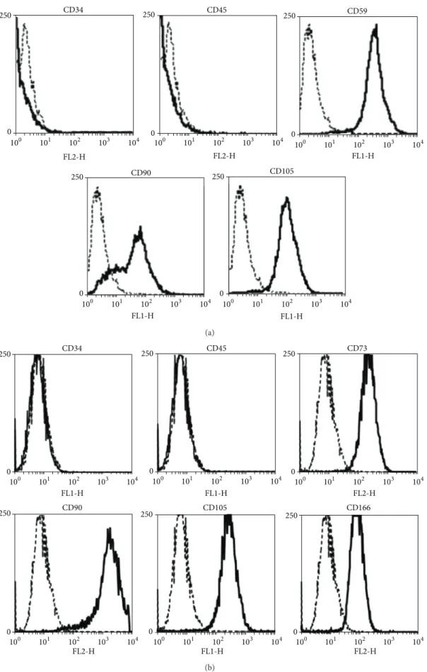
Human adipose-derived stem cells were characterized by mesenchymal (CD90, CD105, and the stem cell antigen 1 (Sca-1) homolog CD59) and hematopoietic (CD34, CD45) markers with flow cytometry in order to confirm their lineage.
Human bone-marrow-derived stem cells(hBMSCs) were isolated from samples gathered from young patients (aged 2–20) during standard orthopedic surgical procedures with the informed consent of the patients or their parents under approved ethical guidelines set by the Ethical Committee of the Hungarian Medical Research Council. All procedures were approved by the Ethical Committee of Semmelweis University. Only such tissues were used that otherwise would have been discarded. The bone marrow was flushed into T75 flasks and diluted with low-glucose (1.0 g/L) DMEM culture medium containing 10% FCS, 100 U/mL penicillin, 100𝜇g/mL streptomycin, and 4 mM L-glutamine. The flasks were incubated at 37∘C in fully humidified atmosphere of 5%
CO2 and 95% air for 3 days. After the incubation period, the hBMSCs adhered to the surface of the flasks and the remaining components of bone marrow were eliminated by washing with PBS. The used hBMSCs were cultured in the same conditions as hASCs. Human bone-marrow-derived stem cells were characterized by mesenchymal (CD73, CD90, CD105, and CD166) and hematopoietic (CD34, CD45) mark- ers with flow cytometry in order to confirm their lineage.
Characterization was performed on cells cultured under standard culture conditions and growing as monolayers while displaying constant cell proliferation rates over the entire culture period.
For preparingconditioned media(ACM) adipose-derived stem cells were used because their proliferation capabilities are much better compared to hBMSCs which helped to achieve the highest possible concentrations of paracrine molecules in the medium in the given period of time [39].
Human ASCs were seeded at 10.000 cells/cm2 in 100 mm Petri dishes using 8 mL low-glucose (1.0 g/L) DMEM cul- ture medium containing 10% FCS, 100 U/mL penicillin,
(a)
DiD 0
45
100 101 102 103 104
(b)
Analysis Reperfusion cocultivation
H9c2 cells
Simulated ischemia
160min 30min
hASCs or hBMSCs Fresh media
or ACM
24-hours
(c)
Figure 1: Ischemia-reperfusion model. (a) Representative fluorescent microscopic picture showing H9c2 cells injured with our ischemia- reperfusion model and treated with Vybrant DiD (ex/em: 644/665 nm, blue) labeled cells. The cytoplasm of the living cells is stained with calcein-AM (ex/em: 494/517 nm, green), the nuclei of the necrotic cells are ethidium homodimer-2 stained (ex/em: 536/624 nm, red). (b) Flow cytometric histogram on the distinction between stem cells and H9c2 cells based on DiD staining. (c) Schematic representation of the experimental protocol.
100𝜇g/mL streptomycin, and 4 mM L-glutamine. The dishes were incubated at 37∘C in fully humidified atmosphere of 5%
CO2 and 95% air, and cell-free supernatants were collected for further experimental use after 48 hours.
2.2. In Vitro Ischemia-Reperfusion Model. Ischemia-reperfu- sion was simulatedin vitroby oxygen and glucose deprivation as described previously in our earlier publications [40–42].
Briefly, 30.000/well H9c2 cells in 12-well plates were incu- bated in glucose-free DMEM in an atmosphere of 0.5% O2 and 99.5% N2for 160 minutes. This procedure was performed on an established incubation system (PeCon, Erbach-Bach, Germany). After incubation, the cells were reoxygenated and glucose was provided by immediate replacement of the media with fresh high-glucose DMEM, and the cells were kept in standard cell culture conditions till further experimen- tal actions. Representative fluorescence microscopy pictures were taken to follow the cell viability during the model using a Zeiss LSM 510 META (Carl Zeiss, Jena, Germany). We
used calcein-AM (ex/em: 494/517 nm, Invitrogen, Carlsbad, CA, USA) and ethidium homodimer-2 (ex/em: 536/624 nm, Invitrogen, Carlsbad, CA, USA) labeling to mark live/dead cells. Added mesenchymal stem cells were dyed with Vybrant DiD (ex/em: 644/665 nm, Invitrogen, Carlsbad, CA, USA) (Figure 1(a)).
2.3. Experimental Protocol. Four experimental groups were investigated in which postischemic cells received: (1) nor- mal medium (I-R-model); (2) hASC conditioned medium (ACM); (3) hASCs; and (4) a group that received hBMSCs.
Cell-treated groups were given 20.000 cells 30 minutes after the reoxygenation, and the added cells were labeled with Vybrant DiD fluorescent membrane dye to enable differen- tiation from the postischemic cells (Figure 1(b)). Cells were cocultivated for 24 hours in standard cell culture conditions.
In the case of ACM group at the end of simulated ischemia, the glucose-free medium was changed to same volume of cell- free hASC conditioned media (Figure 1(c)).
2.4. Cell Viability Measurement with Flow Cytometry.
Twenty-four hours after reoxygenation, cells were harvested by trypsinization and resuspended in 500𝜇L PBS containing 5 nM calcein-AM and 350 nM ethidium-homodimer-2 for flow cytometric analysis [43]. Controls were prepared as follows: for live control, cells were cultured in standard conditions; for dead control, cells were treated with 100 mM H2O2 for 1 hour immediately before trypsinization. For the measurements, FACSCalibur flow cytometer (Becton Dickinson, Franklin Lakes, NJ, USA) was used and the data was analyzed with the Weasel program (The Walter and Eliza Hall Institute, Parkville, VIC, Australia). Using flow cytometry, we could distinguish the therapeutically given cells from the postischemic cells on the basis of their DiD labeling, and these cells were gated in or out as appropriate for further analysis.
2.5. Metabolic Activity Measurement. For the evaluation of the metabolic activity in the groups, we used the Presto- Blue Cell Viability reagent (Invitrogen, Carlsbad, CA, USA), according to the manufacturer’s instructions. Because of the relative low cell numbers used in the experiments, we chose a 24-hour incubation time in 37∘C and measured absorbance as instructed.
In the hASC and hBMSC groups to exclude the influ- ence of the added stem cells metabolism, we subtracted the metabolic activity value of 20.000 stem cells from the measured value. The obtained results are compared to the metabolic activity of 30.000 H9c2 cells cultured in standard cell culture conditions (control).
2.6. Lactate Dehydrogenase (LDH) Cytotoxicity Assay. The measurement was performed using LDH Cytotoxicity Kit II (PromoCell, Heidelberg, Germany) according to the manu- facturer’s instructions, with 30-minute incubation period and absorbance measurement at 490 nm. For the LDH measure- ments, the previously described experimental groups were used 24 hours after ischemia-reperfusion. The LDH enzyme level was determined in the supernatant of 30.000 H9c2 cells cultured in standard conditions (control). The absorbance results were normalized with the total cell number in each sample.
2.7. Statistics. Statistical analysis of data was carried out either with one-way analysis of variance with Newman- Keuls multiple comparison post hoc test or unpaired𝑡-test as appropriate. All data are expressed as mean±SEM. A𝑃value of<0.05 was accepted as statistically significant.
3. Results
3.1. Characterization of hASCs and hBMSCs. Analyses of cell surface markers by flow cytometry demonstrated that hASCs (Figure 2(a)) were positive for the mesenchymal stromal (stem) cell markers CD90 and CD105 as well as the stem cell antigen 1 (Sca-1) homolog CD59 and were negative for the lymphohaematopoetic markers CD34 and CD45. Flow cytometry analysis of cultured bone-marrow-derived stem
cells (Figure 2(b)) exhibited the lack of hematopoietic mark- ers (CD34−, CD45−), but revealed mesenchymal stem cell lin- eage specific cell surface markers (CD73+, CD90+, CD105+, and CD166+). With respect to cell surface marker expression, our findings were consistent with previous reports [44,45].
3.2. Flow Cytometric Viability Analysis. Our experimental results regarding the postischemic cells showed that without any treatment live cells amounted to 12.13 ± 0.75% after 24 hours. However, the percentage of live cardiomyoblasts after 24 hours was significantly increased both with hASC (24.66 ± 2.49%) and hBMSC (25.41 ± 1.99%) treatments but not with the addition of ACM (13.94 ± 1.44%). There was no significant difference between the cell-treated groups. Cell- treatments led to a significantly increased percentage of live cells compared to ACM treatment as well (Figure 3(a)).
The percentage of the dead cells in the I-R model group was 87.71 ± 0.82%, while this was significantly smaller in the hASC and hBMSC treated groups (hASC:75.24 ± 2.49%;
hBMSC:74.62 ± 1.99%), but was not statistically different in the ACM treated group (ACM:85.75 ± 1.57%). There was no significant difference between the hASC- and hBMSC-treated groups (Figure 3(b)).
Putting the added cells into consideration, we found that most of the stem cells were alive in both the hASC- treated group (70.30 ± 2.35%) and the hBMSC-treated group (73.30 ± 1.92%), and there was no statistically significant difference between the groups (Figure 3(c)). Furthermore, no difference was found considering the dead cell population (hASC:29.20 ± 2.42%, hBMSC:25.81 ± 1.89%;Figure 3(d)).
3.3. Metabolic Activity Measurement. Using the PrestoBlue colorimetric assay, we strengthened our earlier findings with the flow cytometric analysis. The reducing capability of the cells reflecting their viability was significantly higher after treatment with hASCs (0.652 ± 0.089AU, arbitrary units) and hBMSCs (0.607 ± 0.059AU) compared to the I-R model (0.065 ± 0.033AU). Moreover, the treatment with ACM was also able to increase the metabolic activity of the postischemic cells (0.225±0.013AU). No difference was observed between the beneficial effects of the two different stem cell lines (Figure 4(a)).
3.4. Lactate Dehydrogenase Cytotoxicity Assay. Cellular ne- crosis expressed by LDH release decreased significantly com- pared to I-R model (0.225±0.006AU) when the postischemic cells were treated with hASC and hBMSC (hASC:0.148 ± 0.005AU; hBMSC:0.146 ± 0.004AU). Conditioned media could decrease the LDH levels only very slightly (0.208 ± 0.009AU). In case of hASC and hBMSC treatments, the necrosis was not significantly different from the control (Figure 4(b)).
4. Discussion
We report here that human adipose- and bone-marrow- derived cells directly improve the survival of postischemic cardiomyoblasts in anin vitroreductionist model. Metabolic
100 101 102 103 104 0
250 CD90
100 101 102 103 104 0
250 CD105
FL1-H FL1-H
100 101 102 103 104 0
250 CD34
100 101 102 103 104 0
250 CD45
100 101 102 103 104 0
250 CD59
FL2-H FL2-H FL1-H
(a)
100 101 102 103 104 FL1-H
CD34
0 250
100 101 102 103 104 FL1-H
CD45
0 250
100 101 102 103 104 FL2-H
CD73
0 250
100 101 102 103 104 FL2-H
250
0
CD90
100 101 102 103 104 FL1-H
250
0
CD105
100 101 102 103 104 FL2-H
250
0
CD166
(b)
Figure 2: Characterization of adult stem cells. Flow cytometric analysis revealed a CD34−, CD45−and CD59+, CD90+, CD105+pattern for hASCs (a) and a CD34−, CD45− and CD73+, CD90+, CD105+, CD 166+ pattern for hBMSCs (b). The isotype controls are indicated with dashed lines.
0 10 20 30
I-R model ACM hASC hBMSC
Live cells (%)
∗∗∗
∗∗∗
∗∗∗ ∗∗
(a)
90
60 80 100
70
I-R model ACM hASC hBMSC
Dead cells (%)
∗∗∗
∗∗∗
∗∗∗
∗∗
(b)
0 20 40 60 80 100
hASC hBMSC
Live cells (%)
(c)
0 20 40 60 80 100
hASC hBMSC
Dead cells (%)
(d)
Figure 3: Flow cytometric analysis of the postischemic cells and the therapeutic cells after 24 hours. Flow cytometric cell death analysis of the postischemic cells revealed that cell treatment increased the percentage of live cells (a), while ACM did not (I-R model:12.13 ± 0.75%;
ACM:13.94 ± 1.44%; hASC:24.66 ± 2.49%; hBMSC:25.41 ± 1.99%). (b) The percentage of dead cells decreased when therapeutic cells were added (I-R model:87.71 ± 0.82%; ACM:85.75 ± 1.57%; hASC:75.24 ± 2.49%; hBMSC:74.62 ± 1.99%). The percentages of live (c) and dead (d) cells among the therapeutically added cells were not significantly different (𝑛= 17–31,∗∗𝑃 < 0.01, ∗∗∗𝑃 < 0.001).
∗∗∗
∗
∗∗∗
∗∗∗ ∗∗∗
I-R model ACM hASC hBMSC Control
0 0.2 0.4 0.6 0.8 1
(AU)
(a)
∗∗∗
∗∗∗
I-R model ACM hASC hBMSC Control
0 0.05 0.1 0.15 0.2 0.25
∗∗∗
∗ ∗∗∗
(AU)
(b)
Figure 4: Metabolic activity measurement and LDH assay. (a) The metabolic activity measured 24 hours after the ischemia-reperfusion injury significantly decreased in the cells after the ischemic conditions compared to the control group, but the metabolic activity was enhanced with all the applied treatments (control:0.858 ± 0.021AU; I-R model:0.065 ± 0.033AU; ACM:0.225 ± 0.013AU; hASC:0.652 ± 0.089AU; hBMSC:
0.607 ± 0.059AU;𝑛 = 3,∗𝑃 < 0.05,∗∗∗𝑃 < 0.001). (b) LDH levels in the cell culture supernatant were significantly lower when ACM, hASC, or hBMSC therapy was carried out (I-R model:0.225±0.006AU; ACM:0.208±0.009AU; hASC:0.148±0.005AU; hBMSC:0.146±0.004AU;
𝑛 = 3, ∗𝑃 < 0.05,∗∗∗𝑃 < 0.001). The stem cell treated groups are not significantly different from each other and also not different from the control.
activity measurement and the evaluation of necrosis strength- ened the beneficial effect of cell treatment. Importantly, there was no difference in these direct effects between the adipose- and bone-marrow-derived stem cells.
Furthermore, the percentage of live mesenchymal stem cells after 24 hours was the same, so their survival prop- erties are also likely to be similar. These observations are important because many publications indicate a better result with adipose-derived stem cells, but the underlying mech- anisms of action are not clearly understood yet, and, to our knowledge, this is the first report on the comparison of the direct effects of adult stem cells on the parenchymal cells of the damaged tissue and on their survival in a standardized situation. Rasmussen et al. have shown that adipose-derived stem cells had preserved cardiac function following myocardial infarction in their animal model while bone-marrow-derived stem cells from the same source had not [26]. They reported that neither of these cell types induced angiogenesis. Thus, based on a recent report [39], they argued that the potential difference between them could be explained by differences in senescence properties of the cells. Others showed in an investigation on spinal cord injury that adipose-derived cells increased angiogen- esis more than cells from other mesenchymal sources and expressed higher amounts of VEGF while having similar migration properties to the bone-marrow-derived stem cells [46]. Adipose-derived stem cells were also found to be more effective on cutaneous wounds upregulating fibroblast migration and proliferation [47]. However, this may prove to be problematic in case of myocardial regeneration due to increased possibility of scar formation. Finally, a compara- tive study indicated ASCs to be a more promising source because of its more favorable immunomodulatory effects [48].
We have drawn a few conclusions from our data on the possible mechanisms of actions, also. First, LDH levels decreased to control levels after cell treatment. This means that necrosis was practically blocked by the added cells, indicating that the dead cells in our study were apoptotic cells. This is beneficial as apoptotic cells were shown to be immunoregulatory, and some researchers argue that the main effect of the current cytotherapy is aspecific and is the conse- quence of this apoptotic pool [36]. However, it must be real- ized that this possibility does not explain our results on cell viability as our model is completely reductionist and contains no immune cells. Second, the conditioned medium slightly increased metabolic activity and decreased LDH levels; thus, it had an antinecrotic effect. It means that the paracrine cocktail released from mesenchymal stem cells contains substances that act directly on the ailing postischemic cells.
The enhanced metabolic activity may relate to slightly better functionality of the surviving cells while the decreased LDH- levels indicate that the postischemic cells are directed from necrosis towards apoptotic cell death because the ACM had no effect on the cell viability in the flow cytometric measurements which is in accordance with our previous work using bone-marrow-derived cells in cell culture inserts [40].
In our study, the ineffectiveness of the stem cell condi- tioned media versus the stem cell treatment in increasing live cell numbers means either that the cell-cell contact is particularly important in the direct beneficial effect or the reached concentration of paracrine molecules is not high enough for their effect. The importance of cell-cell contact in the actions of therapeutically added cells was highlighted in earlier studies where the mechanism was related to intercellular tubular connections that potentially lead to mitochondrial exchange between the cells [40, 49].
Cell fusion is another phenomenon which can be observed in coculture studies, and in some cases it was also observed in in vivoanimal studies of stem cell grafting [30,50, 51].
The possibility of cell fusion in our model was addressed in our first publication and its frequency was found to be extremely low [40]. However, it must be noted that the extent of cell fusion shows extremely high variation among different culture and detection techniques, and it cannot be ruled out that extensive cell fusion is an in vitro artifact [28,52,53]. Still, recent studies suggest that despite the low frequency cell fusion may exert relevant impact on stem cell programming or reprogramming in the heart [54]. In view of the recent literature, it is more probable that the mechanism is mediated via paracrine factors, but the stem cells have to be induced by the microenvironment or by contact with injured cells to release these beneficial factors in necessary amounts. It is also possible that during the production of the conditioned media the concentration of paracrine factors in the conditioned media did not reach the levels necessary to be effective. No wonder, studies of late started to concentrate the conditioned medium to achieve higher concentrations and found promising results [55]. Our approach raises the possibility that the secreted molecules are effective only in close proximity to the affected cells where their local concentration can reach high levels. It is highly possible that only direct cell-to-cell contact can provide the necessary distance. It is interesting to note, that such “microparacrine” mechanism exists in relation of stem cells in the bone marrow, where the so-called “endosteal niche-stem-cell synapses” are formed [56]. A final, additional concern could be that conditioned media has a predominant role in angiogenesis; thus, it is ought to be ineffective in our reductionist model [26].
The relative role of the observed direct mechanism in the in vivo setting is difficult to measure, but it may be quite robust if we consider that we observed a doubling in the number of live cells. However, it must be realized, that in our experimental model the majority of the therapeutically added stem cells survived unlike thein vivosituation where most of the injected cells die soon after the transplantation [36]. Thus, the added cells had a prolonged time for exerting their effect.
At this point, it may be useful to consider some exper- imental points in our study. Our experimental model was devised to investigate acute effects of therapeutic stem cells on severely damaged cells to give room for the potential effects of stem cells. For this reason, we have set the length of simulated ischemia to a level where only 5–20% of cells survived without any treatment, which reflects the conditions found at the site of the injury in the heart after myocardial infarction.
We demonstrated the suitability of this model by detecting significantly elevated levels of oxidative stress and cellular necrosis after the simulation of ischemia-reperfusion in our earlier publication [42]. The effects were analyzed at 24 hours because we wanted to rule out the potential differentiation of the added stem cells, which occurs over longer time periods.
Some limitations must be accounted for considering our study. In our experiments, we used H9c2 cells which are derived from rat embryonic heart tissue. Obviously, there are differences between these cells and human adult cardiomy- ocytes, but H9c2 cells are frequently used in studies dealing with reperfusion injuries, and we have ample experience with these cells in our model [57–59]. Furthermore, as our aim was to analyze the direct effects of cells or media on postischemic cells, using H9c2 cultures instead of adult cardiomyocyte cell cultures we could avoid the possibility that inflammatory cells would contaminate the cultures and affect the results.
Still, it has to be kept in mind that we used human cells for treatment, but as no immune functions were involved in our model this fact must not had any major effect on our results, and human therapeutic cells are widely used in the literature in animal disease models [60–62]. Also, in our experiments we used an in vitro approach to the much more complex issue of stem cell therapy in myocardial infarction, with all the disadvantages and advantages of such model. Anin vitro transplantation model in a cell culture system cannot mimic the 3-dimensional tissue where cell-to-cell connections are different and it cannot reflect the complex (e.g., immuno- logical) events taking place during and after myocardial infarction. However, a limitation of thein vivomodels in cell treatment studies is a lack of separation between the direct effect on the treated parenchymal cells and the indirect effect caused by alteration of the environment (e.g., inactivation or reduced migration of leukocytes, angiogenesis, etc.). We believe that our model was appropriate for the scope of our study because it can focus on the direct effects of the added cells on the postischemic cells. The similar benefits achieved with hASC treatments strengthen that hASCs can be an alternative to the most commonly used hBMSCs in the emerging field of cell therapy. Subcutaneous adipose tissue is an attractive source for obtaining autologous mesenchymal stem cells as it can be harvested easily by liposuction which is performed routinely on thousands of people per year. The yield of stem cells per gram of adipose tissue is reported to be superior to that which can be achieved per milliliter of bone marrow [63], and adipose tissue can be harvested safely in much higher quantity. This is important as stem cells consti- tute only a small portion of cells in bone marrow and their number and differentiation capacity correlate inversely with age [64]. Similarly to hBMSCs, hASCs were shown to be able to differentiate toward osteogenic, adipogenic, myogenic, and chondrogenic lineages [6, 37, 64] and to secrete a host of paracrine factors that can increase angiogenesis and act as antiapoptotic signals [18,31]. As their direct effects are at least as good as the effects of hBMSCs, our results strengthen the assumption that they constitute a better and more practical source for therapies using adult stem cells. No evidence is available to date, but two Phase I clinical trials have been
recently completed to test the safety and feasibility of adipose- derived mesenchymal stem cell treatment in myocardial infarction and in chronic myocardial ischemia (APOLLO, NCT00442806; PRECISE, NCT00426868) [65].
5. Conclusions
Our results highlight that adipose-derived and bone-mar- row-derived stem cell treatments can directly save damaged cardiomyoblasts with the same efficacy. The survival of these cells in the noxious, oxidative environment is also similar.
These results may indicate that if these cells arrive to the injury site the resulting direct effect will be similar on the cardiac cells so the observed differences in efficacy found in in vivo experiments and in clinical trials may relate to different properties in homing, angiogenesis induction, fibroblast regulation, or immunomodulation.
Authors’ Contribution
M´onika Szepes and Zsolt Benk˝o contributed equally to this work.
Acknowledgments
This work was supported by T´ET-SIN, T ´AMOP 4.2.2-08/1/
KMR-2008-0004, T ´AMOP-4.2.1/B 09/1/KMR-2010-0001, OTKA 83803, and Bolyai fellowships. The authors are grateful to Gabriella V´acz for her help in the isolation of human bone- marrow-derived stem cells and to Anna Tutino, Eleni Dong´o, and ´Aron Farkas for their kind assistance in maintaining the cell lines.
References
[1] S. Bajada, I. Mazakova, J. B. Richardson, and N. Ashammakhi,
“Updates on stem cells and their applications in regenera- tive medicine,”Journal of Tissue Engineering and Regenerative Medicine, vol. 2, no. 4, pp. 169–183, 2008.
[2] S. M. Wu and K. Hochedlinger, “Harnessing the potential of induced pluripotent stem cells for regenerative medicine,”
Nature Cell Biology, vol. 13, no. 5, pp. 497–505, 2011.
[3] C. Leeb, M. Jurga, C. Mcguckin et al., “New perspectives in stem cell research: beyond embryonic stem cells,”Cell Proliferation, vol. 44, supplement 1, pp. 9–14, 2011.
[4] A. C. Brignier and A. M. Gewirtz, “Embryonic and adult stem cell therapy,”Journal of Allergy and Clinical Immunology, vol.
125, supplement 2, no. 2, pp. S336–S344, 2010.
[5] R. Sanz-Ruiz, E. Guti´errez Iba˜nes, A. V. Arranz, M. E.
Fern´andez Santos, P. L. S. Fern´andez, and F. Fern´andez-Avil´es,
“Phases I-III clinical trials using adult stem cells,”Stem Cells International, vol. 2010, Article ID 579142, 12 pages, 2010.
[6] P. A. Zuk, M. Zhu, P. Ashjian et al., “Human adipose tissue is a source of multipotent stem cells,”Molecular Biology of the Cell, vol. 13, no. 12, pp. 4279–4295, 2002.
[7] A. Wilson, P. E. Butler, and A. M. Seifalian, “Adipose-derived stem cells for clinical applications: a review,”Cell Proliferation, vol. 44, no. 1, pp. 86–98, 2011.
[8] F. Hildner, C. Albrecht, C. Gabriel, H. Redl, and M. van Griensven, “State of the art and future perspectives of articular cartilage regeneration: a focus on adipose-derived stem cells and platelet-derived products,”Journal of Tissue Engineering and Regenerative Medicine, vol. 5, no. 4, pp. e36–e51, 2011.
[9] J. M. Gimble, W. Grayson, F. Guilak, M. J. Lopez, and G.
Vunjak-Novakovic, “Adipose tissue as a stem cell source for musculoskeletal regeneration,”Frontiers in Bioscience, vol. 3, pp.
69–81, 2011.
[10] S. Lendeckel, A. J¨odicke, P. Christophis et al., “Autologous stem cells (adipose) and fibrin glue used to treat widespread traumatic calvarial defects: case report,” Journal of Cranio- Maxillofacial Surgery, vol. 32, no. 6, pp. 370–373, 2004.
[11] C. M. Cowan, Y.-Y. Shi, O. O. Aalami et al., “Adipose-derived adult stromal cells heal critical-size mouse calvarial defects,”
Nature Biotechnology, vol. 22, no. 5, pp. 560–567, 2004.
[12] M. Cherubino, J. P. Rubin, N. Miljkovic, A. Kelmendi-Doko, and K. G. Marra, “Adipose-derived stem cells for wound healing applications,”Annals of Plastic Surgery, vol. 66, no. 2, pp. 210–
215, 2011.
[13] B. L´eobon, J. Roncalli, C. Joffre et al., “Adipose-derived car- diomyogenic cells:in vitroexpansion and functional improve- ment in a mouse model of myocardial infarction,”Cardiovascu- lar Research, vol. 83, no. 4, pp. 757–767, 2009.
[14] R. Sanz-Ruiz, M. E. F. Santos, M. D. Mu˜noa et al., “Adipose tissue-derived stem cells: the friendly side of a classic cardiovas- cular foe,”Journal of cardiovascular translational research, vol. 1, no. 1, pp. 55–63, 2008.
[15] X. Bai, Y. Yan, Y.-H. Song et al., “Both cultured and freshly isolated adipose tissue-derived stem cells enhance cardiac function after acute myocardial infarction,” European Heart Journal, vol. 31, no. 4, pp. 489–501, 2010.
[16] M. Mazo, J. J. Gavira, B. Pelacho, and F. Prosper, “Adipose- derived stem cells for myocardial infarction,”Journal of Cardio- vascular Translational Research, vol. 4, no. 2, pp. 145–153, 2011.
[17] K. Schenke-Layland, B. M. Strem, M. C. Jordan et al., “Adipose tissue-derived cells improve cardiac function following myocar- dial infarction,”Journal of Surgical Research, vol. 153, no. 2, pp.
217–223, 2009.
[18] N. N. Hoke, F. N. Salloum, K. E. Loesser-Casey, and R. C.
Kukreja, “Cardiac regenerative potential of adipose tissue- derived stem cells,”Acta Physiologica Hungarica, vol. 96, no. 3, pp. 251–265, 2009.
[19] C. Valina, K. Pinkernell, Y.-H. Song et al., “Intracoronary administration of autologous adipose tissue-derived stem cells improves left ventricular function, perfusion, and remodelling after acute myocardial infarction,”European Heart Journal, vol.
28, no. 21, pp. 2667–2677, 2007.
[20] A. Abdel-Latif, R. Bolli, I. M. Tleyjeh et al., “Adult bone marrow- derived cells for cardiac repair: a systematic review and meta- analysis,”Archives of Internal Medicine, vol. 167, no. 10, pp. 989–
997, 2007.
[21] M. J. Lipinski, G. G. L. Biondi-Zoccai, A. Abbate et al., “Impact of intracoronary cell therapy on left ventricular function in the setting of acute myocardial infarction. A collaborative sys- tematic review and meta-analysis of controlled clinical trials,”
Journal of the American College of Cardiology, vol. 50, no. 18, pp.
1761–1767, 2007.
[22] E. Chavakis, M. Koyanagi, and S. Dimmeler, “Enhancing the outcome of cell therapy for cardiac repair: progress from bench to bedside and back,”Circulation, vol. 121, no. 2, pp. 325–335, 2010.
[23] M. Mazo, M. Ara˜na, B. Pelacho, and F. Prosper, “Mesenchymal stem cells and cardiovascular disease: a bench to bedside roadmap,”Stem Cells International, vol. 2012, Article ID 175979, 11 pages, 2012.
[24] M. T. Elnakish, F. Hassan, D. Dakhlallah et al., “Mesenchymal stem cells for cardiac regeneration: translation to bedside reality,”Stem Cells International, vol. 2012, Article ID 646038, 14 pages, 2012.
[25] M. Mazo, V. Planat-B´enard, G. Abizanda et al., “Transplantation of adipose derived stromal cells is associated with functional improvement in a rat model of chronic myocardial infarction,”
European Journal of Heart Failure, vol. 10, no. 5, pp. 454–462, 2008.
[26] J. G. Rasmussen, O. Frobert, C. Holst-Hansen et al., “Compar- ison of human adipose-derived stem cells and bone marrow- derived stem cells in a myocardial infarction model,” Cell Transplantation. In press.
[27] M. Alvarez-Dolado, R. Pardal, J. M. Garcia-Verdugo et al.,
“Fusion of bone-marrow-derived cells with Purkinje neurons, cardiomyocytes and hepatocytes,”Nature, vol. 425, no. 6961, pp.
968–973, 2003.
[28] J. Kajstura, M. Rota, B. Whang et al., “Bone marrow cells dif- ferentiate in cardiac cell lineages after infarction independently of cell fusion,”Circulation Research, vol. 96, no. 1, pp. 127–137, 2005.
[29] C. E. Murry, M. H. Soonpaa, H. Reinecke et al., “Haematopoi- etic stem cells do not transdifferentiate into cardiac myocytes in myocardial infarcts,”Nature, vol. 428, no. 6983, pp. 664–668, 2004.
[30] J. M. Nygren, S. Jovinge, M. Breitbach et al., “Bone marrow- derived hematopoietic cells generate cardiomyocytes at a low frequency through cell fusion, but not transdifferentiation,”
Nature Medicine, vol. 10, no. 5, pp. 494–501, 2004.
[31] J. Rehman, D. Traktuev, J. Li et al., “Secretion of angiogenic and antiapoptotic factors by human adipose stromal cells,”
Circulation, vol. 109, no. 10, pp. 1292–1298, 2004.
[32] T. P. Lozito and R. S. Tuan, “Mesenchymal stem cells inhibit both endogenous and exogenous MMPs via secreted TIMPs,”Journal of Cellular Physiology, vol. 226, no. 2, pp. 385–396, 2011.
[33] S. Sadat, S. Gehmert, Y.-H. Song et al., “The cardioprotective effect of mesenchymal stem cells is mediated by IGF-I and VEGF,”Biochemical and Biophysical Research Communications, vol. 363, no. 3, pp. 674–679, 2007.
[34] T. Kinnaird, E. Stabile, M. S. Burnett et al., “Marrow-derived stromal cells express genes encoding a broad spectrum of arteri- ogenic cytokines and promotein vitroandin vivoarteriogenesis through paracrine mechanisms,”Circulation Research, vol. 94, no. 5, pp. 678–685, 2004.
[35] H. K. Haider and M. Ashraf, “Strategies to promote donor cell survival: combining preconditioning approach with stem cell transplantation,”Journal of Molecular and Cellular Cardiology, vol. 45, no. 4, pp. 554–566, 2008.
[36] I. B. Copland and J. Galipeau, “Death and inflammation follow- ing somatic cell transplantation,”Seminars in Immunopathol- ogy, vol. 33, no. 6, pp. 535–550, 2011.
[37] P. A. Zuk, M. Zhu, H. Mizuno et al., “Multilineage cells from human adipose tissue: implications for cell-based therapies,”
Tissue Engineering, vol. 7, no. 2, pp. 211–228, 2001.
[38] K. M. Kompisch, C. Lange, D. Steinemann et al., “Neurogenic transdifferentiation of human adipose-derived stem cells? A critical protocol reevaluation with special emphasis on cell
proliferation and cell cycle alterations,”Histochemistry and Cell Biology, vol. 134, no. 5, pp. 453–468, 2010.
[39] M. A. Vidal, N. J. Walker, E. Napoli, and D. L. Borjesson,
“Evaluation of senescence in mesenchymal stem cells isolated from equine bone marrow, adipose tissue, and umbilical cord tissue,”Stem Cells and Development, vol. 21, no. 2, pp. 273–283, 2012.
[40] A. Cseleny´ak, E. Pankotai, E. M. Horv´ath, L. Kiss, and Z.
Lacza, “Mesenchymal stem cells rescue cardiomyoblasts from cell death in anin vitroischemia model via direct cell-to-cell connections,”BMC Cell Biology, vol. 11, article 29, 2010.
[41] A. Cseleny´ak, Z. Benko, M. Szepes, L. Kiss, and Z. Lacza, “Stem cell transplantation in anin vitrosimulated ischemia/reperfu- sion model,”Journal of Visualized Experiments, no. 57, Article ID e3575, 2011.
[42] M. Szepes, Z. Janicsek, Z. Benko et al., “Pretreatment of therapeutic cells with poly(ADP-ribose) polymerase inhibitor enhances their efficacy in anin vitromodel of cell-based ther- apy in myocardial infarct,”International Journal of Molecular Medicine, vol. 31, no. 1, pp. 26–32, 2013.
[43] M. A. King, “Detection of dead cells and measurement of cell killing by flow cytometry,”Journal of Immunological Methods, vol. 243, no. 1-2, pp. 155–166, 2000.
[44] R. de La Fuente, J. L. Abad, J. Garc´ıa-Castro et al., “Dediffer- entiated adult articular chondrocytes: a population of human multipotent primitive cells,”Experimental Cell Research, vol.
297, no. 2, pp. 313–328, 2004.
[45] J. Oswald, S. Boxberger, B. Jørgensen et al., “Mesenchymal stem cells can be differentiated into endothelial cellsin vitro,”Stem Cells, vol. 22, no. 3, pp. 377–384, 2004.
[46] X. Liu, Z. Wang, R. Wang et al., “Direct comparison of the potency of human mesenchymal stem cells derived from amnion tissue, bone marrow and adipose tissue at inducing der- mal fibroblast responses to cutaneous wounds,”International Journal of Molecular Medicine, vol. 31, no. 2, pp. 407–415, 2013.
[47] Z. Zhou, Y. Chen, H. Zhang et al., “Comparison of mesenchymal stromal cells from human bone marrow and adipose tissue for the treatment of spinal cord injury,”Cytotherapy, vol. 15, no. 4, pp. 434–448, 2013.
[48] Z. Xishan, H. Baoxin, Z. Xinna et al., “Comparison of the effects of human adipose and bone marrow mesenchymal stem cells on T lymphocytes,”Cell Biology International, vol. 37, no. 1, pp. 11–
18, 2013.
[49] E. Y. Plotnikov, T. G. Khryapenkova, A. K. Vasileva et al.,
“Cell-to-cell cross-talk between mesenchymal stem cells and cardiomyocytes in co-culture,”Journal of Cellular and Molecular Medicine, vol. 12, no. 5A, pp. 1622–1631, 2008.
[50] F. Ishikawa, H. Shimazu, L. D. Shultz et al., “Purified human hematopoietic stem cells contribute to the generation of car- diomyocytes through cell fusion,”The FASEB journal, vol. 20, no. 7, pp. 950–952, 2006.
[51] Z. Lacza, E. Horv´ath, and D. W. Busija, “Neural stem cell trans- plantation in cold lesion: a novel approach for the investigation of brain trauma and repair,”Brain Research Protocols, vol. 11, no.
3, pp. 145–154, 2003.
[52] J. Garbade, A. Schubert, A. J. Rastan et al., “Fusion of bone marrow-derived stem cells with cardiomyocytes in a heterol- ogous in vitro model,” European Journal of Cardio-Thoracic Surgery, vol. 28, no. 5, pp. 685–691, 2005.
[53] P. Menasch´e, “You can’t judge a book by its cover,”Circulation, vol. 113, no. 10, pp. 1275–1277, 2006.
[54] N. A. Kouris, J. A. Schaefer, M. Hatta et al., “Directed fusion of mesenchymal stem cells with cardiomyocytes via VSV-G facilitates stem cell programming,”Stem Cells International, vol.
2012, Article ID 414038, 13 pages, 2012.
[55] L. Timmers, S. K. Lim, I. E. Hoefer et al., “Human mesenchy- mal stem cell-conditioned medium improves cardiac function following myocardial infarction,”Stem Cell Research, vol. 6, no.
3, pp. 206–214, 2011.
[56] A. Wilson and A. Trumpp, “Bone-marrow haematopoietic- stem-cell niches,”Nature Reviews Immunology, vol. 6, no. 2, pp.
93–106, 2006.
[57] G.-Q. Huang, J.-N. Wang, J.-M. Tang et al., “The combined transduction of copper, zinc-superoxide dismutase and cata- lase mediated by cell-penetrating peptide, PEP-1, to protect myocardium from ischemia-reperfusion injury,” Journal of Translational Medicine, vol. 9, article no. 73, 2011.
[58] K. T. Keyes, Y. Ye, Y. Lin et al., “Resolvin E1 protects the rat heart against reperfusion injury,”The American Journal of Physiology, vol. 299, no. 1, pp. H153–H164, 2010.
[59] D. K. Singla and D. E. McDonald, “Factors released from embryonic stem cells inhibit apoptosis of H9c2 cells,” The American Journal of Physiology, vol. 293, no. 3, pp. H1590–
H1595, 2007.
[60] D. Yang, W. Wang, L. Li et al., “The relative contribution of paracine effect versus direct differentiation on adipose-derived stem cell transplantation mediated cardiac repair,”PLoS One, vol. 8, no. 3, Article ID e59020, 2013.
[61] S. Alshammary, S. Fukushima, S. Miyagawa et al., “Impact of cardiac stem cell sheet transplantation on myocardial infarc- tion,”Surgery Today, 2013.
[62] A. R. Williams, K. E. Hatzistergos, B. Addicott et al., “Enhanced effect of combining human cardiac stem cells and bone marrow mesenchymal stem cells to reduce infarct size and to restore cardiac function after myocardial infarction,”Circulation, vol.
127, no. 2, pp. 213–223, 2013.
[63] J. K. Fraser, I. Wulur, Z. Alfonso, and M. H. Hedrick, “Fat tissue:
an underappreciated source of stem cells for biotechnology,”
Trends in Biotechnology, vol. 24, no. 4, pp. 150–154, 2006.
[64] L. Peng, Z. Jia, X. Yin et al., “Comparative analysis of mesenchy- mal stem cells from bone marrow, cartilage, and adipose tissue,”
Stem Cells and Development, vol. 17, no. 4, pp. 761–773, 2008.
[65] P. Diez Villanueva, R. Sanz-Ruiz, A. Nunez Garcia et al.,
“Functional multipotency of stem cells: what do we need from them in the heart?”Stem Cells International, vol. 2012, Article ID 817364, 12 pages, 2012.
Submit your manuscripts at http://www.hindawi.com
Hindawi Publishing Corporation
http://www.hindawi.com Volume 2014
Anatomy
Research International
Peptides
Hindawi Publishing Corporation
http://www.hindawi.com Volume 2014
Hindawi Publishing Corporation http://www.hindawi.com
International Journal of
Volume 2014
Zoology
Hindawi Publishing Corporation
http://www.hindawi.com Volume 2014
Molecular Biology International
Genomics
International Journal of
Hindawi Publishing Corporation
http://www.hindawi.com Volume 2014
The Scientific World Journal
Hindawi Publishing Corporation
http://www.hindawi.com Volume 2014
Hindawi Publishing Corporation
http://www.hindawi.com Volume 2014
Bioinformatics
Advances inMarine Biology
Journal ofHindawi Publishing Corporation
http://www.hindawi.com Volume 2014
Hindawi Publishing Corporation
http://www.hindawi.com Volume 2014
Signal TransductionJournal of
Hindawi Publishing Corporation
http://www.hindawi.com Volume 2014
BioMed
Research International
Evolutionary Biology
International Journal of
Hindawi Publishing Corporation
http://www.hindawi.com Volume 2014
Hindawi Publishing Corporation
http://www.hindawi.com Volume 2014
Biochemistry Research International
Archaea
Hindawi Publishing Corporation
http://www.hindawi.com Volume 2014
Hindawi Publishing Corporation
http://www.hindawi.com Volume 2014
Genetics
Research International
Hindawi Publishing Corporation
http://www.hindawi.com Volume 2014
Advances in
Virology
Hindawi Publishing Corporation http://www.hindawi.com
Nucleic Acids
Journal ofVolume 2014
Stem Cells International
Hindawi Publishing Corporation
http://www.hindawi.com Volume 2014
Hindawi Publishing Corporation
http://www.hindawi.com Volume 2014
Enzyme Research
Hindawi Publishing Corporation
http://www.hindawi.com Volume 2014
International Journal of


![Imidazo[1,2-b]pyrazole-7-Carboxamide Derivative Induces Di ff erentiation-Coupled Apoptosis of Immature Myeloid Cells Such as Acute Myeloid Leukemia and Myeloid-Derived Suppressor Cells](data:image/gif;base64,R0lGODlhAQABAIAAAP///wAAACH5BAEAAAAALAAAAAABAAEAAAICRAEAOw==)