1
MTA DOCTORAL THESIS
BONE MARROW DERIVED STEM CELLS IN HEALTH AND DISEASE
Éva Mezey
Budapest, 2011
Szüleim Emlékére
dc_219_11
2
FREQUENTLY USED ABBREVIATIONS:
BM bone marrow
HSC hematopoietic stem cell
MSC/BMSC mesenchymal stem cell (used as a synonym for bone marrow derived stem cell or bone marrow derived stromal cell)
GFP green fluorescent protein
GCSF granulocyte colony stimulatory factor SCF Stem cell factor
GPCR G protein coupled receptor
dc_219_11
3
Table of Contents I) Introduction
II) Contribution of circulating cells to tissue regeneration 1) CNS
1a) Rodent brains
a(1) Uninjured brain a(2) Injured brain 1b) Human brains 2) Epithelial Tissues 2a) Mouse uterus
2b) Human cheek cells and salivary glands
III) Immune-regulatory effects of bone marrow stromal cells 1) Septic environment
2) Th2 dominant (allergic) environment 3) Histamine rich environment
IV) Conclusions
1) Contribution of circulating cells to tissue regeneration 2) Immune-regulatory effects of bone marrow stromal cells V) Acknowledgements
dc_219_11
4
I.) Introduction
This thesis summarizes my work of the last 15 years on the role of bone marrow derived cells in the maintainance of the healthy environment in the body.
My interest in these cells stemmed from the observation that they are able to move from the blood into the brain. This observation stood against a longstanding dogma that all the cells that constitute the CNS we are either born with or they derive from neural stem cells that seed the brain during embryonal development. The sig- nificance of the observation was that - if held true for humans - it would constitute new means to get pharmaceutical agents into or replace missing natural agents in the central nervous system. My first experiments showed that stem cells in the blood could indeed give rise to microglia and astrocytes in the rat brain. Subsequently, we showed that cells from gender-mismatched transplanted bone marrow make their way into the brain and differentiate into all neural linages in the mouse. These results were confirmed independently by other groups before we also demonstrated the same phenomenon in humans. In human transplant recipients, we found that bone marrow derived cells also colonize the oral mucosa and salivary glands.
In addition to their regenerative capacity, in the last years new evidence emerged that non-hematopoietic cells in the bone marrow might modulate the function of the immune system and these data prompted us to study this new phenomenon as well.
Our published data indicate that administration of bone marrow stromal cells to animals corrects imbalances in the immune system. Based on our work and studies by other groups, a number of clinical trials are underway to determine whether these immunomodulatory effects of BMSCs can be exploited to treat diseases.
I have separated the description of my work into two parts: First I have described our studies demonstrating the role of circulating BM cells in tissue regeneration.
Then I have summarized our work on the immuno-regulatory properties of bone marrow derived stromal cells.
dc_219_11
5
II.) Contribution of circulating cells to tissue regeneration
The first attempt in medical history to use BM for tissue regeneration was to renew the BM itself. Around the middle of the 20th century scientists realized that certain organs might be transplanted from one person into another - but problems associated with transplantation quickly surfaced. Sir Peter Brian Medawar’s work shed light on graft rejection and this eventually allowed clinicians to use matched donated organs to recipients and/or use immune-suppression to prevent rejection.
However, there were way too few organs to meet the demand, and the need to manufacture organs in vitro arose. To imagine making organs we need to understand how they develop in the embryo and how tissues are maintained throughout life physiologically. A logical choice of cells that contribute to tissue regeneration is those that circulate and thus can easily access all organs of the body, such as blood cells. These cells arise in the BM known to have two populations of stem cells: the hematopoietic (HSC) and the stromal (BMSC) stem cells. HSCs are generally accepted to give rise to the different classes of blood cells (myeloid, erythroid, lymphoid, platelets, and mast cells), while BMSCs give rise to the structural elements of the skeleton, such as bone, cartilage, and marrow fat.
1.) CNS
1a)Rodents
a(1) Uninjured brain
Our first sets of experiments was aimed at determining whether circulating blood cells can enter the CNS and differentiate into cells of the neural lineage, e.g., microglia, microglia, neurons, or vascular endothelial cells. We worked out a technique to detect the Y chromosome in cells and to colocalize it with a variety of protein markers that confirm the identity of differentiated cells.
Once the technique was working reliably, we irradiated female rats to destroy their BM and then injected them with genetically identical male bone marrow.
dc_219_11
6
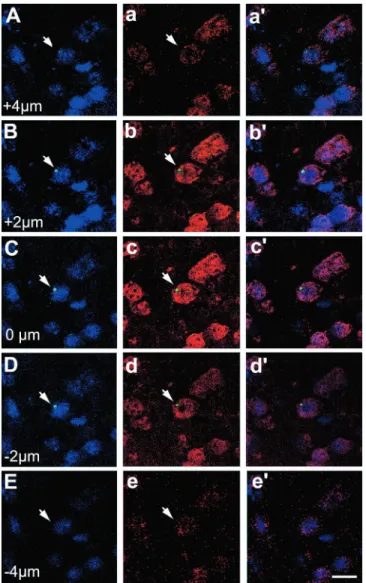
Subsequently, we examined the rats’ brains for the presence of Y chromosome positive (i.e., male donor) cells. We found both macroglia and microglia that contained the Y chromosome, suggesting that these cells derived from the donor BM and entered the brain through the general circulation. After we published these results in 1997 (1), we wondered whether bone marrow cells could contribute to the neuronal population of the brain as well as the glial populations. Since radiation might injure the blood-brain barrier and make it leaky, we designed a new experiment that circumvented this problem. To achieve this, we collaborated with a group of scientists who developed a special transgenic mouse that lacks a gene (PU1) vital for the development of the white cell lineage. PU1-/- mice have to be given BM transplants at birth in order to survive, and we transplanted them with gender mismatched BM within 48 hours of their births (2). At different time points following these transplantations we perfused the mice and used Y chromosome in situ hybridization histochemistry (ISHH) to colocalize the Y chromosome and NeuN, a specific marker of neurons (Fig.1). We found that 0.3-2.3% of all neurons were positive for both markers. The shortest survival time following BM transplant was 1 month, the longest was 4 months. We did not find a correlation between the elapsed time post-transplant and the number of double-positive cells. We also did not find any specific regional localization; the double-positive cells could be found in all brain regions examined in a seemingly random distribution.
Fig.1.
Y chromosome staining in the CNS. Coronal sections from 4-month-old nontrans- planted (A) female and (B) male brains were mounted and processed together. The panels show the over- lay of the NeuN (red) immunostaining, Y chromosome nonradioactive ISH [visualized with tyramide-FITC conjugate (green)], and DAPI stain- ing of cell nuclei (blue). The Y chromosome was restricted to the male brain, demonstrating hybridization specifici- ty. (C) Confocal image of coronal sections from a 4-month-old recipient female striatum that was double-immunostained for the neuron-specific antigens NeuN and NSE (neuron specific enolase). All NeuN- expressing cells (red) were also immunoreactive for NSE (green). (D) Sagittal section from a 1-month-old female PU.1 knockout mouse brain transplanted at birth with male bone marrow. The Y chromosome was visualized with BCIP/NBT (dark purple dots) to identify anatomical landmarks. cc, corpus callosum; cx, cerebral cortex; CPu, caudate putamen; fi, fimbria hippocampi; hi, hippocampus; LV, lateral ventricle. (E to G) Identical fields showing NeuN, Y chromosome, and DAPI nuclear triple staining in the hypothalamic dorsomedial nucleus of a 3-month-old female recipient. Colocalization of the Y chromosome [visualized with tyramide-
dc_219_11
7
FITC conjugate (green)] to a NeuN immunopositive (red) nucleus is shown in (E).
In (F), DAPI staining identifies all cell nuclei (blue). Overlays of the NeuN, Y chromosome, and DAPI fluorescence are shown in (G). The arrow identifies a cell nucleus that contained both the Y chromosome (indicating the bone marrow origin) and NeuN. Scale bar in (G) represents the following sizes: 30 µm, (A) and (B); 10 µm, (C); 250 µm (D); and 12 µm, (E) to (G).
a(2) Injured brain
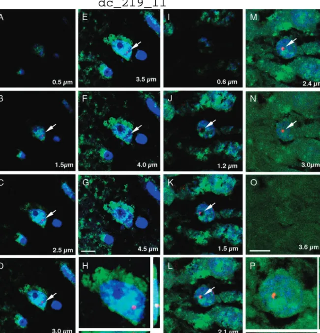
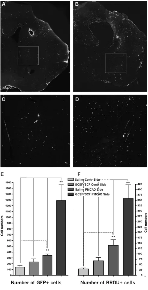
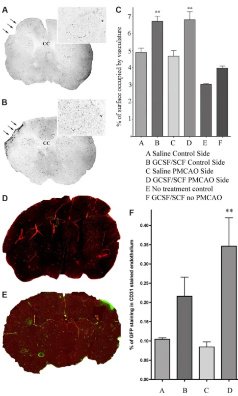
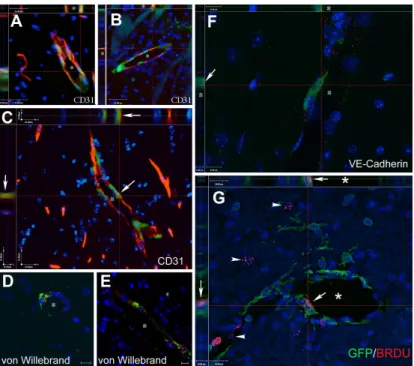
After we established that circulating cells can enter the CNS and differentiate into neural cells, we asked whether this process might be used physiologically for repair following brain injury. A month after transplanting PU1-/- mice with GFP expressing, gender mismatched bone marrow, we induced hypoxic brain injury using a permanent ligation of the middle cerebral artery (MCAO) and examined the brains 1-6 months later (3). We used both the GFP and the Y chromosome as markers of donor cells. We observed that few neurons, but numerous astrocytes and many new vessels were derived from cells of the transplanted BM.
Mobilizing BM cells with a combination of GCSF/SCF induced the formation of many new vessels and significantly decreased the infarct volume (Fig.2) in the mice.
These results suggested that mobilized circulating cells might contribute to brain healing
dc_219_11
8
Fig.2.
in a variety of ways, e.g., by stimulating neovascularization and also generating new cells.
1b) Human Brains
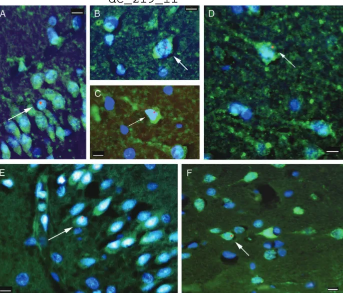
Following our rodent studies we wanted to know whether BM cells could colonize the brains of humans. In a collaboration with scientists at the Johns Hopkins School of Medicine, we collected samples from brains of four female patients who died after receiving bone marrow transplants from male donor relatives (4). The brains were immersion fixed in formaldehyde and paraffin embedded, which made it difficult to use the staining methods that we had employed earlier. However, once again, we worked out a suitable method to colocalize the human Y chromosome with specific nuclear (NeuN) and cytoplasmic (Kv2.1) neuronal markers. Using the new techniques we found that, indeed, cells that derived from the transplanted male donor blood entered the brains and did bear neuronal markers in all four patients. We found the greatest number of donor-derived neurons (7 in 10,000) in the youngest patient, a 2 year old child, who also lived the longest time after transplantation. The distribution of the labeled cells in the brains was not homogeneous. There were clusters of Y-positive cells, suggesting that single progenitor cells underwent clonal expansion and differentiation.
dc_219_11
9
Fig.3. Neuronal markers colocalized with the Y chromosome in human postmortem brain samples. Fluorescent microscopic images of neocortex from patients 2 (A–C) and 1 (E) and hippocampus from patients 1 (D) and 3 (F) are shown. The green color represents the immunostaining for neuronal markers Kv2.1 (A–D) and NeuN (E and F), and the Y chromosome is represented by the red fluorescent dots. All cell nuclei are stained with 4!,6-diamidino-2- phenylindole, a chromosomal marker that shows up as blue fluorescence. All images are overlays of the images seen through the three separate filters to show all colors. Arrows point to cells that are labeled with neuronal markers and are also Y chromosome-positive. In the Kv2.1 immunostaining the initial axons of some neurons can also be visualized. (Scale bars, 10 µm.)
2) Epithelial tissues
2a) Mouse uterus
Since the uterine epithelium needs to regenerate after every cycle (in the mouse every 4 days), we decided that this tissue would be ideal to look for contribution of BM derived cells to epithelial regeneration (5). Out of the two major populations of the BM stem cells only the hematopoietic cells have an accepted marker, CD45, also called common leukocyte antigen. We created a novel transgenic mouse where we introduced Cre recombinase cDNA into a
dc_219_11
10
bacterial artificial chromosome (BAC) containing the complete mouse CD45 gene (Fig.4).
Fig. 4. The recombinase cDNA was inserted downstream of an internal ribosomal entry site (IRES), which in turn was placed downstream of the last coding exon of CD45. The resulting BAC was used to make transgenic mice (CD45/Cre) where Cre recombinase is made in any cell that expresses CD45. GFP is expressed in CD45+ cells of the double-transgenic (CD45/Cre-Z/EG) mice. (A): Upon homologous recombination, the IRES-Cre recombination cassette flanked by two homologous fragments (A and B) was inserted into exon 33 of the CD45 gene in a BAC containing the entire coding region. The recombinant construct was confirmed to be correct by restriction mapping, PCR with primers flanking the recombination fragments, and sequencing. (B): To track the fate of CD45+ cells, we created a double-transgenic strain by breeding CD45/Cre mice with the double-reporter Z/EG mice. In the crossbred mice, every CD45+ cell expresses the Cre recombinase that will excise the floxed lacZ cassette, thus enabling the activation of GFP. Regardless of the future fate of CD45+
cells, GFP will be expressed continuously throughout their life spans. (C):
dc_219_11
11
Fluorescence-activated cell sorting of peripheral blood of a double-transgenic animal demonstrating a high percentage (85%) of double-positive blood cells using green fluorescence for GFP and red for CD45. (D–F): White blood cells immunostained with GFP (green) (D), CD45 (red) (E), and an overlay of (D) and (E) with added 4,6- diamidino-2-phenylindole (nuclear-blue) staining (F). Scale bar = 15 µm.
Abbreviations: BAC, bacterial artificial chromosome; bp, base pairs; EGFP, enhanced green fluorescent protein; GFP, green fluorescent protein; IRES, internal ribosomal entry site; PCR, polymerase chain reaction.
Once we crossed this novel transgenic mouse with the double reporter mice we looked at the uterine epithelium and found that the more cycles the mice had the more GFP+ epithelial cells we found. These green cells were always in patches suggesting a clonal origin, i.e. that a circulating cell becomes a uterine stem cell and continues to repopulate the mucosa. The most striking
finding in this study was that following pregnancy - at which event the surface area of the uterus has to enlarge by a factor of 20X, about 80% of the uterine epithelium became GFP+.
Fig.5.GFP-expressing uterine epithelial cells from double transgenic mice of different ages and pregnant mice. All nuclei were stained in blue with DAPI. (A, B): No GFP- positive (green) uterine epithelial (red staining represents L. tetragonolobus, the epitheli-al marker) cells were detect-ed in 6- week-old animals. (C, D): Sporadic GFP- positive uterine epitheli-al cells were present at 12 weeks of age. Arrow indicates a GFP"
uterine epithelial cell. (E, F): At 20 weeks of age, 6% of uterine epithelial cells expressed GFP. (G, H): In 12-week-old pregnant mice, there was a robust increase in the number of GFP-expressing cells: 82%
of the uterine epithelial cells were GFP".
Scale bars : 60 µm (left column) and 20 µm (right column)
dc_219_11
12
Since our work was published, other groups confirmed that human uterine epithelium behaves very similarly and that circulating BM cells contribute to its regeneration physiologically. Our findings also suggest a new possible explanation of endometriosis, a painful, difficult to treat human disease, when ectopic uterine epithelium is present in the abdominal cavity and responds to hormonal changes.
2b) Human cheek cells and salivary glands
The oral mucosa is constantly exposed to a variety of insults, such as extreme heat and cold, spices, mechanical injuries. All of these cause cell death and the dead cells need continuous replacement throughout life. Fortunately, the oral cavity is also easily accessible and sampling the mucosa is quick and painless. For all of the above reasons we decided to use the buccal mucosa to test whether BM derived cells contribute to the regeneration of this epithelial tissue. We took cheek scrapings from five females who had received either a bone-marrow transplant or an allogeneic mobilized peripheral-blood progenitor-cell transplant (enriched in CD34+ cells) from male donors years before (6).
Fig.6. In-situ hybridization and immunohistochemistry in human buccal cells (A) Y- chromosome autoradiography on a smear of buccal cells. The arrows point to the autoradiographic grains (black dots) above the nuclei of Y-chromosome containing cells. (B, C, D) The cells were first immunostained for cytokeratin 13 (green), then hybridized with a human Y-chromosome riboprobe (red fluorescent dots shown by the arrows). The nuclei of the cells are stained blue with DAPI. (E, F, G, H) FISH with X-chromosome and Y-chromosome probes. Individual buccal cells are shown.
The green fluorescent dot in the nucleus represents the X probe and the red dot the Y probe. The nuclei of the cells are stained blue with DAPI. The scale bar represents 10 µm in all panels, except 100 µm for panel A.
dc_219_11
13
We then performed in-situ hybridization on the cheek cells with Y and X chromosome probes. In separate experiments we combined fluorescent ISH for the Y chromosome in the buccal epithelial cells with immunohistochemistry to label cytokeratin 13, an accepted specific epithelial marker. When examined 4-6 years after male-to-female marrow-cell transplantation, all female recipients had Y- chromosome-positive buccal cells between 0.8-12.7%. In more than 9700 cells studied, we detected only one XXXY-positive cell (0·01%) and one XXY cell (0·01%), both of which could have arisen when an XY cell fused with an XX cell.
These results suggested that BMD cells migrate into the cheek and differentiate into epithelial cells, an occurrence that does not depend on fusion of BMD cells to recipient cells. This finding might be an example of transdifferentiation of haematopoietic or stromal progenitor cells. The HSCs seem more likely to be the cells responsible for this effect because:
1. There is no solid proof yet that the BMSCs from the bone marrow circulate;
2. Even if they do circulate, their numbers in the BM is orders of magnitude lower than their hematopoietic counterparts; and
3. Three out of the five patients received stem cell enriched peripheral blood instead of BM transplant, and the former are enriched in CD34+, CD38- hematopoietic stem cells and progenitors. These patients showed as high a percentage of chimerism (11%), as the ones receiving full BM (including the stromal stem cells)
Fig.7. Blood from a non-transplanted female control, buccal cells from recipient 5, her donor’s blood, and her son’s blood, were analyzed with four sets of Y- chromosome markers. X axis shows product length in bases, and Y axis shows signal strength.
dc_219_11
14
Finally, we wanted to test the hypothesis that the Y chromosome positive cells did not derive from the transplanted tissues (BM or PB), but from a previous pregnancy with a male fetus. We succeeded in collecting samples from the donor and the son of one of the patients and used microsatellite markers to identify the Y chromosome that we found in the cheek cells. The DNA analysis proved that the buccal cell Y chromosome was identical to that of the male donor versus the son of the patient.
We also tested several normal volunteers, who did not receive BM transplants, but were pregnant and gave birth to male children. None of the samples from these women had Y chromosome positive buccal cells. As a follow-up to this study we investigated the presence of Y chromosome-positive cells in salivary gland biopsies of 5 females who had received a marrow or blood stem cell transplant from male donors (7). One to 16 years after transplantation all scattered Y chromosome-positive cells were found in the acini, ducts, and stroma of their salivary glands (mean of 1.01%). Potentially, if their numbers can be boosted, these cells could be used for treatment of Sjögren’s syndrome and salivary glands damaged by therapeutic irradiation for cancers of the head and neck.
c. References
1. Eglitis MA and E Mezey. (1997). Hematopoietic cells differentiate into both microglia and macroglia in the brains of adult mice. Proc Natl Acad Sci U S A 94:4080-5
IF: 9.040 9108108 Cited: 631
2. Mezey E, KJ Chandross, G Harta, RA Maki and SR McKercher. (2000).
Turning blood into brain: cells bearing neuronal antigens generated in vivo from bone marrow. Science 290:1779-82.
IF: 23.872 11099419 Cited: 1091
3. Mezey E, S Key, G Vogelsang, I Szalayova, GD Lange and B Crain.
(2003). Transplanted bone marrow generates new neurons in human brains. Proc Natl Acad Sci U S A 100:1364-9
IF: 10.272 Cited: 307
4. Toth ZE, Leker RR, Shahar T, Pastorino S, Szalayova I, Asemenew B, Key S, Parmelee A, Mayer B, Nemeth K, Bratincsák A, Mezey E
The combination of granulocyte colony-stimulating factor and stem cell factor significantly increases the number of bone marrow-derived endothelial cells in brains of mice following cerebral ischemia.
Blood 111:(12) pp. 5544-5552. (2008) IF: 10.432 Cited: 23
dc_219_11
15
5. Bratincsak A, MJ Brownstein, R Cassiani-Ingoni, S Pastorino, I Szalayova, ZE Toth, S Key, K Nemeth, J Pickel and E Mezey. (2007).
CD45-positive blood cells give rise to uterine epithelial cells in mice.
Stem Cells 25:2820-6 IF: 7.531 Cited:14
6. Tran SD, SR Pillemer, A Dutra, AJ Barrett, MJ Brownstein, S Key, E Pak, RA Leakan, A Kingman, KM Yamada, BJ Baum and E Mezey. (2003).
Differentiation of human bone marrow-derived cells into buccal epithelial cells in vivo: a molecular analytical study. Lancet 361:1084-8.
IF: 18.316 Cited: 91
7. Tran SD, Redman RS, Barrett AJ, Pavletic SZ, Key S, Liu Y, Carpenter A, Nguyen HM, Sumita Y, Baum BJ, Pillemer SR, Mezey E (2011) Micro- chimerism in salivary glands after blood- and marrow-derived stem cell transplantation. Biology Of Blood And Marrow Transplantation 17:(3) pp.
429-433.
IF: 3.149
III. Immune-regulatory effects of bone marrow stromal cells
A group from the Karolinska Institute headed by Dr. Katarina LeBlanc had a young patient who developed graft vs. host disease (GVHD) as a complication of a BM transplantation. After all known treatment strategies failed, they decided to request permission for a new therapeutic intervention. It was known that BM derived stromal cells (BMSCs or also called mesenchymal stem cells-MSCs) suppress T cell proliferation. Thus, they infused isolated and cultured BMSCs from the patient’s mother and saw a dramatic improvement in the GVHD. They published their results in Lancet and started a whole new chapter in the clinical use of BMSCs.
1) Septic conditions
After reading LeBlanc's Lancet paper, we became interested in the biology of BMSCs and the mechanisms by which they regulate immune responses. One of the most complicated of these occurs in sepsis, when the response to microbial invasion frequently overshoots and ends up killing the host as well. We wondered if a live cellular therapy could be beneficial in a septic environment that changes very quickly
dc_219_11
16
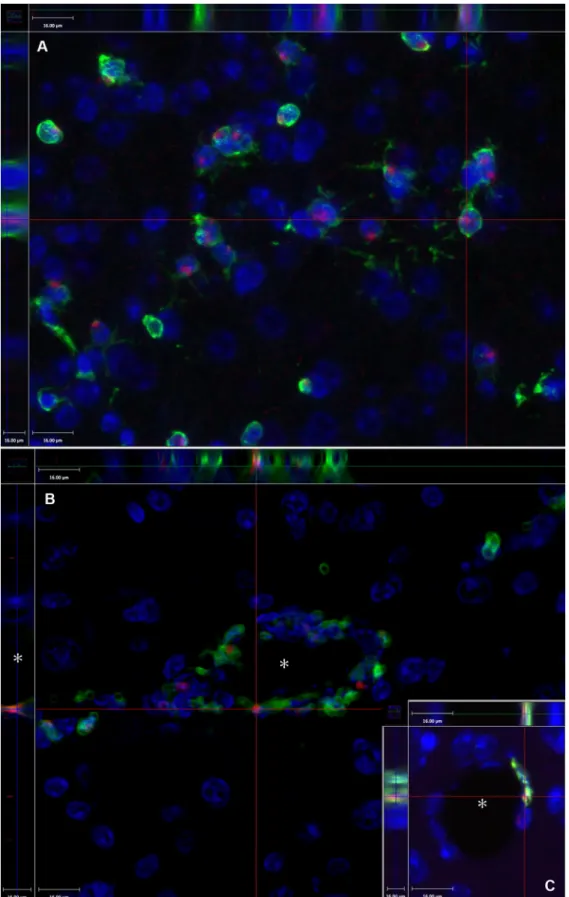
and the appropriate immune response at any given timepoint could make the difference between life and death. We used a mouse model of human peritonitis, called cecal ligation and puncture (CLP) and either injected the mice with BMSCs of different sources or vehicle intravenously at the time of septic injury (8). We found that the injected cells significantly improved the survival of the mice due to an improvement in organ functions. We then studied the mechanism of this action using a wide variety of techniques and using BMSCs from many transgenic mice that lacked certain factors. With this approach we determined that the injected BMSCs induce pro-inflammatory macrophages to become anti-inflammatory and produce large amounts of IL-10. The BMSCs were stuck in the capillaries of the lung where they were surrounded by monocytes/macrophages. Monocytes and/or macrophages from septic lungs made more IL-10 when prepared from mice treated with BMSCs versus untreated mice. In vitro, lipopolysaccharide (LPS)-stimulated macrophages produced more IL-10 when cultured with BMSCs, but this effect was eliminated if the BMSCs lacked the genes encoding Toll-like receptor 4 (TLR4), myeloid differentiation primary response gene-88 (MyD88), tumor necrosis factor (TNF) receptor-1a or cyclooxygenase-2 (COX2). Thus we concluded that BMSCs (activated by LPS or TNF-a) can reprogram macrophages by releasing prostaglandin E2 that acts through the prostaglandin EP2 and EP4 receptors.
Fig.8. Summary of studies of the molecular pathways involved in the interaction between BMSC and macrophages. (a) IL-10 concentration changes in supernatants of
dc_219_11
17
cocultures in a variety of treatment conditions after LPS stimulation. Colored graphs (except for the blue color that labels the bone marrow macro-phages as the source of all consecutive experiments) show treatments that eliminate the effect of BMSCs on macrophages. Black graphs show IL-10 levels after LPS stimulation, whereas open graphs show the control (nonstimulated) values. The experiments where the conditions eliminated the effect are colored. Purple color shows the effect of septic environment, green color shows agents and cellular compartments related to the PGE2 pathway and pink color shows agents related to the nitric oxide pathway. Three separate kinds of macrophages (bone marrow macrophages; peritoneal macrophages and the RAW264.7 cell line) were examined initially. Because they behaved identically in the assay, we used bone marrow derived macrophages (BM MF) for the rest of the experiments. In the box labeled 1, the effect of septic environment on the BMSCs is studied in BMSCs from TLR4-, MyD88-, TNFR1- and TNFR2- deficient mice, or antibody to TNF- α was used to neutralize the effect of TNF. The box labeled with 2 shows the cytokines and agents that have been implicated in the literature in immunomodulation of T cells by BMSCs, including COX1/2 and iNOS inhibitors. The box labeled 3 shows studies of the COX2 pathway, including the prostaglandin receptors EP1–EP4. Finally, in the box labeled with 4, we show studies related to nitric oxide. (b) A summary of our current hypothesis about the mechanisms that underlie the interactions between BMSCs and macrophages in the CLP sepsis model. Bacterial toxins (for example, LPS) and circulating TNF-α act on the TLR4 and TNFR-1 receptors of the BMSCs, respectively. This results in the translocation of NFkB into the nucleus. This activation process seems to be nitric oxide dependent.
Activated NFkB induces the production of COX2, resulting in increased production and release of PGE2. PGE binds to EP2 and EP4 receptors on the macrophage, increasing its IL-10 secretion and reducing inflammation.
2) Th2 dominant (allergic) environment
We learned from the sepsis study that BMSCs are able to correctly respond to environmental clues and regulate the immune system according the needs of the body.
Proliferating helper T cells differentiate into two major subtypes of cells known as Th1
dc_219_11
18
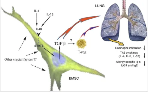
and Th2 lymphocytes. Our study of sepsis and others as well had shown that BMSC- driven immunomodulation is mediated by the suppression of proinflammatory Th1 responses, rebalancing the Th1/Th2 ratio toward Th2. We wondered whether BMSCs could act in a Th2-dominant environment—e.g., the one that prevails in the ragweed induced mouse asthma model. Intravenous injection of BMSCs at the time of the antigen challenge protected the animals from the majority of asthma-specific pathological changes (9), including inhibition of eosinophil infiltration and excess mucus production in the lung, decreased levels of Th2 cytokines (IL-4, IL-5, and IL-13) in the bronchial lavage (BAL), and lowered serum levels of Th2 immunoglobulins (IgG1 and IgE). We used BMSCs derived from a variety of transgenic mice; performed in vivo blocking of cytokines; studied how asthmatic serum and BAL from ragweed challenged animals effect the BMSCs of in vitro to find the mechanism of the effect. Our results suggest that IL-4 and/or IL-13 activate the STAT6 pathway in the BMSCs that in turn causes an increase of their TGF-b production, which seems to mediate the beneficial effect, either alone, or in concert with regulatory T cells, that might also be recruited by the BMSCs.
Fig. 9. Schematic drawing shows the mechanism of effect based on data of the present study. BMSCs “sense” the allergic environment, and as a result of the increased levels of IL-4/IL-13, they respond by producing higher amounts of TGF-β that, either alone or by recruiting regulatory T cells, will ultimately lead to a decrease of lung eosinophil infiltration, as well as allergy-specific cytokine and Ig production.
dc_219_11
19
3) Histamine rich environment
In humans mast cells (MCs) play a major role in the reaction to allergens. Thus we examined the effect of MC degranulation and histamine on the function of BMSCs.
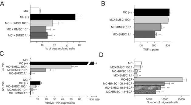
Mast cells (MCs) have a central role in allergic responses, including certain types of asthma and in the development of autoimmune disease. These cells represent an important link between innate and acquired immunity. We studied the interaction between mouse bone marrow-derived stromal cells and mouse bone marrow derived MCs (10) and found that BMSCs can efficiently suppress several MC functions in vitro as well as in vivo. When MCs are cocultured with BMSCs directly (allowing cell to cell contact), the BMSCs suppress MC degranulation, proinflammatory cytokine production, chemokinesis, and chemotaxis.
Fig. 10. In vitro studies of the interactions between MCs and BMSCs
A: Different ratios (1:1; 1:10 and 1:100) of IgE sensitized MCs and BMSCs were cultured together for 24 hours prior to IgE specific antigen challenge. -hex release was used as a marker of MC degranulation. BMSCs attenuate MC degranulation in all ratios tested. B: Different ratios (1:1; 1:10 and 1:100) of IgE sensitized MCs and BMSCs were cultured together for 24 hours prior to IgE specific antigen challenge for 12 hours. The BMSCs decreased the amount of released TNF-α in a ratio dependent manner as measured by ELISA. C: The experiment in B was repeated to measure TNF-α mRNA levels at two time-points (30 and 120 min) following antigen challenge. Similar to the
dc_219_11
20
levels of TNF-α protein, mRNA synthesis also decreased in response to the presence of BMSCs at both time- points in a ratio dependent manner. D: The migration of MCs was affected by the presence of BMSCs in the culture – with increasing numbers of BMSCs within the co-culture, the spontaneous (upper four columns) as well as SCF induced migration (chemokinesis and chemotaxis, respectively) of MCs were significantly reduced. In all Figures:*=p<0.05; **=p<0.01 and ***=p,0.001
Similarly, MC degranulation within mouse skin or the peritoneal cavity was suppressed following in vivo administration of BMSCs. Further, we demonstrate that these inhibitory effects were dependent on upregulation of COX2 in BMSCs and facilitated through the activation of EP4 receptors on MCs.
Fig.11. In vivo confirmation of COX2 mediated mechanism. The peritoneal assay was repeated using COX2 KO BMSCs followed by challenging the sensitized resident MCs with antigen (Ag). In contrast to wild-type (WT) BMSCs, COX2 deficient BMSCs failed to suppress hexosaminidase release by peritoneal MCs.
Based on these data we suggest that BMSCs might represent a novel cell based therapeutic approach in the treatment of MC driven allergic diseases.
d. References
8. Nemeth K, A Leelahavanichkul, PS Yuen, B Mayer, A Parmelee, K Doi, PG Robey, K Leelahavanichkul, BH Koller, JM Brown, X Hu, I Jelinek, RA Star and E Mezey. (2009). Bone marrow stromal cells attenuate sepsis via prostaglandin E(2)-dependent reprogramming of host macrophages to increase their interleukin-10 production. Nat Med 15:42-9.
IF:27.146 Cited: 107
dc_219_11
21
9. Nemeth K, A Keane-Myers, JM Brown, DD Metcalfe, JD Gorham, VG Bundoc, MG Hodges, I Jelinek, S Madala, S Karpati and E Mezey. (2010).
Bone marrow stromal cells use TGF-beta to suppress allergic responses in a mouse model of ragweed-induced asthma. Proc Natl Acad Sci U S A 107:5652-7.
IF: 9.432 Cited: 14
10. Brown JM, Nemeth K, Kushnir-Sukhov NM, Metcalfe DD, Mezey E (2011) Bone marrow stromal cells inhibit mast cell function via a COX2-dependent mechanism Clin.Exp. Allergy 41:526-534
IF: 4.084
dc_219_11
22
IV. Conclusions
I. Contribution of circulating cells to tissue regeneration 1) CNS
1a) Rodents
Cells that circulate in the bloodstream can gain access to the brain in rodents. In animals without brain injury these cells are evenly distributed and can become neural cells including microglia, macroglia, endothelial cells and neurons.
When a brain injury (stroke) occurs, the number of circulating cells entering the brain seems to increase. Many of these cells become vascular endothelial cells and form new vessels. The neovascularization facilitates healing, decreasing the necrotized volume and increasing the number of surviving cells.
1b) Humans
In samples of postmortem human brains we determined the presence of donor derived cells in the CNS following gender mismatched bone marrow transplants. As in rodents, we found that cells from the donor bone marrow entered the brains and differentiated into neural cells there. We found the greatest numbers of these cells in the youngest patient (2 years old) who also survived the longest following BM transplantation. Although we can not do functional tests in postmortem human samples, based on morphology and multiple neuronal markers, the donor derived cells were undistinguishable from the host neurons. Another important observation is that statistical analysis of the donor derived cells suggest that they are clonal, i.e. a cell from the circulation seeds the brain and multiplies and differentiates there.
Significance of the above findings: the demonstration that circulating cells cross the blood-brain suggests that these cells might be used as vehicles to introduce growth factors/differentiation factors or enzymes into the CNS. The enzymes might correct or slow the progression of genetic neurological diseases. The finding that GCSF/SCF treatment significantly enhances neovascularization indicates that this treatment may be beneficial in patients with stroke
dc_219_11
23
2) Epithelial tissues
2a) Mouse uterus
Based on the human cheek cell data we used a genetic tool to see if hematopoetic progenitors can indeed seed epithelial tissues. Using genetic marking of hematopoetic progenitors we have demonstrated that these cells get into the endometrium and participate in replenishing lost cells during the cycle. In some mice 70% of the epithelium was of hematopoetic origin following a pregnancy, which is known to inclrease the uterine surface area by 20 fold.
2b) Human cheek cells and salivary glands
Using a similar approach to the brain studies, we found that donor derived blood cells contribute to the population of cheek epithelium. Due to the unique patient population we were also able to establish the fact that in two patients the new cheek cells derived from hematological progenitors, since these patients did not receive BM, but isolated hematological progenitors from peripheral blood. We also examined the possibility that Y chromosome containing cheek cells might derive from an earlier pregnancy with a male fetus. DNA analysis conclusively showed that the Y chromosome in the cheek cells was from the donor vs the male offspring of the recipient.
Significance of the above findings: the demonstration that circulating BM derived cells can contribute to epithelial tissues supplies a. an alternative explanation to the pathomechanism of endometriosis, a rather common disease in the female population and b. raises the possibility that if this process can be boosted, than oral mucosa and salivary gland function could be restored in people who lose them due to autoimmune disease (Sjogren’s Syndrome) or head- and neck irradiation.
dc_219_11
24
III. Immunoregulatory effect of BMSCs
1) Septic environment
We analyzed the role that BMSCs play in regulating immune function in sepsis. Our results demonstrated that iv injected BMSCs are entrapped in the lungs where they communicate with monocytes/macro-phages that surround them. In the septic environment, these monocytes/macrophages are proinflammatory and make TNF-α. When they come in contact with BMSCs they change character and becomy anti-inflammatory. This tunes down the immune attack on body organs and allows animals to survive the septic process.
The crosstalk between BMSCs and the monocytes/macrophages involves the production and release of PDG2 by the BMSCs and a resulting increase of the monocytes/macrophages IL-10 production. The final outcome is a decreased number of neutrophils in body organs and less oxidative damage. On the other hand, there are more circulating neutrophils, and consequently more efficient clearence of bacteria.
2) Th2 dominant (allergic) environment
Using a ragweed induced allergy mouse model we determined that BMSCs are able to “sense” the allergic environment (increased levels of IL-4 /IL-13) and respond by producing large amounts of TGF-β that (either alone or by recruiting regulatory T cells) will ultimately lead to a decrease of lung eosinophil infiltration, and the allergy-specific cytokine and Ig production.
3) Histamine rich environment
As a continuation of the above studies, since mast cells (MCs) play a significant role in humans in allergic settings, we examined how BMSCs interact with MCs and how they respond to histamine, which is the major MC mediator. We demonstrated that BMSCs have the ability to affect the biology of MCs by limiting their activation and migration. Our data additionally show that these effects are mediated through the EP4 receptor on MCs.
dc_219_11
25
Significance of the above findings:
Our results in the sepsis model indicate that BMSCs might be a very effective therapeutic intervention in human sepsis – when no other treatment option is available. Live cells may be superior to drugs, because the cells respond to their environment while drugs can only play the role that they were designed for.
Since a large number of people die of sepsis every year (over 250000 in the US alone), a new therapy is badly needed.
The second set of studies indicate that in steroid resistant asthmatic conditions the use of BMSCs should be considered, since they are able to mitigate the allergic response and to counter the effects of MC degranulation.
dc_219_11
26
V. ACKNOWLEDGEMENTS
Looking back at my scientific carreer makes me realize that one needs a village (in my case a city and two continents..) to raise a scientist just as one does to raise a child. I have to start with my family, who supported all my adventures from childhood. My parents were always there to encourage me and help me find my way. My Mom planted the seeds of scientific curiosity in me at a very young age and my Dad helped me develop independent (and sometimes unusual) patterns of thinking. My brother, András, kept me in line and taught me many life skills, and both he and his wife, Lasy, gave me tremendous support after my parents were gone.
My husband, Michael Brownstein, was an incredible force, whose scientific advice and input shaped my career in numerous ways. Without him I would not be writing this thesis. And as far as family goes, I also have to thank my daughter, Anna, who gave me a reason to go on even when things were bad, and whose unconditional love and support was always there for me. Last but not least I want to thank Dr.
Judit Futo, my best friend since childhood, for all the times we spent discussing life and death, fun and misfortunes. She made me feel that if I fell, she would always be there to pick me up.
Trying to think of all of my mentors (many of whom also became close friends) I have to start with Janos Szentagothai, whose lectures during my years in medical school attracted me to his Department at the beginning of my student research carreer. Later on he was always supportive and encouraging, and never had a problem with untraditional ways of thinking. He permanently engraved in me he love of the miracle of the nervous system. In the Anatomy Department I had the fortune of having Miklos Palkovits as my immediate supervisor. There is not enough space in this thesis to list all the ways he shaped my life: my approach to problems, the numerous pitfalls of experimental designs and analysis of the results and the lessons about how to avoid them (after he let me fall first..). He knew when to keep close control, when to point out my mistakes, and also when to let me go and succeed or fail, and take the consequences. I want to mention Endre Csanda, the Chair of Neurology, and a very close personal friend, who was always willing to
dc_219_11
27
answer my sometimes silly and unusual questions about the brain and life and acted as a surrogate father after I lost mine. My first foreign mentor, Prof. David DeWied helped me grow scientifically and remaind supportive long after I left his Institute in Utrecht. After I moved to the US, once again, I met many wonderful people of the older and younger generation (including my fellows). In the interest of saving space, I will list their names without giving a detailed description of our interactions, but I want to emphasize that each and every one of them helped me in many ways to be what I am today as a scientist and as a person. I want to thank Tomas Hokfelt, Ronald DeKloet, Irvin Kopin, Julie Axelrod, Bruce Baum, Hal Gainer, Lana Skirboll, Pam Robey, Stefan Hansson, Andreas Zimmer, Janet Clark, Ruth Siegel, Joe Martin, Harry Webster, William Paul, Martin Eglitis, Karen Chandross, Gyongyi Harta, Bela Hunyady, Nancy Buckley, Tal Shahar, Andras Bratincsak, Zsuzsanna Toth, Krisztian Nemeth, Sarolta Karpati, Balazs Mayer, Miklos Krepuska, Sharon Key, Ildiko Szalayova. Many additional colleagues helped me along the way and I apologize to everyone I could not mention here. In addition, I have not listed the many personal friends who stood by me and helped me through hard times. These include my beloved dog P.H. who served as both a psychiatrist and exercise therapist for more than eleven years and is sorely missed.
dc_219_11
Proc. Natl. Acad. Sci. USA Vol. 94, pp. 4080–4085, April 1997 Neurobiology
Hematopoietic cells differentiate into both microglia and macroglia in the brains of adult mice
(gene transferybone marrow transplantationystem cellsylineage analysis)
MARTINA. EGLITIS*†ANDE´VAMEZEY‡
*Laboratory of Cell Biology, National Institute of Mental Health, and‡Clinical Neuroscience Branch, National Institute of Neurological Disorders and Stroke, Bethesda, MD 20892
Communicated by Richard L. Sidman, Harvard Medical School, Southborough, MA, January 28, 1997 (received for review October 9, 1996)
ABSTRACT Glial cells are thought to derive embryolog- ically from either myeloid cells of the hematopoietic system (microglia) or neuroepithelial progenitor cells (astroglia and oligodendrocytes). However, it is unclear whether the glia in adult brains free of disease or injury originate solely from cells present in the brain since the fetal stage of development, or if there is further input into such adult brains from cells originating outside the central nervous system. To test the ability of hematopoietic cells to contribute to the central nervous system, we have transplanted adult female mice with donor bone marrow cells genetically marked either with a retroviral tag or by using male donor cells. Using in situ hybridization histochemistry, a continuing inf lux of hemato- poietic cells into the brain was detected. Marrow-derived cells were already detected in the brains of mice 3 days after transplant, and their numbers increased over the next several weeks, exceeding 14,000 cells per brain in several animals.
Marrow-derived cells were widely distributed throughout the brain, including the cortex, hippocampus, thalamus, brain stem, and cerebellum. Whenin situhybridization histochem- istry was combined with immunohistochemical staining using lineage-specific markers, some bone marrow-derived cells were positive for the microglial antigenic marker F4y80. Other marrow-derived cells surprisingly expressed the astroglial marker glial fibrillary acidic protein. These results indicate that some microglia and astroglia arise from a precursor that is a normal constituent of adult bone marrow.
Besides the cells of the vasculature, the brain comprises two general cell types: neurons and glial cells. Glial cells provide physiological support to neurons and repair neuronal damage due to injury or disease. Macroglia (astroglia and oligoden- droglia) are generally considered to be derived from neuro- ectoderm and are believed to be developmentally distinct from microglia (1). However, the developmental origin of microglia remains debatable (2, 3), the two major views being that they derive either from neuroepithelial cells (4–6) or from hema- topoietic cells (i.e. monocytes) (7, 8). The extent to which cells outside the central nervous system (CNS) contribute to the maintenance of microglia in adults remains debatable (com- pare refs. 9 and 10), and no such contribution to adult neurons or macroglia has been previously described.
To learn if cells of the hematopoietic system are a source of progenitor cells for the CNS, we have used genetically tagged bone marrow cells and monitored their appearance in the brain by in situ hybridization histochemistry (ISHH). We combined ISHH and immunohistochemistry, and performed double- ISHH with digoxigenin and radioactively labeled probes to
analyze which cell types might be derived from bone marrow stem cells.
MATERIALS AND METHODS
Gene Transfer and Bone Marrow Transplantation.Gene transfer into hematopoietic precursors was done as previously described (11, 12), with the addition of stem cell factor to optimize transduction of reconstituting hematopoietic stem cells (13). C57BLy6J mice (The Jackson Laboratory), 6–8 weeks old, were used as donors. Forty-eight hours before marrow harvest, the mice were injected with 5-fluorouracil at a dose of 150 mgykg to ablate mature blood cells and thereby induce progenitor cells into cycle. Upon harvest, marrow was placed into liquid culture in suspension dishes and grown in DMEM containing 15% fetal bovine serum (BioWhittaker) and supplemented with interleukin 3 (50 ngyml), interleukin 6 (100 ngyml), and stem cell factor (100 ngyml). Growth factors were used to maintain early hematopoietic cells in cycle (13).
All were obtained from R & D Systems. After 48 h in culture with growth factors, marrow cells were collected and added to tissue culture dishes containing the F5B producer cell line at subconfluent density. F5B cells shed the N2 retroviral vector, packaged with the ecotropic envelope and carrying the bac- terial gene for neomycin resistance (neoR) (14). After 48 h coculture with F5B cells, bone marrow cells were collected by gentle aspiration, suspended to 13107cells per ml in PBS (in all cases 0.1 M phosphatey140 mM NaCl, pH 7.6) and injected intravenously (2–33106cells per mouse) via the tail vein into sublethally irradiated (4.5 Gy) female WBB6F1yJ-KitW/KitW-v mice. WBB6F1yJ-KitW/KitW-v mice are particularly good re- cipients for bone marrow transplantation because they have genetically defective stem cells (15). This gives normal C57BLy6J donor stem cells a strong repopulating advantage.
In transplants of male donor marrow into female recipients, some marrow was marked with retroviral vector as described.
In other cases, marrow was harvested, washed with PBS, and transplanted directly into recipient mice without culturing in growth factor-containing medium or irradiation of recipient animals.
A total of 46 mice were transplanted, 38 with vector-tagged marrow and 8 with male marrow. Five of the transplants with vector-tagged marrow used male donor cells. Mice were sacrificed at various times after transplantation. At least 2 animals were analyzed at each time point, although more were used at the 14-day (n510), 35-day (n514), and 70-day (n5 6) time points. Tissues were collected and immediately frozen
The publication costs of this article were defrayed in part by page charge payment. This article must therefore be hereby marked ‘‘advertisement’’ in accordance with 18 U.S.C. §1734 solely to indicate this fact.
0027-8424y97y944080-6$0.00y0
PNAS is available online at http:yywww.pnas.org.
Abbreviations: ISHH, in situ hybridization histochemistry; neoR, neo- mycin resistance; GFAP, glial fibrillary acidic protein; CNS, central nervous system; FITC, f luorescein isothiocyanate; DAPI, 49,6- diamidino-2-phenylindole.
†To whom reprint requests should be sent to the present address:
Experimental Therapeutics Branch, National Institute of Neurolog- ical Disorders and Stroke, Building 10, Room 5C211, 10 Center Drive, MSC 1406, Bethesda, MD 20892-1406.
4080
dc_219_11
on dry ice for subsequent sectioning. Some animals underwent cardiac perfusion with PBS before tissue harvest. Animals for perfusion were anesthetized with carbon dioxide, then their chests were opened, and PBS was introduced through a cannula placed in the left ventricle. The right atrium was incised to allow release of blood. Animals were perfused with 50 ml of ice-cold PBS over a period of 5 min.
In SituHybridization Histochemistry. Tissues were evalu- ated with both oligonucleotide and RNA probes. To detect neoRtranscripts, two oligonucleotide probes were prepared, complementary to the sequence of the neoRgene either from nucleotides 222–269 or nucleotides 447–494 (numbering with the A of the initiation codon as 1). The oligonucleotides were labeled using terminal transferase (Boehringer Mannheim) and [35S]thio-dATP (New England Nuclear) as described previously (16). An RNA probe, complementary to the entire neoRcoding region, was labeled with [35S]thio-UTP using SP6 polymerase (17). Labeling with radioactive probes was de- tected by dipping hybridized sections in photographic emul- sion. Emulsion was exposed for 14 days, then developed and sections were stained, air dried, and coverslipped for micro- scopic examination. To detect male bone marrow cells trans- planted into female recipients, sequences specific to the donor mouse Y chromosome were detected using a complementary RNA probe derived from the plasmid pY353yb (18). Glial fibrillary acidic protein (GFAP) gene expression was detected using an RNA probe complementary to the entire GFAP coding region. The Y chromosome and GFAP probes were labeled using digoxigenin-UTP (19), and digoxigenin labeling was developed for GFAP using alkaline phosphatase as de- scribed (19). For detection of the donor Y chromosome, before overnight hybridization with digoxigenin-labeled probes at 558C, the slides were heated at 908C for 10 min in hybridization buffer containing the probes to improve access to nuclear DNA. The digoxigenin-labeled Y chromosome was visualized using a modification (20) of an immunostaining amplification method (21), which results in green fluorescein isothiocyanate (FITC) fluorescence.
Twelve-micrometer thick frozen sections were cut in a cryostat, and ISHH was performed as described previously (16, 17). The sections were fixed, dehydrated, and delipidated in ethanol and chloroform, and then hybridization buffer containing the probe(s) was put on the sections. Slides were incubated overnight in a humidified chamber at 378C (for oligonucleotide probes) or 558C (for riboprobes).
Nuclear Staining.To confirm that Y chromosome ISHH coincided with cell nuclei, sections were counterstained with ethidium bromide or 49,6-diamidino-2-phenylindole (DAPI).
Staining was detected by illumination with a mercury lamp using a microscope equipped for fluorescence micrography.
Immunohistochemical Analysis. For combined ISHHy immunohistochemical analysis, sections were fixed as de- scribed previously (22). They then were incubated for 30 min at room temperature in 3% normal goat serum diluted in PBS (containing 0.6% Triton-X 100) to block nonspecific binding.
Then, the sections were exposed for 1 h at room temperature to either (i) a polyclonal rabbit antibody that detects the mouse F4y80 monocyteymacrophage marker (23) or (ii) a polyclonal CY-3-labeled rabbit antibody against the astroglial marker GFAP (Sigma) used at a dilution of 1:2,000. Binding of nonlabeled primary antisera was detected with a biotinylated goat anti-rabbit IgG (Jackson ImmunoResearch) diluted 1:500. To detect biotinylated secondary antibody, the sections were incubated for 1 h in an avidin-biotin-peroxidase complex diluted 1:250 in PBS with 0.6% Triton-X 100 (24). The slides then were transferred into 0.1 M TriszHCl (pH 7.6) and were developed using diaminobenzidine as a substrate. After a thorough wash, the sections were processed for ISHH. Cola- beling of cells was determined using a combination of bright- field, polarized, fluorescent, and epi-illumination microscopy.
Controls for the immunostaining included leaving out the primary antibodies and using several secondary antibodies (from different species) to confirm that there was no nonspe- cific binding.
RESULTS
Detection of Donor Cells in the Brain After Bone Marrow Transplantation.To evaluate the appearance and distribution of donor cells in the brains of recipient mice, animals were sacrificed 3, 5, 7, 14, 28, 35, 42, and 70 days after transplan- tation with bone marrow cells. At least two animals trans- planted with retrovirally tagged marrow were studied at each time point. Mice transplanted with male marrow were analyzed at 35 days (n59) and 70 days (n54) after transplantation.
Using probes specific to the vector neoRtranscripts, donor cells were detected beginning with day 3, the earliest time of analysis. Many cells were easily detected throughout the brain by day 7, and cells continued to be detected at all subsequent times. To estimate total number of neoR-positive cells in a brain, every 25th section was collected, and all labeled cells in the sections were counted. The number of labeled cells was multiplied by 25 to arrive at the approximate total number of marked cells in a brain. These calculations showed that the overall number of marrow-derived cells per brain gradually increased with increasing time after transplantation. Three days after transplant, 500 cells were detected per brain. Two to four weeks after transplant the number of cells present had increased to at least 2,000 per brain. In several animals more than 10,000 cells per brain were seen, and in one animal the number of cells was over 30,000.
At 1 week, and occasionally at later times, concentrations of neoR-marked cells were observed in the basal subarachnoid space. Cells marked by the retroviral vector were detected in the hippocampus (Fig. 1A and B), septum (Fig. 1C), and hypothalamus (Fig. 1D). Cells were also detected, among other regions, in the cortex, habenula, pons, and cerebellum (data not shown). Labeled cells were detected after PBS perfusion,
FIG. 1. Detection of donor cells in the brain after bone marrow transplantation with retrovirally tagged bone marrow cells. Arrows indicate representative cells positive by ISHH with35S-labeled oligo- nucleotide (A–C) or riboprobe (D). (A and B) Bright (A) and dark (B) field photographs of the same section. ISHH-positive cells (arrows) detected in the hippocampus of an animal 14 days postbone marrow transplantation. (C) Positive cells in the region of the septum of an animal sacrificed 14 days after bone marrow transplantation. The photograph is a double exposure of a bright field image with a dark field image of the same area. The dark field image was photographed using a red filter so that the autoradiographic grains would appear red.
(D) A cell (arrow) within the ependyma of the third ventricle. [Bars 510mm (A–C) and 40mm (D).]
Neurobiology: Eglitis and Mezey Proc. Natl. Acad. Sci. USA 94 (1997) 4081
dc_219_11
indicating that bone marrow-derived cells were an integral part of the brain parenchyma.
Similar regional distribution of donor marrow cells was seen using the Y chromosome probe to detect male donor cells (Fig.
2). Ethidium bromide counter-staining to highlight the nucleus confirmed the nuclear localization of the Y chromosome probe. Many male donor-derived cells were easily detected throughout the brain 35 days after transplantation, and cells continued to be detected at all subsequent times. Cells positive for the Y chromosome marker were detected in the mesen- cephalon (Fig. 2 A–C), septum (Fig. 2D), striatum (Fig. 2E), and habenula (Fig. 2F). Cells also were detected in the cortex, pons, and cerebellum, among other regions (data not shown).
Ex vivo manipulation of the bone marrow cells was not necessary, because male cells were detected in female recip- ients’ brains even when the transplant was done immediately after marrow harvest.
Several parameters were used to verify that the labeling observed after ISHH was specific. First, no labeling was detected in any tissues of animals transplanted with non- marked bone marrow cells. That is, without retroviral tagging, probes for the neoR gene exhibited no background labeling, and the Y chromosome probe did not label female tissues.
With the Y chromosome riboprobe, we also confirmed that both sense and antisense probes exhibited the same distribu- tion, as expected when hybridizing to chromosomal DNA. The pattern of retrovirally labeled cells was identical in all tissues analyzed, both qualitatively and quantitatively, regardless of which probe was used. Finally, we found donor cells in hematologic organs such as bone marrow and spleen at all time points analyzed (data not shown). The pattern of engraftment was qualitatively similar between retrovirally tagged and male donor cells. However, when female mice were transplanted with retrovirally tagged male marrow, more donor cells were detected with the Y chromosome probe than with the neoR probe. This suggests that not all of the cells migrating from the bone marrow into the brain expressed the retrovirally intro- duced neoR gene at a level high enough to be detected by ISHH.
Labeling of Brain Sections after ISHH with the Microglial Marker F4y80.The F4y80 antibody detects the plasma mem- brane protein F4y80 expressed exclusively on macrophages and microglia (23). Colocalization in brain sections revealed cells labeled by the N2 retroviral vector that also expressed the F4y80 antigen (Fig. 3), confirming that bone marrow-derived cells do contribute to the microglial population in the adult brain. However, only a small percentage of ISHH-positive cells were labeled by immunostaining. Similarly, the minority of antigen-positive cells was doubly labeled by ISHH. The dis- tribution of doubly labeled cells reflected the distribution of cells labeled only by ISHH or by immunoshistochemistry, i.e., they were widely distributed throughout the brain.
Labeling of Brain Sections for Both the Astroglial Marker GFAP and the neoRRetroviral or Y Chromosome Donor Cell Tag.The ISHH-positive, F4y80 negative cells could be cells of the myeloid lineage that had not differentiated to express the F4y80 antigen. Or, they could represent a contribution of bone marrow-derived cells to other than myeloid cell lineages. To distinguish between these alternative possibilities, ISHH- positive cells were examined for the expression of another lineage marker, GFAP, specific for astroglia. Surprisingly, we found occasional cells (Fig. 4A) which were labeled both by ISHH (for the donor marrow neoR marker) and by indirect immunohistochemistry (for GFAP). Counting all of the donor cells present in every 25th section obtained from recipient mice 4 weeks after transplantation (n 53), we calculated that as many as 3 3104neoR-marked donor cells were present per brain. Of that total donor cell number, we estimated between 0.5% and 2% exhibited GFAP expression.
To confirm that GFAP mRNA was present in some neoR- positive cells, we also did double-ISHH analysis. Cells coex- pressing GFAP and neoR mRNAs were identified using a digoxigenin-labeled riboprobe against GFAP mRNA together with a35S-labeled probe for the neoRgene marking the donor marrow. As illustrated in Fig. 4 B and C, we found cells labeled with both probes. Their frequency was approximately equal to the frequency of the ISHHyGFAP immunostained double cells.
FIG. 2. Detection of donor cells in several brain regions of a female recipient 6 weeks after transplantation with male bone marrow cells.
Arrows indicate representative cells positive for the Y chromosome by ISHH. (A–C) Photomicrographs of a section through the ventral mesencephalon. A is photographed using a rhodamine filter to excite ethidium bromide staining of the nucleus; B is photographed using a FITC filter to excite Y chromosome-specific FITC staining; and C is photographed with a double-pass filter to show overlap of Y chromo- some labeling and nucleus-specific ethidium bromide staining. Arrows indicate some of the double-labeled cells. (D–F) Photomicrographs demonstrating Y chromosome positive cells in other brain regions. (D) Septum. (E) Striatum. (F) Habenula. (Bars510mm.)
FIG. 3. Double-labeling of brain sections detects cells coexpressing the microglial marker F4y80 and the neoRretroviral tag. The F4y80 monocyteymacrophage antigen was detected by indirect immunoflu- orescent antibody labeling; 35S-radiolabeled probes were used to hybridize to neoRmRNA. The photomicrograph is of a representative field from an animal sacrificed 35 days after bone marrow transplan- tation. A cell in the center stains positive for the F4y80 antigen (red) and exhibits labeling with radioactive probe to neoRtranscripts. The dark field image was photographed using a green filter so that autoradiographic grains would appear green (yellow where they overlap red immunostaining). (Bar510mm.)
4082