Transdifferentiation and regenerative medicine
Manifestation of Novel Social Challenges of the European Union in the Teaching Material of
Medical Biotechnology Master’s rogrammes at the University of Pécs and at the University of DebrecenP
Dr. Balogh Péter – Dr. Engelmann Péter
Transdifferentiation and regenerative medicine
Dr. Péter Balogh – Dr. Péter Engelmann
“Manifestation of Novel Social Challenges of the European Union
in the Teaching Material of
Medical Biotechnology Master’s Programmes at the University of Pécs and at the University of Debrecen”
Identification number:TÁMOP-4.1.2-08/1/A-2009-0011
University of Pécs – Pécs, 2011
© Dr. Péter Balogh, Dr. Péter Engelmann, 2011
The project is funded by the European Union and co-financed by the European Social Fund.
Manuscript completed: 11 November 2011
Editor in charge: University of Pécs
Editor in charge: Dr. Péter Balogh, Dr. Péter Engelmann, Rita Bognár Technical editor: Zsolt Bencze, Veronika Csöngei and Szilvia Czulák
Lector: Dr. György Miskei Length: 88 pages
Identification number:
TÁMOP-4.1.2-08/1/A-2009-0011
3
Contents
List of figures ... 5
I Definition of stem cells and stem cell groups, their maintenance and homeostasis ... 7
II Regeneration in animal models ... 13
III Epigenetic factors in transdifferentiation ... 17
IV Genomic and other cell-tracing approaches, reprogramming ... 23
V Stem cells and transdifferentiation during haematopoesis ... 31
VI Muscle regeneration ... 37
VII Liver regeneration ... 43
VIII Differentiation and regeneration in the pancreas ... 51
IX Transdifferentiation and metaplasia in the central nervous system ... 59
X Cardiovascular regeneration ... 67
XI Kidney regeneration ... 71
XII Cancer stem cells ... 75
XIII Ethical background of stem cell research and therapy ... 81
Further reading ... 87
Identification number:
TÁMOP-4.1.2-08/1/A-2009-0011
5
List of figures
Figure I-1: Sources of embryonic stem cells ... 8
Figure I-2: Cell membrane markers for ESCs ... 9
Figure I-3: Reprogramming: Induction of pluripotency in iPS cells ... 10
Figure I-4: Sequential maturation and pluripotency ... 11
Figure II-1: Similarities in regeneration ... 14
Figure II-2: Imprinting in regeneration ... 15
Figure II-3: Neural stem cells differentiation capacity ... 16
Figure III-1: Epigenetic gene regulation of stem cell genome ... 18
Figure III-2: DNA methylation in stem cells ... 19
Figure III-3: miRNA and stem cell differentiation. ... 21
Figure IV-1: Origin of stem cells and reprogramming ... 24
Figure IV-2: Cell tracing in stem cell biology ... 26
Figure IV-3: Molecular mechanisms of self-renewal ... 27
Figure V-1: Ontogeny of embryonic haemopoietic tissues ... 32
Figure V-2: Transcriptional regulation of early haemopoietic commitment ... 33
Figure V-3: Transcriptional regulation of myeloid differentiation ... 34
Figure V-4: Transcriptional regulation of lymphoid differentiation ... 35
Figure V-5: Steady-state and activated haemopoiesis ... 36
Figure VI-1: Structure and regeneration of skeletal muscle ... 38
Figure VI-2: Non-SCs contributing to muscle regeneration ... 39
Figure VI-3: Kinetics of muscle repair ... 40
Figure VII-1: Developmental relationship between hepatic-pancreatic differentiation ... 44
6 The project is funded by the European Union and co-financed by the European SocialFund.
Figure VII-2: Transcriptional control of hepatoblast development ... 45
Figure VII-3: Structure of hepatic lobe ... 46
Figure VII-4: Main phases of liver regeneration ... 48
Figure VIII-1: Embryonic pancreas development ... 54
Figure VIII-2: β cell and autoimmune processes of diabetes ... 55
Figure VIII-3: Differentiation of insulin producing β cells from ES cells ... 57
Figure X-1: Sources of cells for cardiac regeneration ... 67
Figure X-2: iPS reprogramming for myocardial regeneration ... 69
Figure XI-1: Renal progenitor cells ... 71
Figure XI-2: Repair mechanisms of stem cells in kidney regeneration ... 73
Figure XII-1: Cancer and cancer stem cell theory ... 76
Figure XII-2: CSC development ... 77
Figure XII-3: Altered niche for CSCs ... 78
Figure XII-4: AML niche characteristics ... 79
Figure XII-5: Combined treatment of cancers – CSCs and their niche ... 80
Identification number:
TÁMOP-4.1.2-08/1/A-2009-0011
7
I Definition of stem cells and stem cell groups, their maintenance and
homeostasis
According to their origins, stem cells (SCs) can be defined either by their origin (embryonic/ESCs vs. adult) or differentiation capacity (spontaneous/pluripotent, induced pluripotent, or committed – hemopoietic, mesenchymal stem cells, etc).
Following conception the zygote will cleave into morula, followed by transformation into early blastocyst in pre-implantation embryos, a structure containing external cell layer (trophoectoderm, promoted by Cdx2 transcription factor) and an internal cluster of undifferentiated cells, regulated by Oct3/4 transcription factor. The cells comprising the inner cell mass (ICM) upon the effect of Nanog transcription factor (and also Oct4 and Sox2) progress to the late blastocyst stage where, importantly, two cell layers differentiate. In one layer the epiblast cells will form the embryonic body through giving rise to the three germ layers (ectoderm, endoderm and mesoderm) during the gastrulation in the post-implantation embryos, while the the hypoblast cells will develop into the extraembryonic membranes. Of these the ICM cells and epiblast cells are used for harvesting ESCs.
Transdifferentiation and regenerative medicine
8 The project is funded by the European Union and
co-financed by the European SocialFund.
Figure I-1: Sources of embryonic stem cells
The ESCs can under suitable conditions be maintained over a prolonged period in undifferentiated stage, thus they can give rise to all three germ layers and, importantly, the germ cells. In this way their manipulation can be transferred through the germ line in transgenic mice. On the other hand, cells with similar differentiation spectrum may form highly malignant tumors (teratocarcinomas) containing the derivates of all three germ layers.
For the identification and developmental analysis of ESCs several cell surface markers can be employed. These include stage-specific embryonic antigens (SSEA3-6) as oligosaccharide antigens expressed in a differentiation, and also tumor rejection antigens (TRAs), complex proteoglycane antigens. Other markers include CD34, a shared endothelial-hematopoietic marker, and also alkaline phosphatase positivity.
Morula Early blastocyst
Inner cell mass (ICM)
Late blastocyst
Epiblast
Egg cylinder stage
Primitive
ectoderm Germ cell lineage
Trophectoderm Blastocyst cavity
Primitive endoderm
Parietal endoderm
Visceral endoderm
Extraembryonic ectoderm
Somatic cell lineages Ectoderm Mesoderm Endoderm
Proamniotic cavity Oct3/4
Cdx2
Gata6 Nanog
Definition of stem cells and stem cell groups, their maintenance and homeostasis
Identification number:
TÁMOP-4.1.2-08/1/A-2009-0011
9 Figure I-2: Cell membrane markers for ESCs
The number, functional activity and differentiation status of stem cells is determined by external and internal stimuli and signals. These signals must co- operatively ascertain both the self-renewal (thus the preservation of pluripotency) and the commitment (thus the acquisition of differentiation traits). In this process a threshold level of resistance must be generated within the uncommitted daughter cell, whereas for commitment the sensitivity needs to be increased in the differentiating progeny. The external milieu comprises the stem cell niche, which varies between different organs. Generally the stem cell niche is defined as the surrounding comprised of ECM components, immobilized growth or differentiation factors, and niche support cells, corresponding to the specific characteristics of the tissue (bone marrow, brain, skin, ovary, etc) as well as mature cells.
The internal regulation involves a highly complex network of transcription factors influencing (antagonizing or promoting) each other. Crucial transcription factors include the Oct4, Sox2, Nanog and Stat3 factors, antagonized by Cdx2 transcription
Sia Gal Glc
Man GlcNAc
GlcA IdoA
Fuc Xyl GalNAc Tra 1-60 (KSPG)
NG2 and 473HD (CSPG)
Lewis X
PSA-NCAM
CD34
SSEA-3
SSEA-4
Transdifferentiation and regenerative medicine
10 The project is funded by the European Union and
co-financed by the European SocialFund.
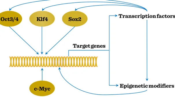
factor. Their effects manifest in a variety of pathways and mechanisms, including Wnt signaling, epigenetic modification, telomere regulation, mRNA metabolism and interference and also cell cycle control. Using iPS cells as model it is possible to dissect the interaction pattern between various transcription factors, affecting direct or indirect gene regulation by target gene expression induction or further transcription factor- mediated enhancement, and also epigenetic modification, also influencing the efficiency of TF-mediated processes.
Figure I-3: Reprogramming: Induction of pluripotency in iPS cells
The capacity to induce stem cell-like potential in differentiated cells has led to the revision of mature cells’ status of pluripotency and transdifferentiation capacity. Thus the maintenance of pluripotency requires an increased threshold against differentiation signals, while responsiveness to these differentiation signals needs to be sequentially provided, thereby increasing resistance against pluripotency. However, this stage is reversible, as enforced expression of pluripotency-related transcription factors may re-
Target genes
Epigenetic modifiers Transcription factors Sox2
Oct3/4 Klf4
c-Myc
Definition of stem cells and stem cell groups, their maintenance and homeostasis
Identification number:
TÁMOP-4.1.2-08/1/A-2009-0011
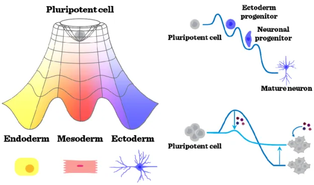
11 establish higher levels of resistance against differentiation signals, and a reduced induction level of pluripotency.
Figure I-4: Sequential maturation and pluripotency
Identification number:
TÁMOP-4.1.2-08/1/A-2009-0011
13
II Regeneration in animal models
Many organisms are capable of restoring by regrowth, parts of their body that have been lost by injury or by autotomy. Physiological regeneration includes seasonal or hormonal cycles of tissues such as antlers of deer and replacement of blood and epithelial cells.
Tissue regeneration is defined as the replacement of damaged tissues without the mediation of a blastema, whereas epimorphic regeneration refers to a type of regeneration mediated by the blastema. Hypertrophy is thought to be a type of regeneration, in which there is a compensatory increase in the size of a paired organ such as kidneys and lungs after its pair has been lost or damaged. The regenerative type refers to restoration of the mass of damaged internal organs such as liver and pancreas.
Morphallaxis refers to reconstruction of form after severe damage by remodeling the body.
It is well known that invertebrates are capable to extreme regeneration. Hydras, planarians are able to regenerate after serious injury their entire body. While annelids have also a huge regeneration capacity, some insects (flies) have this capacity only during larval stage. In opposite, some other insects kept this capacity till adulthood.
Organ regeneration capability is varied among the vertebrates. For instance limb regeneration in mammals is restricted mainly to fingertips, while amphibians are able to regenerate at any levels in proximal / distal (PD) axis. This limb regeneration has several important steps.
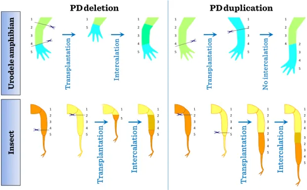
The concept of positional information along an axis during limb regeneration has been established by many experimental studies in amphibians and insects. In typical epimorphic regeneration both in amphibians and hemimetabolous insects, the stump region of the appendage forms a regeneration blastema and starts elongating along the
Transdifferentiation and regenerative medicine
14 The project is funded by the European Union and
co-financed by the European SocialFund.
PD axis. In such regeneration processes, for example, we can presume the sequential values along the PD axis.
Confrontation between normally non-adjacent positional values in the proximal–
distal (PD) sequence can be produced by transplantation experiments. When the proximal and distal positional values confront with deletion of the intermediate region along the PD axis, intercalary regenerative response occurs in both newt limb and insect leg. In this intercalary response, a minimum sequence of positional values will be intercalated (the shortest intercalation rule) and the continuity of adjacent positional values will be restored. When the confrontation is made with duplication of the intermediate region along the PD axis, intercalary response does not occur in newt limb while it occurs in insect leg with formation of a longer leg.
Figure II-1: Similarities in regeneration
Re-expression of genes involved in pattern formation is essential for complete limb regeneration, but the circumstances for re-expression are not always the same
1 2 3 4 5 5
1 1
2 3 4 5
1 2 3 4 5 1
2 3 4 5
1 2 3
4 2
3 4 5
1 5
1 2 3 4 5
UrodeleamphibianInsect Transplantation Intercalation Transplantation No intercalation
1 2 3 4 3 2 3 4 5 1
2 3 4 2 3 4 5
Transplantation Intercalation Transplantation Intercalation
1 2 3 4 5
PD deletion PD duplication
1 2 3 4 5
1 2 3 4 5
1 2 3 4 5
Regeneration in animal models
Identification number:
TÁMOP-4.1.2-08/1/A-2009-0011
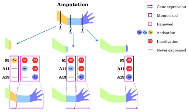
15 because restoration is needed for only the lost part of the limb. Amphibians can regenerate the lost distal part of a limb after amputation at any position along the PD axis. Therefore, it has been thought that the remaining cells in the stump tissues must remember the information about their original positions along the three axes of the limb and this memorized positional information may enable blastema cells to regenerate only the missing part. More distal positional values are newly provided by a de novo process.
Figure II-2: Imprinting in regeneration
The origin of the cells that are involved in regeneration is of some debate. It is possible that regeneration arises by transdifferentiation of existing cells. It is equally possible that a reserve of undifferentiated cells, or in other words adult stem cells exist, which, when subjected to the right signals can be activated to give rise to a whole array of cell types. One good example for that are the stem cells of central nervous system.
In vitro neural differentiation of pluripotent stem cells that mimics the temporal changes in the differentiation capacity of NSCs. Pluripotent stem cells, including both
Amputation
M A11 A13
M A11 A13
M A11 A13
Inactivation
Off
Activation
On On On
Never expressed Memorized Renewed Gene expression
On Off
Off On Off On
Off On
Off Off On
Off Off On
Transdifferentiation and regenerative medicine
16 The project is funded by the European Union and
co-financed by the European SocialFund.
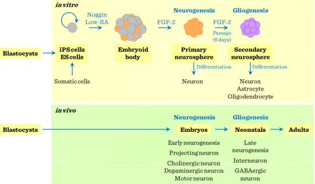
ES and iPS cells, can be induced to adopt a neural fate under the same culture conditions using neurally-biased embryoid body (EB) formation and the subsequent serial passages of neurospheres to give rise to primary and secondary neurospheres.
While the primary neurospheres predominantly differentiate into early projection neurons, secondary neurospheres differentiate into neurons, astrocytes, and oligodendrocytes, thus recapitulating the temporal changes in the differentiation capacity of NSCs.
Figure II-3: Neural stem cells differentiation capacity
Differentiation Noggin
Low-RA FGF-2 FGF-2
Passage (6 days)
in vitro
in vivo Somatic cells
Neurogenesis
Neurogenesis
Early neurogenesis Projecting neuron Cholinergic neuron Dopaminergic neuron
Motor neuron Neuron
Gliogenesis
Gliogenesis
Late neurogenesis
Interneuron GABAergic
neuron Neuron Astrocyte Oligodendrocyte
Differentiation iPS cells
ES cells
Embryoid body
Primary neurosphere
Embryos
Secondary neurosphere
Neonatals Adults Blastocysts
Blastocysts
Identification number:
TÁMOP-4.1.2-08/1/A-2009-0011
17
III Epigenetic factors in transdifferentiation
Epigenetics refers to the non-sequence based structural changes of chromosomal regions that can alter the gene expression activities in response to the external signals.
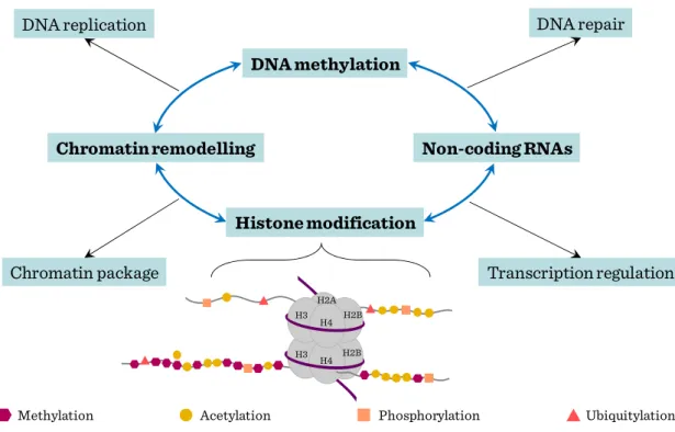
Major types of epigenetic modifications include DNA methylation, histone modifications, chromatin remodeling and noncoding RNAs. Distinct histone modifications which carry regulatory information were dubbed ‘histone code’, based on the analogy with the ‘genetic code’ of genomic DNA sequence. Histones are the subjects of over 8 major distinct classes of modifications with dynamic natures and consequences. Such covalent modifications play fundamental roles in chromatin condensation, replication, DNA repair, and transcriptional regulation. Among them, methylation and acetylation are the most studied and best understood histone modifications. Histone acetylation is generally associated with actively transcribed genomic domains and the degree of modification correlates with the level of transcription, whereas histone methylation can have different effects on gene expression depending on which residue is modified. Among the diverse histone modifications, methylations in histone 3 lysine 4 and lysine 27 are of particular importance. These modifications are catalyzed by Trithorax (TrxG) and Polycomb (PcG) group complexes respectively, which establish developmental decisions and mediate the expression programs of lineage specific genes. Lysine 4 methylation positively regulates gene expression by serving as a binding platform to recruit nucleosome remodeling enzymes, whereas lysine 27 methylation negatively regulates gene expression by generating a compact chromatin domain. Similarly, H3K36me3 is associated with actively transcribed regions whereas H3K9me3 is involved in inactive genomic domains.
Particularly, H3K4me3 is regarded as a hallmark for the promoters of actively
Transdifferentiation and regenerative medicine
18 The project is funded by the European Union and
co-financed by the European SocialFund.
transcribed genes and H3K36me3 is an indicator of transcription elongation. The different preferences of these histone modification occupancies on genome-wide landscape could be used to define transcription units.
The mechanism adopted by histone modifications to regulate transcription and chromatin organization is not clearly defined. An attractive hypothesis is that epigenetic factors, including modifying enzymes or remodeling factors, could potentially tether chromatins to mediate cis and trans interactions. Such interactions presumably cause the structural changes of underlying DNA and chromatins. Conformation changes could even be mediated by specific protein complexes recruited by these modifications. These interactions can alter the physical property of the chromatin and affect its high order structures.
Figure III-1: Epigenetic gene regulation of stem cell genome
In mammalian cells, both specific histone modification and DNA methylation are involved in chromatin silencing. DNA methylation and histone modification are
DNA replication DNA repair
Chromatin package Transcription regulation
Histone modification Chromatin remodelling
DNA methylation
Non-coding RNAs
Methylation Acetylation Phosphorylation Ubiquitylation
H3 H4
H2B H2A H3
H4 H2B
Epigenetic factors in transdifferentiation
Identification number:
TÁMOP-4.1.2-08/1/A-2009-0011
19 believed to be interdependent processes. Recent studies suggest that a combination of histone acetylation and DNA demethylation induces neuronal stem cells (NSC) to trans- differentiate into hematopoietic cells.
In ES cells, the overall nuclear architecture is globally decondensed and condensation reoccurs during differentiation. Specific changes in histone modifications have been shown to accompany ES cell differentiation and mammalian development.
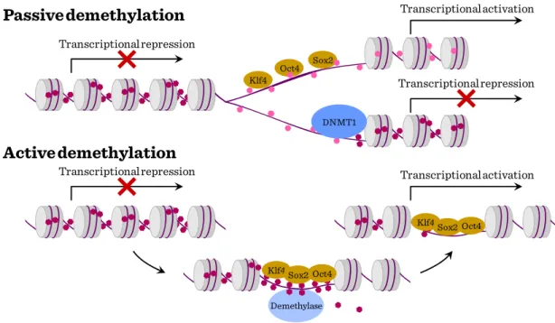
The establishment of symmetric DNA methylation patterns could be prevented passively during replication by the steric hindrance of Dnmt1 due to the stochastic binding of the reprogramming factors to target sites or by inhibiting Dnmt1 function indirectly. Hemimethylation of the DNA would result in a progressive loss of methylation upon further rounds of cell division. Alternatively, DNA methylation could be actively removed by the recruitment of a demethylating enzyme.
Figure III-2: DNA methylation in stem cells Passive demethylation
Active demethylation
Transcriptional repression
Transcriptional repression
DNMT1 Klf4
Oct4 Sox2
Transcriptional activation Transcriptional repression
Klf4Sox2Oct4
Demethylase Klf4Sox2Oct4
Symmetrically methylated DNA Hemi methylated DNA
Transcriptional activation
Transdifferentiation and regenerative medicine
20 The project is funded by the European Union and
co-financed by the European SocialFund.
A new integrated global regulatory network is currently emerging based on the dynamic interplay of chromatin remodeling components, TFs, and small ncRNAs.
These three mechanisms synergize to choreograph stem cell self-renewal and the generation of cell diversity. Mammalian cells harbor numerous
small ncRNAs, including small nucleolar RNAs, (snoRNAs), microRNAs (miRNAs), short interfering RNAs (siRNAs) and small double-stranded RNAs, which regulate gene expression at many levels including chromatin architecture.
Most show distinctive temporal- and tissue-specific expression patterns in different tissues, including embryonic (ESC) stem cells and the brain, and some are imprinted.
Many miRNAs are specifically expressed during ESC differentiation, embrygenesis, neuronal differentiation and hematopoetic lineage commitments.
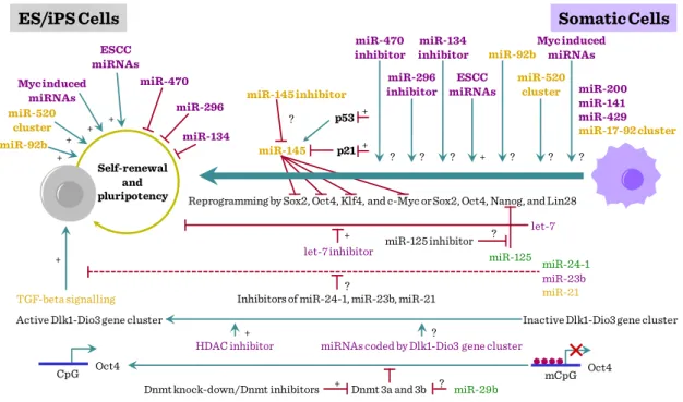
Several miRNAs, including ESC miRNAs, Myc-induced miRNAs, miR-92b, and the miR-520 cluster, have been shown to positively regulate the self-renewal and pluripotency of ES cells. Among these, only ESC miRNAs have been tested for their ability to promote reprogramming. Additionally, a number of tissue-specific miRNAs, such as let-7, miR-134, miR-470, miR-296, and miR-145, have been shown to interfere with the self-renewal and pluripotency of ES cells. However, with the exception of let- 7, the effects of inhibiting the activity of these miRNAs on reprogramming are not known. Recent study demonstrated that inhibition of let-7 activity promotes reprogramming. miR-125, which inhibits the expression of Lin28, is also expected to positively influence reprogramming.
Additionally, miRNAs that target specific signaling pathways (e.g. TFG-beta signaling) and epigenetic processes (e.g. DNA methylation) can also be tested for their
Epigenetic factors in transdifferentiation
Identification number:
TÁMOP-4.1.2-08/1/A-2009-0011
21 ability to promote reprogramming. miRNAs encoded by Dlk1–Dio3 gene cluster are also attractive candidates for promoting reprogramming because activation of imprinted Dlk1–Dio3 gene cluster is essential for generating fully reprogrammed iPS cells, which are functionally equivalent to ES cells.
Figure III-3: miRNA and stem cell differentiation
+ +
+ +
+ +
+ + miR-520
cluster miR-92b
Myc induced miRNAs
ESCC miRNAs
Self-renewal and pluripotency
miR-296 miR-470
miR-134
HDAC inhibitor miRNAs coded by Dlk1-Dio3 gene cluster
Dnmt knock-down/Dnmt inhibitors Dnmt 3a and 3b miR-29b TGF-beta signalling Inhibitors of miR-24-1, miR-23b, miR-21
Reprogramming by Sox2, Oct4, Klf4, and c-Myc or Sox2, Oct4, Nanog, and Lin28
let-7 inhibitor miR-125 inhibitor
miR-125 miR-145 inhibitor
miR-145 p53 p21
miR-200 miR-141 miR-429 miR-17-92 cluster
ES/iPS Cells Somatic Cells
Active Dlk1-Dio3 gene cluster Inactive Dlk1-Dio3 gene cluster
miR-24-1 miR-23b miR-21 let-7 ESCC
miRNAs
+ miR-470
inhibitor
? miR-296 inhibitor
? miR-134 inhibitor
?
miR-92b
? miR-520
cluster
?
?
?
+ ?
?
?
Myc induced miRNAs
?
+
mCpG Oct4 CpG Oct4
Identification number:
TÁMOP-4.1.2-08/1/A-2009-0011
23
IV Genomic and other cell-tracing approaches, reprogramming
Embryonic stem cells (ESCs) are derived from the inner cell mass (ICM) of the early embryo. Similar to cells of the ICM, ESCs hold great developmental potential and are able to differentiate to all cell lineages of an organism except for extraembryonic tissues, a unique property termed as pluripotency. In addition, ESCs can self-renew and be cultured indefinitely in vitro. ESCs are promising sources of cells for regenerative therapy, hence an enhanced understanding of the molecular mechanisms that regulate their propagation and pluripotency will allow us to better utilize them for clinical treatments.
Oct4, Sox2 and Nanog constitute the core regulatory network that governs ESC pluripotency. These core transcription factors concertedly regulate common downstream genes that promote pluripotency and self-renewal, while inhibiting differentiation processes. On the other hand, epigenetic modifications such as histone and DNA methylation can occur synergistically with the transcription factor network to regulate the expression of genes that maintain the ESC state. Together, both transcriptional and epigenetic processes specify gene expression programs critical for the maintenance of ESC properties (pluripotency and/or self-renewal). Conversely, a loss of pluripotency involves switching the transcriptional program to one that promotes differentiation.
Transdifferentiation and regenerative medicine
24 The project is funded by the European Union and
co-financed by the European SocialFund.
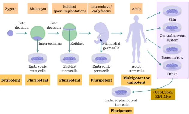
Figure IV-1: Origin of stem cells and reprogramming
Currently, a series of stem cell labeling techniques are applied in stem cell transplants fields, which include the BrdU, fluorescent dye, green fluorescent protein, magnetic, and isotope labeling techniques. Moreover, different tracing techniques applied in stem cell transplants were accorded with the different aims of the experiments and the characteristics of the stem cells.
BrdU incorporation and fluorescent dyes (CFSE, DiI, PKH26) were firstly used as stem cell tracers for their convenience. However, their labeling intensity will be gradually decreased, and they cannot detect the labeled stem cells in vivo for a prolonged time. In recent years, GFP is widely used for stem cell tracing for its stable expression, high specificity, and easy tracability in vivo, but it is limited by the fact that high levels of GFP have certain cell toxicity. In addition other recombinant marker such as LacZ reporter expression was used. Y chromosome marker detection by FISH is also another option.
+ Oct4, Sox2, Klf4, Myc Blastocyst
Embryonic stem cells
Pluripotent Zygote
Totipotent
Adult Epiblast
(post-implantation)
Epiblast stem cells
Pluripotent
Late embryo/
early foetus
Embryonic germ cells
Pluripotent
Adult stem cells Multipotent or
unipotent
Skin
Centralnervous system
Bone marrow
Other
Induced pluripotent stem cells Pluripotent Inner cell mass Epiblast Primordial
germ cells Fate
decision
Fate decision
Genomic and other cell-tracing approaches, reprogramming
Identification number:
TÁMOP-4.1.2-08/1/A-2009-0011
25 Currently MRI and isotope labeling techniques were attempted to trace the transplanted stem cells in vivo for their non-invasive nature. However, MRI labeling can cause false positive results and isotope labeling was limited by the specific stem cell markers.
Development of in vivo-time lapse / two-photon microscopy gave further advance for in vivo imaging of stem cells. Time-lapse microscopy is a powerful way of probing the behaviour of live stem cells in artificial niches. Stem cells can be imaged at various time points and locations to generate time-lapse movies, and automated image analysis and statistical analyses are used to quantify the dynamic behaviour of cells. A number of different read-outs, corresponding to different stem-cell functions, are also available.
Together with cell migration, changes in cell shape and changes in proliferation kinetics, the recording and automated analyses of changes in the fate of individual stem cells are crucial: cell death (1); quiescence (that is, non-cycling; 2); symmetrical self- renewal divisions (proliferation behaviour imposed in response to stress or trauma; 3);
asymmetrical self-renewal divisions generating one daughter cell that retains stem-cell identity and one already partly differentiated (a behaviour thought to be dominant during homeostatic conditions; 4); and symmetrical depletion divisions, in which both daughter cells lose stem-cell function (the default behaviour of adult stem cells grown in vitro; 5).
Transdifferentiation and regenerative medicine
26 The project is funded by the European Union and
co-financed by the European SocialFund.
Figure IV-2: Cell tracing in stem cell biology
Cells are described as pluripotent if they can form all the cell types of the adult organism. If, in addition, they can form the extraembryonic tissues of the embryo, they are described as totipotent. Multipotent stem cells have the ability to form all the differentiated cell types of a given tissue. In some cases, a tissue contains only one differentiated lineage and the stem cells that maintain the lineage are described as unipotent. Postnatal spermatogonial stem cells are unipotent in vivo but are pluripotent in culture.
z
y x
Single-cell fate analyses
Migration Proliferation Cell-shape change
t1
t2
tn
Automated image analyses
and
statistical analyses
Genomic and other cell-tracing approaches, reprogramming
Identification number:
TÁMOP-4.1.2-08/1/A-2009-0011
27 Figure IV-3: Molecular mechanisms of self-renewal
Four strategies for reprogramming differentiated cells to an embryonic state:
somatic cell nuclear transfer (SCNT), cell fusion, culture-induced reprogramming and iPSC generation. Culture-induced reprogramming of embryonic germ cells and spermatogonial stem cells, although important, does not constitute an example of radical reprogramming. Cell fusion has not yet resulted in the production of pluripotent diploid cells, although it might do so in the future. By contrast, both SCNTand iPSCformation have succeeded in this task. SCNT, cell fusion and iPSC formation can all be interrogated to address the kinetics and mechanisms of the observed reprogramming.
SCNTand iPSCgeneration—the latter in the absence of chemical supplements—are both inefficient procedures. The best recorded efficiencies using mouse fibroblasts are similar: 3.4% for SCNT and 1–3% for iPSCgeneration. iPSC lines can arise over a protracted timeline and, therefore, inappropriate changes might be reversed over time.
In other ways, SCNT, cell fusion and iPSC reprogramming are clearly different G2
G1 M
S
Cell-cycle regulation
Prevention of differentation Sox2 Nanog
Oct3/4
Klf4 Tbx3
STAT3
Akt MAPK
Jak
PI3K Grb2
Lif
Cdx2 Gata4
c-Myc b-Myb
Transdifferentiation and regenerative medicine
28 The project is funded by the European Union and
co-financed by the European SocialFund.
processes. In SCNT, the time required for reprogramming before initiation of development is short. OCT4 was detectable in mouse SCNT blastocysts within 12–24 h although the reactivation of many embryonic genes was erratic and variable between embryos. Rapid OCT4 and SSEA4 expression is also seen in the somatic nucleus of 13–
16% of mouse ESC-somatic cell heterokaryons. In both processes, significant and rapid nuclear swelling, which is thought to indicate chromatin decondensation, precedes reprogramming. By contrast, during iPSC formation, OCT4 becomes detectable only after roughly 2 weeks and only in a small proportion of treated cells. These data indicate that the processes share a stochastic nature but that, in SCNT and cell fusion, reprogramming occurs much faster. In all situations, it remains unclear whether the reactivation of the embryonic genome seen during reprogramming requires DNA replication and cell division. In the frog, the activation of embryonic or specific pluripotency-associated genes does not require the host cell or donor nucleus to progress through the cell cycle or undergo DNA replication. Similarly, the expression of various pluripotency-associated genes can be detected in the somatic components of heterokaryons before nuclear fusion. By contrast, iPSCs have always been made from proliferative somatic cell populations, and whether DNA replication and/or cell division are essential for reprogramming remains untested. In conclusion, cell fusion and SCNT initiate a faster activation of pluripotency-associated genes in the absence of cell division. This could be a consequence of the oocyte or ESC cytoplasm and/or nucleoplasm providing a more complete set of factors that facilitate more rapid reprogramming than a defined group of transcription factors, or of inherent differences in the reprogramming mechanism. So far, the design of iPSC-reprogramming experiments has made it impossible to exclude the necessity of DNAreplication and cell division. Owing to methodological differences, it is difficult to determine from
Genomic and other cell-tracing approaches, reprogramming
Identification number:
TÁMOP-4.1.2-08/1/A-2009-0011
29 published results whether such a discernible difference in reprogramming efficiencies applies to iPSCs that are made from a diverse range of cell types.
Identification number:
TÁMOP-4.1.2-08/1/A-2009-0011
31
V Stem cells and transdifferentiation during haematopoesis
The significance of haemopoietic differentiation from stem cells has three main implications in regenerative medicine. One is the evolution of haemopoiesis itself, demonstrating a considerable difference of differentiation spectrum as determined by various progeny populations generated at distinct tissues (yolk sac, embryonic liver or adult bone marrow). Second, one of the few broadly used approaches with substantial clinical impact in regenerative medicine concerns the re-establishment of haemopoiesis in leukemic or other conditions. Third, the regeneration of several non-haemopoietic tissues has also been attempted following degenerative or necrotic tissue damages, such as myocardial infarct or neuronal lesions.
The first site with haemopoietic activity in a developing embryo is located in the blood island in yolk sac, an extraembryonic component. At this location parallel haemopoietic (erythropoietic) and endothelial differentiation takes place, indicating the existence of a common haemangioblast precursor. Similar relatedness can be observed later in the embryo proper at the ventral aspect of dorsal aorta, in the region termed aorto-gonad-mesopnephros (AGM), where developing blood cells show close physical relationship with the vascular endothelium. Subsequently parallel to the emergence of circulation the haemopoiesis is established in the fetal liver and, to a lesser extent, in the fetal spleen. Blood cells may also be generated from hemogenic endothelial cells, and depending on the period where hemopoiesis is established, these can be dissected into pro-definitive, meso-definitive, meta-definitive and finally, adult definitive, phases. The yolk sac haemopoiesis is referred to as “primitive hemopoiesis”, which thus precedes the various stages of definitive hemopoiesis. Towards the end of pregnancy the
Transdifferentiation and regenerative medicine
32 The project is funded by the European Union and
co-financed by the European SocialFund.
developing bone marrow is colonized by osteoclast-like cells which, together with the osteoblasts and fibroblastic stroma cells, will establish the bone morrow niche capable of receiving circulatory HSCs.
Figure V-1: Ontogeny of embryonic haemopoietic tissues
At these locations the haemopoietic stem cells (HSCs) can be defined by their cell surface markers, which in mouse include Sca-1, c-kit, CD45, and several shared haemopoietic/endothelial antigens, including CD31, CD34 and VE-cadherin antigens.
For initiating haemopoiesis the HSCs express Runx, Scl and GATA-2 transcription factors. In addition to endogeneous cell programming, a certain topographic preference within the embryonic body is required for the haemopoesis to manifest. This morphogenic factors facilitating the ventralisation within the dorsal aortic segments, such a VEGF, bFGF, TGFβ and BMP4, will promote the establishment of haemopoiesis, whereas factors promoting ectodermal/dorsalizing effect, such as EGF and TGFα, will inhibit it.
Pro-definitive
Primitive Meso-definitive Meta-definitive Adult-definitive E10.5
E7.5 E8.25 E9.0
Hemangioblast Hemogenic Endothelium
Myeloid Lymphoid-Myeloid CFU-s
Neonatal HSC
HSC Yolk sac
Allantois Chorion
Allantois
YS blood islands Placenta
AGM
Liver Placenta
AGM
Liver Yolk sac
Embryo pSP
Umbilical artery
Vitelline artery
Umbilical artery
Vitelline artery
Stem cells and transdifferentiation during haematopoesis
Identification number:
TÁMOP-4.1.2-08/1/A-2009-0011
33 Subsequent to the initiation of haemopoiesis, several decision steps are necessary for the specification of lineage commitment of HSCs. One important checkpoint is the preservation of pluripotency, by the concerted action of Notch-1, GATA-2, HoxB4 and Ikaros transcription factors. Furthermore, cell cycle inhibitor p21 is necessary to keep a large fraction of stem cells out of proliferation. Next the enhanced expression of PU1 will favor myeloid comitment, whereas low-to-intermediate expression of PU1 together with GATA-3 and Ikaros transcription factors will commit HSCs towards lymphoid lineage.
Figure V-2: Transcriptional regulation of early haemopoietic commitment
Hemangioblasts Hemogenic endothelium
Apoptosis
SCL AML-1 GATA-2 Lmo
PU.1/GATA-3/Ikaros PU.1/GATA-1
CMP
HSC
CLP Notch1
Ikaros HoxB4 GATA-2
Bcl-2 p21
Transdifferentiation and regenerative medicine
34 The project is funded by the European Union and
co-financed by the European SocialFund.
Once the myeloid dominance has been established through increased PU1 or GATA-1 expression, further transcriptional control will determine the commitment along erythroid/megakaryocytic (GATA-1/2 dominance) or myelomonocytic (PU1 dominance) lineages. In addition, the induction of C/EBPα or C/EBPβ will modulate the fate of myeloid-committed cells towards granulocytic branches.
Figure V-3: Transcriptional regulation of myeloid differentiation
In contrast to either PU1 or GATA-1 polarity leading to myeloid or erythroid differentiation, their balanced low-to-medium expression promotes the lymphoid commitment of common myeloid-lymphoid precursors (CMLP). Importantly, the transcriptional control for promoting lymphoid commitment crucially involves the upregulation of IL-7 receptor, with the parallel decrease of receptors for myeloid growth factors, including GM-CSF and G-CSF, in addition to the downregulation of c-kit receptor for stem cell factor.
Monocyte
Neutrophil
Eosinophil
Erythrocyte
Megakaryocyte
GMP
CMP HSC
EMP PU.1 & GATA-1
PU.1
GATA-1/FOG
ICSBP, PU.1
C/EBPβ, GATA-1
GATA-1
GATA-1, 2 C/EBPα
Stem cells and transdifferentiation during haematopoesis
Identification number:
TÁMOP-4.1.2-08/1/A-2009-0011
35 The common lymphoid progenitor (CLP) may commit itself along T-lineage, mediated by Notch1-mediated signaling, or may follow the B-lineage commitment by upregulating the E2A transcription factor. At this point a parallel upregulation of the Id2 transcription factor may divert the differentiation towards NK-linege within the lymphoid pathway. CLPs at this (still flexible) stage may enter the thymus where the local microenvironment rich in Notch1 ligands (Jagged, etc.) may promote T-lineage commitment. Interestingly, some B-cell precursors may retain some flexibility, even the possibility for macrophage-like reversal.
Figure V-4: Transcriptional regulation of lymphoid differentiation
In addition to the steady-state balanced haemopoiesis, external stimuli (hypoxia, inflammation, etc) coupled with the peripheral release of soluble mediators (erythropoietin, or inflammatory cytokines such as TNFα) may significantly alter the leukocyte output, by eliciting increased production of G-CSF and GM-CSF, leading to accelerated myelopoiesis.
CLP
Pro-T
PU.1 IL-7R
GM-CSFR
Notch1
Pu.1, E2A
PU.1
Pax5?
EBF Pax5
Early pro-B Late pro-B
Pre-
pro-B D-JH V-D-JH
B cell Monocyte
HSC
Transdifferentiation and regenerative medicine
36 The project is funded by the European Union and
co-financed by the European SocialFund.
Figure V-5: Steady-state and activated haemopoiesis
In addition to maintaining or correcting haemopoiesis, HSCs have also been tested for their ability to promote regeneration of non-hamopoetic cells. These tissues include muscle, liver, and neural tissues; however, the exact mechanism how these cells may contribute to the healing effect remains to be determined. The possibilities include direct transdifferentiation into tissue-stem-cell like entities, or differentiation into some cells facilitating the removal of degenerative tissues, and also the enhanced vessel formation by regional angiogenesis, thus the restoration of blood supply.
HSC
HSC
HSC
HSC Endothel
Fibroblast
Osteoblast
G-CSF, GM-CSF TPO
TGFβ
EPO EPO
IL-1α TNFα Macrophage
Bacterial infection Inflammation
Anemia Hypoxia
HSC
HSC
HSC
HSC SCF, FLT-3I, TPO
Endothel
G-CSF, GM-CSF Fibroblast
Osteoblast
Blood vessel
Identification number:
TÁMOP-4.1.2-08/1/A-2009-0011
37
VI Muscle regeneration
The skeletal muscle represents the largest mass of mesodermal/connective tissue, with highly variable developmental characteristics and origins. Thus the external ocular muscles develop from the cranial unsegmented mesoderm, the tongue muscles derive from the occipital somites, the hypaxial limb muscles and the epaxial back muscles develop from the dermomyotome of somites. The myogenic programming in the mesoderm involves the upregulation of Pax3 transcription factor, inducing directly or indirectly (with the involvement of Myf5 and Myf6 transcription factors) the expression of MyoD transcription factor. As microenvironmental regulators, BMP4 produced by the neural tube and lateral plate mesoderm inhibits, whereas Sonic Hedgehog (Shh) and several Wnt’s produced by the notochord and surface ectoderm promote myogenic differentiation. The need for regenerating skeletal muscle or using muscle tissues for regeneration purposes can be grouped in three main conditions:
(1) Skeletal regeneration due to trauma or genetic disorder (most frequently in Duchenne’s muscular dystrophy, DMD). Duchenne’s muscular dystrophy is caused by the X-linked mutation of the dystrophin gene, the largest known mammalian gene, with the prevalence of 1:3,500 male neonates. The disease manifests in early-mid childhood, and is characterized by progressive muscle-wasting including limb muscles, diaphragm, and heart leading to cardiorespiratory failure, and death usually occurs in the teenage years or early 20s.
(2) Cardiac regeneration using skeletal muscle stem cells (satellite cells – SCs) in myocardial infarcts;
(3) Sphincter muscle regeneration in urinary incontinentia using SCs.
Transdifferentiation and regenerative medicine
38 The project is funded by the European Union and
co-financed by the European SocialFund.
The structural unit of skeletal muscle is the myofibre, a synctitial meshwork of myocytes ensheathed by basal lamina or endomysium. The myofibres contain fused cells with myonuclei and mitochondria, and, importantly, myofibrils with contractile myofilaments. Between the basal lamina and the plasma membrane of myofibrils are located the mononuclear SCs, first described in 1961 by Alexander Mauro using electron microscopy.
Figure VI-1: Structure and regeneration of skeletal muscle
Depending on their tissue location and developmental features of the muscular segment, various skeletal muscle SCs display a diverse array of cell surface characteristics and transcription factor mRNA expression profile, including variable expression of CD34. Sca-1, M-cadherin, CD56, c-met receptor for HGF/Scatter factor as surface molecules or Pax3 and Pax7 myogenic transcription factors are expressed in a region-dependent fashion.
Myofibril
Hematopoietic cells
Pericyte
Endothelial cell
Arteriole and capillaries
Interstitial cell Basal lamina
Satellite cell (SC)
Muscle fiber Myonucleus
Quiescent SC Pax7+
Activated SC Pax7+ Myf5+MyoD+
Fusion and differentiation Return to
quiescence
Myoblast Pax7- Myf5+MyoD+ Expansion
(symmetric division) Asymmetric
division Activation
Myocyte MyoD+
Muscle regeneration
Identification number:
TÁMOP-4.1.2-08/1/A-2009-0011
39 As potential candidates for muscle regeneration other than SCs, several other cell types were suggested. These include haemopoietic stem cells, endothelial cell subsets, mesenchymal stem cells as well as some non-SC pluripotent stem cells found within the muscles. For regeneration either allogenic precursor/stem cell transfer or autologous gene-manipulated stem cells have been proposed.
Figure VI-2: Non-SCs contributing to muscle regeneration
The mechanism how SCs may contribute to skeletal muscle regeneration may include various forms of differentiation from SCs, or regeneration from diverse (haemopoietic, mesenchymal, etc.) stem cells, or even fusion between SCs and residual myofibres. In vivo myogenic lesion by intramuscular injection of cardiotoxin is first followed by SC activation by soluble factors, including IGF, FGF, HGF/scatter factor, leading to SC proliferation in the first 2–3 days. Subsequently, smaller diameter myofibres with centrally positioned nuclei form during the next 5–7 days, followed by the maturation upon the fusion of myocytes into regenerated myofibres, resulting in
Expansion
Commitment(if needed) Allogeneic transplantation Autologous transplantation
(after genetic correction)
Mesenchymaldifferentation
Adipose-derivedstem cells MyoD-converted cells
HSCs Side population Mesenchymal stem cells
MAPCs SCs and subpopulations
MDSCs CD133+stem cells
HSCs Side population CD133+stem cells
MABs/pericytes Myoendothelialcells
EPCs MSCs
iPS cells Reprogramming Dermis or other tissues
Skeletal muscle
Bone marrow
Other sources Blood Vessels
Characterization
Transdifferentiation and regenerative medicine
40 The project is funded by the European Union and
co-financed by the European SocialFund.
larger diameter and nuclei located peripherally approximately 10–12 days after the injury.
Figure VI-3: Kinetics of muscle repair
The use of allogeneic cells requires the administration of immunosuppressant drugs, which may also cause myoblast apoptosis. Using autologous myogenic stem cells for correcting DMD expressing the normal dystrophin gene may also elicit immune reactivity in individuals born with the aberrant gene. In addition, due to the special location and broad tissue distribution of skeletal muscle, the administration of cells also poses a substantial obstacle, as the short migratory capacity of myogenic cells would require as many as 100 injections in a 1 cm2 area.
The use of skeletal SC as source for myocardial regeneration has resulted in a moderate improvement of left ventricle function and cardiac output, however the rate of arrythmias has also increased, which could be ameliorated with antiarrhythmic drugs.
The use of skeletal SCs trogether with fibroblasts and other support cells to correct Activation
Proliferation
Differentiation
Maturation
0 1 2 5 10 14
Days post injury
Muscle regeneration
Identification number:
TÁMOP-4.1.2-08/1/A-2009-0011
41 sphincter muscle insufficiency has resulted in some improvement, although its effects manifest after a considerable delay.
Identification number:
TÁMOP-4.1.2-08/1/A-2009-0011
43
VII Liver regeneration
Liver transplantation is currently the only therapeutic option for patients with end-stage chronic liver disease (caused by inflammation, genetic or other causes for metabolic abnormalities, such as storage diseases, etc) and for severe acute liver failure. Because of limited donor availability, attention has been focused on the possibility to restore liver mass and function through cell transplantation. Although hepatocytes can substantially regenerate the liver, further improvements are expected from using stem cells.
For liver regeneration several stem cell candidates have been assayed, including mesodermal lineage progenitors, hepatic progenitors at various degrees of differentiation as well as hemopoietic progenitors. Considerable advances have been made in identifying cells in the fetal, neonatal and adult liver with stem-cell properties.
Cell lines have also been established, including ES cells, fetal liver cells, and oval (progenitor) cells that also exhibit stem cell properties and differentiate into hepatocytes and/or bile ducts in vitro and in vivo However, all of these cells and cell lines have shown only limited repopulation of the normal liver at the current state-of-the-art, except for rat fetal liver stem/progenitor cells that produce substantial long-term replacement and function.
The liver develops from the ventral endodermal epithelium, which can give rise to the hepatocytes, cholangiocytes and a subset of pancreatic cells, while the remaining part of the pancreas develops from the dorsal endoderm.
Transdifferentiation and regenerative medicine
44 The project is funded by the European Union and
co-financed by the European SocialFund.
Figure VII-1: Developmental relationship between hepatic-pancreatic differentiation
Various members of the FoxA and GATA transcription factor families promote the commitment towards hepatoblast lineage, a common precursor for hepatocytes and cholangiocytes, assisted by several members of the FGF and BMP families as soluble regulators. Following the upregulation of a variety of transcription factors as well as Notch and Wnt mediators the hepatoblast differentiates into either hepatocytes or cholangiocytes, where subsequent maturation will establish the hepatic cords as well as the bile ducts.
Oval cell progenitor
Hepatic oval cell
Bile duct Hepatocyte
Pancreatic oval cell
Endocrine cell Pancreatic duct Acinar cell Pancreatic progenitor(s)
?
Liver regeneration
Identification number:
TÁMOP-4.1.2-08/1/A-2009-0011
45 Figure VII-2: Transcriptional control of hepatoblast development
This maturation process is accompanied by tissue reorganization via migration and vascular patterning, where the cellular movement involves the hepatoblasts, substantially promoted by Prox-1, Tbx3 and WT1 transcription factors. Further maturation will establish the complex structure of the hepatic lobule, including the differential gene expression and other characteristics of periportal (6–8 cells), centrilobular (8–10 cells) or most centrally positioned GS (1–3 cells with glutamine synthetase enzyme) hepatocytes.
Hepatocyte maturation cords
Hepatocyte
Core transcription factor network:
HNF-1α LRH-1
HNF-6 Foxa2 HNF-4α
HNF-1β
Jagged
Cholangiocyte
Parenchyma Periportal HNF-1β
Sox9 HNF-6/OC-2 TGFβ
Hex C/EBPα
Hepatoblast
HNF-6 HNF-1β
Notch2 HNF-4α
C/EBPα Tbx3
Albumin HGF
Cholangiocyte maturation ducts
?
Wnt BMP+FGF
FoxM1B ECM ECM
Transdifferentiation and regenerative medicine
46 The project is funded by the European Union and
co-financed by the European SocialFund.
Figure VII-3: Structure of hepatic lobe
Following injury, exposure of perisinusal (stellate or Ito’s) cells to the apoptotic or necrotic hepatocytes stimulates these quiescent Vitamin A-storing mesenchymal cells through their Toll-like receptors. These cells play fundamental role in the fibrotic regeneration of the liver. Coupled with extrahepatic fibrocyte immigration, presence of activated T cells and epithelial-to-mesenchymal transition of the hepatocytes together with the altered extracellular matrix composition, the transformation of stellate cells as tissue-specific pericytes significantly increases the myofibroblast compartment of the liver. During their activation their increased responsiveness to PDGF and enhanced migratory capacity toward the site of injury and contractile capacity of myofibroblasts significantly contribute to the altered microcirculation, increased fibrosis and scar formation.
The parenchymal regeneration following loss of liver mass involves a rapid transformation of resting hepatocytes into proliferating cell. However, in case of massive liver loss, toxicity or blocked hepatocyte regeneration (in chronic hepatitis), the
Glutamine synthetase + (1-3 cells)
Centrilobular (8-10 cells) Limiting plate
Periportal (6-8 cells)
Portal tracts (triads) Central vein
Bile duct
Bile canaliculi
Sinusoids
Branch portal vein
Branch hepatic artery
Central vein
Portal tract