A new method for identification and isolation of human embryonic stem cell-derived cardiac progenitors and
cardiomyocytes
Ph.D. dissertation
Kornélia Szebényi
Semmelweis University
Doctoral School of Molecular Medicine
Doctoral supervisor: Dr. Balázs Sarkadi, member of HAS
Official opponents: Dr. Gábor Földes, Ph.D.
Dr. Melinda Pirity, Ph.D.
President of the examination committee: Dr. Erzsébet Ligeti, member of HAS Members of the examination committee: Dr. Szabolcs Sipeki, Ph.D.
Dr. László Hiripi, Ph.D.
Budapest
2014
Table of Contents
1. List of Abbreviations ... 3
2. Introduction... 6
2.1. Human pluripotent stem cells ... 6
2.1.1 Generation of human pluripotent stem cells ... 6
2.1.2 Culture and characterization of human pluripotent stem cells ... 8
2.1.3 Application of human pluripotent stem cells and their derivatives ... 10
2.2. Generation of cardiomyocytes from human pluripotent stem cells ... 11
2.3. Systems allowing the isolation of human pluripotent stem cell-derived cardiac cells ... 14
1.3.1 Genetic methods for isolation of hPSC-derived cardiac cells ... 16
1.3.2 Non-genetic methods for isolation of cardiac cells ... 18
3. Aims ... 21
4. Materials and Methods ... 22
4.1. hESC culture and differentiation ... 22
4.2. Flow cytometry ... 23
4.3. Cell sorting ... 24
4.4. Fluorescence plate reader measurements ... 24
4.5. Immuncytochemistry ... 25
4.6. Real-time quantitative PCR analysis ... 26
5. Results ... 27
5.1. The CAG promoter allows the identification of cardiomyocytes in spontaneous differentiation cultures of human embryonic stem cells ... 27
5.1.1. Characterization of the hESC lines HUES9-CAG-EGFP and BG01V-CAG-EGFP . 27 5.1.2. In differentiation cultures of HUES9-CAG-EGFP and BG01V-CAG-EGFP cardiomyocytes show exceptionally high EGFP expression... 31
5.2. Directed cardiac mesoderm differentiation of CAG-EGFP expressing human embryonic stem cells ... 37
5.3. The CAG promoter allows identification and isolation of human embryonic stem cell- derived cardiac progenitors ... 42
5.3.1. Isolation and mRNA expression profiling of the CAG-EGFPhigh subpopulation ... 42
5.3.2. Comparing CAG-EGFP-based isolation of cardiac progenitors to other methods known from the literature ... 46 5.4. Examining different culture conditions for isolated CAG-EGFPhigh cardiac progenitors . 50
5.5. CAG-EGFPhigh cardiac progenitors give rise to a relative pure population of
cardiomyocytes ... 53
5.6. Enhanced culture conditions for supporting the growth of CAG-EGFPhigh rEBs without losing cardiac commitment ... 59
5.6.1. Modest growth of 3D aggregates generated from isolated CAG-EGFPhigh cells ... 59
5.6.2. Thiazovivin enhances reaggregation and survival of the sorted cells ... 61
5.6.3. Isoproterenol enhances cardiac differentiation of CAG-EGFPhigh cells cultured in END-2 conditioned medium... 65
6. Discussion ... 67
7. Conclusions ... 73
8. Summary ... 75
9. Összefoglalás ... 76
10. References ... 77
11. List of publications related to this thesis ... 95
12. List of publications not directly related to this thesis ... 96
13. Acknowledgement ... 97
1. List of Abbreviations
ACTB – human β-actin AF – AlexaFluor
AFP – alpha-fetoprotein
ALCAM/CD166 - activated leukocyte cell adhesion molecule APC - allophycocyanin
bFGF – basic fibroblast growth factor βIII-Tub – beta-III tubulin
BMP4 - bone morphogenetic protein 4 CAPG – capping protein gelsolin-like CD – cluster of differentiation
C-KIT/CD117 - tyrosine kinase Kit CM – cardiomyocyte
CMSC – cardiomyocyte supporting cell CMV - cytomegalovirus
CPC – cardiac progenitor cells cTnI – cardiac troponin I D - day
DM – differentiation medium
DMEM – Dulbecco’s Modified Eagle’s Medium EB – embryoid body
EMT – epithelial-to-mesenchymal transition
END-2 - mouse visceral endoderm-like cells END2-CM – END-2 conditioned medium FACS - fluorescence activated cell sorting FBS – fetal bovine serum
FITC - fluorescein isothiocyanate GFP – green fluorescent protein hESC – human embryonic stem cell
hiPSC – human induced pluripotent stem cell hPSC – human pluripotent stem cell
ISL1 - insulin gene enhancer protein
KDR/VEGF-R - vascular endothelial growth factor receptor 2 MEF – mouse embryonic fibroblast
mESC – mouse embryonic stem cell MTG – monothioglycerol
PAX6 - paired box 6
PDGFRA - platelet-derived growth factor receptor PE - phycoerythrin
PECAM-1 – platelet endothelial cell adhesion molecule-1 PGK - phosphoglycerate kinase
PI - propidium iodide
QPCR - real-time quantitative PCR RCM – ReproCardio Medium
rEBs – reaggregated embryoid bodies SIRPA/CD47 - signal regulatory protein α SMA – alpha-smooth muscle actin
SSEA4 - stage-specific embryonic antigen-4 TBX5 - T-Box Protein 5
TMRM - tetramethylrhodamine methyl ester perchlorate TNNT2 - troponin T type 2
UbC - ubiquitinC
VCAM1/CD106 - vascular cell adhesion molecule 1
2. Introduction
Stem cells are capable of renewing themselves through cell division for unlimited times and divide asymmetrically, generating two different daughter cells. Under certain physiological or experimental conditions, stem cells can be induced to become tissue- specific cells with special functions through differentiation. Stem cells can have different potentials (e.g. toti-, pluri- or multipotency), limiting the number of tissue types that can be differentiated from them.
Features of pluripotency include the potential of unlimited cell growth and self- renewal, as well as the capacity to generate all cell types of the body. However, pluripotent cells are restricted in their potential compared to totipotent stem cells, since human pluripotent stem cells (hPSCs) cannot give rise to cells of the extra-embryonic tissues (amnion, chorion, yolk sac and the allantois). Multipotent stem cells have even lower potential and only give rise to tissue-specific cells. Therefore multipotent stem cells are often termed as multipotent progenitor cells of certain developmental lineages.
This dissertation will focus on hPSCs and their in vitro differentiation capabilities, with a specific focus on the cardiac lineage to provide a method for identifying and isolating cell types at different stages of cardiac commitment.
2.1. Human pluripotent stem cells
2.1.1 Generation of human pluripotent stem cells
The derivation of mouse pluripotent stem cell lines from blastocyst-stage embryos revolutionized biological research through allowing generation of knock-out animal models (2007 Nobel Prize) and opening new possibilities in studying early developmental events. The much awaited next step, the establishment of the first human embryonic stem cell (hESC) line was reported by Thomson et al. seventeen years later, in 19981. The generation of hESC lines proved to be as revolutionary as the establishment of mouse ESC lines earlier, providing an excellent platform for human developmental studies and toxicological screenings, as well as the possibility of regenerative therapeutic applications.
Human ESC lines are usually derived from the inner cell mass of blastocyst stage embryos (see Figure 1). Unfortunately, ethical concerns regarding the use of in vitro- fertilized human embryos for research purposes strongly hinder the application of hESCs in basic research, drug development and medical therapy, even though the embryos are otherwise supposed to be destroyed. In addition to the ethical concerns, several other drawbacks exist. One of these is the potential immunological incompatibility that might limit the use of hESC-derived cells for regenerative purposes2. However, immunological problems may be prevented by hESC-banking3, 4 or transplantation of encapsulated hESC-derivatives secreting the necessary factors to compensate for deficits5.
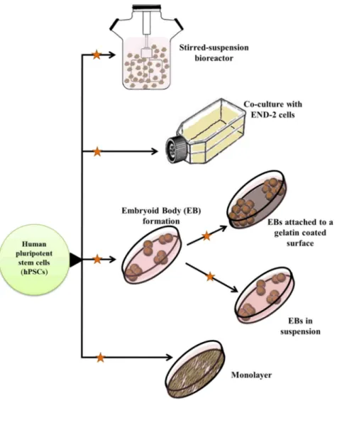
Figure 1. Methods for the generation and differentiation of human pluripotent stem cell lines. The image is taken from Szebényi et al.6
In 2006, another milestone of stem cell research was reached when induced pluripotent stem cells (iPSC) were generated from mouse fibroblast cultures by forced
expression of four transcription factors (Oct3/4, Sox2, Klf4 and c-Myc)7. This study was immediately followed by numerous others, documenting the generation of human induced pluripotent stem cells (hiPSCs) with viral or non-viral delivery of the reprogramming factors8 (see Figure 1). The discovery that mature cells can be reprogrammed to become pluripotent was honoured by the 2012 Nobel Prize.
By now, hundreds of hPSC lines exist and many of them are available from tissue banks, while cell-line specific information has been collected and made accessible in Stem Cell Registries (reviewed by Borstlap et al.9).
2.1.2 Culture and characterization of human pluripotent stem cells
Human iPSCs are similar to hESCs regarding their morphological and proliferative characteristics, as well as cell surface marker and gene expression profiles or differentiation potential. Small colonies of hESCs and hiPSCs are usually maintained on mitotically inactivated feeder cells (mouse or human10, 11), or on extracellular matrix- coated surfaces (e.g. Matrigel12, Laminin13, 14 or Vitronectin15, 16) (see Figure 1). Feeder cells secrete necessary factors for maintaining the pluripotent state, therefore a special type of medium is needed for culturing hPSCs on extracellular matrix-coated surfaces (e.g. mTeSR, MEF conditioned medium or Essential 8 medium), to compensate for the absence of the feeder layer.
Several widely accepted markers for testing the undifferentiated (pluripotent) state of hPSC cultures exist, including alkaline phosphatase activity, expression of cell surface molecules (SSEA3, SSEA4, TRA1-60 TRA1-81, E-Cadherin), and transcription factors e.g. Nanog, Oct3/4 and Sox2 17. During long-term cell culture chromosomal abnormalities are frequently acquired, therefore methods to monitor and suppress abnormal karyotype acquisition are crucial18-21. The problem of large-scale production of high quality hPSCs has been also addressed, resulting in different solutions such as robotic systems22 or 3D culture expansion in stirring suspension bioreactors23-26.
Under adequate conditions hPSCs differentiate spontaneously to cells of all three primitive germ layers (endoderm, ectoderm and mesoderm)27. In vivo this leads to the
development of benign tumours (teratomas), containing a random mixture of partially developed tissues. In vitro hPSCs can spontaneously differentiate into specific cell types, including neurons, cardiomyocytes, epithelial and mesenchymal cells, although the output of each differentiation may vary depending on lot-to-lot variation of the reagents, e.g.
foetal bovine serum (FBS), used to provide necessary differentiation signals. Therefore, several well-defined and reproducible methods for hPSC differentiation have been developed, allowing generation of cell populations by applying defined growth factors or chemical inducers instead of FBS or co-culture of hESCs with cells capable of lineage specific induction (more to this topic in Chapter 1.4)28. By using these methods, designated collectively as directed differentiation, clinically relevant cells types could be generated, including neural tissues29, 30, cardiomyocytes31, insulin producing islet cells32,
33, and various blood cells34. However, it is important to emphasize, that in vitro developing cells usually reach a maturation status resembling rather the embryonic than the adult phenotype, therefore some studies aimed to develop techniques for maturation of specific cell types35-38.
There is one key point where hiPSCs and hESCs may differ from each other, and that is the epigenetic memory of hiPSCs inherited from the reprogrammed somatic cells and gained through the reprogramming itself (reprogramming-specific epigenetic signature), eventually keeping away iPSCs from ground state pluripotency39-41. Even functional differences among hESC and hiPSC-derivatives have been reported42, 43 . Therefore the existence of iPSC technology does not mean that there is no need for hESCs anymore - actually the use of hiPSCs makes hESCs indispensable as controls for hiPSCs-based models. A more technical issue is that development of new methods (genetic or non-genetic), allowing the isolation of a specific cell type, should be carried out on several types of hPSCs, since these methods have to be equally valid for hESCs and hiPSCs.
Still, hPSCs and their derivatives provide an excellent basis for studying early events of human development and for the establishment of human cellular drug screening model systems. New medical regenerative therapies based on various hESCs are already in clinical trials44.
2.1.3 Application of human pluripotent stem cells and their derivatives
A major point where hiPSCs and hESCs are different is that the generation of hiPSCs does not require the use of human embryos, and therefore ethical concerns attenuate. An additional benefit of hiPSCs over hESCs is that the iPSC technology can provide patient- and disease-specific pluripotent stem cells and derivatives, allowing personalized in vitro disease modelling, drug screening, and even cellular replacement therapies without immunological problems.
The potential of hiPSCs in therapeutic approaches was examined in several proof-of-concept studies, e.g. in models of sickle cell anaemia45, Fanconi anaemia46 or Parkinson’s disease47. In the case of the sickle cell anaemia model it was demonstrated that patient-derived hiPSCs can be corrected by targeted gene modification, while in the case of Fanconi anaemia the authors corrected patient-derived somatic cells before reprogramming and generated phenotypically normal haematopoietic progenitors of the myeloid and erythroid lineages from the corrected hiPSCs, thereby providing evidence for the potential of the iPSC technology in future therapeutic applications.
Disease-specific hPSC derivatives can also be used in screening applications to find drug candidate molecules allowing the rescue of the disease phenotype. In the case of hiPSCs derived from a patient with familial dysautonomia the authors revealed not only disease specific defects in neurogenesis but also found a compound that positively affected the otherwise defective migration of neural crest precursors48.
Besides hiPSC-based disease models, disease-specific hESC lines also exist and are mostly derived from embryos diagnosed to carry mutations causing human diseases in Preimplantation Genetic Diagnosis49. Several hESC lines have been developed providing in vitro models for adrenoleukodystrophy, Duchenne and Becker muscular dystrophy, Fanconi anaemia, complementation group A, fragile-X syndrome, Huntington disease, Marfan syndrome, myotonic dystrophy, neurofibromatosis type I and thalassaemia50.
Another application of hPSCs is to provide human cardiomyocytes (CMs) for the drug development process, in order to test for cardiac side effects of drugs. These effects are the most common causes of withdrawal of already approved drugs from the market,
and result from the species differences (e.g. in ion channel drug sensitivities) between humans and the animal models used for preclinical toxicity studies 51. In addition, cardiac disease-specific human CMs can be used in screening applications, either aiming to find drug candidate molecules allowing to rescue the disease phenotype, or to detect disease- specific cardiotoxic side effects of any type of drugs.
Several hiPSC lines were derived from patients with cardiac channelopathies, such as the Long-QT syndrome type 152, Long-QT syndrome 253 and Timothy syndrome54, while other congenital diseases affecting the heart such as the catecholaminergic polymorphic ventricular tachycardia55, the arrhythmogenic right ventricular dysplasia/cardiomyopathy56, the glycogen storage disease type II (Pompe) disease57 or the LEOPARD syndrome58 could also be modelled by hiPSC-derived CMs.
A more detailed review of existing cardiac disease-specific iPSC lines can be found in the attached book chapter by Szebényi et al., “Human Stem-Cell-Derived Cardiomyocytes in Drug Discovery and Toxicity Testing” (attached to the dissertation).
2.2. Generation of cardiomyocytes from human pluripotent stem cells
Human PSCs provide an unlimited source for differentiated cells through their unlimited self-renewal ability. However, directed differentiation protocols are needed to allow the enrichment of the cell type of particular interest, and to render its production economical, enabling the use of hPSC-derivatives in large-scale applications such as drug screening or therapeutic approaches.
Figure 2. Differentiation methods to generate cardiomyocytes from human pluripotent stem cells.
Figure 2 shows various methods applied for cardiac differentiation of hPSCs.
The most commonly used method to induce cardiomyocyte differentiation in vitro is the formation of three-dimensional aggregates, termed as embryoid bodies (EBs), consisting of cells representing the three germ layers59 EBs are usually formed in suspension cultures in the presence of bovine or human serum, followed by either plating on a gelatine-coated surface or further differentiated in suspension culture.
Serum is used to provide the necessary differentiation signals, however its lot-to-lot
variations result in stochastic output of the differentiation, usually with a low yield of cardiomyocytes. Therefore growth factors and morphogenes are often used to direct the differentiation of hPSCs31, 60, 61
, in the attempt to mimic the steps of in vivo cardiogenesis, namely mesoderm induction through Wnts, BMPs, or Nodal62-64, patterning of the mesoderm towards cardiogenic mesoderm, formation of cardiogenic mesoderm presumably through Wnt inhibition61 and Notch activity65, and finally maturation to early cardiomyocytes. In addition, small molecules such as 5- azacytidine60, 66-68, cyclosporin-A69, or ascorbic acid67 are also often used to enhance cardiomyocyte differentiation from hPSC.
In another widely used approach, undifferentiated hPSCs are co-cultured with mouse visceral endoderm-like (END-2) cells70, 71. While direct contact was reported to be important between END-2 cells and mouse ESCs or iPSCs for inducing cardiomyogenesis72, cardiac differentiation of enzymatically passaged hESC lines could be enhanced in END-2 conditioned medium (END2-CM) as well73, presumably due to factors secreted by END-2 cells. Biochemical analysis revealed elevated levels of prostaglandin I2 in END2-CM and its cardio-inductive effect was confirmed when a fully synthetic medium supplemented with prostaglandin I2 resulted in a cardiogenic activity equivalent to END2-CM74.
An alternative protocol applied for cardiac differentiation is the monolayer culture of hPSCs on Matrigel-coated tissue culture plates in a feeder-free system31. To induce cardiac differentiation, MEF-conditioned medium used to maintain the pluripotent state is replaced with serum free RPMI-B27 medium supplemented with growth factors, such as BMP4 and activin A (as a mimic of Nodal).
Recently several scalable systems have been developed to adequately supply the large numbers of cardiac cells required for drug screening or therapeutic applications.
These systems are usually based on the use of a stirred-suspension bioreactor, providing a stable physicochemical environment, well-controlled aggregate sizes and yielding an order of magnitude more CMs than conventional differentiation methods75, 76.
Besides directed differentiation cultures, other methods also exist to guide the differentiation of hPSCs towards the cardiac lineage, e.g. transgenic modification
through the delivery of cardiac specific transcription factors77 or recombinant proteins78. Since these methods are not closely related to the topic of the dissertation, further details are not provided here. However, a detailed review can be found in the attached book chapter Szebényi et al., “Human Stem-Cell-Derived Cardiomyocytes in Drug Discovery and Toxicity Testing”.
Another strategy to achieve an increased yield of CMs is to induce proliferation of mature hPSC-CMs that undergo progressive cell-cycle withdrawal during maturation79. Different cardiomyocyte cell-cycle reentry inducing extracellular factors have been reported, e.g. the fibroblast growth factor-1 (FGF-1) together with p38 MAP kinase inhibition80, periostin81 and Neuregulin-1β (NRG-1β)82, a cardioactive growth factor released from endothelial cells of the ventricular endocardium83. After it was shown that Neuregulin-1, induces proliferation of mononucleated, but not binucleated CMs in vivo82 another study demonstrated that inhibition of the signalling pathway involving NRG-1β and its tyrosine kinase receptor, ErbB4 enhances the proportion of cells showing nodal phenotype among hESC-derived CMs (60% nodal versus 40%
ventricular subtype), while addition of exogenous NRG-1β resulted in the enhanced generation of CMs with ventricular phenotype (10% nodal versus 90% ventricular subtype)84. Direct differentiation of atrial and ventricular myocytes from hESCs has also been achieved by regulating Noggin and retinoid signals85.
2.3. Systems allowing the isolation of human pluripotent stem cell-derived cardiac cells Applicability of hPSC-derived cardiomyocytes depends not only on the large number of cells needed to be produced, but also on the purity of the cell population obtained at the end of differentiation. Therefore, the development of methods, allowing distinction between cardiac and other cell types is at least as relevant as the directed differentiation protocols. Isolation of living cells needed for drug screening, transplantation studies or basic research can be based on the use of genetically engineered reporter systems or cell surface markers. Genetically modified features are specially required when markers of the cell type of interest are not located on the cell surface (e.g. transcription factors or sarcomeric proteins) and therefore selection would not be possible without disruption of the cell membrane. This was the case for
cardiomyocytes for a long time, previously to the identification of several surface markers co-expressing with cardiac-specific transcription factors and sarcomeric proteins72, 86, 87
. Moreover, a cardiac-specific reporter hESC line could be successfully used for identification of surface markers of the cardiac lineage86, 88.
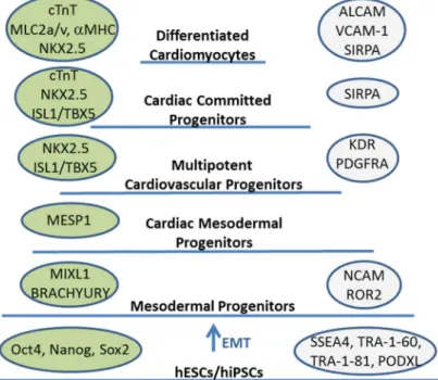
Increased yield of CMs as well as purification could also be achieved by isolation of progenitors (e.g. mesodermal, cardiac mesodermal, cardiovascular or cardiac progenitors, see Figure 3). Progenitors possess high proliferation capacity and restricted differentiation potential, allowing first the expansion of these cells by providing stage-specific renewal signals and then CM differentiation by cardio- inductive signals. A culture condition supporting continuous self-renewal and proliferation of cardiovascular progenitors by inhibition of BMP, Activin/Nodal and glycogen synthase kinase 3 (GSK3) pathways have already been described89.
Figure 3. Proposed progenitor stages of the differentiation of human pluripotent stem cells into cardiomyocytes. The progenitor stages are identified by transcription factors and structural proteins (shown in green bubbles) and cell surface markers (shown in grey bubbles). The image is taken from Szebényi et al. 148, with some modifications.
1.3.1 Genetic methods for isolation of hPSC-derived cardiac cells
Genetic modification of stem cells is less efficient than that of tumour-derived or immortalized cell lines, therefore generation of reporter-expressing cardiomyocytes raises some technical challenges (for further review see the attached book chapter Szebényi et al., “Human Stem-Cell-Derived Cardiomyocytes in Drug Discovery and Toxicity Testing”). However, by now several techniques have been documented to allow the generation of transgenic hPSC lines, without impairing self-renewal capacity and differentiation potential6, 90-92. Moreover, in most cases the studies demonstrated that the obtained cardiomyocytes were physiologically normal, regardless of the integration of the transgenes or the selection procedures applied for CM enrichment.
Constitutive promoters, such as ACTB, UbC, EF1α, PGK, CMV or the original variant of the CAG provide nearly uniform transgene expression in all tissues; therefore, theoretically, these types of promoters cannot be used for isolation of CMs. However, it was demonstrated in differentiated hESCs that the activity of several commonly used constitutive promoters can be restricted to specific cell lineages, since ACTB was only active in undifferentiated and neuronal cells, while UbC and PGK were inactive in endothelial cells 93. Moreover, the CMV promoter became active only after mESCs differentiated into neuronal precursor cells 94, while the CAG promoter showed long- term activity during mESC differentiation towards mesoderm 95. Still, reporter systems driven by various constitutive promoters (ACTB or CAG) could be reliable used for identification of transplanted transgenic hESC-derived CMs in infarcted mouse96 and rat97, 98 or healthy guinea pig heart99 to provide evidence for successful and functional engraftment.
Cardiac tissue specific promoters allow the isolation of cardiomyocytes, while progenitors able to give rise to cardiac cells can be obtained when the promoter of an early mesoderm or cardiac lineage-specific gene is used. The first well-defined cells able to give rise to the cardiac (besides of hematopoietic, vascular, and skeletal muscle) lineage are mesodermal progenitors, expressing the T-box factor BRACHYURY and the homeodomain protein MIXL1 (see Figure 3 for markers characteristic for specific stages of the cardiac differentiation). To allow selective isolation of mesodermal progenitors GFP expression cassette under the control of the BRACHYURY promoter
was randomly inserted into the genome100 or were targeted into the MIXL1 locus by homologous recombination101. More recently, a dual reporter system has been established where mesoderm posterior 1 (MESP1), a transcription factor characteristic for the pre-cardiac mesoderm is labelled with mCherry, while NKX2.5 (expressed later than MESP1) is labelled with EGFP, allowing isolation of progenitors at different stages of cardiac commitment102. For this dual reporter system a previously established hESC reporter line provided the basis, in which the GFP cDNA was introduced into the NKX2.5 locus by homologous recombination (NKX2.5eGFP/w)88. This NKX2.5eGFP/w reporter cell line allows the selection of multipotent cardiovascular progenitors, displaying cardiac, endothelial and vascular smooth muscle potential, similarly to the Cre/loxP system-based labelling of ISL1 expressing cells with the DsRed fluorescent reporter protein103. This system allows not only the selection of ISL+ cells, but also the lineage tracing of these cells, since the labelling is irreversible and DsRed expression does not attenuate with the downregulation of ISL1 expression.
An alpha myosin heavy chain (αMHC) promoter-based system was used to follow maturation of hESC-derived cardiomyocytes over a year long36, and also to identify hESC-derived early myocardial precursors (αMHC-GFPpos) able to give rise to atrial ventricular and specialized conduction CM subtypes, but not to non-muscle cardiac cells104. Besides fluorescent proteins, drug resistance genes can also be used for enrichment of targeted cell populations. For example, hESC derived cardiomyocytes could be enriched to high purity (80-90%) when the puromycin resistance gene was driven by the αMHC promoter105, 106.
Myosin light chain-2v (MLC-2V) is expressed later in CM differentiation than αMHC, therefore it does not allow the same insight into early CM differentiation, however, when the enhanced GFP was expressed under the control of the MLC-2V promoter, GFP expression-based identification and isolation of electrically active hESC-derived CMs was possible, according to Huber et al.107. It was also suggested that a short fragment of the cardiac troponin I (TNNI3) promoter in combination with the human cardiac alpha-actin enhancer is sufficient to identify hESC-derived CMs108.
The disadvantage of these systems is that the activity of the transgene can only be detected at a certain stage of differentiation. To overcome this issue, Fu et al. applied
the combination of a constitutive and a tissue-specific promoter109. In this system GFP expression was driven by the constitutive EF1α promoter to allow selection of successfully transduced undifferentiated hESCs, while MLC2V promoter-driven dsRed expression was applied to enrich for CMs with ventricular phenotype109109109109109106105
. Stage-specific reporter systems offer several advantages, for example these systems can be used as readouts of culture optimization in a stage-specific manner or can be utilized in the search for new cell-surface markers of the different stages of the cardiac lineage.
1.3.2 Non-genetic methods for isolation of cardiac cells
Mesodermal progenitors derived from hPSCs via epithelial-to-mesenchymal transition (EMT) can be selected not only by genetic methods, but also based on a cell surface marker combination, in which the neuronal cell adhesion molecule (NCAM or CD56) is used as positive, while the epithelial cell adhesion molecule (EpCAM or CD326) is used as negative marker110. However, the CD56posCD326neg marker combination was also documented to identify hESC-derived neuronal cells111, suggesting that markers or marker combinations may identify a progenitor population only in a culture dependent manner and when the differentiation is directed into another lineage or not directed at all, than specificity of the markers may diminish. In addition, upregulation of the tyrosine kinase transmembrane receptor (ROR2) can also be used to identify and select mesodermal progenitors112.
Similarly to the above mentioned marker combination, the stage-specific embryonic antigen-1 (SSEA-1) has also been proposed to be a suitable marker, in this case for isolation of multipotent cardiovascular progenitors113, while others suggested SSEA-1 to be expressed on hESC-derived epithelial1 or neuroectodermal committed cell types114. Thus SSEA-1 may only allow identification of multipotent cardiovascular progenitors when the differentiation has already been efficiently directed into the cardiac lineage.
It was shown that low level expression of the vascular endothelial growth factor receptor 2 (VEGFR-2/KDR) together with the absence of the mast/stem cell
growth factor receptor, also known as proto-oncogene c-Kit or tyrosine kinase Kit (C- KIT or CD117) identifies a population of cardiovascular progenitor cells (KDRlowC- KITneg), expressing ISL1 and low levels of NKX2-5 and TBX561. This finding was verified not only on hESCs, but also in the case of hiPSCs115 and showed that mouse models are not always adequate for establishing human protein expression profiles, since in mice cardiac progenitors are c-KitposNkx2-5pos116, while during human cardiac differentiation the emergence of a C-KITposNKX2-5pos population could not be observed at all117.
In addition, it was also suggested that cardiovascular progenitors can be identified based on the co-expression of KDR and PDGFRA64. However, the same research group demonstrated later that not all of the KDRposPDGFRApos cells give rise to cells of the cardiac lineage86. In addition, isolation based on KDR expression may result in an impure population of cardiovascular progenitors, since KDR is also expressed on hPSCs61.
The NKX2-5eGFP/w reporter system was used to screen for new cell-surface markers of the cardiac lineage, resulting in the discovery of two early CM markers, namely vascular cell adhesion molecule 1 (VCAM1) and signal regulatory protein α (SIRPA)88. Further experiments showed that VCAM1 positivity appears slightly later than SIRPA, and NKX2-5posSIRPAposVCAM1pos cardiomyocytes arose from an NKX2- 5posSIRPApos intermediate117.
Upregulation of SIRPA happened parallel to the emergence of the first NKX2- 5–GFPpos cells (around day 8 of the differentiation), a day before the spontaneous contractile activity started in the differentiation cultures of HES2 hESCs86. Thus SIRPApos cells isolated at EB day 8 were considered as cardiac precursors and were shown to be able to give rise to troponin expressing SIRPApos CMs. Therefore it was concluded that SIRPA is a cell surface marker able to identify cardiac precursors and cardiomyocytes.
VCAM1 was shown to allow isolation of CMs but not cardiac progenitors, since troponin expression preceded VCAM1 appearance during differentiation118. VCAM1 expression based selection on day 11 of the differentiation resulted in a population of highly purified (>95%) CMs in the case of all four hPSC lines examined,
although VCAM1pos cells represented only a subset within troponin T-positive cells (33.4%-63.5%).
The activated leukocyte cell adhesion molecule (ALCAM or CD166) is another marker, that have been suggested to enable isolation of hESC-derived CMs87, and later it was also shown that ALCAM allows the high-purity enrichment of hiPSC-derived CMs as well115. In this latter work first KDRlowC-KITneg multipotent cardiovascular progenitors were isolated on EB day 6 and recultured as monolayer, than on day 20 of the differentiation CD166 was used to select CD166pos CMs and CD166neg smooth muscle cells. However, it is important to mention that there is only a short, early developmental stage-specific time window when ALCAM expression is restricted to cardiomyocytes, and beyond this particular developmental stage ALCAM expression is widespread among different types of tissues115, 119-121
.
The first non-genetic method reported in the literature allowing the isolation of living cardiomyocytes was based on the high mitochondrial content of CMs, resulting in that tetramethylrhodamine methyl ester perchlorate (TMRM), a fluorescent dye specifically labelling mitochondria could be used for cardiomyocyte enrichment with a purity of 99%122. However, it was shown that this method allows only the selection of late-stage CMs, while early CMs could not be discriminated from undifferentiated hESCs88.
3. Aims
The goal of the research presented in the dissertation was to establish a novel in vitro method for the purification of human embryonic stem cell-derived cardiomyocytes and cardiac progenitors. In order to achieve this objective, the aims of the present study were:
1. To demonstrate that the CAG promoter provides the opportunity to identify cardiomyocytes in spontaneous differentiation cultures of human embryonic stem cells.
2. To induce cardiac differentiation of human embryonic stem cells in order to enrich the differentiation cultures for cardiac progenitors.
3. To demonstrate that the CAG promoter provides the possibility to identify and isolate cardiac progenitors during directed differentiation of human embryonic stem cells.
4. To find optimal culture conditions for isolated cardiac progenitors to maintain cardiac commitment during further differentiation.
5. To demonstrate that the isolated human embryonic stem cell-derived cardiac progenitors can be differentiated into relatively pure population of cardiomyocytes.
6. To optimize reaggregation and survival properties of isolated human embryonic stem cell-derived cardiac progenitors to enhance cardiomyocyte yield.
4. Materials and Methods
4.1. hESC culture and differentiation
The original HUES9 human embryonic cell line was kindly provided by Dr.
Douglas Melton from the Harvard University, while BG01V was purchased from ATCC. The Sleeping Beauty (SB) transposon based gene delivery method was applied to genetically modify HUES9 and BG01V hESCs with a plasmid containing the cDNA of the fluorescent protein EGFP, under the control of a specific variant of the CAG promoter (SB-CAG-EGFP construct) 90. This specific variant contains a CMV enhancer region, two sequences from the chicken β-actin promoter and one short part of the rabbit β1-globin promoter 90.
HUES9-CAG-EGFP and BG01V-CAG-EGFP hESC colonies were cultured on mitomycin-C treated mouse embryonic fibroblast (MEF) feeder cells (MERCK Millipore), in a medium consisting of Knockout DMEM (Life Technologies), supplemented with 20% Knockout Serum Replacement (Life Technologies), 2 mM glutamine, 0.1 mM nonessential amino acids, 0.1 mM β-Mercaptoethanol and 15 ng/ml recombinant human basic fibroblast growth factor (bFGF).
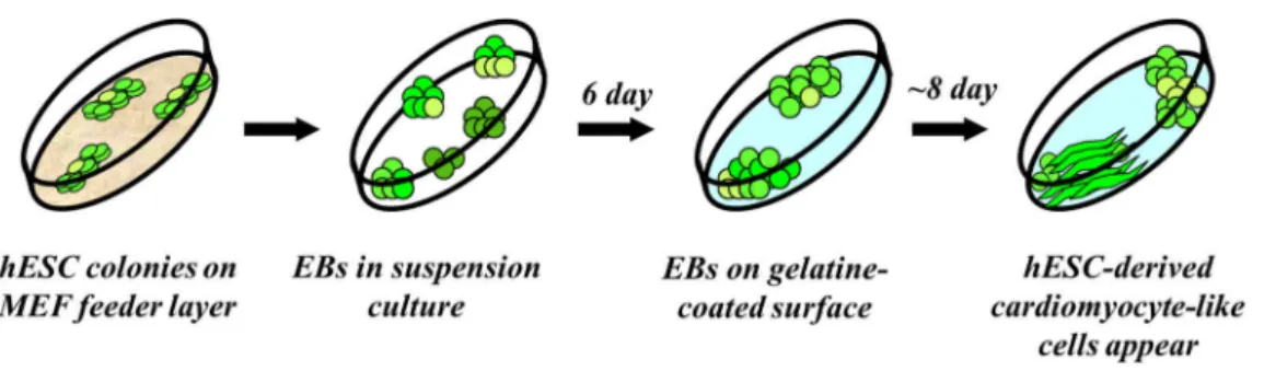
Figure 4. Schematic outline of spontaneous differentiation through the EB formation method.
Figure 4 shows the schematic outline of the spontaneous differentiation protocol, designated as the embryoid body (EB) formation method. Differentiation was initiated via EB formation under suspension culture conditions, ensured by an ultra-low
attachment surface of the culture plate and an EB medium containing Knockout DMEM, 20% ES-tested FBS (Life Technologies), 1% NEAA, 0,2% β-merkaptoethanol and 1% L-glutamin. On the 6th day of differentiation EBs were plated onto gelatine coated 24 well plates, in a density of 5-10 EBs/well. From this time point EBs were kept in differentiation medium (DM) containing DMEM (Life Technologies) and 10% EU- tested FBS (Life Technologies).
Directed differentiation of HUES9-CAG-EGFP and BG01V-CAG-EGFP cells was initiated via EB formation in suspension, as previously reported 61, with some modifications, as shown in the Results. Spontaneously contracting cardiomyocytes appeared at day 14-16 of the differentiation in both cases.
4.2. Flow cytometry
Undifferentiated HUES9-CAG-EGFP colonies were harvested from mouse feeder cells by enzymatic digestion with 0.05% trypsin-EDTA (Gibco), HUES9-CAG-EGFP EBs were dissociated with 0.25% trypsin-EDTA (Gibco). Single cell suspension was washed with PBS containing 0.5% bovine serum albumin and incubated for 30 min at 37 °C with the following directly labelled monoclonal antibodies: anti-human SSEA4- PE antibody (R&D System Inc., Minneapolis, MN) for specific labeling of pluripotent cells, anti-human CD166-A647 antibody (AbD Serotec) for specific labeling of activated cell adhesion molecule (ALCAM) expressing cells, CD106-PE for specific labeling of Vascular Cell Adhesion Molecule-1 (VCAM1) (BD Pharmingen) and anti- mouse Sca-1 (Ly-6A/E)-PECy5.5 (Beckton-Dickinson, San Jose, CA) antibody for specific labelling of mouse feeder cells in the case of undifferentiated cells (sample D0) and 6 day-old EBs (sample D6). The unconjugated monoclonal antibody SIRPA (BioLegend) was labelled with Alexa Fluor 647-conjugated goat anti-mouse IgG antibody (Invitrogen). For intracellular staining the cells were fixed and permeabilized with 4% paraformaldehyde (PFA) in PBS, subsequently the staining with anti-Troponin I (Monoclonal Anti-TNNI3, 1:500 dilution, Sigma) labelled with Alexa Fluor 647- conjugated goat anti-mouse IgG antibody (Invitrogen) was performed in PBS with 2%
BSA and 0.75% Saponin (Sigma). Control staining with appropriate isotype-matched
control antibodies or background levels of fluorescence of the fluorochrome-conjugated secondary antibody was included. Dead cells were gated out based on 7AAD (Sigma) staining. HUES9 cells were measured to set the level for EGFP-positivity of undifferentiated HUES9-CAG-EGFP cells. Samples were analyzed by a FACSCalibur flow cytometer (Beckton-Dickinson, San Jose, CA) equipped with a 488 nm argon laser and a 635 nm red diode laser with BD CellQuest acquisition software (BDIS) or by FACSAria High Speed Cell Sorter (Beckton-Dickinson, San Jose, CA) with BD FACSDiva software.
4.3. Cell sorting
HUES9-CAG-EGFP EBs were dissected with 0.25% Trypsin-EDTA and single cell suspension was sorted into artificial fractions (low, mid and high) based on EGFP fluorescent signal intensity using the fluorescence based FACSAria High Speed Cell Sorter (Beckton-Dickinson, San Jose, CA). Minimum linear value within the CAG- EGFPlow fraction was 3000, maximum linear value was 15500; for the CAG-EGFPhigh fraction minimum linear value was 29500, while maximum linear value was 262000.
Precision of the sorting procedure was monitored by EGFP expression profiling of the sorted fractions with flow cytometry, immediately after sorting (see Results). Cells obtained from the different fractions were washed with sterile PBS and either recultured or immediately resuspended in Trizol (Invitrogen, Carlsbad, CA, http://www.invitrogen.com) for further gene expression analysis. Isolated cells were placed into NUNC HydroCell Surface 96 well plates in a density of 30,000 cells/well, unless stated otherwise.
4.4. Fluorescence plate reader measurements
For fluorescence plate reader measurements a VICTOR X3 2030 Multilabel Reader (Perkin Elmer) was used. EBs reaggregated from the sorted cells (rEBs) were cultured in NUNC HydroCell Surface 96 well plates and fluorescence measured 3 and 20 days after sorting (see Results). Excitation wavelength was 490 nm, and a F535 emission
filter was used for detection of EGFP fluorescence. Fluorescence intensity levels were calculated as averages of 9 independent measurements.
Propidium iodide (PI) staining was carried out 3 and 20 days after sorting, in order to assess the growth of the rEBs and estimate changes in cell numbers. The rEBs were fixed with methanol on ice for 15 minutes and, after a washing step, were stained with PI. After incubation with PI for 15 minutes the rEBs were transferred into fresh PBS in a 96 well plate (Greiner). Excitation wavelength was 540 nm, and a F660 emission filter was used for detection of PI fluorescence. Fluorescence intensity levels were calculated as averages of 6 independent experiments.
4.5. Immuncytochemistry
For immunostaining, whole EBs were differentiated either on gelatin-coated 8 chamber dishes with glass bottom (Imaging chambers, PAA) or on 24 well culture plates, while EBs reaggregated from the sorted cells (rEBs), were plated onto gelatin- coated 8 chamber dishes with glass bottom, 6-7 days after the sorting procedure. To achieve adequate adherence of the rEBs, a 5 minute trypsinization procedure was performed with a 0.25% Trypsin-EDTA solution. EBs and rEBs were fixed with 4%
paraformaldehyde in Dulbecco's modified Phosphate Buffered Saline (DPBS) for 15 min at room temperature. After three washing steps with DPBS, nonspecific antibody binding was blocked for 1 h at room temperature in DPBS containing 2 mg/ml bovine serum albumin, 1% fish gelatin, 5% goat serum and 0.1% Triton-X 100. The samples were then incubated for 1 h at room temperature with the primary antibodies. The primary antibodies used were: anti-Troponin I (Monoclonal Anti-TNNI3, 1:500 dilution, Sigma), Pecam (Anti-Human PECAM-1, 1:500 dilution, eBioscience), SMA (Monoclonal Anti-Actin, Alpha-Smooth Muscle, 1:100 dilution, Sigma), AFP (Monoclonal Anti-alpha-Fetoprotein, 1:500 dilution, Sigma), βIII-Tubulin (Monoclonal Anti-Neuron-specific beta-III Tubulin, 1:2000 dilution, R&D Systems). After washing with DPBS, the cells were incubated for 1 hour with the secondary antibodies at room temperature. Secondary antibodies were diluted in the blocking solution at 1:250 in each case. Alexa Fluor 568-conjugated goat anti-mouse IgG antibody (Invitrogen) was
applied to detect troponin I, AFP, βIII-Tubulin. DAPI (Invitrogen, Madison, WI) was used for nuclear staining (10 µM, 10 minute-long incubation in DPBS). Samples stained on 8 chamber dishes with glass bottom were examined by an Olympus FV500-IX confocal laser scanning microscope, while samples stained on 24 well plastic culture plates were examined by fluorescence microscopy. At least two independent samples were used in each measurement.
4.6. Real-time quantitative PCR analysis
Total RNA was isolated from cells using TRIzol™ Reagent (Invitrogen) following the manufacturer’s instructions. cDNA samples were prepared from 0.2 µg total RNA using the Promega Reverse Transcription System Kit as specified by the manufacturer.
For real-time quantitative PCR (QPCR) the following Pre-Developed TaqMan® assays were purchased from Applied Biosystems: NANOG as undifferentiated stem cell marker; BRACHYURY as early mesodermal marker; ISL1, TBX5, NKX2.5 and GATA4 as early markers of cardiac differentiation; TNNT2, PLN and NPPA as cardiac specific marker; ALCAM as stage specific cardiac marker; MYL2 as ventricular-, MYL7 as atrial-, HCN4 as nodal subtype specific marker; P0 ribosomal protein as endogenous control for quantification. QPCR analyses were carried out using the StepOne™ Real-Time PCR System (Applied Biosystems), according to the manufacturer’s instructions. The fold- change of mRNA in experimental and control cells was determined using the 2-∆∆Ct method. Relative mRNA levels were presented as mean values ± S.E.M. of 3 independent experiments. Levels of significance were calculated by the two-tailed Student t-test.
5. Results
5.1. The CAG promoter allows the identification of cardiomyocytes in spontaneous differentiation cultures of human embryonic stem cells
5.1.1. Characterization of the hESC lines HUES9-CAG-EGFP and BG01V-CAG-EGFP
EGFP expressing HUES9 and BG01V cells were generated by transfection with the SB-CAG-EGFP construct (see Materials and Methods). The genetic modification was carried out by the research group of Tamás Orbán. Transgenic hESC lines (HUES9-CAG-EGFP and BG01V-CAG-EGFP) were established through the enrichment of EGFP expressing hESCs by sorting or cloning. Several cell lines have been established with different EGFP expression intensities, either completely free of transgene-negative (CAG-EGFPneg) cells or containing a small population of CAG- EGFPneg cells.
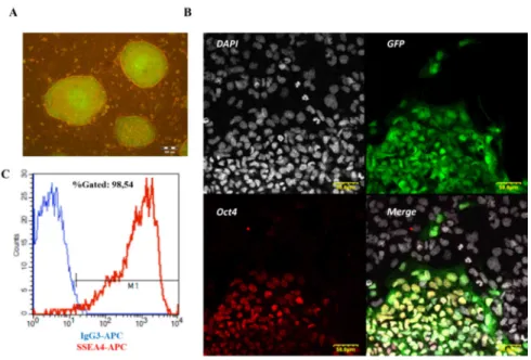
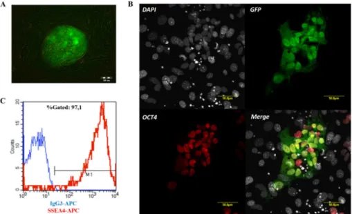
Figure 5. Characterization of HUES9-CAG-EGFP cells. (A) Fluorescence microscopy image of HUES9-CAG-EGFP colonies on MEF feeder layer, showing hESC-like morphology. (B) Confocal microscopy images of HUES9-CAG-EGFP colony on MEF feeder layer, showing expression of the pluripotency marker OCT4. (C) Flow cytometry measurement of SSEA4-APC and its isotype control IgG3-APC in HUES9-CAG-EGFP cells.
HUES9-CAG-EGFP cells were cultured on a mouse embryonic fibroblast (MEF) feeder layer and maintained pluripotency during long-term culture, as demonstrated by the colony (clump) morphology characteristic for pluripotent hESCs (Figure 5A), by immunostaining against the pluripotency marker OCT4 (Figure 5B) and by flow cytometry measurement detecting SSEA4 expression (Figure 5C).
Similarly to the HUES9-CAG-EGFP hESC line, BG01V-CAG-EGFP clumps were also cultured on MEF feeder layer (Figure 6A) and expressed OCT4 (Figure 6B) and SSEA4 (Figure 6C), respectively. Flow cytometry measurements detecting SSEA4 expression were routinely carried out to monitor the pluripotent state of hESCs during long term culture. Only hESCs with more than 95% SSEA4 positivity were used for experiments.
Figure 6. Characterization of BG01V-CAG-EGFP cells. (A) Fluorescence microscopy image of BG01V-CAG-EGFP colonies on MEF feeder layer, showing hESC-like morphology. (B) Confocal microscopy images of BG01V-CAG-EGFP colony on MEF feeder layer, showing expression of the pluripotency marker OCT4. (C) Flow cytometry measurement of SSEA4-APC and its isotype control IgG3-APC on BG01V-CAG-EGFP cells.
Pluripotency of hESC lines is also characterized by the ability of differentiating into the three germ layers, namely into the endoderm, ectoderm and mesoderm lineages.
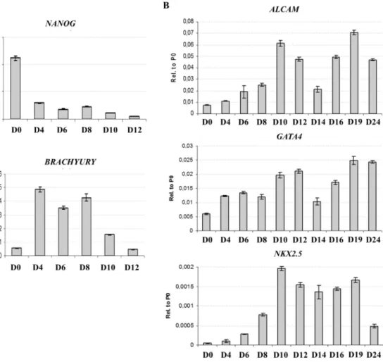
HUES9-CAG-EGFP cells were differentiated through the EB method, which is often used for initiating spontaneous differentiation. Loss of pluripotency and mesodermal commitment was investigated by QPCR, detecting the downregulation of the pluripotency marker NANOG and the upregulation of the early mesodermal marker gene BRACHYURY during the first 12 days of differentiation (Figure 7A). Downregulation of BRACHYURY was followed by the upregulation of the early cardiac marker genes GATA4, NKX2.5 and ALCAM, indicating the emergence of cardiac-committed cells in the differentiation culture around the10th day of the differentiation (Figure 7B).
Figure 7. Transcriptional profile of spontaneous differentiation of HUES9- CAG-EGFP cells. (A) QPCR analysis of the mRNA expression of NANOG and BRACHYURY in whole EBs at early stages of differentiation. (B) QPCR analysis of the mRNA expression of ALCAM, GATA4 and NKX2.5 in whole EBs at different stages of spontaneous differentiation.
Figure 8. Immunocytochemistry analysis of spontaneously diffferentiating HUES- CAG-EGFP cells. Green: CAG-EGFP. Blue: DAPI. (A) Anti-alpha-fetoprotein (AFP) and anti-neuron-specific beta-III-tubulin (βIII-Tub) staining of HUES9-CAG-EGFP EBs on day 12 of the differentiation. (B) Anti-platelet endothelial cell adhesion molecule (PECAM-1) and anti-alpha-smooth muscle actin (SMA) staining of HUES9- CAG-EGFP EBs on day 22 of the differentiation. The image is taken from Szebényi et al.123, with some modifications.
The emergence of cells of the other two germ layers was demonstrated by immunocytochemistry studies (Figure 8A). On day 12 of the differentiation (D12) EBs
were stained against the early endoderm marker alpha-fetoprotein (AFP) and the ectoderm marker beta-III-tubulin (βIII-Tub), and both endodermal and ectodermal cells were found to be present in the differentiation culture. Cells of the mesodermal lineages were identified based on the expression of alpha-smooth muscle actin (SMA), characteristic for smooth muscle cells and based on platelet endothelial cell adhesion molecule (PECAM-1) expression, characteristic for endothelial cells, respectively (Figure 8B). The expression of SMA and PECAM was investigated on day 22 (D22) of the differentiation, since cells positive for these markers emerge only at later stage of the differentiation. Besides smooth muscle and endothelial cells, cardiomyocytes are also of mesodermal origin. Cardiac differentiation was demonstrated by the emergence of numerous spontaneously contracting areas during later stage of differentiation (after 14 days).
5.1.2. In differentiation cultures of HUES9-CAG-EGFP and BG01V-CAG-EGFP cardiomyocytes show exceptionally high EGFP expression
Spontaneous differentiation of HUES9-CAG-EGFP hESCs usually resulted in numerous spontaneously contracting areas with extremely high EGFP fluorescence signal intensity, masking dimmer fluorescence of the surrounding tissues (Figure 9, white arrows pointing on the contracting areas with high EGFP signal). Extremely high EGFP expression intensities were predominantly observed onsite of the contracting areas (Supplementary video 1) and allowed the identification of cardiomyocytes in the differentiation culture based alone on their extremely high EGFP expression intensities.
Two different hESC lines (HUES9-CAG-EGFP and BG01V-CAG-EGFP) and a mouse ESC line (R1-CAG-EGFP, kindly provided and differentiated by Elen Gócza) were used to demonstrate that this effect is cell line and species-independent (Figure 9).
Moreover, it was shown by Tamás Orbán and co-workers that this phenomenon is exclusively dependent on the CAG promoter and independent of the transgene integration site and copy number, the transgene sequence, as well as the method of gene delivery. A detailed description of the experiments resulting in this conclusion can be found in the attached paper, Orbán et al.90.
Figure 9. Fluorescence microscopy images of spontaneously contracting areas. The images show spontaneously contracting areas differentiated from two different human embryonic stem cell lines (HUES9-CAG-EGFP and BG01V-CAG-EGFP) and from a mouse embryonic stem cell line (R1-CAG-EGFP) at two different magnifications.
To elicit whether the extremely high EGFP expression intensities of cardiomyocytes are the result of higher transcription or translation rate, or certain posttranslational modifications, HUES9-CAG-EGFP differentiation cultures were separated into three fractions based on EGFP fluorescence by a FACSAria High Speed Cell Sorter at the 30th day of differentiation (Figure 10A). The data presented in Figure 10 are the result of a differentiation, repeated independently from the experiments published by Orbán et al. In this particular experiment the applied HUES9- CAG-EGFP hESC line contained a subpopulation of EGFP negative cells (Figure 10A, yellow population), superseding the use of the HUES9 cell line as negative control for setting up the detection of the EGFP signal. EGFP negative cells were not included in further analysis since this population contained a mixture of all types of cells emerging during differentiation. The fraction containing cells with exceptionally high EGFP fluorescent signal intensity was designated as the CAG-EGFPhigh subpopulation (Figure
10A, green), while cells with low EGFP intensity as the CAG-EGFPlow subpopulation (Figure 10A, blue). Cells between these two fractions were designated as CAG- EGFPmid (Figure 10A, purple).
Sorted cells were resuspended in TRIzol Reagent to isolate total cellular RNA from each of the separated fractions. Real-time quantitative PCR analysis revealed that EGFP transcription levels closely correlated with the fluorescent signal intensities of the fractions, thereby providing evidence for increased transcription through increased promoter activity as the cause of enhanced EGFP fluorescent signal intensity (Figure 10B). Therefore higher nucleus-cytoplasm ratio in cardiomyocytes compared to other cell types could also be excluded as the cause of this phenomenon.
Figure 10. The CAG-EGFPhigh subpopulation is enriched in cardiomyocytes.
(A) Isolation of CAG-EGFPlow (blue), CAG-EGFPmid (purple) and CAG-EGFPhigh (green) fractions from HUES9-CAG-EGFP EBs based on the CAG-EGFP signal intensity on day 32 of the differentiation. (B) QPCR analysis of the mRNA expression of GFP, ALCAM, NKX2.5, GATA4, PLN and NPPA in EBs at day 32 of spontaneous differentiation. (C) QPCR analysis of the mRNA expression of PAX6 and CAPG in EBs at day 32 of spontaneous differentiation.
Transcriptional profiles of cardiac-specific (NKX2.5, GATA4, ALCAM, PLN and NPPA), early neuron-specific (PAX6), and skin-specific (CAPG) marker genes measured by QPCR revealed that the CAG-EGFPhigh fraction contained higher level of cardiac-specific mRNA than the other two fractions, while neuron and skin-specific mRNA was underrepresented in this sample compared to the other two fractions (Figure 10B). These findings proved that apart from being constitutively functional in all cell types (including undifferentiated hESCs, see Figure 5 and 6) this specific variant of the CAG promoter was transcriptional extremely active in cardiac tissues, providing the possibility for selection of transgene expressing hESCs and hESC-derived CMs, and therefore it was named as “double-feature” promoter90.
Figure 11. Non-contracting areas expressing CAG-EGFP at a very high intensity are cardiomyocytes, but not all of the cardiomyocytes express CAG-EGFP at an exceptionally high level. (A) Fluorescence microscopy images at two different magnifications of a non-contracting area expressing CAG-EGFP at an exceptionally high level. (B) Fluorescence microscopy images at two different magnifications of cardiac troponin I (cTnI) staining of the same area. Blue: DAPI. Green: CAG-EGFP.
Red: cTnI. The white arrow indicates a cTnI positive area with lower EGFP signal intensity. The image is taken from Szebényi et al.123, with some modifications.
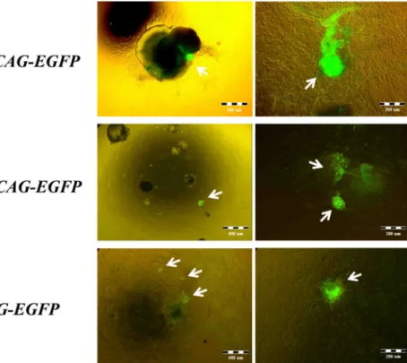
Besides of numerous contracting areas several non-contracting areas with exceptionally high EGFP signal intensities were spotted by fluorescent microscopy during examination of the differentiation cultures, even at later stages of the differentiation (Figure 11A). To further support the previous finding, namely that the transcriptional activity of the CAG promoter is higher selectively in cardiomyocytes, wells of 24-well plates containing such non-contracting areas were stained against cardiac troponin I (cTnI). Figure 11B shows a representative cTnI staining, demonstrating good colocalization with the enhanced EGFP signal, as well as the existence of cardiomyocytes (cTnI positive cells) with lower EGFP signal intensity (Figure 11B, white arrow).
Figure 12. Fluorescence microscopy images of the same area showing exceptionally high EGFP expression during the course of spontaneous differentiation of HUES9- CAG-EGFP culture. Onset of the spontaneous contractile activity is denoted by a white heart symbol. White arrows indicate the areas with extremely high EGFP signal.
Next, the appearance of areas with exceptionally high EGFP signal was investigated by fluorescent microscopy at early stages of spontaneous differentiation and selected areas were followed during the course of differentiation to evaluate the
ability of the CAG promoter to identify progenitors of cardiomyocytes (Figure 12).
This was important because the selection of mature cardiomyocytes is limited by their poor reaggregation and survival properties as single cells, while cardiac progenitors possess better reaggregation and survival properties. In addition, cardiomyocytes undergo progressive cell-cycle withdrawal during maturation79, while progenitor cells are still able to divide, therefore cardiomyocyte yield of the differentiation can also be enhanced by selective propagation of cardiac progenitors.
Day 10 was found as the earliest time point when cells further differentiating to contracting cardiomyocytes could be identified based on their high EGFP expression (Figure 12). These findings suggested that the CAG promoter would allow not only the isolation of cardiomyocytes, but also that of cardiac progenitors, since the contractile activity, indicating the appearance of cardiomyocytes started only later, after day 14 in the differentiation cultures.
5.2. Directed cardiac mesoderm differentiation of CAG-EGFP expressing human embryonic stem cells
To facilitate the examination of the ability of the CAG promoter to identify cardiac progenitors, spontaneous differentiation was needed to be replaced by a differentiation protocol which is able to enrich the output of the differentiation for cells with mesoderm origin. The original protocol was obtained from the literature61 and was optimized for CAG-EGFP expressing hESCs, however, some basic modifications were introduced to render it affordable for routine use in a small academic laboratory.
Figure 13 shows the outline of the protocol used to induce mesodermal differentiation. Bone morphogenetic factor-4 (BMP4), Activin A, ascorbic acid and monothioglycerol (MTG) was used to differentiate cells towards the mesoderm, while further commitment to the cardiac mesoderm was supported by the addition of ascorbic acid and MTG alone. Formation of cells of the cardiovascular lineages was achieved in differentiation medium (DM) supplemented with ascorbic acid and MTG, added until the 14th day of the differentiation.
Figure 13. Schematic representation of the protocol for directed differentiation.
The image is taken from Szebényi et al.123, with some modifications.
Directed differentiation was monitored by fluorescence activated cell sorting (FACS) measurements and its kinetics was compared to that of spontaneous differentiation (Figure 14). The loss of pluripotency demonstrated by the decrease of SSEA4 expression could be detected earlier, already on day 6 in the directed differentiation culture, while during spontaneous differentiation SSEA4 expression started to decline only around D10 (Figure 14A).
ALCAM can be designated as a cardiomyocyte marker, but it is important to mention that ALCAM is expressed solely on cardiomyocytes at an early developmental stage-specific manner, while beyond this particular developmental stage ALCAM expression is widespread among different types of tissues115, 119-121
. Therefore it was important to examine the kinetics of ALCAM expression during the course of differentiation of HUES9-CAG-EGFP cells. Directed differentiation resulted ALCAM positive cells around day 9, while during spontaneous differentiation ALCAM positive cells emerged later (around day 12).
To further examine mesoderm differentiation the cell surface expression of KDR (VEGF-R) and CD117 (C-KIT) was monitored during the first 12 days of the differentiation (Figure 14B), based on the paper of Yang et al., showing that the KDRlow/CD117neg subpopulation indicate the presence of cardiovascular progenitors during directed differentiation of hESCs61. In the directed differentiation cultures increased presence of this population could be observed between day 6 and day 12, while in spontaneous differentiation cultures the KDRlow/CD117neg subpopulation evolved only later, around day 9 and composed a smaller fraction of the culture as shown in Figure 14B.
Counting of contracting areas served as a readout for cardiac differentiation efficiency. Directed differentiation resulted more contracting areas than spontaneous differentiation (Figure 14C). Figure 14D shows that this difference could be demonstrated by FACS measurement of undifferentiated (light grey) and differentiated HUES9-CAG-EGFP samples (dark grey for spontaneous or black for directed differentiation) stained against cTnI.
Figure 14. Comparison of spontaneous and directed differentiation. (A) Flow cytometry analysis of SSEA4 and ALCAM/CD166 on cells from whole HUES9-CAG- EGFP EBs at different stages of spontaneous (upper row) and directed differentiation (lower row). (B) Flow cytometry analysis of C-KIT/CD117 and KDR on cells from whole HUES9-CAG-EGFP EBs at different stages of spontaneous (upper row) and directed differentiation (lower row). (C) Table summarizing the outcome of a spontaneous and a directed differentiation in terms of contracting areas (CAs)-content on a 24 well plate at day 30 (D30). (D) Flow cytometry analysis of cardiac troponin I (cTnI) staining of undifferentiated and differentiated (spontaneous or directed) samples.
For flow cytometry analysis the FACSCalibur flow cytometer was used; dot plot gates were set based on the isotype controls.