Stem Cell Research 50 (2021) 102134
Available online 18 December 2020
1873-5061/© 2020 The Author(s). Published by Elsevier B.V. This is an open access article under the CC BY-NC-ND license
Lab resource: Stem Cell Line
Establishing a human embryonic stem cell clone with a heterozygous mutation in the DGCR8 gene
D ora Re ´ ´ e
a, Adrienn Borsy
a, Abel F ´ ´ othi
a, Tam ´ as I. Orb ´ an
a, Gy ¨ orgy V ´ arady
a, Zsuzsa Erdei
a, Bal ´ azs Sarkadi
a, J ´ anos R ´ ethelyi
b,c, N ora Varga ´
a,1, Agota Ap ´ ´ ati
a,*,1aInstitute of Enzymology, Research Centre for Natural Sciences, Budapest, Hungary
bDepartment of Psychiatry and Psychotherapy, Semmelweis University, Budapest, Hungary
cMolecular Psychiatry Research Group, National Brain Research Program (NAP), Hungarian Academy of Sciences and Semmelweis University, Budapest, Hungary
A B S T R A C T
DiGeorge Syndrome (DGS) Critical Region 8 (DGCR8) is a primary candidate gene in they DGS. The DGCR8 microprocessor complex subunit is an essential cofactor in the canonical miRNA biogenesis which is involved in diverse cellular functions such as cell fate decisions, apoptosis and different signaling pathways. However, the role of DGCR8 in these processes or development of DGS is not fully understood. Here we present a heterozygous DGCR8 mutant human embryonic stem cell line (HuES9DGCR8+/−) created by the CRISPR/Cas9 system. The generated HuES9DGCR8+/− cells maintain normal karyotype, morphology, pluripotency and differentiation capacity into all three germ layers.
Resource Table Unique stem cell line
identifier HVRDe009-A-1
Alternative name(s) of
stem cell line HuES9DGCR8+/-
Institution Research Centre for Natural Sciences, Eotv¨ ¨os Lor´and Research Network
Contact information of distributor
´Agota Ap´ati, apati.agota@ttk.hu Type of cell line ESC
Origin Human
Additional origin info Sex: female
Cell Source Blastocyst
Clonality Single cell clone Method of reprogramming NA Genetic Modification YES
Type of Modification Insertional mutagenesis, heterozygous Associated disease DiGeorge Syndrome
Gene/locus DGCR8
Method of modification CRISPR/Cas9 Name of transgene or
resistance GFP and Puromycin Inducible/constitutive
system NA
Date archived/stock date 28.05.2020.
Cell line repository/bank NA
(continued on next column)
(continued) Unique stem cell line
identifier HVRDe009-A-1
Ethical approval HuES9 NIH Approval number: NIHhESC-09-0022 and Health Care Research Council, Human Reproduction Committee in Hungary (in Hungarian: Eg´eszs´egügyi Tudom´anyos Tan´acs, Hum´an Reprodukci´os Bizotts´ag (ETT HRB) Approval number: 6681/2012-EHR Resource utility
The generated HuES9DGCR8+/− cell line may help the better under- standing of the molecular mechanisms underlying the complex symp- toms of the DGS. Furthermore, this in vitro model system can be used for examinations of miRNA processing, pharmacological testing and drug screening.
Resource details
The DG syndrome is the most common micro-deletion syndrome associated with a broad range of developmental features affecting the cardiovascular, nervous, and immune systems. These abnormalities are caused by heterozygous deletions of chromosome 22q11.2 affecting about 40–50 protein-coding genes and about 40 non-protein coding genes. The exact cellular and biological phenotype of this chromosomal disease is difficult to understand due to the complex genetic back- ground. The primary candidate gene in the deleted region is the DG syndrome critical region gene 8 (DGCR8), which is the essential cofactor
* Corresponding author.
E-mail address: apati.agota@ttk.mta.hu (A. Ap´ ´ati).
1 Agota Ap´ ´ati and N´ora Varga contributed equally to this work.
Contents lists available at ScienceDirect
Stem Cell Research
journal homepage: www.elsevier.com/locate/scr
https://doi.org/10.1016/j.scr.2020.102134
Received 21 September 2020; Received in revised form 21 November 2020; Accepted 14 December 2020
for Drosha in primary miRNA processing (Tomari and Zamore, 2005).
We aim to study the effect of DGCR8 on cell differentiation and the function of mature cell types, to understand its role in the disease. For this purpose, we generated a heterozygous mutant human embryonic
stem cell line by a CRISPR/Cas9-based knock-out/knock-in method (Fig. 1A). Unique sequences in Exon 3, which were applied to knock out DGCR8 expression in mouse embryonic stem cells (Yeom et al., 2006.), were used to design an appropriate sgRNA for targeting DGCR8 Fig. 1. Generation and characterization of a DGCR8 mutant human embryonic stem cell line HVRDe009-A-1.
(Supplementary file A). The synthesized oligo was cloned into a px330 vector (Addgene #42230), carrying the Cas9 nuclease, meanwhile, a specific, self-cleaving NHEJ donor vector (T´alas et al., 2017) containing CAG promoter-driven GFP and Puromycin resistance genes (Kolacsek et al., 2011) was constructed to disrupt DGCR8 expression. HuES9 cells were electroporated with the 2 plasmids, and after puromycin selection, GFP expressing single-cell clones were established and expanded in puromycin containing mTeSR medium on Matrigel coated plates. Clones were genotyped using the inverse PCR method and one out of 20 single- cell clones showed the targeted integration in a heterozygous manner.
Sanger sequencing of the integrated transgene showed a 16 bp long NHEJ repair product upstream of the donor DNA (Fig. 1A and Supple- mentary file B). To exclude possible off-target insertions, predicted sites were sequenced, moreover, one copy of GFP was demonstrated by quantitative real-time PCR (Supplementary file G and C) (Kolacsek et al., 2011) providing uniform GFP expression (Fig. 1F). The cells maintained stem cell-like morphology and normal karyotype after transfection, antibiotic selection, and single-cell cloning (Fig. 1B and C). The cell line was also free of mycoplasma (Supplementary file D). Western blot analysis of DGCR8 in the HuES9DGCR8+/− hESCs showed decreased DGCR8 expression compared to the wild-type HuES9 cells (Fig. 1D and Supplementary file E). HuES9 DGCR8+/− cells constantly expressed plu- ripotency markers OCT4 and NANOG (Fig. 1G and I left panel). More- over, flow cytometry analysis showed over 99% expression of SSEA4 (Fig. 1E). We further investigated the differentiation capacity of the HuES9DGCR8+/− cells by in vitro embryoid body (EB) formation. Im- munostaining and real-time qPCR analysis confirmed continuous decline of pluripotency marker NANOG and the trilineage differentia- tion potential of HuES9DGCR8+/− by the expression of ectoderm (TUJ1, PAX6) mesoderm (TBXT, SMA) and endoderm (AFP) markers (Fig. 1H and I). Short tandem repeat (STR) analysis confirmed the identity of HuES9DGCR8+/− cells (Supplementary file F). Here we describe a single cell derived, heterozygous DGCR8 knock-out/ GFP knock-in, human embryonic stem cell line (Table 1 and 2).
1. Materials and methods 1.1. Cell culturing
HuES9 and the generated HuES9DGCR8+/− cell lines were maintained on Matrigel®-coated plates (Corning) in mTeSR1 (Stem Cell Technolo- gies) media. HuES9DGCR8+/− culture media was supplemented with 0.8 µM puromycin (Thermofisher Scientific). Cells were passaged with Accutase (Thermofisher Scientific) and were replaced in mTeSR1-Y
(mTeSR1 supplemented with 10 µM Y27632-2HCl (Selleckchem)).
1.2. CRISPR/Cas9 genome editing
The sgRNA for the precise genome editing was designed using the Zhang lab’s guide design tool (http://crispr.mit.edu/). The sgRNA oli- gonucleotides were cloned into pX330-U6-Chimeric_BB-CBh-hSpCas9 (Addgene #42230) plasmid. The GFP and Puromycin resistance genes were cloned into the donor plasmid (T´alas et al., 2017). 4 ×10⁶ HuES9 cells were electroporated in Amaxa Nucleofector using the Human Stem Cell Nucleofector™ Kit (Lonza) with the A-23 program, then plated onto Matrigel®-coated plates, in mTeSR1-Y. For cloning, GFP expressing cells were plated in Matrigel®-coated 96-well plates, in mTeSR1-Y supple- mented with 30% MEF-CM (DMEM, supplemented with 15% FBS), harvested from mouse embryonic fibroblast (MEF) culture (Thermo- fisher Scientific). Single-cell clones were expanded and screened by PCR and Sanger sequencing for determination of integration site and po- tential off-target modifications.
1.3. Western blot
After briefly sonicated, samples were run on 8% acrylamide gels, then electroblotted onto PVDF membranes (BioRad). Membranes were blocked by 5% milk/TBS-Tween, and incubated with Anti-DGCR8 antibody (Table 2) overnight at 4 ◦C. Membranes were then incubated with Anti-Rabbit IgG secondary antibody (Table 2) for 60 min at room temperature (RT). For signal detection ECL reagent (Thermofisher Sci- entific) was used, and the membranes were exposed to Agfa films. Anti- beta Actin antibody (Table 2) was used to normalize DGCR8 expression.
DGCR8 protein level in HuES9DGCR8+/− cell line relative to parental cell line and normalized to beta-actin endogenous control is indicated.
1.4. In vitro spontaneous differentiation
hESC colonies were first dissociated with Collagenase (Thermofisher Scientific) and cultured in suspension on low attachment plates in EB medium (KO-DMEM supplemented with 20% FBS, 1 mM L-GLU, 1%
non-essential amino acids, and 0,1 mM ß-mercaptoethanol) (Thermo- fisher Scientific) for 6 days. Then embryoid bodies (EBs) were trans- mitted onto 0.1% gelatin (Thermofisher Scientific) coated 24 well tissue culture plates or confocal chamber slides (Nalgene) and allowed to differentiate for another 12 days in DMEM supplemented with 10% FBS.
The derivatives of EBs were characterized by immunocytochemical staining and by qPCR.
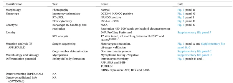
Table 1
Characterization and validation.
Classification Test Result Data
Morphology Photography normal Fig. 1 panel B
Phenotype Immunocytochemistry OCT3/4, NANOG positive Fig. 1 panel G
RT-qPCR
Flow cytometry NANOG positive
SSEA-4: <99% Fig. 1 panel I
Fig. 1 panel E
Genotype Karyotype (G-banding) and
resolution 46XX,
Resolution 450–500 bands per haploid chromosome set Fig. 1 panel C
Identity DNA Profiling Performed Supplementary file panel F
STR analysis 17 sites tested, all matching between HuES9wt and HuES9DGCR8+/−
Mutation analysis (IF
APPLICABLE) Sanger sequencing Heterozygous mutation,
off-target validation Fig. 1 panel A and Supplementary file panel B, G
Copy number determination One insertion in genome Supplementary file panel C
Microbiology and virology Mycoplasma Mycoplasma testing /Negative Supplementary file panel D
Differentiation potential Embryoid body formation Immunocytochemistry:
AFP, SMA and B-III- TUBULIN
mRNA expression: AFP, BRY and PAX6
Fig. 1 panels H and I
Donor screening (OPTIONAL) NA Genotype additional info
(OPTIONAL) NA
1.5. Immunofluorescent staining and flow cytometry
Cells were fixed, blocked, and permeabilized as described previously (Erdei et al., 2014.). Next, cells were incubated separately with primary antibodies (Table 2) for 60 min at RT. Cells were then incubated with corresponding secondary antibodies for 60 min at RT. Cell nuclei were stained with DAPI. SSEA4 Flow Cytometry analysis was performed as described previously (Erdei et al., 2014.).
1.6. Real-time PCR analysis (RT-PCR)
Total RNA was isolated using TRIzol Reagent (Thermofisher Scien- tific). cDNA samples were synthesized from 400 ng of total RNA using the Promega Reverse Transcription System. RT-PCR analyses were performed using TaqMan® assays (Table 2) and analyzed by the Ste- pOne™ Real-Time PCR System (Thermofisher Scientific). All quantita- tive gene expression data were normalized to the expression level of RPLP0.
1.7. Karyotype and STR analysis
Karyotyping and STR analysis were performed by UD-GenoMed Medical Genomic Technologies Ltd.
1.8. Mycoplasma test
MycoAlert™ Mycoplasma Detection Kit was used according to the manufacturer’s instructions (Lonza).
Declaration of Competing Interest
The authors declare that they have no known competing financial interests or personal relationships that could have appeared to influence the work reported in this paper.
Acknowledgements
The authors thank Be´ata Haraszti, Korn´elia N´emethy, L´aszl´o Homolya, Andr´as T´alas and Orsolya Kolacsek for advice and technical assistance. The HuES9 cell line was kindly provided by Douglas A.
Melton, Harvard University. The Addgene plasmid pX330-U6- Chimeric_BB-CBh-hSpCas9 (#42230) was originally deposited by Feng Zhang.
Funding
This study was funded by the National Brain Research Program (NAP) of Hungary (grant numbers: 2017-1.2.1-NKP-2017-00002 to AA ´ and JMR), the Higher Education Institutional Excellence Programme of the Ministry of Human Capacities in Hungary, within the framework of the Neurology thematic programme of Semmelweis University and the National Research, Development and Innovation Office, Hungary, (OTKA-K128369 to AA). ´
Table 2 Reagents details.
Antibodies used for immunocytochemistry/flow-cytometry
Antibody Dilution Company Cat # and RRID
Targeted marker (ICC/
WB)
Rabbit anti-DGCR8 1:1000 Abcam, Cat#
ab191875, RRID: n/a Loading control
(WB) Anti-beta Actin 1:10000 Abcam Cat# ab20272, RRID:AB_445482 Pluripotency
marker (ICC) Mouse anti-Oct3/4 1:50 Santa Cruz
Biotechnology Cat# sc- 5279, RRID:AB_628051 Pluripotency
marker (ICC) Goat anti-Nanog 1:100 R and D Systems Cat#
AF1997, RRID:
AB_355097 Pluripotency
marker (Flow)
Mouse anti-SSEA-4-APC Isotype control: IgG3- APC
1:100
1:100 R and D Systems Cat#
FAB1435A, RRID:
AB_494994 R and D Systems Cat#
IC007A, RRID:
AB_952035 Endoderm
marker (ICC) Mouse anti-AFP 1:500 Sigma-Aldrich Cat#
A8452, RRID:
AB_258392 Mesoderm
marker (ICC) Mouse anti-SMA 1:500 Abcam Cat# ab7817, RRID:AB_262054 Ectoderm
marker (ICC) Mouse anti-ß-III-Tubulin 1:2000 R and D Systems Cat#
MAB1195, RRID:
AB_357520 Secondary
antibody (ICC)
Goat-Anti-Mouse (H + L) Cross-Adsorbed Secondary Antibody, Alexa Fluor 488
1:250 Thermo Fisher Scientific Cat# A- 11029, RRID:
AB_2534088 Secondary
antibody (ICC)
Donkey-Anti-Goat (H + L) Cross-Adsorbed Secondary Antibody, Alexa Fluor 488
1:250 Thermo Fisher Scientific Cat# A- 11055, RRID:
AB_2534102 Secondary
antibody (WB)
Anti-Rabbit IgG 1:5000 Thermo Fisher Scientific, Cat# G- 21234, RRID:
AB_2536530 Primers
Target Forward/Reverse primer (5′-3′) Pluripotency
Marker(qPCR) NANOG Hs02387400_g1 (Thermo Fisher Scientific) Endoderm
differentiation marker (qPCR)
AFP Hs00173490_m1 (Thermo Fisher Scientific)
Mesoderm differentiation marker (qPCR)
TBXT Hs00610080_m1 (Thermo Fisher Scientific)
Ectoderm differentiation markers (qPCR)
PAX6 Hs00240871_m1 (Thermo Fisher Scientific)
House-Keeping
Gene (qPCR) RPLP0 Hs99999902_m1 (Thermo Fisher Scientific) Genotyping and
sequencing DGCR8 Fwd: AGTTTGGCCCATGGGTAGG/Rev:
GGAACACCCACTGCTTCTGAC Inverse PCR CRISPR-
targeted gDNA
Fwd: GCGACTCTAGAGTCGTGGCCTTGGC / Rev: AGGCGGGCCATTTACCGTAAG Off-target
validation predicted off-target sites
#1 CREB5-For/
TTACCACACACCAAACCATAGGC
#1 CREB5-Rev/
CTCTGCACTGCCTTTCATTCAC
#2 ANAPC11-For/
GGCCCATTTGAGATCTTTGAAG
#2 ANAPC11-Rev
CTACGGGAGAACAGCAAGCC
#3 DYNC-For/
TGACAATGTGCATGAATTTGCC
#3 DYNC-Rev/
CAATGTGCCACGGAAAGTTTG
#4 ZMAT3-For/
Table 2 (continued) Primers
Target Forward/Reverse primer (5′-3′) GTACCCTAATGGACACATGGACG
#4 ZMAT3-Rev/
CAGATAAGGGTGGACTGCTGTACTC
#5 OR2L13-For/
TTTATCATCTTCCGCACCTGC
#5 OR2L13-Rev/
CTGACAGTCGGGAGCAAGAAG
Appendix A. Supplementary data
Supplementary data to this article can be found online at https://doi.
org/10.1016/j.scr.2020.102134.
References
Erdei, Z., L˝orincz, R., Szeb´enyi, K., P´entek, A., Varga, N., Liko, I., V´ ´arady, G., Szak´acs, G., Orb´an, T.I., Sarkadi, B., Ap´ati, A., 2014. Expression pattern of the human ABC ´ transporters in pluripotent embryonic stem cells and in their derivatives: ABC Transporters in Pluripotent hESC. Cytometry 86 (5), 299–310. https://doi.org/
10.1002/cyto.b.21168. Epub 2014 Apr 11. PMID: 24729538.
Kolacsek, O., Krízsik, V., Schamberger, A., Erdei, Z., Ap´ati, A., V´arady, G., M´at´es, L., Izsv´ak, Z., Ivics, Z., Sarkadi, B., Orb´an, T.I., 2011. Reliable transgene-independent
method for determining Sleeping Beauty transposon copy numbers. Mob. DNA 2 (1), 5. https://doi.org/10.1186/1759-8753-2-5. PMID: 21371313.
T´alas, A., Kulcs´ar, P.I., Weinhardt, N., Borsy, A., T´oth, E., Szeb´enyi, K., Krausz, S.L., Husz´ar, K., Vida, I., Sturm, ´A., Gordos, B., Hoffmann, O.I., Bencsura, P., Nyeste, A., Ligeti, Z., Fodor, E., Welker, E., 2017. A convenient method to pre-screen candidate guide RNAs for CRISPR/Cas9 gene editing by NHEJ-mediated integration of a ’self- cleaving’ GFP-expression plasmid. DNA Res. 24 (6), 609–621. https://doi.org/
10.1093/dnares/dsx029. PMID: 28679166.
Tomari, Y., Zamore, P.D., 2005. MicroRNA biogenesis: drosha can’t cut it without a partner. Curr. Biol. 15 (2), R61–R64. https://doi.org/10.1016/j.cub.2004.12.057.
PMID: 15668159.
Yeom, K.H., Lee, Y., Han, J., Suh, M.R., Kim, V.N., 2006. Characterization of DGCR8/
Pasha, the essential cofactor for Drosha in primary miRNA processing. Nucleic Acids Res. 34 (16), 4622–4629. https://doi.org/10.1093/nar/gkl458. Epub 2006 Sep 8 PMID: 16963499.