Three dimensional tissue cultures and tissue engineering
Dr.. Domokos Bartis, University of Pécs Medical School
Dr.. Judit Pongrácz, University of Pécs Medical School
technicaleditor: Zsolt Bencze, Veronika Csöngei, Szilvia Czulák
Three dimensional tissue cultures and tissue engineering
by Dr.. Domokos Bartis and Dr.. Judit Pongrácz
technicaleditor: Zsolt Bencze, Veronika Csöngei, Szilvia Czulák Publication date 2011
Copyright © 2011 University of Pécs Copyright 2011, Dr. Judit Pongrácz
Table of Contents
1. Three dimensional tissue cultures and tissue engineering ... 1
1. Three dimensional tissue cultures and tissue engineering – Basic principles ... 1
2. Cells and tissue types in tissue engineering – Stem cells ... 3
2.1. Characteristic stem cell types ... 4
3. Bioreactors ... 10
3.1. Cells to be used in bioreactors ... 11
3.2. Bioreactor design requirements ... 11
3.3. Main types of bioreactors ... 12
3.4. Drawbacks to currently available bioreactors ... 16
4. Biomaterials ... 17
4.1. Natural biomaterials ... 17
4.2. Synthetic biomaterials ... 18
5. Scaffold fabrication ... 20
5.1. Methods for scaffold construction ... 20
6. Biocompatibility ... 23
7. Cell-scaffold interactions ... 23
8. Biofactors ... 25
8.1. Representative Growth Factors ... 26
8.2. Delivery of Growth Factors ... 27
9. Controlled release ... 27
9.1. Delivery methods of growth factors ... 27
9.1.1. Integration of bioactive molecules into scaffolds ... 28
9.2. Additional protein delivery systems in tissue engineering ... 29
9.3. Biodegradable and non-degradable platforms ... 29
10. Biosensors ... 29
11. Aggregation cultures ... 30
11.1. Types of aggregation cultures ... 30
11.1.1. Natural aggregates ... 31
11.1.2. Synthetic cell aggregation ... 33
12. Tissue printing ... 33
13. Tissue repair ... 35
13.1. Engineered tissues for transplantation ... 35
14. Commercial products ... 41
14.1. Products for cardiovascular diseases ... 42
14.2. Products for cartillage ... 43
14.3. Products for liver tissues ... 43
14.4. Products for skin therapies ... 44
15. Clinical trials ... 45
15.1. Types of clinical trials ... 46
15.2. Phases of clinical trials ... 46
15.3. Adult stem cells in tissue therapy ... 47
16. Research applications and drug testing ... 51
16.1. Examples of aggregate cultures in research applications ... 51
17. Ethical issues ... 51
18. Economic significance ... 52
Recommended reading ... 53
Chapter 1. Three dimensional tissue cultures and tissue engineering
1. Three dimensional tissue cultures and tissue engineering – Basic principles
Tissue damages caused by mechanical injuries or diseases are frequent causes of morbidity and mortality.
Tissue injuries are normally repaired by “built-in” regeneration mechanisms. However, if the tissue regeneration process malfunctions or the extent of the injury is too large, organ transplant can potentially be the only solution. Lack of transplantable organs when other therapies have all been exhausted adversely affects the quality and length of patients‟ life, and is severe financial burden on the individual and society. Tissue injury associated diseases would become treatable using targeted tissue-regeneration or transplantation therapies. To provide tissues for therapy or for research to study tissue specific physiological mechanisms and diseases processes, the discipline of tissue engineering has evolved.
Originally, tissue engineering was categorized as a sub-field of engineering and bio-materials, but having grown in scale and significance tissue engineering has become a discipline of its own. To regenerate or even to re- create certain parts of the human body, tissue engineering uses combinations of various methods of cell culture, engineering, bio-materials and suitable biochemical and biophysical factors. While most definitions of tissue engineering cover a broad range of applications, in practice the term is closely associated with applications that repair or replace portions of whole tissues including bone, cartilage, blood vessels, skin, etc.
Figure I-1: Basic principles of tissue engineering
Tissues created in vitro frequently originate from embryonic or adult cells. Furthermore, in vitro generated tissues often need certain mechanical support and complex manipulation to achieve the required structural and physiological properties for proper functioning. To achieve complex tissue structures, conventional cell cultures (Figure I-2) where cells grow as monolayers in two-dimension (2D) can no longer be used.
Figure I-2: 2D tissue cultures
Particularly, as in monolayer cultures stretched cells form a single layer only network, that is incapable to perform complex functions. In tissue engineering, traditional cell culture technology is replaced by three- dimensional (3D) cell cultures (Figure I-3) where cultured cells assume a more natural morphology and physiology.
Figure I-3: 3D tissue cultures
Various three dimensional tissue culture technologies have developed as tissue engineering gained impetus in medical research and therapy. Three dimensional culture technologies frequently apply various bio-materials where cells are provided with the necessary interactions to form the required tissue or organ.
If tissues are not needed immediately, both differentiated adult primary cells as well as adult and embryonic stem cells can be stored in liquid nitrogen, below -150°C.
The following chapters shall provide a brief overview of various aspects of tissue engineering including cell types, bio-materials, culture methods and application of engineered tissues. Clinical trials as well as ethical issues will also be discussed.
2. Cells and tissue types in tissue engineering – Stem cells
Various types of cells are used in tissue engineering that can be categorized in several ways. In broad terms, technologies in tissue engineering make use of both fully differentiated as well as progenitor cells at various stages of differentiation generally known as stem cells.
Stem cells are undifferentiated cells with the ability to divide in culture and give rise to different forms of specialized cells.
According to their source, stem cells are divided into two main groups: “adult” and “embryonic” stem cells (Figure II-1).
Figure II-1: Stem cell types
While adult stem cells are multipotent, embryonic stem cells are mostly pluripotent except of cells in the earliest stages of embryonic development (fertilized egg) that are totipotent. Stem cell niches where stem cells reside in an embryo or any given organ are defined as stem cell microenvironments. The fate of stem cells depends on the effect of the microenvironment. Stem cells – depending on the stimuli – have two main ways to replicate: either symmetrically resulting in two daughter cells with stem cell characteristics or asymmetrically yielding a daughter cell with stem and another daughter cell with differentiating cell characteristics (Figure II-2).
Figure II-2: Types of stem cell replications I
With the increasing number of divisions, the proliferation capacity of stem cells is decreasing favouring differentiated phenotypes (Figure II-3).
Figure II-3: Types of stem cell replications II
As a result, continuous proliferation signals can lead to depletion of stem cell sources.
2.1. Characteristic stem cell types
Embryonic stem cells (ES). Although ethical debate is ongoing about using embryonic stem cells, embryonic stem cells are still being used in research for the repair of diseased or damaged tissues, or to grow new organs for therapy or for drug testing. Especially, as rodent embryonic stem cells do not behave the same way as human embryonic stem cells and require different culture conditions to be maintained. Recently, it has been discovered, that if stem cells are taken at a later stage not at the early blastocyst stage (3–4 days after conception), when the developing embryo implants into the uterus, epiblast – the innermost cell layer – stem cells form (Figure II-4), and these cells resemble human embryonic stem cells and had many of the same properties making it possible to use rodent rather than human cells in some research.
Figure II-4: Epiblast stem cells
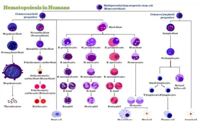
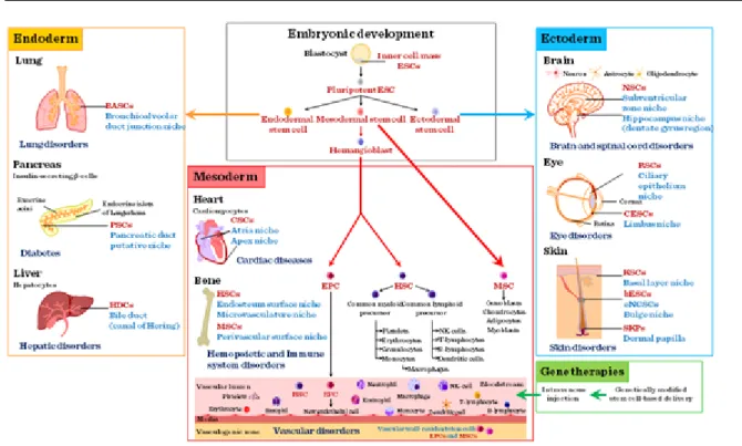
Adult stem cells. Adult or somatic stem cells (ASC) (Figure II-5) are present in every organ including the bone marrow, skin, intestine, skeletal muscle, brain, etc. (Figure II-6) where tissue specific microenvironments provide the necessary stem cell niches where stem cells reside and where the tissue gains its regenerative potential from.
Figure II-6: Hematopoietic stem cells (HSCs)
Mature cells, when allowed to multiply in an incubator, ultimately lose their ability to function as differentiated cells. To proliferate, differentiate and function effectively in engineered tissues, cells must be easily procured and readily available; they must multiply well without losing their potential to generate new functional tissue;
they should not be rejected by the recipient and not turn into cancer; and they must have the ability to survive in the low-oxygen environment normally associated with surgical implantation. Mature adult cells fail to meet many of these criteria. The oxygen demand of cells increases with their metabolic activity. After being expanded in the incubator for significant periods of time, they have a relatively high oxygen requirement and do not perform normally. A hepatocyte, for example, requires about 50 times more oxygen than a chondrocyte consequently much attention has turned to progenitor cells and stem cells. True stem cells can turn into any type of cell, while progenitor cells are more or less committed to becoming cell types of a particular tissue or organ.
Somatic adult stem cells may actually represent progenitor cells in that they may turn into all the cells of a specific tissue. If somatic stem cells are intended to be used in regenerative therapy, healthy, autologous cells (one‟s own cells) would be the best source for individual treatment as tissues generated for organ transplant from autologous cells would not trigger adverse immune reactions that could result in tissue rejection. Also, organs engineered from autologous cells would not carry the risk of pathogen transmission. However, in genetic diseases suitable autologous cells are not available, while autologous cells from very ill (e.g. suffering from severe burns and require skin grafts) or elderly people may not have sufficient quantities of autologous cells to establish useful cell lines. Moreover, since this category of cells needs to be harvested from the patient, there are also some concerns related to the necessity of performing such surgical operations that might lead to donor site infection or chronic pain while the result might remain unpredictable. Also, autologous cells for most procedures must be purified and cultured following sample taking to increase numbers before they can be used:
this takes time, so autologous solutions might be too slow for effective therapy.
Recently there has been a trend towards the use of mesenchymal stem cells from bone marrow and fat. These cells can differentiate into a variety of tissue types, including bone, cartilage, fat, and nerve.
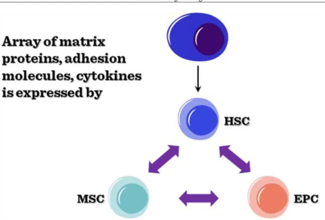
Bone marrow stem cells (MSCs). Bone marrow stem cells can be subdivided into bone marrow stromal (endothelial, mesenchymal) and haematopoietic lineages. All the specific progenitor types can be purified from the bone marrow based on their respective cell surface markers. In the bone marrow microenvironment, these stem cells show a functional interdependency (Figure II-7), secreting factors and providing the necessary cellular interactions to keep the stem cell niche suitable for all the progenitor cells.
Figure II-7: Functional interdependency of bone marrow stem cells The ontogeny of tissue lineages in bone marrow is summarized in 8.
Figure II-8: Ontogeny of tissue lineages in bone marrow
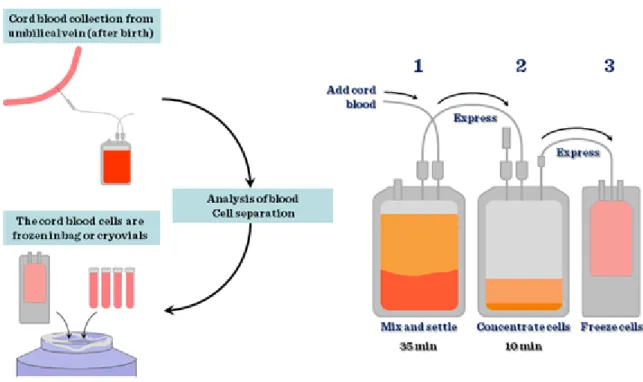
Cord blood stem cells. The largest source of stem cells for regenerative medicine is the umbilical cord stem cell pool. Based on the number of babies born yearly (approximately 130 million), this is not surprising. However, unless the cord blood is collected from the newborn to be used in potential diseases later on in his or her life – mainly childhood haematological disorders –, the application is not autologous, therefore cell types have to be matched just as in any other transplantation procedures. Cord blood is collected at birth and processed
immediately (Figure II-9) to purify stem cells based on their cell surface markers or simply just to be frozen with added DMSO in liquid nitrogen.
Figure II-9: Cord blood stem cells and foetal stem cells
Figure II-10: Stem cell populations in cord blood
In the past 36 years 10,000 patients were treated for more than 80 different diseases using cord blood stem cells.
Ideally, cord blood banks should be set up with active links to international data bases where all the stored cells would be characterized with HLA typing and made available when needed. Unfortunately, storage of stem cells is costly therefore this relatively simple source for progenitor cells is not used to its full potential.
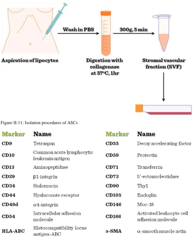
Adipose tissue derived stem cells. Recently there has been a trend to obtain mesenchymal stem cells from fat.
Similarly to other differentiated tissue types, adipose tissues contain tissue specific stem cells. Adipose stem cells (ASC) can be easily isolated from adipose tissues (Figure II-11), and ASCs are multipotent, their
immunophenotype is consistent (Figure II-12) and adipose stem cells are easily manipulated by genetic engineering.
Figure II-11: Isolation procedures of ASCs
Figure II-12: Immunophenotype of ASCs (Positive markers)
The differentiation potential of ASCs is also wide. By using the right combination of extracts and factors, ASCs can differentiate into cardio-myocytes, skeletal myocytes, chondrocytes, osteoblasts, neuronal, endodermal and ectodermal lineages.
Application of stem cells (either embryonic or adult stem cells) (Figure II-13) is becoming broader since cellular manipulation can aid development of the required cell types.
Figure II-13: Application of ESCs and ASCs
Cells can be reprogrammed by modification of their original gene expression patterns or cellular differentiation can be manipulated by applying directed changes into the cellular growth environment in forms of growth factors, cytokines, cellular interactions (e.g. feeder layer). Naturally, the right factors need to be identified prior to any attempts to grow specific tissue types for commercial use. In fact tight regulatory issues surround tissue engineering aimed at clinical application of laboratory generated tissues in regenerative medicine. Apart from ethical issues of using embryonic stem cells, and working with human subjects to purify adult stem cells, the purification of stem cells requires GLP (good laboratory practice) and GMP (good manufacturing practice) conditions that make the production of such tissues very expensive.
Differentiated cells. Previously, it was believed that many differentiated cells of adult human tissues have only a limited capacity to divide. In contrast, some almost differentiated cells have widely been used in clinical tissue engineering. The “almost” status is frequently achieved by culturing purified differentiated cells in 2D culture conditions where they lose their final differentiation characteristics. Among differentiated cells used in tissue engineering are notably fibroblasts, keratinocytes, osteoblasts, endothelial cells, chondrocytes, preadipocytes, and adipocytes. One of the most frequently used differentiated cell types are chondrocytes. In the following, chondrocytes shall serve as an example to describe the use of differentiated cells in tissue engineering applications.
Isolation of autologous chondrocytes for human use is invasive. It requires a biopsy from a non-weight-bearing surface of a joint or a painful rib biopsy. In addition, the ex vivo expansion of a clinically required number of chondrocytes from a small biopsy specimen, which may itself be diseased, is hindered by deleterious phenotypic changes in the chondrocyte. It is important that chondrocytes synthesize type II collagen, the primary component of the cross-banded collagen fibrils. The organization of these fibrils, into a tight meshwork that extends throughout the tissue, provides the tensile stiffness and strength of articular cartilage, and contributes to the cohesiveness of the tissue by mechanically entrapping the large proteoglycans. In growing individuals, the chondrocytes produce new tissue to expand and remodel the articular surface. With aging, the capacity of the cells to synthesize some types of proteoglycans and their response to stimuli, including growth factors, decrease.
These age-related changes may limit the ability of the cells to maintain the tissue, and thereby contribute to the development of degeneration of the articular cartilage. For the organ repair or replacement process the in vitro amplified number of chondrocytes have to be grown in bioreactors to grow as weight bearing tissue. Although differentiated chondrocytes appear to be one of the best sources for engineered cartilage, to date, few tissue- engineered systems provide an autologous, minimally invasive, and easily customizable solution for the repair or augmentation of cartilage defects.
3. Bioreactors
One of the persistent problems of tissue engineering is the difficult production of considerable tissue mass. As engineered tissues normally lack blood vessels, it is difficult for cells in the inside of a larger tissue mass to obtain sufficient oxygen and nutrient supply to survive, and consequently to function properly.
Although self-assembly plays an important role in any tissue engineering methods, tissue growth and functional differentiation needs to be directed by the tissue engineeer. Creation of functional tissues and biological structures in vitro needs to observe some basic requirements for cellular survival, growth and differentiation. In general, the basic requirements include continuous supply of oxygen, correct pH, humidity, temperature, nutrients and osmotic pressure. If the structure of the tissue is important from the point of view of tissue function, suitable scaffolds are often needed. Tissue engineered cultures present additional problems in maintaining culture conditions. In standard, simple maintenance cell cultures, molecular diffusion is often the sole means of nutrient and metabolite transport. However, as a culture becomes larger and more complex, for example in the case of engineered organs with larger tissue mass, other mechanisms must be employed to maintain the culture and to avoid cellular necrosis. It is important to create some sort of capillary networks within the tissue that allows relatively easy transport of nutrients and metabolites.
Similarly to directing growth and differentiation of stem cell cultures, complex tissue cultures also require added factors, hormones, metabolites or nutrients, chemical and physical stimuli to reach the desired functionality.
Just a couple of examples: chondrocytes for example respond to changes in oxygen tension as part of their normal development to adapt to hypoxia during skeletal development. Other cell types, such as endothelial cells, respond to shear stress from fluid flow, which endothelial cells encounter in blood vessels. Mechanical stimuli, such as pressure pulses seem to be beneficial to all kinds of cardiovascular tissue such as heart valves, blood vessels or pericardium. All the special requirements are intended to be resolved by the use of bioreactors.
A bioreactor in tissue engineering is a device that simulates a physiological environment in order to promote cell or tissue growth. A physiological environment can consist of many different parameters such as temperature and oxygen or carbon dioxide concentration, but can extend to all kinds of biological, chemical or mechanical stimuli. Therefore, there are systems that may include the application of the required forces or stresses to the tissue two- or three-dimensional setups (e.g., flex and fluid shearing for heart valve growth). Several general-use and application-specific bioreactors are available commercially to aid research or commercial exploitation of engineered tissues. Bioreactors, however, require a large number of cells to start off with.
3.1. Cells to be used in bioreactors
Static cell cultures are the most frequently applied cell culture methods. The maintenance is easy, cheap and no specialized laboratory equipment is needed. Petri dishes or disposable plastic tissue culture flasks are used most frequently. Basically 2 types of conventional tissue culture exist: Adherent cells which grow in monolayer cultures and non-adherent cells that grow in suspension cultures. In both case, conventional cell cultures are capable to maintain cells at relatively low densities. The main problem rises concerning static cell cultures when large numbers of cells are needed and conventional cultures need to be scaled-up to be added to bioreactors. In the conventional cultures nutrient supply is maintained by frequent and periodic change of culture medium that is time consuming and involves a high risk of infection. The advantages of the dynamic cellular environment of bioreactors include the dynamic and continous supply of nutrients and oxigen. These features make the formation of larger 3D tissue structures possible. However, availability of nutrients and oxygen inside a 3D tissue construct is still problematic. While in 3D cell cultures direct cell-cell contacts enhance cellular communication, transport of oxygen, nutrients and metabolites is still a challenging issue. First, oxygen and nutrients should diffuse from the static medium to the surface cells. This concerns the oxygen content and the oxygen-carrying capacity of the medium. Diffusion from the surface cells to the deeper structures is also important. Critical parameters are the porosity of the cultured cell/tissue construct. Understandably, thickness of dynamic environment of bioreactors, this parameter can be increased several fold.
3.2. Bioreactor design requirements
Although bioreactors cannot recreate the physiological environment, they need to reproduce as many parameters as possible. Bioreactors need to maintain desired nutrient and gas concentration in 3D constructs and to facilitate mass transport into and from 3D tissues. It is also important for bioreactors to improve even cellular distribution, which is also facilitated through the dynamic environment. Exposure of the construct to physical stimuli is also
important during the engineering of load-bearing tissues, like cartilage, bone, tendon and muscle. Without mechanical stimuli these tissues cannot withstand physiological load and strain.
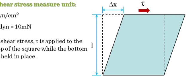
There is an additional critical parameter in dynamic tissue cultures: the shear force. Shear forces are particularly important, as cells are sensitive to shear stress, which may cause dedifferentiation, growth inhibition or apoptosis. Unfortunately, shear stress distribution is uneven in dynamic bioreactors. In dynamic bioreactors the highest stress is located around edges and sides of the moving vessel or around the moving edge of the stirrer and the edges of the scaffold which is static itself but the fluid is moving around it. (The measure unit of shear stress is the dyn/cm2. 1 dyn = 10 mN. The maximum shear stress for mammalian cells is 2.8 dyn/cm2, so in a well-designed bioreactor, shear stress values are well below this number) (Figure III-1).
Figure III-1: Shear forces in dynamic fluids
Perhaps the most difficult task is to provide real-time information about the structure of the forming 3D tissue.
Normally the histological, cellular structure of the 3D construct can only be judged after the culturing period is completed.
In general, bioreactors should be as clear and simple as possible to make cleaning easy and to reduce the risk of infections. The assembly and disassembly of the device should be also simple and quick. It is extremely important that all parts of the bioreactor that comes into contact with the cell culture is made of biocompatible or bioinert materials. For example no chromium alloys or stainless steel should be used in biorecators. Also the material should withstand heat or alcohol sterilization and the presence of the continuously humid atmosphere.
The design should also ensure the proper embedding of instruments like the thermometer, pH meter, the pump or rotator motor, etc.
3.3. Main types of bioreactors
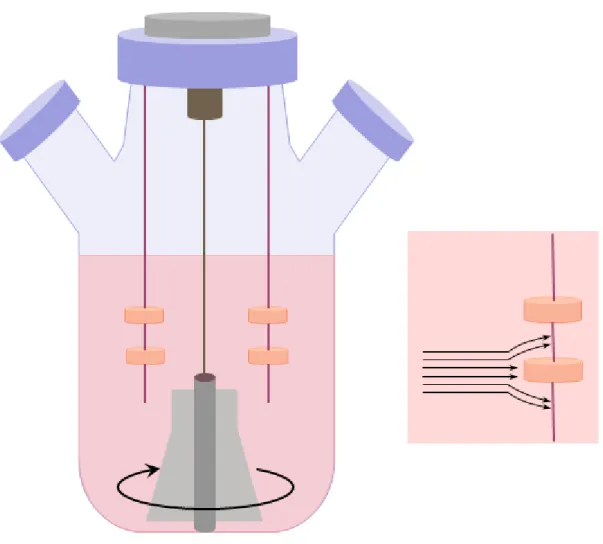
Spinner flask bioreactors (Figure III-2) are maybe the simplest and the most frequently used bioreactor types.
Figure III-2: Spinner flask bioreactors
Spinner bioreactor types mix the oxygen and nutrients throughout the medium and reduce the concentration boundary layer at the construct surface. In a spinner flask, scaffolds are suspended at the end of needles in a flask of culture media. A magnetic stirrer mixes the media and the scaffolds are fixed in place with respect to the moveing fluid. Flow across the surface of the scaffolds results in turbulences and flow instabilities caused by clumps of fluid particles that have a rotational structure superimposed on the mean linear motion of the fluid particles. Via these added fluid motions fluid transport to the centre of the scaffold is thought to be enhanced.
Typically, spinner flasks are around 120 ml in volume (although much larger flasks of up to 8 liters have also been used). The most frequent stirring speed is 50–80 rpm and generally 50% of the total medium is changed every two days. The efficiency of the enhancement of mass transport is indicated that cartilage constructs have cell seeding efficiency is typically low in spinner flask bioreactors, this method usually fails to deliver homogeneous cell distribution throughout scaffolds and cells predominantly reside on the construct periphery.
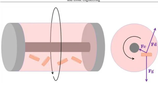
The rotating wall bioreactor (Figure III-3) was originally developed by NASA.
Figure III-3: Rotating wall bioreactors
It was designed with a view to protect cell culture experiments from high forces during space shuttle take off and landing. The device has proved useful in tissue engineering laboratories on Earth too. In a rotating wall bioreactor, scaffolds are free to move in media in a vessel. A rotating wall vessel bioreactor consists of a cylindrical chamber in which the outer wall, inner wall, or both are capable of rotating at a constant angular speed. The vessel wall is then rotated at a speed such that a balance is reached between the downward gravitational force and the upward hydrodynamic drag force acting on each scaffold. The wall of the vessel rotates, providing an upward hydrodynamic drag force that balances with the downward gravitational force, resulting in the scaffold remaining suspended in the media. Dynamic laminar flow generated by a rotating fluid environment is an alternative and efficient way to reduce diffusional limitations of nutrients and wastes while producing low levels of shear compared to the stirring flask. Culture medium can be exchanged by stopping the rotation temporarily or by adding a fluid pump whereby media is constantly pumped through the vessel. Fluid transport is enhanced in a similar fashion to the mechanism in spinner flasks and the rotational devices also provide a more homogeneous cell distribution compared to static or spinner bioreactor cultures. Gas exchange occurs through a gas exchange membrane. Typically, the bioreactor is rotated at speeds of 15–30 rpm. Cartilage tissue of 5 mm thickness has been grown in this type of bioreactor after seven months of culture. As tissue mass increases while cells grow in the bioreactor, the rotational speed must be increased in order to balance the gravitational force and to ensure that the scaffold remains in suspension.
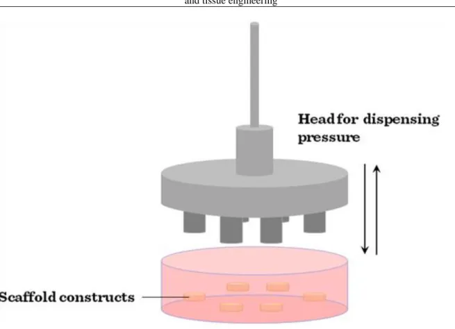
Compression bioreactors (Figure III-4) are another widely used type of bioreactors.
Figure III-4: Compression bioreactors
This class of bioreactor is generally used in cartilage engineering and can be designed so that both static and dynamic loading can be applied. In contrast to static loading that has a negative effect on cartilage formation dynamic loading is more beneficial and more representative of physiological tissue deposition. In general, compression bioreactors consist of a motor, a system providing linear motion and a controlling mechanism to provide different magnitudes and frequencies. A signal generator can be used to control the system including loading of cells while transformers can be used to measure the load response and imposed displacement. The load can be transferred to the cell-seeded constructs via flat platens which distribute the load evenly. However, in a device for stimulating multiple scaffolds simultaneously, care must be taken that the constructs are of similar height or the compressive strain applied will vary as the scaffold height does. Mass transfer is improved in dynamic compression bioreactors over static culture (as compression causes fluid flow in the scaffold) which results in the improvement of the aggregate modulus of the resulting cartilage tissue to levels approaching those of native articular cartilage.
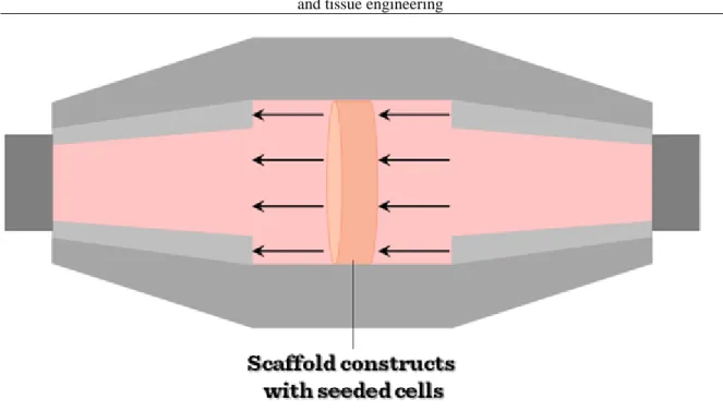
Tensile strain bioreactors have been used in an attempt to engineer a number of different types of tissues including tendon, ligament, bone, cartilage and cardiovascular tissue. Some designs are very similar to compression bioreactors, only differing in the way the force is transferred to the construct. Instead of flat platens as in a compression bioreactor, a way of clamping the scaffold into the device is needed so that a tensile force can be applied. Tensile strain has been used to differentiate mesechymal stem cells along the chondrogenic lineage. A multistation bioreactor was used in which cell-seeded collagen-glycosaminoglycan scaffolds were clamped and loaded in uniaxial tension. Alternatively, tensile strain can also be applied to a construct by attaching the construct to anchors on a rubber membrane and then deforming the membrane. This system has been used in the culture of bioartificial tendons with a resulting increase in Young‟s modulus over non-loaded controls. (Young‟s modulus is a numerical constant that was named after an 18th-century English physician and physicist. The constant describes the elastic properties of a solid material undergoing tension or compression forces in one direction only).
Culture using flow perfusion bioreactors (Figure III-5) has been shown to provide more homogeneous cell distribution throughout scaffolds.
Figure III-5: Flow perfusion bioreactors
Collagen sponges have been seeded with bone marrow stromal cells and perfused with flow. This has resulted in greater cellularity throughout the scaffold in comparison to static controls, implying that better nutrient exchange occurs due to flow. Using a calcium phosphate scaffold, abundant extracellular matrix (ECM) with nodules of calcium phosphate was noted after 19 days in steady flow culture. In comparisons between flow perfusion, spinner flask and rotating wall bioreactors, flow perfusion bioreactors have proved to be the best for fluid transport. Using the same flow rate and the same scaffold type, while cell densities remained the same using all three bioreactors, the distribution of the cells changed dramatically depending on which bioreactor was used. Histological analysis showed that spinner flask and static culture resulted in the majority of viable cells being on the periphery of the scaffold. In contrast, the rotating wall vessel and flow perfusion bioreactor culture resulted in uniform cell distribution throughout the scaffolds. Flow perfusion bioreactors generally consist of a pump and a scaffold chamber joined together by tubing. A fluid pump is used to force media flow through the cell-seeded scaffold. The scaffold is placed in a chamber that is designed to direct flow through the interior of the scaffold. The scaffold is kept in position across the flow path of the device and media is perfused through the scaffold, thus enhancing fluid transport. Media can easily be replaced in the media reservoir. However, the effects of direct perfusion can be highly dependent on the medium flow rate. Therefore optimising a perfusion bioreactor for the engineering of a 3D tissue must address the careful balance between the mass transfer of nutrients and waste products to and from cells, the retention of newly synthesised ECM components within the construct and the fluid induced shear stresses within the scaffold pores.
Flow perfusion reactors in bone tissue engineering. Flow perfusion bioreactors have proved to be superior compared to rotating wall or spinner flask bioreactors to seed cells onto scaffolds. The expression levels of bone differentiation markers (namely Alkaline Phosphatase, Osteocalcin and the transcription factor Runx2) proved to be consistently higher in flow perfusion reactors than in any other type of bioreactors. Additionally, the mineralization of the scaffolds is also higher. To avoid the disadvantageous effects of high shear stress, the flow rate in the reactor needs to be set carefully. Experiments demonstrated that intermittent dynamic flow is more favourable than steady speed flow in bone tissue engineering.
Two chamber bioreactor – The most current and revolutionary achievement in tissue engineering was the implantation of a tissue engineered trachea which was developed in a special two-chamber bioreactor. The external surface of the de-cellularized donor trachea was seeded with autologous chondrocytes differentiated from hemapoetic SCs of the recipient and airway epithelium was seeded to the inner surface in a rotation wall- like bioreactor. The application of two separate “chambers” allowed simultaneous culture of different cell types.
The construct was then used for the surgical replacement of the narrowed trachea in a tuberculosis patient.
3.4. Drawbacks to currently available bioreactors
Tissue engineering methodology is very labor intensive, specialized equipment and specially trained technicians are needed to perform the work. The current bioreactors are highly specialized devices which are difficult to
assemble and disassemble. The cell output is low and the culturing times are long. Moreover, real-time monitoring of tissue structure and organization is not yet available. Problems with compression bioreactors involve mainly the mechanical parts, which are prone for leakage. Understandably, infection is a problem when such bioreactors are used. Added problem is the type of the scaffold to grow the cells on, as the applied scaffolds have to withstand mechanical stimulation, so strong scaffolds are needed, which may have longer degradation time once implanted. Naturally, this is not preferred. So a compromise has to be achieved between scaffold stiffness and resorption time.
4. Biomaterials
The requirements for biomaterials used in tissue engineering are quite strictly defined. Biocompatibility for example is high on the agenda, as scaffold and bioreactor materials have to be tissue friendly and not eliciting immunoresponse. Moreover, at best, the biomaterial should support cellular and tissue functions like adhesion, differentiation and proliferation via its special surface chemistry. Porosity is an important requirement concerning scaffolds. Generally the porosity should reach and even exceed 90% to allow even seeding of cells and to support vascular ingrowth after implantation. Controlled biodegradation is also an important issue in some cases when the healthy tissue replaces the implanted biomaterial and the biomaterial gradually degrades in the body of the host. Biomaterials can be divided into natural and synthetic biomaterials.
4.1. Natural biomaterials
The advantages of natural biomaterials (Figure IV-1) are that they mostly come from an in vivo source therefore large quantities are constantly available at a reasonable price.
Figure IV-1: Types of natural biomaterials
Further advantages of natural biomaterials are that they already have binding sites for cells and adhesion molecules so the biocompatibility is not a major issue. However, there are also some disadvantages. Due to natural variability in the in vivo source, the lot-to-lot variability is always a concern. Additionally, potential impurities may also result in unwanted immune reactions, while their mechanical properties are also limited.
Collagen is the most frequently used and therefore the most studied biomaterial. There are rich in vivo sources, as all connective tissues of animals are rich in this ubiquitous protein. Collagen has a fibrous structure, and its amino acid composition is unique. It provides binding sites for integrins called RGD sites (from the amino acid sequence arginine-glycine-aspartic acid). Collagen has a superior biocompatibility being a conserved protein.
Additional benefit, that the immune system well tolerates collagen. It is capable of supporting a large spectrum cellular differentiation types, therefore collagen is well preferred as scaffold.
Another, easily accessible type of scaffold is fibrinogen. Fibrinogen is obtained from (human) plasma. Although in its uncleaved form is a soluble protein, upon cleavage with thrombin fibrinogen sets as a gel and forms a 3D meshwork which is 100% biocompatible and physiological in wound healing. It is often used as a biological glue when cells to be seeded onto scaffolds (e.g. non-woven mesh or fleece or other porous materials) are suspended first in a fibrinogen-containing solution. Then the solution is applied onto the scaffold and upon the addition of thrombin, a hydrogel is formed which enhances the cells‟ ability to attach to the 3D scaffold. Fibrin is also suitable for supporting ES cell differentiation as well as keeping differentiated cells in culture. Recent applications of fibrin include cardiovascular, cartilage, bone and neuronal tissue engineering.
Silk that is also used as scaffold material, is a protein produced within specialized glands of some arthropods. It has a special tertiary structure consisting of repeating amino acid motifs forming an overlapping beta-sheet structure giving the unique sturdiness for this protein. There have been many industrial attempts to mimic the features of silk. As a result the availability of recombinant analogues is increasing.
The silkworm‟s (Bombix mori) silk consists of two different protein components, namely Fibroin and Sericin.
Sericin forms the outer layer on the fibroin core making it slippery and elastic. Fibroin that is biocompatible and possesses excellent mechanical properties is also used as tissue engineering scaffold. Its use is widespread in bone, cartilage and ligament engineering. Additionally, silk fibroin can be modified chemically, e.g. the attachment of RGD groups provides binding sites for osteoblasts and enhances Ca++ deposition and bone cell differentiation. Moreover, silk promotes more intensive chondrogenesis than collagen used as a scaffold material for cartilage engineering. The degradation rate of silk is very slow but finally bone tissue gradually replaces the silk scaffold.
Polysaccharide-based biomaterials are polymers consisting of sugar monomers. Those used for tissue engineering purposes are of plant (seaweed) or animal origin. Some polysaccharides may trigger unwanted immune reactions so a careful selection is advised considering polysaccharide scaffold materials.
Polysaccharides are most frequently used as hydrogels, which per se form a 3D meshwork so they can provide a scaffold for seeded cells. These hydrogels are frequently used as injectons: they can be dispensed directly to the site of injury so it supports wound healing and also cell growth and differentiation.
Main source of agarose, another scaffold material for tissue engineering, are red algae and seaweed. Agarose is the most frequently used polysaccharide scaffold consisting of a galactose-based backbone. It is immunologically inert, so no immune response is triggered. One of its great advantages lies in its versatility: the stiffness and mechanical parameters can be easily manipulated of agarose gels. It has been used for scaffolding cartilage, heart, nerve tissues and it also supports stem cell differentiation.
Alginate is the polysaccharide component of the cell walls of brown algae. It is an acidic compound, so in tissue engineering various cationic alginate salts are used. Sodium-alginate is a frequently used food additive (E-401) and its use is also widespread in gastronomy. Besides of gastronomic applications, sodium alginate is used in industry as a heavy metal-binding or fat-binding agent. The potassium salt is also used in the food industry as an emulsifier and stabilizer. In tissue engineering its calcium salt or Calcium-alginate has gained widespread application. Calcium alginate is a water-insoluble gel-like material, and it is generally used in the industry or laboratory for enzyme immobilization or encapsulation. In tissue engineering calcium-alginate proved to be useful for encapsulation of whole living cells, thus isolating them from the immune system preventing rejection after transplantation. In a clinical trial, calcium-alginate was used for the encapsulation of pancreatic islet cells which preserved their ability to produce insulin according to the needs of the host as the encapsulation prevented the immune reaction against the grafted cells.
Hyaluronan which is also termed as Hyaluronic acid is an animal-derived polysaccharide which is extensively used as a scaffold material in tissue engineering. It is a non-sulfatated Glucose-Amino-Glycan (GAG) molecule and exists as a major component of the ECM in hyalinic cartilage and skin but it is present in other organs as well. As a natural ECM component, multiple cell surface receptor binding and cell adhesion sites available on the macromolecular complex. Hyaluronan has an important role in wound healing and tissue repair. Moreover, it supports embryonic stem cell differentiation, survival and proliferation. Like other polysaccharides, hyaluronan is used as a gel in nerve, cartilage and skin tissue engineering.
Chitosan is derived from the deacetylation of chitin which is a strongly cationic polysaccharide which is the main component of the arthropod exoskeleton. Chitosan is commercially derived from sea-dwelling crustaceans and it is widely used for bandages and wound dressing, utilizing its ability to enhance blood clotting. Chitosan scaffolds are mainly used in bone tissue engineering, as chitosan supports osteoblast differentiation. Moreover, chitosan-calcium-phosphate composite scaffolds form a pliable, injectable hydrogel at slightly acidic pH. Upon transition to physiological pH, it gels thus anchoring osteocytes to the scaffold. Native and collagen-modified chitosan can be used for tissue engineering, as both forms support progenitor differentiation into osteoblasts.
4.2. Synthetic biomaterials
In tissue engineering a large scale of synthetic biomaterials (Figure IV-2) are used besides that of natural origin.
Figure IV-2: Types of synthetic biomaterials
Their main advantages over natural biomaterials are the high reproducibility, availability on demand and constant quality supporting industrial-scale production. Moreover, by application of slight changes of production, an easy control of mechanical properties, degradation rate, shape, composition, etc. can be adjusted to current needs. However, synthetic materials often lack sites for cell adhesion and the biocompatibility is frequently questionable. Biocompatibility and support of stem cell differentiation is not clear either, while immune reactions are also possible.
Poly (lactic-co-glycolic acid) PLGA is an FDA approved scaffold material. PLGA is a mixed polymer consisting of lactic and glycolic acids in various ratios. PLGA is a biodegradable material and its degradation rate can be modulated. It is one of the most frequently used biomaterials applied in neural tissue, bone and cartilage tissue engineering. PLGA is biocompatible, triggers no immune reaction and supports embryonic stem cell differentiation, proliferation and survival. However one has to consider that the degradation products are acidic, therefore may alter cell metabolism.
Poly (ethylene glycol) PEG is a commonly used biocompatible polymer. This molecule has amphophilic properties having a hydrophilic head and a variably long hydrophobic tail. The PEGylation of proteins is a commonly used pharmaceutical technology to modulate the degradation and absorbtion of bioactive proteins like interferons. The chemical modification of PEG is also available e.g. heparin, peptides, or RGD motifs. PEG is also a frequently used scaffold material in bone, cartilage, nerve, liver and vascular tissue engineering.
Application of PEG-formed hydrogels is very versatile. The chemical nature, the extent of crosslinking, and the potential application of modifications, like RGD or other amino-acid groups, makes PEG widely used. PEG- based gels can be applied not only to entrap or anchor cells but also in storing and delivering bioactive molecules like BMP or TGFβ.
Peptide-based biomaterials consist of short amino acid sequences which usually are ampholitic thus making peptides capable of self-assembly. The application of these peptides allows the combination of the advantages of synthetic materials with that of natural scaffolds. Synthetic nature of the peptides eliminates batch-to-batch variability and consistent purity and quality can be achieved. Moreover, known binding sites for cell-surface adhesion molecules can be included in the sequence. For example, IKVAV amino acid sequence is from laminin and it facilitates neurite outgrowth. RGD sequence is a binding site for integins which promotes cellular adherence and migration.
Ceramic-based biomaterials are used in bone tissue engineering only. These scaffolds are totally or partly inorganic. Mostly they are shaped with heat forming porous, brittle materials. For example, bioactive glass is slowly biodegradable. With an ion-exchange mechanism it slowly turns to natural hydroxyapathite via surface degradation. So bioglass is biocompatible and used as an implant material. Hydroxyapatite is the inorganic compound of bone. Sometimes it is used as a completely inorganic, porous scaffold, more often it is combined with other biocompatible, organic polymers, like PLGA, collagen, or chitosan. This combination also enhances the drug delivery capacity of these scaffolds to enhance bone formation.
Metals are used as implant materials. Mostly alumina and titanium alloys are used because of their biocompatibility. The high durability of metals is needed where implants are subjected to extreme mechanical load, like articular prostheses, dental implants or heart valves. These metals are bioinert materials, however, sometimes metallic implants may cause immunological reactions like metal allergy in sensitive individuals.
Also, as they are not biodegradable it is questionable whether metals can be listed amongst tissue enginnering scaffolds.
5. Scaffold fabrication
Scaffolds, as it has been mentioned in previous chapters, are natural or synthetic materials which provide the basis for 3D tissue engineered constructs. There are some basic criteria for scaffolds. Biocompatibility is an important issue: non-biocompatible materials trigger immune reactions in the host so a chronic inflammation may occur upon implantation. The surface chemistry is also critical: cells and ECM molecules need to come into contact with the scaffold surface through its surface molecules. The scaffold surface should support cellular functions such as adhesion and migration. Cells which are seeded onto scaffolds should populate it evenly, and scaffolds should also allow vascularization of the implanted construct that is vital for successful function and integration into the body of the host. So scaffold materials should be rich in interconnected pores, which allow cell infiltration and support vascularization. Controlled biodegradability should also be considered. Ideally, the scaffold degrades in the host, where the scaffold material is replaced by ECM material produced by implanted and physiologically present cells leading to formation and integration of the new tissue.
Upon engineering tissues that are expected to be exposed to mechanical load – e.g. cartilage or bone –, consideration of mechanical properties of the scaffold materials is especially important. The scaffolds used for generating such tissues should withstand mechanical stress upon the preparation of the tissue in the compression or strain bioreactor and also resist to physiological stresses shortly after implantation. A balance has to be stricken, however, as stronger scaffolds usually degrade slower.
Scaffolds are often generated to be able to hold and to slowly release drugs or biomolecules. Cells growing on scaffolds in bioreactors often need biomolecules or growth factors to stimulate their proliferation, differentiation and formation of the new tissue. It is important that the scaffold material should interact with the ECM so that the replacement of the formation of new ECM after implantation goes swiftly. Sometimes it is important, that scaffolds mimic the ECM providing support for cell growth and attachment.
Scaffold characteristics are needed to be carefully considered when constructing a new tissue in vitro. Primarily, scaffolds provide the 3D environment for cells therefore they should support cellular functions. Also, scaffolds temporarily replace the ECM after implantation and have key role in directing cellular differentiation. Both during the construction and after implantation, the structure of the scaffold determines cell nutrition and mass transport into the newly formed or implanted engineered tissue.
5.1. Methods for scaffold construction
Solvent casting & particulate leaching (SCPL) (Figure V-1) is the easiest and cheapest way of scaffold formation.
Figure V-1: Solvant casting – particulate leaching
Basically, the mold is filled with a pore-forming material and the dissolved scaffold material is poured into the mold. After the evaporation of the solvent the scaffold material solidifies. In order to form porous scaffolds, the pore-forming particles should be dissolved. The SCPL technique is simple, easy and inexpensive technique and no special equipment is needed to perform this methodology. However, there are some drawbacks which come usually from the nature of the solvent in which the scaffold material was dissolved in. Usually organic solvents are applied, which are often toxic. The contaminations are difficult to eliminate and cells seeded onto these scaffolds can affected by the toxicity of the solvent remnants.
Phase separation methods are also very often applied in the fabrication of scaffold materials. The scaffold- forming polymer is dissolved in the mixture of 2 non-mixing solvents then saturated solutions are produced by heating. The polymer-lean and polymer-rich phase separates at a higher temperature. When the temperature is lowered, the liquid-liqiud phase is separated again and the dissolved polymer precipitates on the phase-border from the over-saturated solutions. Then the solvent is removed by extraction, evaporation or sublimation. This method usually provides scaffolds of high porosity.
Gas foaming is a special technology for the production of tissue engineered scaffolds using supercritical CO2. A specialized pressure chamber is needed which is filled with the scaffold material. CO2 is then slowly let into the chamber, under very high pressure, so that it reaches supercritical state. In the supercritical gas, the scaffold material is practically dissolved. When the pressure is lowered, the CO2 turns into gas again and the phase separation of dissolved scaffold occurs. Scaffold foams with particularly high porosity can be produced while no toxic organic solvents are used during the procedure. Moreover, recent research results demonstrated that for a short period of time even living cells can survive the high-pressure conditions without significant damage, so cells can be added during the preparation phase ensuring even cellular distribution.
Electrospinning (Figure V-2) is used not only in scaffold fabrication for tissue engineering but also for other materials such as industrial filters.
Figure V-2: Electrospinning
A special injecting device is required for production which injects the dissolved scaffold polymer into the air.
Opposite to the injector, an electrically charged plate collects the discharged material so that a non-woven textured material will be formed consisting of very thin fibres. The electrospinning techinque is very versatile and no extreme conditions (heat, coagulation, etc.) required to produce scaffold materials. Additionally, many types of polymers are applicable, e.g. PLA, PLGA, silk fibroin, chitosan, collagen, etc. This methodology allows the easy regulation of the thickness, aspect ratio, porosity, fiber orientation of the produced material consisting of non-woven fibres.
The scaffolds produced with fiber-mesh technology consist of (inter)woven fibres. This structure allows both 2D and 3D scaffold structures, and pore size can be easily manipulated via adjusting weaving and fiber parameters.
This is a versatile technique, since the scaffold material is broadly applicable and combinations of materials can be applied too.
Self assembly is the spontaneous organization of molecules into a defined structure with a defined function. For tissue engineering purposes mainly amphiphilic peptides are used. These molecules contain a charged head and a hydrophobic tail part and capable of assemble into pre-defined structures in aqueous solutions via forming non-covalent bonds. Self-assembling peptide apmholites can also be modified. For example, addition of a phosphoserine group to the peptide enhances the mineralization in bone tissue, the presence of RGD groups provide binding sites for cell surface integrins, and cysteines are capable to form intermolecular bridges thus increasing stability. GGG linker amino acids are inserted frequently between the head and tail groups to increase flexibility of the peptides.
Rapid prototyping is the automatic construction of physical objects using additive manufacturing technology.
This technology is used not only in industrial manufacturing processes but also for production of tissue engineering scaffolds. Rapid prototyping allows fast (scaffold) fabrication with consistent quality, texture and structure. However, negative side of this technology that it needs special equipment: expensive and specialized computer-controlled machinery.
There are multiple technologies within Rapid prototyping, like fused deposition modeling (FDM). This applies a robotically guided machine which extrudes filament consisting of a polymer or other material through a nozzle.
Multiple layers can be formed where the object is solid and the use of cross-hatching (using a different substance) is also possible for areas that will or can be removed later.
Another rapid prototyping method is selective laser sintering (SLS) (Figure V-3).
Figure V-3: Selective laser sintering (SLS)
When this technology is used, scaffold material (usually a polymer) is initially in powder form, slightly below its melting temperature. One layer of the heated powder is laid on the mold and a computer-guided laser beam provides heat for the powder particles to sinter (weld without melting) together. As one layer is completed, more and more new powder layers will be sintered, as the piston moves downward. So with this method the 3D structure of the object will be formed layer-by-layer.
6. Biocompatibility
Biocompatibility of an engineered tissue is closely related to the behavior of the used biomaterials in various contexts. A biomaterial can be considered biocompatible if it performs with the desired host response in any particular application. The term “biocompatibility” may refer to specific properties of a given material without specifying where or how the material is used. The basic requirements towards every biomaterial are the following: the biomaterial or its degradation products are not cytotoxic, elicits little or no immune response, and is able to integrate with a particular cell type or tissue. The ambiguity of the term reflects how biomaterials interact with the human body and eventually how those interactions determine the clinical success of the engineered tissue. As tissue engineering often uses biomaterials to provide scaffolds for the required tissues, biocompatibility also refers to the ability to perform as a substrate that will support the appropriate cellular activity, including the facilitation of molecular and mechanical signaling systems, in order to optimize tissue regeneration, without eliciting any undesirable effects in those cells, or inducing any undesirable local or systemic responses in the eventual host.
For example, the basic minimal requirements, for an artificial vascular graft is not just a measurement of its ability to act as a passive tool for blood flow that doesn‟t trigger immune response, platelet adhesion, activation, aggregation and coagulation as well as complement cascade activation, but also how it integrates into the surrounding tissues.
Similarly, in a bone implant that was generated on a polymer scaffold structure it has to support cell growth, provide sufficient temporary mechanical support to withstand in vivo stresses and loading while scaffold degradation and resorption kinetics have to be controlled in such a way that the bioresorbable scaffold retains its physical properties for at least 6 months (4 months for cell culturing and 2 months in situ). Thereafter, it will start losing its mechanical properties and should be metabolized by the body without a foreign body (immune or toxic) reaction after 12–18 months.
Understanding of the anatomy and physiology of the action site is essential for a biomaterial to be effective. An additional factor is the dependence on specific anatomical sites of implantation. It is thus important, during design, to ensure that the implement will fit complementarily and have a beneficial effect with the specific anatomical area of action.
7. Cell-scaffold interactions
Scaffolds have previously been regarded as passive components of a tissue. Scaffolds, however, turn out to have an active role in generating functional properties of the engineered tissue (Figure VII-1 and Figure VII-2).
Figure VII-2: Primary SAEC in matrigel
Even if a scaffold just simply binds two or more types of cells, the enforced proximity of individual cells immediately results in several interesting properties. The scaffolded complex can be regarded as a micro- environment on its own, where the concentrations of participants as well as their metabolites and secreted factors are vastly enriched in a small location that gives rise to complex, dynamic behaviour of cells.
Methods employed in the course of tissue engineering often offer unique opportunities to observe cell-matrix interactions that cannot otherwise be viewed. These observations may provide insights into cell behavior than can contribute important new knowledge about cell biology. One such set of observations led to the discoveries that musculoskeletal connective tissue cells express a contractile muscle actin isoform, alpha-smooth muscle actin, and can contract. This knowledge may help to explain how these cells generate forces required for certain physiological and pathological functions, and this information may inform future approaches to regulate this function to advance tissue engineering. Tissue engineering science thus emerges as an important force that can both contribute to cell and molecular biology and add to the fund of knowledge supporting the production of tissues in vitro or in vivo to improve the management of a wide variety of disorders. Recent advances in micro- and nano-fabrication technologies offer the possibility to engineer scaffolds with a well defined architecture providing a control of cellular spatial organization, mimicking the microarchitectural organization of cells in native tissues.
ECM, the natural medium in which cells proliferate, differentiate and migrate, is an absolute requirement for tissue regeneration. Cell-ECM interaction is specific and biunivocal. Cells synthesize, assemble and degrade ECM components responding to specific signals and, on the other hand, ECM controls and guides specific cell functions. Motifs are locally released according to cellular stimuli. Cells are attached to ECM through molecules belonging to the integrin family and recognize specific aminoacid sequences through cell surface receptors.
Moreover, solid state, structural ECM molecules act as reservoirs for secreted signaling molecules for their on- demand release. Since ECM plays an important role in a tissue‟s mechanical integrity, crosslinking of ECM may be an effective means of improving the mechanical properties of tissues. The ECM crosslinking can result from the enzymatic activity of lysyl oxidase (LO), tissue transglutaminase, or nonenzymatic glycation of protein by reducing sugars. The LO is a copper-dependent amine oxidase responsible for the formation of lysine- derived crosslinks in connective tissue, particularly in collagen and elastin. Desmosine, a product of LO-mediated crosslinking of elastin, commonly is used as a biochemical marker of ECM crosslinking. The LO-catalyzed crosslinks that are present in various connective tissues within the body – including bone, cartilage, skin, and lung – are believed to be a major source of mechanical strength in tissues. Additionally, the LO-mediated
enzymatic reaction renders crosslinked fibers less susceptive to proteolytic degradation. ECM fibres can also store and facilitate release of biofactors in insoluble/latent forms through specific binding with glycosaminoglycans (e.g. heparins), and biofactors can elicit their biological activity once released.
8. Biofactors
Bioactive molecules are frequently required for tissue engineering to regulate cellular functions such as proliferation, adhesion, migration, and differentiation (Figure VIII-1).
Figure VIII-1: Signalling networks in tissue development and maintenance
The most frequently applied biofactors are various growth factors. The term “growth factors” include differentiation factors and angiogenic factors in addition to growth factors and other regulatory factors, including bone morphogenetic proteins. The redundancy found in most biological structures is so great that precise characterization of a growth factor that falls into just one category is misleading. Many growth factors may provide a host of functions and according to the biochemical, cellular, and biomechanical context may modulate cell attachment, cell growth and/or death, cell differentiation, cell migration, vascularization, etc. Both growth factors and differentiation factors are essential to establish functional cells on an appropriate architecture.
Growth factors may be exogenously added, or the cells themselves may be induced to synthesize them in response to chemical and/or physical stresses. Although cytokines and growth factors are present within the ECM in minute quantities, they are potent modulators of cell behavior.
The most frequently used growth factors with their multiple isoforms are vascular endothelial growth factor (VEGF), platelet-derived growth factor (PDGF), keratinocyte growth factor (KGF), transforming growth factor β (TGF-β), bone morphogenic protein (BMP) and fibroblast growth factor (FGF), each with its specific biological activity. While it is clear that these factors are necessary in various combinations, determination of the optimal dose is difficult. Additionally, sustained and localized release of the required factor at the desired site, and the inability to turn the factor “on” and “off” as needed during the course of tissue repair is extremely difficult.
Growth factors have been studied in numerous ways in the field of tissue engineering, and some of the growth factors have produced promising results in a variety of preclinical and clinical models.
During tissue morphogenesis the presence of soluble GFs guides cellular behaviours, thus governing neo-tissue