Cell and Tissue Disintegration
Benno Hess
I. General Information II. Methods
1. Surviving tissue a) Tissue slices b) Tissue brei
c) Heterogeneous cell suspensions 2. Tissue fixation
3. Homogenates a) W e t homogenates
a) Mechanical homogenization
(3) Sonic homogenization and disintegration by pressure y) Thermal disintegration
8) Chemical disintegration
e) Biological-enzymatic disintegration b) Dry homogenates
4. Tissue and cell fractionation
I. General Information 1
)
One aim of enzymatic analysis is to obtain information on the concentration and localization of metabolites in the living cell
2 )
. It is therefore desirable to prepare the living material in a form suitable for the measurements, but without altering the structure or the relative amounts of the substances to be analysed. However, two difficulties stand in the way of this ideal situation:
1. The destruction of the physiological state is accompanied by a change in the physico- chemical state and the concentration of the metabolites ("operational isomers").
2. When a tissue is disintegrated the compartmental concentration gradients may be disturbed (non-linear change of the compartmental conditions).
These two complications are interdependent and in consequence the analytical results provide a more or less distorted picture of the conditions in the living cell. Every assay of metabolites 1)
General information and reviews on this subject: E. Bamann and K. Myrback: D i e Methoden der Fermentforschung. Thieme, Leipzig 1941, Vol. 2; S. P. Colowick and N. O. Kaplan: Methods in Enzymology. Academic Press, N e w York 1955, Vol. I; K.Lang and E. Lehnartz in Hoppe- Sey ler-Thierfelder: Handbuch der physiologisch- und pathologisch-chemischen Analyse, Springer, Berlin-Gottingen-Heidelberg 1955, Vol. II, p. 545; W. W. Umbreit, R. H. Burris and / . F. Stauf- fer: Manometric Techniques. Burgess Publishing Co., Minneapolis 1959; H. M. Rauen: Biochemi
sches Taschenbuch, Springer, Berlin-Gottingen-Heidelberg 1956; M. Dixon and E. C. Webb:
Enzymes. Longmans, Green & Co. Ltd., London, N e w York, Toronto 1958.
2
) For a review and definition of the terms concentration, reacting concentration, content and level, activity of metabolites, cellular concentration, tissue concentration, status in vivo and altered status, s e e
3 - 7 3) ) .
O. Meyerhoff and N. Geliazkowa, Arch. Biochem. Biophysics 12, 405 [1947].
4
> F. Lynen and R. Koenigsberger, Liebigs A n n . Chem. 573, 60 [1951].
5) Th. Biicher and M. Klingenberg, Angew. Chem. 70, 552 [1958].
6
) B. Hess and B. Chance, Naturwissenschaften 46, 248 [1959].
7) H. J. Hohorst, F. H. Kreutz and Th. Biicher, Biochem. Z. 332, 18 [1959].
in biological material is therefore an "operational test", i.e. the results are influenced by the property of the compound tested for and by the method used. A comparison of the results of different workers is only possible if the methods used are well defined.
Apart from these main obstacles, it is important to consider the possible instability of the metabolites during the disintegration of biological material. They may be acid labile (ATP, ADP, DPNH, TPNH)*), alkali labile (triosephosphate, DPN, TPN), oxidizable or easily denatured (proteins). They can also be transformed enzymatically during the relatively long disintegration process. For example, the cooling of guinea pig kidney from 38 to 0°C with the "quick-freeze" tongs takes 90 msec.
8
). However, 22 msec, is sufficient for a 10% change in the steady state concentration of FADH and FAD in ascites tumour cells
9
). Usually the steady state levels of low molecular weight metabolites are shifted during the disintegration towards the true position of equilibrium. Anaerobiosis, aerobiosis and dilution may be contri
butory factors in changing the amounts of substrate (e.g. displacement of the aldolase equi
librium 1 0
), displacement of the ATP/ADP or lactate/pyruvate ratios on disintegration with acid; decomposition of enzyme-substrate complexes).
The disintegration conditions are be governed by two considerations:
1. Preservation of the chemical structure of the metabolites. For this, the temperature, pH, ionic strength, time, etc., used in the fixation and extraction of the tissue must be compatible with the decay constants and the biological half-life times of the compounds to be deter
mined.
2. Preservation of the localization of the compounds in the tissue and cell compartments.
The tissue is divided by its morphological characteristics; i.e. into structures visible with the light and electron microscopes.
Generally, distinction is made between disintegration methods used for the assay of high molecular weight compounds (e.g. enzymes) and those used for low molecular weight metabolites.
If enzyme activity is to be determined, the speed with which the tissue is fixed, disintegrated and extracted is less important. Any proteolytic decomposition during the disintegration (up to several hours) can usually be ignored. To preserve the structure of enzyme proteins the extraction is carried out at low temperature with distilled water
1 1
), salt solutions 1
), sucrose
1
), periston, dextran or albumin solutions. To avoid denaturation of protein by the acid produced in tissues with a high rate of glycolysis, the solutions used for extraction
*) Abbreviations: A T P = adenosine triphosphate A D P = adenosine diphosphate D P N = diphosphopyridine nucleotide D P N H = reduced diphosphopyridine nucleotide
T P N = triphosphopyridine nucleotide T P N H = reduced triphosphopyridine nucleotide
F A D = flavine adenine dinucleotide F A D H = reduced flavine adenine dinucleotide
8) A. Wollenberger, O. Ristau and G. Schoffa, Pfliigers Arch. ges. Physiol. Menschen, Tiere 270, 399 [I960].
9) B. Chance and B. Hess, J. biol. Chemistry 234, 2404 [1959].
!0) Th. Biicher in: Neuere Ergebnisse aus Chemie und Stoffwechsel der Kohlenhydrate, Springer, Berlin-Gottingen-Heidelberg 1958, p. 185.
n) G. Beisenherz, H. J. Boltze, Th. Biicher, R. Czok, K. H. Garbade, E. Meyer-Arendt and G. Pfiel
der er, Z. Naturforsch. 8b, 555 [1953].
should be buffered at a suitable pH. The addition of chelating agents to prevent oxidation, or of cysteine to protect SH groups is also recommended. The tissue is disintegrated by careful homogenization, sometimes with the addition of compounds which act on the membranes (e.g. digitonin). An example of the extraction of an enzyme is given on p. 53.
In contrast, the disintegration of tissue for the analysis of low molecular weight metabolites requires the fixation of the cellular state within seconds or even milliseconds. For this, the sample is frozen and/or metabolic inhibitors or denaturing agents are used.
Rapid cooling (as in the "quick-freeze" method) brings about nearly ideal fixation of the tissue. It has the advantage that more time is available for the subsequent operations (depro
teinization, extraction). Particularly elegant are direct spectrophotometric measurements on deep-frozen tissue samples (e.g. in liquid a i r )
1 2 ) .
Inhibitors prevent certain metabolic reactions and so fix the concentrations of the metabolites (inhibition of glycolysis by addition of fluoride, iodoacetic acid, cyanide or hydrazine;
production of anaerobic conditions by the addition of cyanide, sodium azide, sodium sulphide or by gassing with nitrogen).
Deproteinizing agents include: acids (trichloroacetic acid, perchloric acid, etc.), alkalies and organic solvents. Metabolic processes can also be stopped by rapid heating (e.g. pouring a culture of algae into hot alcohol
1 3 )).
Generally, it is best to use a combination of deproteinizing agent, metabolic inhibitor and fixation by low temperature in conjunction with the extraction or homogenization of the tissue. An example is given on p. 47.
Occasionally it is possible to make use of the lability of certain metabolites. For example, the instability of DPN in alkali is utilized to destroy this coenzyme in a mixture of DPN and DPNH. Triosephosphate is hydrolysed by alkali and the phosphate liberated can be determined. DPNH is sensitive to acid. The acid degradation of TPNH gives depyridino- TPN, which can be detected chromatographically.
If the metabolites or enzyme proteins are stable or can be stabilized artifically, the tissue can be processed with the cell topography in mind. By homogenization and subsequent differential centrifugation (perhaps in a continuous flow centrifuge) the cell components are separated from one another. Each fraction is then analysed.
When a tissue is being quantitatively processed it is important to check the content of the substance being assayed after every step. This type of check should show any degradative processes or changes in the limits of the cell compartments. Stable properties of biological material such as, dry or fresh weightD, cell count, haematocrit, protein content (biuret method with cell extracts
1 1
), cell, mitochondrial or sarcosome suspensions) serve as reference standards. Relation to the nucleic acid phosphate or to the sum of the adenosine phosphates (ATP, ADP, A M P )
I 3 a)
serves for conversion to cell counts. The concentrations of stable enzymes can also serve as reference standards (cytochrome a in mitochondria or intact cells
9 );
cytochrome c or glyceraldehyde-3-phosphate dehydrogenase for glycolytic enzymes and soluble cytoplasm
1 4
>).
12
) B. Chance and E. L. Spencer jr., Faraday Society Discussions 27, 200 [1959].
13
) A. A. Benson and M. Calvin in S. P. Colowick and N. O. Kaplan: M e t h o d s in Enzymology. Aca
demic Press, N e w Y o r k 1957, Vol. IV, p. 882.
13a
) B. Hess in: Control of Respiration and Fermentation. R o n a l d Press, N e w York 1962.
14) W. Vogell, F. R. Bishai, Th. Biicher, M. Klingenberg, D. Pette and E. Zebe, Biochem. Z. 332, 81 [1959].
The extracellular fluid mixes with the cell contents on extraction and therefore distorts the true concentration ratios. The measured values must therefore be corrected for the metabolite concentration of the extracellular fluid. To correct for blood content the oxyhaemo
globin concentration is determined 7,15.16). The size of the intercellular space can be calculated from a chloride determination.This calculation is based on the assumption that the intracellular chloride concentration is low, while the chloride .concentration of the intercellular tissue fluid and plasma is rather similar
? ).
The distribution of metabolites or other compounds in the intracellular compartments is obtained from the analysis of the cell fractions. The amount of protein (biuret method) or the amount of cytochrome c or a (in mitochondria or sarcosomes) can serve as reference standards for the calculation of the metabolite concentrations. The size of the intracellular space cannot be determined exactly.
Usually a particular method of tissue disintegration cannot satisfy all the requirements.
Only a few methods are optimal. To increase the value of a piece of research, it is better to use several complementary methods of extraction. In view of the heterogeneity of biological structures practically every tissue requires a different treatment, even though the same compound is being estimated.
Special care is required in the treatment of the organisms whose tissue is to be examined.
The nutritional and functional state of experimental animals deserves consideration. The period of hunger, thirst, dark or work before the experiment should also be reported as well as the conditions of narcosis and the removal of blood (arterial or venous blood, with or without pressure) or organs.
II. Methods*)
1. Surviving tissue
a) Tissue slices
Tissue slices are organized, surviving tissue without a blood supply, but allowing free diffusion of oxygen and metabolites. They are usually prepared with a razor blade. The critical thickness of a tissue slice for the diffusion of oxygen is 0.2 mm. (for the calculation of the critical thickness, see
1 , 1 7
)). The free diffusion of metabolites varies and can be limiting (e.g. sodium glutamate
1 8 )
). Tissue slices are suitable for the assay of enzymes (directly or after fractional extraction
1 8 a )
) and for the determination of metabolites (after deproteiniz
ation).
Types of instrument: Blade holder according to Deutsche
Stadie-Riggs tissue slicer 2 0
).
*) The types of instrument and manufacturers mentioned here are meant to be examples. It is not possible to give a complete list,
is) B. Chance, J. biol. Chemistry 797, 557 [1952].
16) H. Holzer, G. Sedlmayr and M. Kiese, Biochem. Z. 328, 176 [1956].
1
7
) O. Warburg, F. Kubowitz and W. Christian, Biochem. Z. 227, 252 [1930].
is) P. P. Cohen and M. Hayano, J. biol. Chemistry 166, 239 [1946].
1 8 a )D . Pette, Biochem Z., in press.
19) W. Deutsch, J. Physiology 87, 56 P H936].
20) W. C. Stadie and B. C. Riggs, J. biol. Chemistry 154, 687 [1944].
b) Tissue brei
Tissue brei is used for metabolic studies, particularly on muscle. The brei is prepared with a tissue mincer (Latapie mincer), with the result that the tissue is broken up more than in the case of slices. The tissue fragments have a diameter of 0.3 to 0.5 mm. The perfusion of the tissue brei with the suspending medium is therefore made easier. A disadvantage is the high proportion of cells which are destroyed. A brei can be rapidly deproteinized. It is used as the starting material for the preparation of muscle sarcosomes (e.g. heart muscle sarcosomes from rats or pigeons). The brei is homogenized with a Potter-Elvehjem homogenizer (see below), followed by differential centrifugation. The metabolism (P/O ratio or respiratory control) of the sarcosomes obtained in this way is not affected.
T y p e of i n s t r u m e n t : The Latapie mincer, which is similar to a household mincer, presses the tissue with a piston through the holes of a metal plate against a rotating blade.
It can be pre-cooled. M a n u f a c t u r e r : Arthur H.Thomas Co., Philadelphia, Penn., USA (in 2 sizes, for 25 g. and more, and for smaller amounts).
c) Heterogeneous cell suspensions
Heterogeneous cell suspensions, such as blood, exudates, transudates (ascites, pleura, etc.) or cell cultures, are best collected with a syringe, preferably under paraffin. In the collection of venous blood the effect of stasis should be taken into account. For the rapid deproteiniz
ation of blood, it should be led directly from the vein (or by use of a syringe) into a weighed centrifuge tube containing ice-cold perchloric acid.
Haemolysis of erythrocytes with digitonin has become popular 2 1
* and rupture of the cells also occurs when they are cooled to — 90° C (e.g. acetone-dry ice). For the preparation of homogeneous suspensions, see p. 49. Heparin, oxalate, citrate, versene or siliconized glass
ware is used to prevent the coagulation of blood. The addition of metabolic inhibitors, such as fluoride or iodoacetate to inhibit glycolysis, is also recommended. The effect of these sub
stances on the activity of enzymes and the concentrations of metabolites must be checked.
2. T i s s u e fixation
To fix tissue rapidly, flat tongs with aluminium jaws are used. These can be pre-cooled with liquid air or liquid nitrogen ("quick-freeze" tongs
7 , 8 )
) . The tissue gripped between the cold jaws is compressed to form a thin layer and protruding tissue is broken off. A test of this method showed that 1.6 g. guinea pig kidney was cooled from 38 to 0° C in 0.09 sec, from 38 to — 40°C took 0.15 sec. and to reach —160°C required only 0.5 sec.
8 )
. The tissue is compressed to a layer 0.7 mm. thick. The method is particularly suitable for fixing tissue containing labile metabolites (liver, kidney, brain, muscle, nerves)
2 l a ) . Procedure according to
7 )
: grip exposed liver or liver lobes with two forceps, lift up slightly and press between two light metal blocks (provided with wooden handles) which have been pre-cooled in liquid air. Separate the frozen piece of liver from the excess tissue with a pair of scissors, break off any pieces of incompletely frozen tissue protruding over the edges of the metal blocks and immerse the tissue contained in the blocks in liquid air. With frequent additions of liquid air, powder the piece of liver in a mortar pre-cooled with liquid air. The
2 0 G. W. Lohr, H. D. Waller and O. Karges, Klin. Wschr. 35, 871 [1957].
2 i a ) £ " . Gerlach, H. J. Doring and A. Fleckenstein, Pfliigers Arch. ges. Physiol. Menschen, Tiere 266,
266 [1958].
tissue should not be allowed to thaw during the process. It is therefore best to work in a cold room. Also the absorption of water by the cold tissue, which is considerable in the highly humid air of laboratories at normal temperature, is minimized by working in a cold room.
Add a portion of the tissue to a weighed amount of ice-cold 6 % perchloric acid (about 1 g.
tissue to 5 ml. HCIO4) and grind rapidly with a glass pestle. The tissue powder must not be allowed to remain on the walls of the vessel during its addition to the perchloric acid. Weigh the vessel containing the tissue suspension, homogenize the contents for 30 sec. and centrifuge for 4 min. in the cold at 2800 g. Decant the supernatant, re-extract the sediment with 3 % perchloric acid and centrifuge again. Combine the supernatants.
The authors 7)
state that powdering tissue and extracting the powder is sufficient for the quantitative recovery of metabolites, and that with five metabolites, subsequent homogeni
zation of the final sediment in an Ultra-Turrax (see below) resulted in no increase in the yields. In spite of this, for safety's sake re-homogenization was advised to guarantee the quantitative extraction of metabolites.
T y p e s of i n s t r u m e n t : "Quick-freeze" tongs according to Wollenberger et al.®.
"Quick-freeze" tongs according to Biicher et al
1
^ 3. H o m o g e n a t e s
Table 1 gives a survey of methods of homogenization. It is followed by a detailed description of methods and types of instrument. In certain cases procedures are given which are generally held to give optimum results. The author's experience confirms this opinion.
Table 1. Methods for the preparation of homogenates and for the disintegration of cells Types of
homogenate Method Tissue
a) Wet homogenates a) mechanical homogenization
grinding with sand, alumina powder,
glass powder, diatomaceous earths muscle, bacteria frozen tissue
pestle homogenizer muscle parenchyma
glass beads homogenizer bacteria, yeast, ascites tumour cells
blade homogenizer (blendor) muscle parenchyma P) sonic homogenization
ultrasonic homogenizer bacteria, yeast y) thermal disintegration
freezing and thawing
[freezing mixtures ( a c e t o n e- C 0 2 ; alcohol-CC>2); liquid air;
liquid nitrogen]
universal application
8) chemical disintegration with isopentanol; butanol;
digitonin; petrol ether erythrocytes, mitochondria, muscle
E) biological-enzymatic disintegration autolysis, maceration;
lysozyme, bacterial proteases yeast, bacteria, muscle b) Dry homogenates homogenization by dehydration
acetone; lyophilization
a) Wet homogenates
a) Mechanical homogenization
G r i n d i n g in a m o r t a r
Grinding biological material in a cooled mortar with sand, alumina, diatomaceous earths or glass powder is an important preparative method which can be followed by homogeniza
tion. It has proved successful for the disintegration of bacteria (with glass powder 22> or alumina 2 3 )) or heart muscle for the isolation of sarcosomes (with alumina or s a n d2 4'2 5 )) . Pulverization in a porcelain mortar is the method of choice for disintegration of pieces of tissue 7> which have been frozen with liquid air or liquid nitrogen.
Pestle h o m o g e n i z e r according to Potter and Elvehjem1®
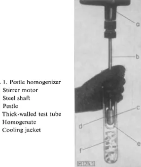
At present the most widely used instrument for the homogenization of tissue (particularly parenchymatous tissue) is the pestle homogenizer. It consists of a tight-fitting pestle (margin ca. 0.2 mm.) made of glass or plastic which is rapidly rotated by a stirrer motor in a thick- walled tes ttube (ca. 1.6 cm. diameter, 15 cm. long) also made of glass or plastic (see Fig. 1).
The tissue is cut up into small pieces with scissors, suspended in a medium, this supension is poured into the test tube and then by rapidly rotating the pestle (about 1000 revolutions per min.) the tissue is homogenized in a few minutes. Fresh tissue (1 — 2 g.) is suspended in 8 —10 ml. water to give a 1: 10 dilution. Usually the test tube is pushed up and down the rotating pestle by hand (see Fig. 1). The test tube is cooled to prevent over-heating during
Fig. 1. Pestle homogenizer a: Stirrer motor b : Steel shaft c: Pestle
d: Thick-walled test tube e: H o m o g e n a t e f: Cooling jacket
22) W. P. Wiggert, M. Silverman, M. F. Utter and C. H. Werkman, Iowa State Coll. J. Sci. 14, 179 [1940].
23) L. A. Mancon and / . O. Lampen, J. biol. Chemistry 193, 539 [1951].
24) K. W. Cleland and E. C. Slater, Biochem. J. 53, 547 [1953].
25) B. Chance and M. Baitscheffsky, Biochem. J. 68, 283 [1958J.
26) V. R. Potter and C. A. Elvehjem, J. biol. Chemistry 114, 495 [1936].
the homogenization. The extent of the homogenization can be varied by using either a loose (e.g. for kidney) or tight-fitting pestle. The uses of pestle homogenization include the prepar
ation of tissue for the subsequent separation of cell fractions by differential centrifugation (see p. 54).
T y p e s of i n s t r u m e n t :
Several types of pestle homogenizers of varying size (5—50 ml.) are available. There are also different types of pestle. M a n u f a c t u r e r : Desaga, Heidelberg, Germany; Arthur H.
Thomas Company, Philadelphia 5, Pa., USA.
G l a s s b e a d h o m o g e n i z e r s
A tissue or cell sample is disintegrated by mechanical vibration, rotation or shaking in the presence of glass beads. The method is particularly suitable for types of cells which are resist
ant to disintegration, such as bacteria, yeast, ascites tumour cells, etc. By variation of the diameter of the glass beads or the frequency of the vibration, rotation or shaking, the degree of homogenization can be varied.
T y p e s of i n s t r u m e n t :
Glass bead homogenizer according to Nossal 27)
(shaking principle). Tightly packed cells are filled into a steel container (2 cm. diameter, 12 cm. long) with a spatula, and glass beads in the ratio 3 : 1 parts by weight are added. The steel capsule is shaken in a longitudinal direction with a frequency of 90 kc. The length of shaking required to disintegrate ascites tumour cells
2 8
* is two 5 sec. periods, and for fresh yeast or bacteria, two 10 sec. periods. M a n u f a c t u r e r : H. Mickle, 4 Ormond Drive, Hampton, Middlesex, England. (The apparatus of this firm is built for considerably lower frequencies than those stated here. The author has had no experience with this apparatus).
Glass bead homogenizers according to Zillig and Holzel 29
^ (vibration principle). Method:
as above. Vibration frequency: 50 kc. The author has had no experience with this apparatus.
Glass bead homogenizer according to Merkenschlager, Schlossmann and Kurz^ (rotation principle). 75 ml. Duran glass containers, which can, for example, hold 20 g. yeast (fresh weight). Rotation frequency: 2000—4000 r. p. m. Cooling: liquid carbon dioxide (the tem
perature of the material remains below 5°C). Time of homogenization: 30—102 sec. for complete homogenization and preparation of cell-free extracts. With shorter homogenization times (10 sec.) the mitochondria remain intact. M a n u f a c t u r e r : B. Braun, Melsungen, Germany.
Glass beads: The glass beads must be washed with dilute nitric or hydrochloric acid, followed by thorough rinsing with distilled water. Diameter of the glass beads: 0.1 mm. for bacteria;
0.5 mm. for yeast. M a n u f a c t u r e r : Superbrite Glass Beads, Minnesota Mining and Manu
facturing Comp., St. Paul 6, Minn., USA; Ballotini beads, English Glass Co. Ltd., Leicester, England.
B l a d e h o m o g e n i z e r ( b l e n d o r )
The tissue is disintegrated in a short time by rapidly rotating blades (10000—40000 r. p. m.), resulting in a fine brei. The method is not suitable for cell fractionation since the intracellular
27) p. M. Nossal, Australian J. exp. Biol. M e d . Sci. 31, 583 [1953].
28) B. Chance and B. Hess, J. biol. Chemistry 234, 2413 [1959].
29) W. Zillig and H. Holzel, Hoppe-Seylers Z. physiol. Chem. 312, 140 [1958].
30) M. Merkenschlager, K. Schlossmann and W. Kurz, Biochem. Z. 329, 332 [1957].
elements, such as mitochondria, are destroyed. It is also not suitable for the homogenization of micro-organisms because only relatively thick suspensions can be disintegrated.
T y p e s of i n s t r u m e n t :
Bottom drive homogenizers: for the preparation of large amounts of tissue (100 g. to 1 kg.).
Usually available with several attachments of different sizes. Speed: 6000 to 12000 r. p. m.
M a n u f a c t u r e r s and trade names: Starmix (Electro-Star, Reichenbach, Fils, Germany), Kenwood-Chief (Kenwood, Stuttgart, Germany), Braun-Mixer (M. Braun, Frankfurt/M., Germany), MSE-Ato-Mix 800 and Ato-Mix 100 (Measuring & Scientific Equipment Ltd., Spencer Street, London, S. W. 1, England).
Top drive homogenizers: for the preparation of small amounts of tissue (up to 100g.).
Available with attachments up to 100 ml. Variable speeds, with some types up to 50000 r. p. m.
Some instruments are available with cooling jackets. M a n u f a c t u r e r s and trade names:
Servall-Omnimixer (Ivan Servall Inc., Norwalk, Conn., USA). Maximum capacity: 200 ml.;
maximum speed 16000 r. p. m.; micro-attachment available with a capacity of 0.5—3.0 ml.
and a speed of 50000 r. p. m. The Virtis homogenizer (The Virtis Comp., 160 Eshborton Ave.,Yonkers, N. Y., USA), dimensions as above. Biihler homogenizer (Biihler, Tubingen, Germany). Speed: max. 50000 r. p. m.; capacity 10 — 150 ml.; can be cooled. MSE homo
genizer (Measuring & Scientific Equipment Ltd., Spencer Street, London, S. W. 1, England).
Dimensions as above. Ultra-Turrax (Janke & Kunkel KG, Stauffen i. Br., Germany).
Maximum speed: 14000 r. p. m.; volume up to 10 ml.
7
*.
/?) Sonic homogenization and disintegration by pressure
The pressure changes of several thousand atmospheres caused by sonics or ultrasonics break cell walls. Acoustic methods are therefore suitable for the preparation of homogenates. Ani
mal tissues (spleen, liver ascites cells, erythrocytes, kidney, Hela tumour cells, thymus and lymph nodes; 10—90 sec.) and micro-organisms (algae, fungi; 2—45 min.) can be disinte
grated. Many enzymes are liberated. Mitochondria are destroyed. It is usually necessary to cool during the disintegration.
P r o c e d u r e according t o 3 1 )
: Prepare a 5 % cell suspension of pressed baker's yeast in 0.1 M potassium phosphate buffer (pH 7.5). Stir this suspension for 20 min. at 0°C before the sonication to obtain an even suspension of cells, so that no air-containing aggregations can occur in the sonication vessel. Sonicate the suspension in 15 ml. portions. Frequency: 19.5 kc;
output: about 50 ± 5 watts/cm.
2
. Cool the sonicator probe to between —5 and — 8°C during sonication with the highest output, with 70 % ethanol as the refrigerant, so that the soni
cated fluid has a final temperature of 1 to 3°C. At lower intensities of sonication, cool just sufficiently to prevent the suspension from freezing. After sonication of a sample, clean the continuous flow chamber by filling with buffer and sonicating for 1 min. (Sonicator: Schoeller Ultra-Disintegrator).
Instead of using the rapid pressure changes brought about by ultrasonics, it is also possible to disintegrate cells by very high static pressures (500—3 500 atm.)
3 0 a )
. This method is partic
ularly suitable for the disintegration of yeast, green algae and plant cells.
30a) E. Ribi, T. Perrine, R. List, W. Brown and G. Codde, Proc. Soc. exp. Biol. Med. 100, 647 [1959].
3 D H. Hubener, H. J. Gollmick, K. Tesser, W. Lippert and L. Rossberg, Biochem. Z. 331, 410 [1959].
Types of i n s t r u m e n t and m a n u f a c t u r e r s :
Schoeller Ultra-Disintegrator (Schoeller & Co., Frankfurt/M-Sud, Morfelder Landstrasse 115, Germany). Frequency: ca. 20kc, the ultrasonic output is freely adjustable. Good results in the disintegration of yeast and bacteria
3 1
*. The instrument can be cooled.
Raytheon Sonic Oscillator (Raytheon Mfg., Comp., Waltham, Mass., USA). Frequency:
9 and 10 kc. It is possible to cool the instrument. With the 10 kc instrument 50 ml. suspen
sions are usually treated for 10 to 30 min.
MSE-Ultrasonic Disintegrator Model 60W and 500 (Measuring & Scientific Equipment Ltd., Spencer Street, London, S . W . I , England); Output: ultrasonic frequency 20kc.
Volume: 20 and 50 ml. (Model 60); 200 ml. and 500 ml. (Model 500). It is possible to cool the instrument.
Pressure: Servall Ribi Refrigerated Cell Fractionator (Servall Inc., Norwalk, Conn., USA).
y) Thermal disintegration
Repeated freezing and thawing is a successful means of disintegrating intact cells, partic
ularly erythrocytes and bacteria (see also "quick-freeze" methods, p. 47). As a general rule
3 2 -
3 3
* slow freezing leads first to the intercellular formation of crystal nuclei. On more rapid freezing intracellular ice crystals are also formed and these destroy the intracellular structure. On thawing, the cells are ruptured osmotically due to the presence of pure water.
If the temperature is lowered very quickly (within seconds), crystallization cannot occur and the tissue becomes vitrified. In this case, if the tissue is thawed quickly to avoid crystal
lization, living cells and tissue are undamaged. Consequently, if a tissue fixed by the "quick- freeze" method is to be prepared for the analysis of metabolites, the thawing and depro
teinization (addition of perchloric acid) of the tissue must be combined. For spectrophoto
metric measurements in liquid air devitrification of the tissue is obtained by addition of glycerol (e. g . 1 part yeast suspension containing 0.3 g. fresh yeast/ml. + 1 part glycerol)
1 2
*.
d) Chemical disintegration
In chemical disintegration methods the cell wall is attacked chemically. The disintegrating agent is allowed to act for the shortest possible time and is usually helped by the use of a mechanical homogenization method (Potter-Elvehjem homogenizer or Waring blendor).
The chemicals may interfere during the analysis, therefore only those which can be easily removed are used. An example is the preparation of erythrocyte haemolysates with digi- tonin
2 1
*. This method depends on the destruction of the erythrocyte membrane by the reac
tion of digitonin with the cholesterol of the cell wall. It has also been used in the disintegration of mitochondria (from liver cell homogenates) for the subsequent extraction of mitochondrial particles
3 4
*. The method is also suitable for the lysis of leucocytes and platelets.
P r o c e d u r e according to 3 5
*: Mix 5 ml. washed erythrocytes, leucocytes or platelets, 2.5 ml.
0.05 M triethanolamine buffer (pH 7.5), 2.5 ml. doubly distilled water and 1.0 ml. saturated, aqueous solution of digitonin. Incubate the mixture for 60 min. at 3°C (until haemolysed).
Centrifuge off the cell stroma (15 min. at 3000 g). In addition, disintegrate leucocytes and platelets mechanically in a Potter-Elvehjem homogenizer.
32
* F.F.Nord and M. Bier in R. Plank: Handbuch der Kaltetechnik. Springer, Heidelberg 1952,
Vol. 9, p. 84.
33) F. N. Furness, Ann. N . Y. Acad. Sci. 85, 501 [I960].
34) C. Cooper, T. M. Devlin and A. L. Lehninger, Biochim. biophysica Acta 18, 159 [1955].
35) G. W. Lohr and H. D. Waller, Klin. Wschr. 37, 833 [1959].
Chemical disintegration methods have been widely adopted for the preparation of cell fractions. They are used to destroy lipoprotein complexes, for example, the butanol extraction of microsomes and mitochondria1'3 6*.
e) Biological-enzymatic disintegration
One of the oldest methods of disintegration is the autolytic decomposition of cellular structures by endogenous proteolytic enzymes. Lebedew described the autolytic decomposi
tion of dried brewer's yeast at 30°C (2 to 3 days)1*. It is the preliminary step before sub
sequent maceration ( = moist autolysis at 35° C, 2 to 3 hours) and for the preparation of maceration juice capable of carrying out fermentation. The maceration is usually carried out with the addition of toluene, ethyl acetate or sodium sulphide.
Recently methods for the enzymatic lysis of bacteria or tissue have been developed. For example, pancreatic juice has been used for the disintegration of Escherichia coli^ and lysozyme for the disintegration of Micrococcus lysodeikticus*®. Biological disintegration of E. coli can also be carried out with bacteriophages3 9*.
Fig. 2. Electron micrograph of heart muscle sarcosomes.
Magnification ca. X 20300.
Partial destruction of the sarcosomes due to the fixing.
(Photograph:
Dr. H. Sitte, Heidelberg).
A special problem is provided by the disintegration of muscle, if intact sarcosomes have to be prepared. In this case, the use of proteolytic enzymes (proteases from Bacillus subtilis) has proved of value4 0*. The p r o c e d u r e is as follows:
36) R. K. Morton, Nature [London] 166, 1092 [1950].
37) L. H. Stickland, Biochem. J. 23, 1187 [1929].
38) M. F. Utter, L. O. Krampitz and C. H. Werkman, Arch. Biochem. Biophysics 9, 285 [1946].
39) /. H. Sher and M. F. Mallette, J. biol. Chemistry 200, 257 [1953].
^o) B. Chance and B. Hagihara, Proceedings V. International Congress for Biochemistry, M o s c o w 1961. Pergamon Press, L o n d o n 1962, S y m p o s i u m V.
Open 5 rat or pigeon hearts with scissors, wash thoroughly with deionized water and cut as small as possible with scissors. Homogenize the tissue in 10 ml. 0.25 M sucrose solu
tion with the addition of bacterial proteinase (10 mg. proteinase "Nagarse" (see below) in 10 ml. 0.25 M sucrose solution) and allow to stand for 20 min. at 4°C. The tissue suspen
sion becomes noticeably more homogeneous due to the proteolytic activity. Homogenize again, allow to stand for 20 min., dilute the homogenate 1: 1 with the sucrose solution and carry out a differential centrifugation to isolate the sarcosomes. Fig. 2 shows a prepa
ration obtained in this way from rat heart.
P r e p a r a t i o n s and m a n u f a c t u r e r s :
Lyophilized, crystalline bacterial protease "Nagarse" (Teikoku Chemical Industry Co., Ltd., Itachibori Minami-dori 1-chome, Osaka, Japan.
Bacterial proteinase "Serva"
Lysozyme "Serva"
All three preparations are obtainable in Germany from Serva Entwicklungslabor, Heidelberg, b) Dry homogenates
The classical method for the preparation of dry homogenates is dehydration with acetone (acetone-dried powder)
l
K It is still used for the preparation of enzymes from bacteria, yeast or animal material. However, it is less suitable for the preparation of extracts for the quanti
tative analysis of enzyme activity. Recently, vacuum freeze-drying of biological material (lyophilization) has been developed and widely introduced.
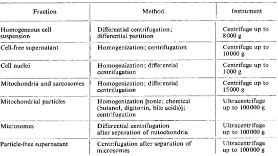
4. T i s s u e a n d cell fractionation
Table 2 gives a survey of the methods for tissue and cell fractionation, which are not dis
cussed individually here.
Table 2. Methods for tissue and cell fractionation
Fraction Method Instrument
H o m o g e n e o u s cell suspension
Differential centrifugation;
differential partition
Centrifuge up to 8 0 0 0 g
Cell-free supernatant H o m o g e n i z a t i o n ; centrifugation Centrifuge up to 10000 g Cell nuclei H o m o g e n i z a t i o n ; differential
centrifugation
Centrifuge up to lOOOg
Mitochondria and sarcosomes H o m o g e n i z a t i o n ; differential centrifugation
Centrifuge up to 1 5 0 0 0 g
Mitochondrial particles Homogenization [sonic; chemical (butanol, digitonin, bile acids)];
centrifugation
Ultracentrifuge up to 100000 g
Microsomes Differential centrifugation after separation of mitochondria
Ultracentrifuge up to 100000 g Particle-free supernatant Centrifugation after separation of
microsomes
Ultracentrifuge up to 100000 g
The preparation of homogeneous cell suspensions (e.g. of thrombocytes, leucocytes) is still a problem. The chief difficulty is the danger of damage to the cell membranes during the usually long isolation procedure. A new method, which is particularly suitable for the sepa
ration of cells and cell particles, consists of differential partition between two aqueous solutions of different high-polymer carbohydrates
4 1
*. A cell-free supernatant obtained after homogenization of a tissue mainly contains mitochondria or sarcosomes, lysosomes and microsomes. The treatment of mitochondria for the preparation of submitochondrial partic
les is mainly sonic homogenization or chemical disintegration. Before the analysis of the cell cytoplasmic compartment for metabolites and enzyme activity, the tissue extract should be freed from particles by ultracentrifugation.
i) P.-A. Albertsson, Nature [London] 182, 709 [1958].