1
Study on factors of vitamin D metabolism in hepatocellular carcinoma
Doctoral thesis Horváth Evelin MD.
Semmelweis University Doctoral School of Clinical Medicine
Supervisor: Ferenc Szalay MD, D.Sc.
Opponents: Katalin Dezső MD, PhD Árpád Patai MD, PhD
Head of the exam committee: Zsuzsa Schaff MD, D.Sc.
Members of exam committee: János Banai MD, D.Sc.
László Herszényi MD, D.Sc.
Budapest 2016
2 1. INTRODUCTION
The relation between the malignant cancer and the biologically active vitamin D, 1α,25(OH)2D3, started with individual observations. Firstly it has been observed that with the lack of hours of sunlight and a decreased level of the serum vitamin D, have caused an increase in the prevalence of colorectal cancer. Later the antitumor effect of vitamin D was proved in several malignant tumors, also in hepatocellular carcinoma (HCC). The initial investigations cleared up that mice with a deficiency of vitamin D are more susceptible to chemically induced hepatocarcinogenesis. There is a wide geographic and ethnic variation of the high incidence of HCC. According to the database of Hungarian Cancer Registry the incidence and the annual mortality rate of HCC is approximately 600 deaths annually. At the 1st Department of Surgery of the Semmelweis University in Hungary between 1996-2009, there were 211 liver resections performed due to the presence of HCC. The treatment of HCC is a frequent problem also in Hungary. According to the latest data of the incidence of HCC, in the last few years it has increased. 70-90% of the cases of HCC develop on cirrhotic livers. The main etiologic factors are alcoholism, the cases of B virus (HBV), hepatitis C virus (HCV) infections and the nonalcoholic steatohepatitis (NASH). The less frequent etiological factors are the hereditary haemochromatosis, alpha-1 antitrypsin deficiency, autoimmune hepatitis, Wilson disease, and some types of porphyries. Abnormal cell proliferation is detected in half of the cirrhotic nodules. Worldwide, the main etiologic factor of HCC is hepatitis B virus. Approximately 350-400 million patients are infected with the virus. The HCC caused by HBV is through liver cirrhosis in 70-90% of the cases. The risk of HCC is higher in cases of the presence of HCV than HBV infection independently of liver cirrhosis. The antiviral treatment of HCV (e.g. pegylated interferon + ribavirin) decreases the risk of the development of HCC. Alcohol abuse is a serious problem in Hungary. In Europe 10.2% of HCC has arosed from alcoholic etiology. It is believed that, the greater part of cryptogenic cirrhosis is nonalcoholic fatty liver disease (NAFLD) and nonalcoholic steatohepatitis (NASH). The majority of these patients are men with a metabolic syndrome. Diagnosing NASH is difficult, if there is a presence of both liver cirrhosis and hepatocellular carcinoma. Worldwide, the aflatoxin toxicity is an important factor in the developement of HCC.
3
The name of vitamin D refers to two compounds: cholecalciferol (vitamin D3), which is produced in the skin from 7-dehydrocholesterol under UV radiation, and the ergocalciferol (vitamin D2) which arises from ergosterol. The mitocondrial CYP27B1 is the vitamin D-activating enzyme, that is mainly expressed in the proximal tubes of the kidney, but also in other tissues. The average life time of the vitamin D in the tissues is short. The vitamin D activates the catabolic enzyme, CYP24A1, through VDR. High CYP24A1 expression is detected in various human malignant tumors (breast, prostate, skin, esophagus, etc.). In some types of tumors the high expression of CYP24A1 amounts worser prognosis, as well as the decreased concentration of vitamin D in the malignant tissue. The low level of serum vitamin D is a risk factor for some malignancies. However, there has been less data collected about HCC. For the first time, in 1991 low vitamin D serum levels were detected in a Japan man with HCC.
There are in vitro, preclinical animal and also clinical studies of the effect that the vitamin D has on HCC. Pourgholami and al. proved that vitamin D has an important anti-proliferative effect on HepG2 and Hep3B cell lines. Treating patients with vitamin D has a limited anti-tumor effect because of hipercalcemia and hipercalciuria. There are many vitamin D analogs without these side effects (EB1089, CB1093, MART-10). The intracellular effects of vitamin D are mostly mediated by the vitamin D receptor (VDR).
2. AIMS
2.1. Epidemiological study on patients with hepatocellular carcinoma in the 1st Department of Internal Medicine of Semmelweis University
1. What are the most important etiological factors in the development of hepatocellular carcinoma (HCC)?
2. How is the distribution of HCC in point of the sex, the age, the presence of liver cirrhosis?
3. How much is the mean survival time of the patients?
4. How is the distribution of serum AFP levels of the patients with HCC?
2.2. Study on the effect of 1α,25(OH)2D3 in different HCC cell lines in vitro
1. Are there any expression of VDR, CYP24A1 and CYP27B1 in the different HCC cell lines?
4
2. How does the mRNA levels change in response to 1α,25(OH)2D3 administration in vitro in the cell lines?
3. Are there any difference of these mRNA levels among the diferent cell lines?
4. How does the expression level of VDR, CYP24A1 and CYP27B1 change in response of the time and the dosis of the treatment of 1α,25(OH)2D3 administration?
2.3. Human study
1. Is there any expression of VDR, CYP24A1 and CYP27B1 in human HCC and surrounding non-tumorous liver tissue samples?
2. Is there any expression difference of these mRNA levels between the HCC and non- tumorous liver tissue samples?
3. Is there any expression difference of these mRNA levels between HCC tissue developed on liver cirrhosis and HCC tissue developed in liver without cirrhosis?
4. Is there any expression difference of these mRNA levels among HCC tissues with different etiology?
5. Is there any relation between the mRNA expression levels of VDR, CYP24A1 and CYP27B1 and the clinicopatological parameters of the patients (sex, grade, age, metastasis, etc.)?
6. Is there any relation between the mRNA expression levels of VDR, CYP24A1 and CYP27B1 levels and the survival time of the patients?
3. MATERIALS AND METHODS
3.1. Patients with HCC for the epidemiological study
Data of patients with HCC, diagnosed at the 1st Department of Medicine, Semmelweis University of Budapest (Hungary) between 2004 and 2009 were collected. We studied the clinicopatological (age, sex, presence of cirrhosis, AFP, etiology, etc.) retrospectively. The data were selected from the official informatics system of Semmelweis University and from medical reports in possession of ethical permission (SE-TUKEB: 199/2009). We were looking for the patients with the following BNO codes: C2200 – liver cell cancer, C2290 – malignant tumor of the liver, C2270 – other cancer of the liver). With this method we found all together 163 patients with HCC in the mentioned time period. Etiological data of 102 patients was available for the study.
5
We named cryptogenic etiology, which patient did not have hepatitis B and C virus infection, alcohol consumption, other toxic effects, and autoimmune liver disease, Wilson disease, and hemochromatosis in the anamnesis.
3.2. Hepatocellular carcinoma cell lines
Experiments were performed on four human hepatocellular carcinoma cell lines:
HepG2, Huh-Neo, Huh5-15 and Hep3B. The Huh-Neo cell line contains the gene for neomycin phosphotransferase (NPT) and is resistant to neomycin. Huh5-15 cells containing the subgenomic hepatitis C virus (HCV) replicon I389hyg-ubi/NS3-3′. HepG2 (ATCC® no. HB-8065 HepG2) was originally derived from the liver tissue of a 15-year-old Caucasian American male with a well-differentiated hepatocellular carcinoma. Hep3B is a well- differentiated human hepatoma cell line from an 8-year-old black male, and contains an integrated HBV genome. Cells were cultured in Dulbecco’s Modified Eagle’s Medium containing 10% fetal calf serum, 1 mM sodium pyruvate, 100 IU penicillin, 100 g/ml streptomycin and 4 mM glutamine at 37°C in a humidified atmosphere of 95% air and 5% CO2. Media were changed every second day. Subcultures were carried out as follows: After removal of medium, cells were rinsed with 2 ml of trypsin-EDTA solution, sitting the flask at room temperature until the cells detached, followed by the addition of fresh culture medium, aspirating and dispensing cells into new culture flasks.
3.3. Culture conditions and 1α,25(OH)2D3 administration
1,25(OH)2D3 was dissolved in ethanol at 100 M and diluted in Opti-MEM to give a final ethanol concentration of 0.1%. Control cultures were treated with Opti-MEM containing ethanol (0.1%) vehicle only. Each HCC cell line was incubated with 1 nM and 10 nM vitamin D for 5 hours in Opti-MEM at 37°C in a humidified atmosphere of 95% air and 5% CO2. In dose-response experiments, cells were incubated with different doses of 1,25-(OH)2D3 (0.256, 0.64, 1.6, 4.0, 10.0 nM) for 5 hours in two parallel series.
To measure the time course of mRNA responses, cells were incubated with 4 nmol of 1,25-(OH)2D3 for 30 minutes, and 1, 2, 5, 8, 10, 12, 14, 24, 26 and 28 hours in two parallel series under normal growth conditions. Treatment solutions containing 1,25(OH)2D3 were prepared in serum-free Opti-MEM for all experiments.
6
3.4. Patients and tissues for the study of CYP24A1 mRNA and protein expression Snap-frozen liver tissues from 13 patients (8 men, 5 women, median age 64±14.8 years, 6 alcoholic, 2 HCV, 1 HBV, 4 cryptogenic etiology) were studied for mRNA and protein expression of CYP24A1. In these cases the surgical samples were immediately snap-frozen in liquid nitrogen and stored at -80°C until the RNA isolation. From the other part of the samples was made paraffin-embedded tissue samples, which we applied to immunochemical studies after nuclei were counterstained with hematoxylin.
3.5. Patients and tissues for the study of CYP27B1 and VDR mRNA expression Paraffin-embedded tissues from 36 patients (27 men, 9 women, median age 64±12.66 years, 12 alcoholic, 8 HCV, 5 HBV, 11 cryptogenic etiology) were used to study mRNA expression of VDR and CYP27B1.
3.6. RNA isolation and, cDNA synthesis and quantitative RT-PCR
Total RNA was isolated from the cell cultures with Roche High Pure Total RNA Isolation Kit. The procedure was performed following the manufacturer’s instructions.
For CYP24A1 mRNA expression was used snap-frozen tumor tissue. Total RNA was extracted with the same High Pure Total RNA Isolation Kit. By following the manufacturer’s instructions of the High Pure RNA Paraffin Kit, CYP27B1 and VDR mRNA were extracted from paraffin-embedded tissues. Five hundred nanograms of total RNA was reverse-transcribed to cDNA in all of the cases. Predesigned and validated gene-specific TaqMan Gene Expression Essays from Applied Biosystems were used in triplicate for quantitative real-time PCR according to the manufacturer’s protocol. Samples were analyzed using ABI Prism 7500 real-time PCR system (Applied Biosystems). Relative quantification (RQ) studies were carried out from collected data (threshold cycle numbers, referred to as Ct) with 7500 System SDS software 1.3 (Applied Biosystems).
3.7. Immuncitology and inmunhistology
Lab-Tek® Chamber SlideTM and Lab-Tek® Chambered Coverglass (Thermo Fisher Scientific GmbH, Bremen, Germany) were used for the immuncytology experiments.
Immunhistochemistry for CYP24A1 was performed using two-step indirect immunoperoxidase technique. WH0001591M7 (monoclonal anti-CYP24A1, clone 1F8
7
antibody produced in mouse; Sigma-Aldrich, dilution 1:200) antibody was used.
Normal kidney sections were used as positive control. Staining was carried out following the manufacturer’s instructions.
3.8. Statistical analysis
Data were analyzed using the SPSS for Windows, release 18 (IBM, Armonk, NY, USA). Final data are presented as the means of two independent measurements. Results are expressed as mean ± standard error of the mean (S.E.M.). Statistical analysis was performed using unpaired Student’s t-test; results with a p-value of 0.05 or less were considered statistically significant.
4. RESULTS
4.1. Epidemiological study on patients with hepatocellular carcinoma
We studied the data of 163 patients all together. The distribution was the following: 113 male (69%), 50 female (31%), mean age 64 years. There was no significant difference between the men and women. Only 3.68% were alive 5 years after the diagnosis. The mean survival time was 12 months. At the end of the study only 17.8% were alive. 89%
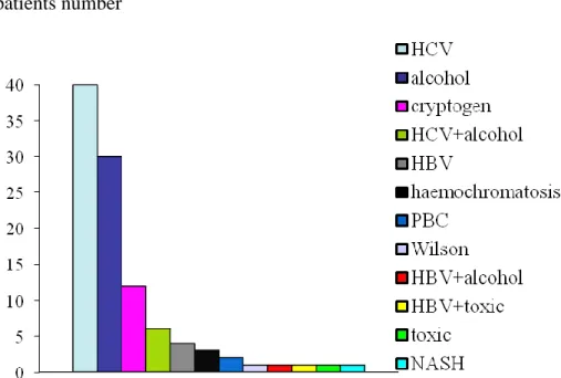
of the patients had liver cirrhosis. In our study group the most frequent etiological factor was the HCV infection, 40 from 102 (39%). The second one was the alcohol (30/102;
29%), after that the cryptogenic etiology (12/102; 11.8%).4 patients had chronic hepatitis B infection (4%), 3 hemochromatosis (3%), 2 primery biliar cirrhosis (2%), and 1-1 patients Wilson disease and non-alcoholic steatohepatitis. There were two toxic anamnestic information. One part of the patients (8/102; 39%) group were two etiologic factor at the same time (alcohol+HCV, alcohol+HBV, HCV+toxic) (Figure 1). Among males the most important etiologic factor is the alcohol consumption. After this the HCV infection, the cryptogenic origin and the HCV chronic hepatitis and the alcohol together were the most frequent factors. Data of AFP was available in 76 patients. Only 45 patients (59%) had abnormal AFP levels in the serum. The mean value was 5635.5 ng/ml, the highest 104800 ng/ml.
8 patients number
Figure 1. Distribution of patients with HCC by the etiology (n=102)
4.2. Study on the effect of 1α,25(OH)2D3 in different HCC cell lines in vitro 4.2.1. VDR mRNA expression in four tipes of HCC cell lines
We detected VDR mRNA expression all of the cell lines, however in very low level.
There was no change in VDR mRNA expressions in any of the examined cell lines after 5 hours of 1,25(OH)2D3 incubation neither at 1 nM nor at 10 nM concentration.
4.2.2. CYP27B1 mRNA expression in four tipes of HCC cell lines
We found CYP27B1 mRNA expression in all of the HCC cell lines. There was no change in CYP27B1 mRNA expressions in any of the examined cell lines after 5 hours of 1,25(OH)2D3 incubation neither at 1 nM nor at 10 nM.
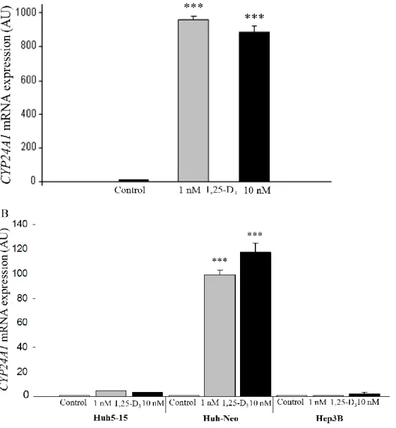
4.2.3. CYP24A1 mRNA expression in four tipes of HCC cell lines
Basic CYP24A1 mRNA expression was very low in all of cell lines. Exposure to 1,25- (OH)2D3 both at doses of 1 nM and 10 nM for 5 hours resulted in a strongly significant (p<0.0001) expression of CYP24A1 mRNA in HepG2 and Huh-Neo cell lines, but had no effect on Huh5-15 and Hep3B cells at the investigated time point (5 h). In HepG2
9
cells, the increase of CYP24A1 mRNA expression was more pronounced, close to 1000-fold compared to Huh-Neo cells, which exhibited a 100-fold increase after 5 hours of incubation (Figure 2). In HepG2 cells, the larger dose of vitamin D did not result in further increase of mRNA expression compared to the effect of 1 nM. In Huh-Neo cells, the magnitude of the mRNA elevation was smaller (p<0.001), and in this set, 10 nM of vitamin D neither did result in further significantly increase of mRNA expression.
Figure 2. CYP24A1 mRNA expression changes in response to 1,25(OH)2D3. HepG2 (A) and Huh-Neo (B) cell lines were incubated with 1 nmol/l and 10 nmol/l active vitamin D for a duration of 5 hours. Cells without 1,25(OH)2D3 incubation were used as controls. ***p<0.0001, AU: Arbitrary units.
10
4.2.4. Change of the mRNA expression of CYP24A1 in response of the concentration of vitamin D on HepG2 and Huh-Neo cell lines
We found a dose-dependent increase in CYP24A1 expression in HepG2 and Huh-Neo cell lines in response to 1,25(OH)2D3 administration for 4 hours. In concordance with the first set of experiments, the response of HepG2 cells was larger than that of Huh- Neo cells at each dose. In Hep G2 cells, 1.6 nM 1,25(OH)2D3 resulted in significant elevation of mRNA (p<0.001), while in Huh-Neo cells, the elevation reached significance only at a dose of 4.0 nM. The difference between the two cell types in CYP24A1 mRNA expression was one order of magnitude using 1.6, 4.0 and 10.0 nM 1,25(OH)2D3. In HepG2 cells, 180-, 820-, and 1010-fold elevations were detected versus 2.0-, 38-, and 140-fold increases in Huh-Neo cells at 1.6, 4.0, and 10 nM, respectively (Figure 3).
11
Figure 3. CYP24A1 mRNA dose response curves of HepG2 (A) and Huh-Neo (B) cell lines in response to 1,25(OH)2D3. The cells were treated with increasing concentrations of 1,25(OH)2D3 from 0.256 nmol/l up to 10 nmol/l for 4 hours. Cells without 1,25(OH)2D3 incubation were used as controls. ***p<0.0001, AU: Arbitrary units.
4.2.5. Change of the mRNA expression of CYP24A1 in response of 4nM concentration of vitamin D on HepG2 and Huh-Neo cell lines subject to the treatment time
Not only was the magnitude of CYP24A1 mRNA expression found to be different in HepG2 and Huh-Neo cells but so was the kinetic of expression. In HepG2 cells, the
12
CYP24A1 mRNA expression exhibited a 5300-fold elevation, reaching its maximum value at 8 hours (Fig. 4A). In Huh-Neo cells, the increase was 152-fold that of the baseline and the maximum was reached at 14 hours (Fig. 4B).
Figure 4. CYP24A1 mRNA time course curves of HepG2 (A) and Huh-Neo (B) cell lines in response to 1,25-dihydroxyvitamin [1,25(OH)2D3]. *p<0.0001, AU: Arbitrary units.
4.2.6. Immunochemical detection of CYP24A1 enzyme expression in HepG2 cell line Immunocytochemistry showed that gene activation with 1,25(OH2)D3 was followed by
13
CYP24A1 protein synthesis, indicating effective translation. Strong CYP24A1 enzyme staining was shown in HepG2 cells after 32 hours of incubation with 4 nM 1,25(OH)2D3, whereas no staining was seen in the untreated control cells.
4.3. Human study
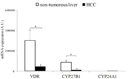
4.3.1. Decreased VDR mRNA expression in HCC versus surrounding non- tumorous liver
VDR mRNA expression was examinated in paraffin-embedded tissue samples (n=36).
VDR mRNA expression was detected in all non-tumorous liver tissues, and also in hepatocellular carcinoma, although at different amounts. Interestingly, the expression level was decreased in HCC, compared to the surrounding liver (p<0.05, Figure 5.).
There was no difference in the expression levels between in HCC based on cirrhotic liver and non-cirrhotic liver (p=0.7), however we encountered significant higher expression of the receptor in HCC with etiology of hepatitis B (HBV) infection compared with etiology of HCV infection (p=0.04) and cryprogenic (p=0.04) (Figure 6.). Furthermore, VDR mRNA expression was significantly decreased in HCC liver tissue compared with the surrounding liver tissue with cryptogenic etiology (p=0.04).
Meanwhile, there was no difference studied the expression in other etiology.
Interestingly we found marked difference between the two genders. Not only the non- tumorous liver samples (p=0.05) but also the HCC samples (p=0.02) derived from women patients showed a significantly lower VDR mRNA expression level, than samples from men independently of the etiology.
4.3.2. Decreased CYP27B1 mRNA expression in HCC versus surrounding non- tumorous liver
CYP27B1 mRNA expression was also exanimate in paraffin-embedded tissue samples (n=36). Similarly to VDR mRNA, CYP27B1 mRNA was also significantly decreased in HCC comparing to the non-tumorous surrounding liver (p<0.05, Figure 5.). There was no difference the CYP27B1 mRNA expression between the HCC based on cirrhotic liver and on non-cirrhotic liver (p=0.9). There was no difference in case of other etiology factor, neither in expression levels of CYP27B1 mRNA. There was no difference in the VDR and CYP27B1 expression levels between the different HCC liver tissues with different grade score. Moreover, non-tumorous liver samples derived from
14
women express lower CYP27B1 mRNA compared with men (p=0.03), but at the same time, we did not found difference between the two genders comparing HCC tissue samples (p=0.8).
Figure 5. Comparison of VDR, CYP27B1, and CYP24A1 mRNA expression profile in hepatocellular carcinoma (HCC) with surrounding non-tumorous liver tissue samples.
*p<0.05, AU: Arbitrary units.
Figure 6. Comparison of VDR, CYP27B1, and CYP24A1 mRNA expression profile in hepatocellular carcinoma (HCC) according to the etiology of HCC. * p<0.05, AU:
Arbitrary units.
15
4.3.3. Relations between CYP27B1 and VDR mRNA expression and the clinicopatological parameters
We have found positive correlation between HCC VDR mRNA expression level and the serum AFP and calcium levels of the patients. In men the CYP27B1 mRNA expression level in the non-tumorous liver tissue was in correlation with the existence of diabetes mellitus. Furthermore, there is a positive correlation between CYP27B1 mRNA level in HCC and the serum bilirubin and cholinesterase levels of the patients. There was no relation between the expression of CYP27B1 and VDR mRNA level and the mean survival time of the patients.
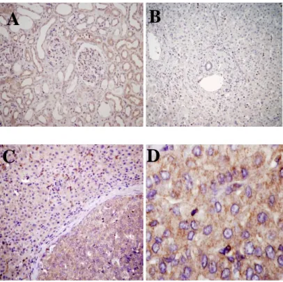
4.3.4. CYP24A1 mRNA and protein expression by immunhistochemistry in HCC
Neither of the non-tumorous liver tissue expressed the mRNA of the inactivating enzyme, but in contrary the majority of HCC tissue samples (8 out of 13) expressed the CYP24A1 enzyme, however likely to the other three types of mRNA we did not found difference between cirrhotic and non cirrhotic HCC tissue (p=0.3). Immunhistochemistry showed that gene activation with 1,25(OH2)D3 was followed by CYP24A1 protein synthesis in HCC tissue samples, indicating effective translation. Strong CYP24A1 enzyme staining was shown in HCC (+++), whereas no staining was seen in non-tumorous liver tissues (Figure 7.). Expression of CYP24A1 was confined to the cytoplasm; the punctuated pattern of the staining is consistent with mitochondrial localization of the enzyme.
16
Figure 7. Immunochemical detection of CYP24A1 enzyme expression in HCC and surrounding liver tissues. Brown color indicates a positive reaction. Nuclei are counterstained with hematoxylin (blue) A. Normal kidney (×200 magnification). B:
Cirrhotic liver tissue without HCC with lack of CYP24 enzyme expression (×200). C:
Margin of cirrhotic liver and HCC with high CYP24A1 protein expression in cancer compared with non-tumorous liver tissue (×200). D: Hepatocellular carcinoma tissue with high CYP24A1 protein expression (×600).
5. CONCLUSIONS
The conclusions of my PhD work are the following:
1. The hepatocellular carcinoma has a poor prognosis. It is aim to screen the patients with risk factors, with liver cirrhosis and the early diagnosis. In Hungary the major risk factors are the HCV infection and the alcohol, and also the two factors together.
According to our data, the diagnosis of HCC is mostly in advanced stage. We do not recommend the AFP alone for the screening of the HCC because of its low sensibility.
2. CYP24A1 mRNA and protein expression significantly increased in response to 1,25(OH)2D3 administration in HepG2 and Huh-Neo cell lines, however there was no significant change in
A
C D
B
17
CYP27B1 and VDR mRNA expression levels in these cell lines. There was no significant change of CYP24A1 mRNA expression in Hep3B and Huh5-15 cell lines. The degree of change of CYP24A1 mRNA level depends of the origin of the HCC cell lines, of the doses and the duration time of vitamin D treatment
3. Expression of VDR and CYP27B1 was significantly lower in HCC compared with non-tumorous liver.
4. The majority of the HCC samples expressed the vitamin D inactivating CYP24A1 mRNA and protein; in contrast neither of the non-tumorous liver expressed this gene.
These novel data indicate decreased bioavailability of 1,25(OH)2D3 in HCC cells, providing an escape mechanism from the anti-tumor effect of 1,25(OH)2D3.
5. Patients with HBV infection expressed the higher VDR mRNA level compared with other etiology. VDR mRNA expression was lower in females HCC and also in non- tumorous liver tissues compared with males. However this difference in case of CYP27B1 mRNA was only detected in non-tumorous liver tissues.
6. There was no difference in VDR, CYP27B1, and CYP24A1 mRNA expression levels between the HCC based on cirrhotic liver and on non-cirrhotic liver, similarly between these mRNA expressions and the grade, TNM stage, and the survival of the patients.
18 6. LIST OF PUBLICATIONS
In connection with theme
1. Horváth E, Lakatos P, Balla B, Kósa JP, Tóbiás B, Jozilan H, Borka K, Horváth HC, Kovalszky I, Szalay F. (2012) Marked increase of CYP24A1 mRNA level in hepatocellular carcinoma cell lines following vitamin D administration. Anticancer Res.
32(11): 4791-4796. IF (2012): 1,713
2. Horváth E, Balla B, Kósa JP, Lakatos PA, Lazáry Á, Németh D, Jozilan H, Somorácz Á, Korompay A, Gyöngyösi B, Borka K, Kiss A, Kupcsulik P, Schaff Zs, Szalay F. (2016) Vitamin D metabolism and signaling in human hepatocellular carcinoma and surrounding non-tumorous liver. Orv. Hetil. 157(48): 1910-1918.
IF (2015): 0,291
Other publications
1. Osztovits J, Horváth E, Tax J, Csihi L, Horvath T, Littvay L, Toth T, Abonyi M, Lakatos PL, Kollai M, Feher J, Szalay F, Blum HE. (2011) Reversible autonomic dysfunction during antiviral treatment in patients with chronic hepatitis C virus
infection: Anti-HCV therapy and autonomic function. Hepat Mon. 11(2): 114-118.
IF (2011): 2,19
2. Bata P, Tarnoki AD, Tarnoki DL, Horváth E, Berczi V, Szalay F. (2012) Acute severe thrombocytopenia following non-ionic low-osmolarity intravenous contrast medium injection. Korean J Radiol. 13(4): 505-509. IF (2012): 1,555
3. Kósa JP, Horváth P, Wölfling J, Kovács D, Balla B, Mátyus P, Horváth E, Speer G, Takács I, Nagy Z, Horváth H, Lakatos P. (2013) CYP24A1 inhibition facilitates the anti-tumor effect of vitamin D3 on colorectal cancer cells. World J Gastroenterol.
19(17): 2621-2628. IF (2013): 2,433
19
4. Tobiás B, Halászlaki Cs, Balla B, Kósa JP, Árvai K, Horváth P, Takács I, Nagy Z, Horváth E, Horányi J, Járay B, Székely E, Székely T, Győri G, Putz Z, Dank M, Valkusz Z, Vasas B, Iványi B, Lakatos P. (2016) Genetic Alterations in Hungarian Patients with Papillary Thyroid Cancer. Pathol Oncol Res. 22(1):27-33.
IF (2015): 1,940

![Figure 4. CYP24A1 mRNA time course curves of HepG2 (A) and Huh-Neo (B) cell lines in response to 1,25-dihydroxyvitamin [1,25(OH) 2 D 3 ]](https://thumb-eu.123doks.com/thumbv2/9dokorg/1342444.108987/12.892.136.645.258.887/figure-cyp-course-curves-hepg-lines-response-dihydroxyvitamin.webp)