Summary. The aim of the present study was to characterise the expression pattern of claudin-7 tight junction protein in canine normal liver, hyperplastic and primary neoplastic lesions of the canine liver and whether this tight junction protein can help differentiate canine cholangiocarcinomas from canine hepatocellular carcinomas. Methods and results: Necropsy samples included 15 canine normal liver tissue samples, 10 hepatocellular nodular hyperplasias, 6 hepatocellular adenomas, 15 well-differentiated and 6 poorly differentiated hepatocellular carcinomas, 6 cholangiocellular hyperplasias, 10 cholangiocellular adenomas, 15 well-differentiated and 6 poorly differentiated cholangiocarcinomas, 6 normal extrahepatic bile ducts, 8 normal gall bladder tissue samples, and 5 cystic mucinous hyperplasias of the gall bladder. In all canine normal liver tissue samples the hepatocytes were negative for claudin-7 and the normal biliary epithelial cells showed intense basolateral membrane claudin-7 positivity. In all cholangiocellular hyperplasia samples and in all cholangiocellular adenoma samples the benign cholangiocytes showed intense basolateral membrane positivity for claudin-7. In all samples of the well-differentiated and poorly differentiated cholangiocarcinomas, the malignant neoplastic biliary epithelial cells showed intense basolateral membrane positivity for claudin-7. Neither the hyperplastic nodules of the liver cells nor the hepatocellular adenomas reacted with claudin-7. The well-differentiated and poorly differentiated hepatocellular cancers were negative for claudin-7. The
epithelial cells of canine normal extrahepatic bile ducts, gall bladder and cystic mucinous hyperplasias of the gall bladder showed intense basolateral membrane positivity for claudin-7. Differences in the intensity of claudin-7 reaction were not apparent among different types of proliferative lesions of cholangiocytes or degrees of cellular differentiation of neoplastic biliary epithelial cells.
Conclusion: Consequently, we hypothesize that claudin-7 is an excellent immunohistochemical marker of the cholangiocellular differentiation in canines and can be used to detect benign and malignant proliferative lesions of the canine biliary tract. It can also help to differentiate canine cholangiocarcinomas from hepatocellular carcinomas.
Key words: Canine cholangiocarcinoma, Canine hepatocellular carcinoma, Immunohistochemistry, Claudin-7
Introduction
The most common primary malignancies of the canine liver are canine hepatocellular carcinomas (cHCCs) and canine cholangiocarcinomas (cCCs). The most common question which arises in connection with the differential diagnosis of the canine liver is whether it is cHCC or cCC (Patnaik et al., 1981; Trigo et al., 1982).
There are several immunohistochemical markers available which help to produce a differential diagnosis in this oncopathological question (De las Mulas et al., 1995; Ramos-Vara et al., 2001).
Claudins are a relatively large family of 17-27 kDa integral membrane tight junction tetraspanin proteins that are classified on the basis of the size of the
Claudin-7 protein differentiates canine
cholangiocarcinoma from hepatocellular carcinoma
Cs. Jakab1, A. Kiss2, Zs. Schaff2, Z. Szabó4, M. Rusvai1, P. Gálfi3, A. Szabára1, Á. Sterczer4and J. Kulka2
1Szent István University, Faculty of Veterinary Science, Department of Pathology and Forensic Veterinary Medicine, Budapest, Hungary, 22nd Department of Pathology, Semmelweis University, Budapest, Hungary, 3Szent István University, Faculty of Veterinary Science, Department of Pharmacology and Toxicology, Budapest, Hungary and 4Szent István University, Faculty of Veterinary Science, Department and Clinic of Internal Medicine, Budapest, Hungary
Offprint requests to: Csaba Jakab, Szent István University, Faculty of Veterinary Medicine, Department of Pathology and Forensic Veterinary Medicine, 1078 István utca 2., Budapest, Hungary. e-mail:
jakab.csaba@aotk.szie.hu
molecules which pass through the paracellular spaces between epithelial and endothelial cells. Tight junctions are the most apical cell-cell contacts and are important for barrier function in epithelial and endothelial cells (Tsukita et al., 2001; Tsukita and Furuse, 2002). The barrier function of the tight junctions and claudins regulates the passage of ions, water, and growth factors through paracellular space. The tight junctions and claudins between epithelial cells form a polarized barrier between luminal and serosal compartments and segregate luminal growth factor from their basal-lateral receptors. This property may promote cancer formation in preneoplastic, premalignant tissues in which the tight junctions and claudins have become chronically leaky to growth factors (Mullin, 1997). Claudin-5 molecule can be used as a differential marker, in the differential diagnosis of canine hemangiosarcomas from other sarcomas (Jakab et al., 2009).
This study has investigated whether claudin-7 antibody, which is commonly used in diagnostic human pathology (Li et al., 2004; Johnson et al., 2005; Hornsby et al., 2007), can be demonstrated by immunohisto- chemical methods in routinely processed tissue samples of canine normal liver, hyperplastic and neoplastic lesions of the canine liver and whether this tight junction protein can help differentiate cCCs from cHCCs. To our knowledge, this is the first study that has examined claudin-7 molecule expression in canine normal liver tissue and different proliferative lesions of canine hepatocytes and cholangiocytes.
Materials and methods Samples of canine tumours
Tissue samples were collected at Szent István University, Faculty of Veterinary Science, Department of Pathology and Forensic Veterinary Medicine (Budapest, HU) between 2004 and 2009. The necropsy samples (n=108) included 15 canine normal liver tissue samples, 10 hepatocellular nodular hyperplasias, 6 hepatocellular adenomas, 15 well-differentiated cHCCs, 6 poorly differentiated cHCCs and 6 cholangiocellular hyperplasias (from cirrhotic liver), 10 cholangiocellular adenomas, 15 well-differentiated cCCs, 6 poorly differentiated cCCs, 6 normal extrahepatic bile ducts, 8 normal gall bladder tissue samples, and 5 cystic mucinous hyperpalsias of the gall bladder. All samples were collected during the necropsy of canines. In none of the cases did we find malignant primary tumours in the pancreas, stomach, small intestine, colorectum, lungs, adrenal glands, mammary glands or kidney.
Samples were fixed in 8% neutral buffered formalin for 24 hours at room temperature, dehydrated in a series of ethanol and xylene, and embedded in paraffin. The 3- 4µm thick sections were routinely stained with hematoxylin and eosin (HE). Each neoplastic case was classified by the pathologists (CSJ and JK).
Immunohistochemistry
Serial sections (3-4 µm) were initially dewaxed in xylene and graded ethanol. After treatment with appropriate antigen retrieval (Target Retrieval Soluton, DAKO, Glostrup, Denmark, pH 6; microwave - 800W - oven for 30 min), the sections were incubated with primary antibody claudin-7 (rabbit polyclonal and diluted 1 in 80, Zymed, Catalog number: 43-9100), for 60 min at room temperature. All primary antibodies were produced by Zymed Inc., San Francisco, CA, USA.
Immunohistochemical labelling was performed using the streptavidin-peroxidase procedure. Antigen-bound primary antibody was detected using standard avidin- biotin immunoperoxidase complex (DAKO, LSAB2 Kit). The chromogen substrate was 3, 3’-diamino- benzidine tetrahydrochloride (DAB substrate- chromogen, DAKO). Sections were counterstained with Mayer’s hematoxylin.
Negative controls were performed by omission of the primary antibody and external positive controls were canine normal colorectum for claudin-7 (Jakab et al., 2010). The internal positive controls were the peritumoural normal biliary epithelial cells (cholangiocytes) for claudin-7, and internal negative controls were the peritumoural normal hepatic cells and portal fibroblasts.
Immunohistochemical assessment
A semiquantitative evaluation of the percentage of positive tumour cells was used. In each case, two independent observers (CSJ and JK) recorded the distribution and intensity of labelling. The immunoreactivity was assessed as follows: negative (-), no immunostaining present; (+), <25% of cells positive;
(++), 25-50% of cells positive; (+++), 50-75% of cells positive; (++++), 75-100% of cells positive. The intensity of reaction for claudin-7 was graded on a scale of 3, where 0 stood for no reaction, 1 for weak reaction, 2 for moderate reaction, and 3 for intense reaction.
Results
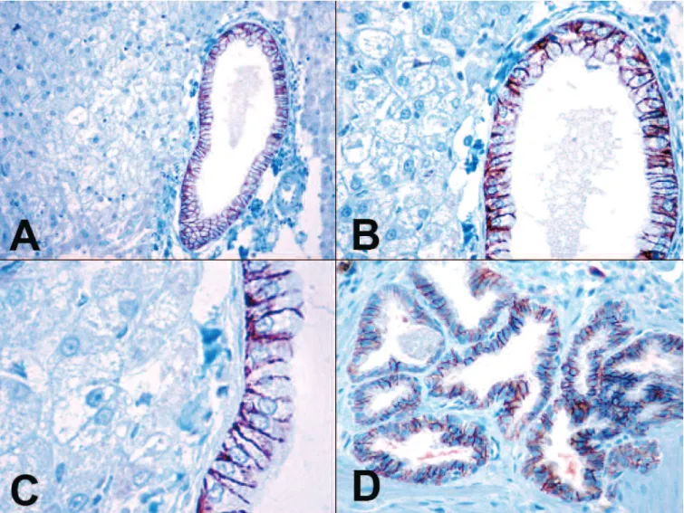
In all canine normal liver tissue samples the hepatocytes, the endothelial cells of the sinuses and portal vessels, the portal fibroblasts, and the Kupffer- cells were negative for claudin-7. In these samples the normal biliary epithelial cells of the bile ducts with variable diameter showed ++++ diffuse, basolateral membrane claudin-7 positivity (15/15; 100%). The reactions were intense (grade 3) (Fig. 1A-C). In all cholangiocellular hyperplasia samples (6/6; 100%) and in all cholangiocellular adenoma samples (10/10; 100%) the benign cholangiocytes showed ++++ diffuse, basolateral membrane positivity for claudin-7 (Fig. 1D).
The reactions were grade 3 in all benign proliferative lesions of the cholangiocytes. In all samples of the well-
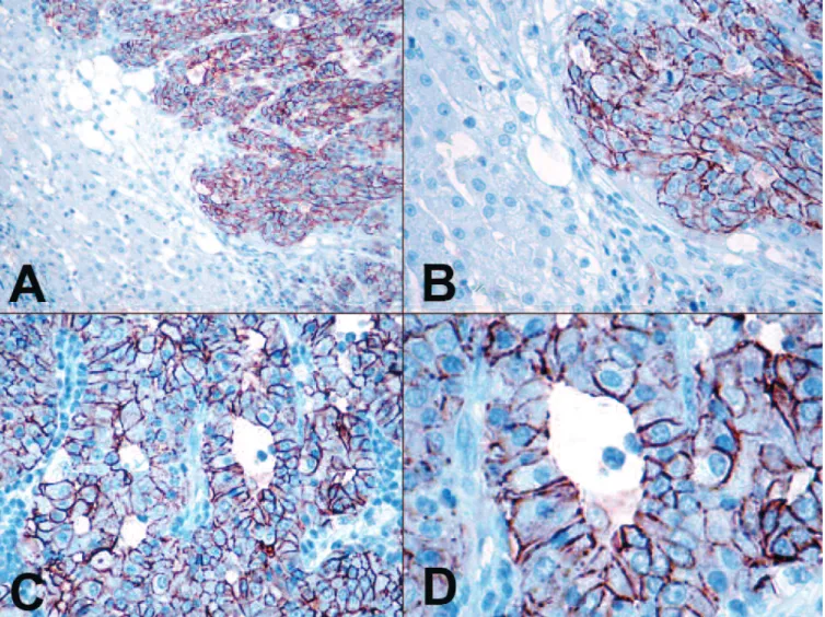
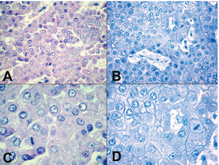
differentiated and poorly differentiated cCCs, the malignant neoplastic biliary epithelial cells showed diffuse, basolateral membrane positivity for claudin-7 (21/21; 100%) (Fig. 2A-D). The reactions were intense in all cases of cCC. Neither the hyperplastic nodules of the liver cells nor any of the hepatocellular adenomas reacted with claudin-7. The well-differentiated and poorly differentiated cHCCs were negative for claudin-7 in all samples (Fig. 3A-D). In canine normal extrahepatic bile ducts the surface epithelial cells and the epithelial cells of the propria glands showed ++++
diffuse, basolateral membrane positivity for claudin-7.
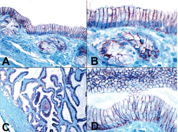
We investigated the claudin-7 immunohistochemical pattern in canine normal gall bladder and in cystic mucinous hyperplasias of the gall bladder. In all tissue samples of the intact gall bladder and its hyperplastic lesions the epithelial cells showed diffuse, intense
basolateral membrane positivity for claudin-7 (Fig. 4A- D). Differences in the intensity of claudin-7 reaction were not apparent among different types of proliferative lesions of cholangiocytes or degrees of cellular differentiation of neoplastic biliary epithelial cells. In the case of hyperplastic and proliferative lesions of the biliary tract the adjacent liver cells were the internal negative controls.
Discussion
The differentiation of canine hepatocellular carcinoma (cHCC) from canine cholangiocarcinoma (cCC) can be difficult.
In veterinary oncopathology, immunohistochemical techniques have been used extensively on liver tumours.
Fig. 1. A-C. The biliary epithelial cells of the canine normal intrahepatic bile ducts showed intense basolateral membrane positivity for claudin-7. On the left hand side of the pictures normal, claudin-7 negative liver cells can be seen. D. The canine cholangiocellular adenomas showed intense membrane claudin-7 positivity. IHC. A, x 100; B, D, x 200; C, x 400.
Frequently used antibodies include hepatocyte paraffin 1 (Hep Par 1) (Ramos-Vara et al., 2001), cytokeratins (CK 7) alpha-fetoprotein (AFP), and carcinoembryonic antigen (CEA) (De las Mulas et al., 1995; Ramos-Vara et al., 2001).
In our present study we investigated the expression pattern of claudin-7 in canine normal liver tissues and primary liver tumours. We detected intense, basolateral membrane claudin-7 expression in the intact biliary epithelial cells of the canine normal liver tissue samples (100%), and in the ductal proliferations from canine cirrhotic liver (100%), and in benign and malignant tumours of the canine cholangiocytes (100%; 100%).
None of the normal hepatocytes and the hyperplastic and neoplastic lesions of the liver cells were recognized by this claudin antibody.
In veterinary oncological study the canine normal
liver cells showed consistently cytoplasmic, disseminated, and granular positivity for Hep Par 1. The eptithelial cells of the bile duct and the non- hepatocellular tissues did not react with this antibody (Ramos-Vara et al., 2001). In our present study the canine normal hepatocytes, the endothelial cells of the sinusoids and portal vessels, the portal fibroblasts, and the Kupffer-cells were negative for claudin-7, but the normal biliary epithelial cells showed intense basolateral claudin-7 membrane positivity. It seems that Hep Par 1 and claudin-7 can help the correct identification of the intact liver cells and biliary epithelial cells, and furthermore they can differentiate between these cells.
Hep Par 1 showed granular and cytoplasmic immunoreactivity in the canine hepatocellular nodular hyperplasias and hepatocellular adenomas, but the cholangicellular adenomas were negative for this
Fig. 2. A,B. Canine cholangiocarcinomas showed intense membrane positivity for claudin-7. On the left hand side of the pictures normal, claudin-7 negative liver cells can be seen. C,D. Claudin-7 expression pattern in the canine poorly differentiated cholangiocarcinoma. IHC. A, x 100; B, C, x 200;
D, x 400.
antibody (Ramos-Vara et al., 2001). In our study the tumor-like lesions of the canine liver cells and the hepatocellular adenomas were negative for claudin-7, but the cholangiocellular hyperplasias (from cirrhotic liver), and cholangiocellular adenomas showed intense membrane claudin-7 positivity. It means that the correct identification of the non-neoplastic proliferations and benign tumoural lesions of the canine liver cells and biliary epithelial cells may be possible with the use of these two antibodies. Earlier studies presented that 92.5% of the cHCCs showed positivity for Hep Par 1, and this antibody did not react with neoplastic cells of the cCCs (Ramos-Vara et al., 2001). In our study we detected that the cHCCs did not react with claudin-7, but all cCCs showed an intense positivity for this antibody.
Our conclusion was that claudin-7 (as a new marker of the biliary epithelium) with Hep Par 1 should be components of the immunohistochemical panel to distinguish cHCC from cCC.
Previous studies described that the canine normal biliary epithelial cells and the benign tumour cells of the cholangiocellular adenomas showed cytoplasmic CK 7 immunoreactivity. Some normal bile ducts were strongly CK 7 positive and others were negative. There was no correlation between the diameter of the bile duct and detection of CK 7 (Ramos-Vara et al., 2001). In our present study the canine normal biliary epithelial cells showed homogenous, strong claudin-7 membrane positivity in all bile ducts with variable diameter. It seems that claudin-7 protein is a better marker than CK 7 for the detection of canine normal biliary epithelial cells.
In our study in all cholangiocellular hyperplasia samples, and in all cholangiocellular adenoma samples, the benign cholangiocytes showed homogenous, membrane positivity for claudin-7, like CK 7. Ramos-Vara et al.
(2001) described that 77% of the cCCs (14/18) were positive for CK 7. 72% of the cCCs (13/18) showed intense (grade 3) CK 7 immunoreactivity (Ramos-Vara
Fig. 3. Canine hepatocellular carcinomas were negative for claudin-7 A.HE. B.IHC. C.HE. D.IHC. A, B, x 200; C, D, x 400.
et al., 2001). Contrarily, in our study we detected that in all samples of the well-differentiated and poorly differentiated cCCs, the neoplastic biliary epithelial cells showed homogenous, intense (grade 3) basolateral, membrane positivity for claudin-7 (21/21; 100%) and 75-100% of tumour cells were claudin-7 positive. Our conclusion was that claudin-7 is a more sensitive immunohistochemical marker than CK 7 for cCCs. Thus claudin-7 with Hep Par 1 should make better components of the immunohistochemical panel than CK 7 with Hep Par 1, to distinguish cHCC from cCC.
Well-differentiated cCCs are readily distinguished from well-differentiated cHCCs from a histological point of view, but the differential diagnostic challenge is the case of poorly differentiated cHCCs versus cCCs. Poorly differentiated cHCCs consist of solid sheets of neoplastic hepatocytes with no apparent pattern. The hepatocellular characteristics are difficult to recognize in poorly
differentiated cHCCs and the differential diagnosis between undifferentiated cHCCs and undifferentiated cCCs is more difficult than between well-differentiated cHCCs and well-differentiated cCCs (Ponomarkov and Mackey, 1976). We think that claudin-7, as an immunohistochemical marker of cholangiocellular differentiation, is available to assist in this differential diagnosis. In this study we detected in all well-, and poorly differentiated cHCCs negativity for claudin-7 protein, and in all well-, and poorly-differentiated cCCs (100%) intense claudin-7 membrane immunoreactivity.
It means that this new immunohistochemical marker, claudin-7, can help to produce the correct hepatopathological diagnosis of canine hepatic neoplasms. There was no adenoid form of cHCCs in our samples. It is difficult to distinguish between adenoid cHCCs and well-differentiated cCCs by routine histological method, but we think that with claudin-7
Fig. 4. A,B. The normal epithelial cells of the canine gall bladder were positive for claudin-7. C,D.Strong diffuse claudin-7 expression in canine cystic mucinous hyperplasias of the gall bladder. IHC. A, x 200; B, D, x 400; C, x 40.
marker the detection of the cholangiocellular characteristic is easy. To our knowledge, this is the first study that has examined claudin-7 expression in canine normal liver and primary liver tumours. According to a veterinary study on rat liver, claudin-2 showed a lobular gradient increasing firm periportal to pericentral hepatocytes, claudin-3 was uniformly expressed, claudin-4 was absent, and claudin-5 was only expressed in endothelial junctions (Rahner et al., 2001).
A veterinary study described that only 1.6 % of the well differentiated cHCCs (1/6) and all poorly differentiated cHCCs (2/2) were positive for AFP, but all moderately differentiated cHCCs were negative for this marker. In this study all cCCs (2/2) were negative for AFP. The cHHCs were negative for CEA, but CEA positive neoplastic cells were present in all cCCs (2/2) and in the cholangiocellular part of the mixed carcinomas (3/3). CEA immunoreactivity was observed within the cytoplasm of tumour cells (Martín De Las Mulas et al., 1995). It seems to be that AFP and CEA immunohistochemical panel can help in the differentiation of cCCs from cHCCs, but we think that, a higher number of investigated samples (normal biliary epithelial cells, cholangiocellular adenomas, cCCs) are needed to compare the sensitivity and specifity of CEA and claudin-7 for cholangiocellular differentiation.
A human oncological study described that claudin-4 expression seems to be a useful marker in differentiating hCCs from hHCCs and could well become a potential diagnostic tool. In this study an intense membranous immunolabeling was found for claudin-4 in all hCCs unrelated to the primary site of origin, namely intrahepatic, extrahepatic or gall bladder cancers. Intact biliary epithelial cells showed weak positivity for claudin-4. In contrast, normal liver cells and tumour cells of hHCCs did not express claudin-4 (Lódi et al., 2006).
Another hepatopathological human study, Németh et al. 2009, used a similar claudin-7 antibody (rabbit polyclonal, Zymed, Catalog number: 43-9100) as we did and a similar dilution (1:80) to ours. In this human study the intensity of the immunoreactions was scored as none, weak, moderate, or strong (Németh et al., 2009). We
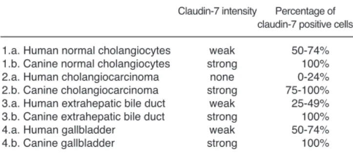
summarised their results in comparison to our findings in table (Table 1). It seems that claudin-7 protein is expressed more strongly in canine normal intrahepatic bile ducts than in human intact intrahepatic bile ducts.
Claudin-7 protein is expressed more strongly in cCCs than in intrahepatic hCCs, and is a more useful specific and sensitive marker in veterinary hepatopathology than in human hepatopathology for the detection of malignancy of the cholangiocytes. It seems that claudin- 7 protein is expressed more strongly in canine normal extrahepatic bile ducts and normal gallbladder than in human intact extrahepatic bile ducts and intact gallbladder. Further morphological studies are needed to investigate the claudin-7 expression pattern in canine extrahepatic bile duct cancers.
In our study we investigated claudin-7 expression in the canine cystic mucinous hyperplasias of the gall bladder. The histopathological hallmarks of this lesion are the hyperplasia of the epithelium with papillary projections and increased mucin production and the abundance of variably sized cystic spaces that distort and thicken the entire mucosa. The numerous 1 to 3 mm cysts are lined with a single layer of epithelial cells. The etiology of this lesion is unknown and may be a response to progestational compounds (Kovatch et al., 1965;
Mawdesley-Thomas and Noel, 1967). The hyperplastic epithelial cells in the cystic hyperplasia of the gallbladder showed intense membrane positivity and the percentage of claudin-7 positive hyperplastic cells was 100%. Further morphological studies are needed to investigate the claudin-7 expression pattern in extremely rare canine gall bladder cancers.
The results of human studies suggest that increased expression of claudin-1 and -2 characterizes the more differentiated foetal component in hepatoblastomas and is a reliable marker for differentiating foetal and embryonal types in hepatoblastomas (Halász et al., 2006). Further studies are needed to investigate claudin- 7 expression in the rare canine hepatoblastomas and other different primary non-hepatocellular and non- cholangiocellular cancers, such as neuroendocrine tumours (carcinoids). The canine liver is a common site of tumour metastasis from mammary gland cancers, adenocarcinoma of small intestine, exocrine part of pancreas, stomach, colorectum, pulmonary carcinomas, and also from cortical cancer of suprarenal gland or phaeochromocytoma of suprarenal gland, but rarely from renal cancers (Patnaik et al., 1981). Our aim in the future is to investigate the expression of claudin-7 in hepatic metastatic tumours, to find out whether this claudin can help in the distinction of cCCs and cHCs from metastatic neoplasms.
In summary, our results show that claudin-7 is an excellent immunohistochemical marker of the cholangiocellular differentiation in canines and can be used to detect benign and malignant proliferative lesions of the canine biliary epthelial cells. It could also be of diagnostic value in the differential diagnosis of cCCs from cHCCs.
Table 1.Comparison of the results of the human and veterinary studies on claudin-7 expression in normal and neoplastic biliary tract and gallbladder.
Claudin-7 intensity Percentage of claudin-7 positive cells 1.a. Human normal cholangiocytes weak 50-74%
1.b. Canine normal cholangiocytes strong 100%
2.a. Human cholangiocarcinoma none 0-24%
2.b. Canine cholangiocarcinoma strong 75-100%
3.a. Human extrahepatic bile duct weak 25-49%
3.b. Canine extrahepatic bile duct strong 100%
4.a. Human gallbladder weak 50-74%
4.b. Canine gallbladder strong 100%
References
De las Mulas J.M., Gomez-Villamandos J.C., Perez J. and Mozos E.
(1995). Immunohistochemical evaluation of canine primary liver carcinomas: distribution of alpha-fetoprotein, carcinoembryonic antigen, keratins and vimentin. Res. Vet. Sci. 59, 124-127.
Halász J., Holczbauer A., Páska C., Kovács M., Benyó G., Verebély T., Schaff Z. and Kiss A. (2006). Claudin-1 and claudin-2 differentiate fetal and embryonal components in human hepatoblastoma. Hum.
Pathol. 73, 555-561.
Hornsby C.D., Cohen C., Amin M.B., Picken M.M., Lawson D., Yin- Goen Q. and Young A.N. (2007). Claudin-7 immunohistochemistry in renal tumors: a candidate marker for chromophobe renal cell carcinoma identified by gene expression profiling. Arch. Pathol. Lab.
Med. 131, 1541-1547.
Jakab Cs., Halász J., Kiss A., Schaff Zs., Rusvai M., Gálfi P., Abonyi T.Zs. and Kulka J. (2009). Claudin-5 protein is a new differential marker for histopathological differential diagnosis of canine hemangiosarcoma. Histol. Histopathol. 24, 801-813.
Jakab Cs., Rusvai M., Gálfi P., Szabó Z., Szabára Á. and Kulka J.
(2010). Expression of claudin-1, -3, -4, -5 and -7 proteins in low grade colorectal carcinoma of canines. Histol. Histopathol. 25, 55- 62.
Johnson A.H., Frierson H.F., Zaika A., Powell S.M., Roche J., Crowe S., Moskaluk C.A. and El-Rifai W. (2005). Expression of tight-junction protein claudin-7 is an early event in gastric tumorigenesis. Am. J.
Pathol. 167, 577-584.
Kovatch R.M., Hildebrandt P.K. and Marcus L.C. (1965). Cystic mucinous hypertrophy of the mucosa of the gallbladder in the dog.
Vet. Pathol. 2, 574-584.
Li W.Y., Huey C.L. and Yu A.S. (2004). Expression of claudin-7 and -8 along the mouse nephron. Am. J. Physiol. Renal. Physiol. 286, 1063-1071.
Lódi Cs., Szabó E., Holczbauer Á., Batmunkh E., Szíjártó A., Kupcsulik P., Kovalszky I., Paku S., Illyés Gy., Kiss A. and Schaff Zs. (2006).
Claudin-4 differentiates biliary tract cancers from hepatocellular
carcinomasMod. Pathol. 19, 460-469.
Martín De Las Mulas J., Gómez-Villamandos J.C., Pérez J., Mozos E., Estrada M. and Méndez A. (1995). Immunohistochemical evaluation of canine primary liver carcinomas: distribution of alpha-fetoprotein, carcinoembryonic antigen, keratins and vimentin. Res. Vet. Scien., 59, 124-127.
Mawdesley-Thomas L.E. and Noel P.R.B. (1967). Cystic hyperplasia of the gall bladder in the beagle associated with administration of progestational compounds. Vet. Rec. 80, 658-659.
Mullin J.M. (1997). Potential interplay between luminal growth factors and increased tight junction permeability in epithelial carcinogenesis.
J. Experim. Zool. 279, 484-489.
Németh Zs., Szász M.A., Tátrai P., Németh J., Győrffy H., Somorácz Á., Szíjártó A., Kiss A. and Schaff Zs. (2009). Claudin-1, -2, -3, -4, -7, -8, and -10 protein expression in biliary tract cancers. J.
Histochem. Cytochem. 57, 113-121.
Patnaik A.K., Hurvitz A.I., Lieberman P.H. and Johnson G.F. (1981).
Canine hepatocellular carcinoma. Vet. Pathol. 18, 427-438.
Ponomarkov V. and Mackey L.J. (1976). Tumors of the liver and biliary system. Bull WHO 53, 187-194.
Rahner C., Mitic L.L. and Anderson J.M. (2001). Heterogeneity in expression and subcellular localization of claudins 2, 3, 4, and 5 in the rat liver, pancreas, and gut. Gastroenterology 120, 411-422.
Ramos-Vara J.A., Miller M.A. and Johnson G.C. (2001). Immuno- histochemical characterization of canine hyperplastic hepatic lesions and hepatocellular and biliary neoplasms with monoclonal antibody hepatocyte paraffin 1 and a monoclonal antibody to cytokeratin 7.
Vet. Pathol. 38, 636-643.
Trigo F.J., Thompson H., Breeze R. and Nash A.S. (1982). The pathology of liver tumours in the dog. J. Comp. Pathol. 92, 21-37.
Tsukita S. and Furuse M. (2002). Claudin-based barrier in simple and stratified cellular sheets. Curr. Opin. Cell. Biol. 14, 531-536.
Tsukita S., Furuse M. and Itoh M. (2001). Multifunctional strands in tight junctions. Nat Reviews Mol. Cell Biol. 2, 285-293.
Accepted January 20, 2010