DOI:10.3748/wjg.v20.i27.9072 © 2014 Baishideng Publishing Group Inc. All rights reserved.
Non-alcoholic fatty liver disease and type 2 diabetes mellitus: The liver disease of our age?
Gábor Firneisz
Gábor Firneisz, 2nd Department of Internal Medicine, Semmel- weis University, H-1088 Budapest, Hungary
Author contributions: Firneisz G contributed solely to this manuscript.
Correspondence to: Gábor Firneisz, MD, PhD, 2nd Depart- ment of Internal Medicine, Semmelweis University, Szentkiralyi St 46, H-1088 Budapest,
Hungary. firneisz.gabor@med.semmelweis-univ.hu Telephone: +36-1-2100278-5520 Fax: +36-1-2661007 Received: November 14, 2013 Revised: March 20, 2014 Accepted: May 12, 2014
Published online: July 21, 2014
Abstract
Non-alcoholic fatty liver disease (NAFLD) is a chronic liver disease that might affect up to one-third of the adult population in industrialised countries. NAFLD incorporates histologically and clinically different non- alcoholic entities; fatty liver (NAFL, steatosis hepatis) and steatohepatitis (NASH-characterised by hepatocyte ballooning and lobular inflammation ± fibrosis) might progress to cirrhosis and rarely to hepatocellular cancer.
NAFL increasingly affects children (paediatric preva- lence is 4.2%-9.6%). Type 2 diabetes mellitus (T2DM), insulin resistance (IR), obesity, metabolic syndrome and NAFLD are particularly closely related. Increased hepatic lipid storage is an early abnormality in insulin resistant women with a history of gestational diabetes mellitus. The accumulation of triacylglycerols in hepa- tocytes is predominantly derived from the plasma non- esterified fatty acid pool supplied largely by the adipose tissue. A few NAFLD susceptibility gene variants are associated with progressive liver disease, IR, T2DM and a higher risk for hepatocellular carcinoma. Although not approved, pharmacological approaches might be con- sidered in NASH patients.
© 2014 Baishideng Publishing Group Inc. All rights reserved.
WJG 20th Anniversary Special Issues (12): Nonalcoholic fatty liver disease
TOPIC HIGHLIGHT
Key words: Non-alcoholic fatty liver disease; Non- alcoholic steatohepatitis; Liver cirrhosis; Hepatocellular cancer; Dysfunctional adipose tissue; Type 2 diabetes mellitus; Insulin resistance; Obesity; Genetics; Therapy Core tip: In this review article, non-alcoholic fatty liver disease (NAFLD) spectrum disease is discussed in detail. The epidemiology of NAFLD/nonalcoholic ste- atohepatitis and the relationship of NAFLD to different forms of diabetes mellitus including type 2 diabetes mellitus and gestational diabetes mellitus are reviewed.
Attention is paid to the main biochemical events, to dysfunctional adipose tissue and visceral adiposity asso- ciated with NAFLD and insulin resistance, to mitochon- drial dysfunction and to the role of the entero-insular axis in NAFLD. Genetics and potential pharmacological approaches are discussed.
Firneisz G. Non-alcoholic fatty liver disease and type 2 diabetes mellitus: The liver disease of our age? World J Gastroenterol 2014; 20(27): 9072-9089 Available from: URL: http://www.wjg- net.com/1007-9327/full/v20/i27/9072.htm DOI: http://dx.doi.
org/10.3748/wjg.v20.i27.9072
HISTORY
The work regarding non-alcoholic fatty liver disease (NAFLD) was launched only three decades ago, when Ludwig et al[1] described an “unnamed” and “poorly understood” liver disease they named non-alcoholic steatohepatitis (NASH) in 20 patients that histologically reminded authors of alcoholic hepatitis with a potential of progression to cirrhosis. Although 20 patients with NAFLD today could likely be recruited within one day in a lobby of a hotel, their observation that NASH is an obesity-associated disease largely accompanied by dia-
betes mellitus presenting with hepatomegaly and mild abnormalities of liver tests still accurately describes the most common clinical findings. There are a number of confounding factors in determining the true prevalence and incidence of NAFLD.
EpIdEmIOlOgY Of NAfld/NASH bASEd ON HISTOlOgY
NAFLD incorporates histologically and clinically differ- ent non-alcoholic entities as fatty liver (NAFL, steatosis hepatis) and steatohepatitis (NASH) with or without fibrosis that might progress to liver cirrhosis and in a few cases to hepatocellular cancer[2,3].
The gold standard methodologies are difficult to apply in a general population based study. Histologi- cal assessment had an exclusive role for decades as the only method for grading hepatic steatosis; however this method has been recently challenged and not only due to the difficulties in everyday use for diagnostic purposes in this spectrum of diseases. Hepatic steatosis is defined as intrahepatic fat content above 5.5%[4,5] or when more than 5% of the hepatocytes contain typically macrove- sicular fat on the histology. Steatosis might be graded as-S1, mild (up to 10% of hepatocytes); S2, moderate (10% to 30% of hepatocytes); S3, severe (more than 30%
of hepatocytes)-according to the proportion of the cells with macrovesicular changes in the liver cells containing fat.Under routine clinical circumstances, liver biopsy is typically indicated when liver tests (LTs) are repeatedly and chronically elevated (e.g., for six months) and are of unexplained origin. A few biopsy-based studies report the NASH prevalence in the general population. Wil- liams et al[6] recruited 400 outpatients aged 18 to 70 years and performed 134 liver biopsies when the screening ab- dominal ultrasound suggested hepatic steatosis. Although ultrasound pre-screening theoretically might implicate a selection bias, the reported prevalence of NAFLD was 46% in this United States cohort. The recalculated NAFLD prevalence-taking into account those who re- fused the biopsy and those with normal liver histology-is 40% based on the biopsy findings. NASH was diagnosed in 12.2% of the entire United States cohort and 29.9%
of the NAFLD patients (Table 1). Differences were found according to ethnic origin, with the highest risk for NAFLD and NASH presented in patients of Hispanic origin (> Caucasian > African-American) and according to the presence of diabetes mellitus (NAFLD prevalence, 74%, NASH prevalence, 22.2%); the NAFLD patients were more likely to be male (58.9%) and of older age, to have a higher BMI and to present with hypertension.
We might conclude that the prevalence of NAFLD and NASH is highly dependent on the structure of the study population due to the significant differences in prevalence among different sub-populations. The dif- ferences in the prevalence and clinical-pathological pre-
sentation are influenced by gender as follows: the males among NAFLD patients are more prone to present with elevated LTs, histologically determined NASH, hepatic fibrosis and higher overall mortality according to the ma- jority of studies[7-9].
The NAFLD spectrum diseases do not differ from the demographic trends observed in metabolic diseases including type 2 diabetes mellitus that affect increasingly younger generations decade by decade[10]. The prevalence of NAFL in Poland and NAFLD in the United States was 4.2% and 9.6% based on histopathology at autopsy in pediatric populations, respectively[11,12]. Excess body- weight was found in 55.6% of the children with NAFL in the European study, and the highest rate of NAFLD was observed in obese United States children (38%); the latter study found a difference in the NAFLD preva- lence among different sub-populations (ethnic origin) in children[11,12].
EpIdEmIOlOgY Of NAfld bASEd ON pROTON mAgNETIC RESONANCE SpECTROSCOpY
Grading hepatic steatosis by histological examination po- tentially holds other biases, including sampling and obser- vation biases. The amount of triglyceride accumulation in the liver could be assumed to be too low to allow the formation of macrovesicles and might not be assessed at histology. Proton magnetic resonance spectroscopy (1HMRS) has appropriate sensitivity for the quantification of the intrahepatic lipid content and correlates better with the biochemical analysis results of liver specimens even if such a small lipid accumulation is in question[13,14]. Compared to traditional histology, 1HMRS based ste- atosis (intrahepatocellular lipid content, IHCL) grading is based on a much larger mass of hepatic tissue that is investigated without the risks of liver biopsy (27 g vs 100 mg), providing additional advantages over the invasive method[13,14].
The NAFLD prevalence from the studies using 1HMRS based IHCL measurements are indicated in Table 1.
The prevalence of fatty liver disease in a population- based study in the United States using a 1HMRS-based measurement for the determination of the intrahepatic triglyceride content (HTGC) was 34%, and in over 90%
of the 2287 enrolled individuals, it was because of non- alcoholic causes[4]. The results of this multi-ethnic study demonstrated differences in the prevalence of hepatic steatosis among the different ethnic groups studied, with a higher prevalence in Hispanic patients, explained by the higher prevalence of obesity and insulin resistance.
The authors concluded that ethnic differences in the prevalence of hepatic steatosis in the study reflected those observed previously for NAFLD-related cirrhosis (Hispanics > whites > blacks). The finding that the ma- jority of subjects (79%) with NAFLD had normal levels of serum alanine aminotransferase should be taken into
account when evaluating population-based studies using score systems that incorporate LT in the diagnosis of NAFLD[4].
In a recent study, biochemically measured hepatic triglyceride levels correlated significantly with the hepatic lipid level (IHCL) measured with 1HMRS. The histologic and 1HMRS grading of fatty liver was in agreement in the majority (65%) of these C-virus infected patients. In contrast, no linear correlation between the biochemically determined liver triglyceride content and histological ex- amination was found[15].
UlTRASOUNd bASEd NAfld pREVAlENCE
The diagnostic plethora of NAFLD is further compli- cated by many studies using ultrasonography data to
identify patients with NAFLD, although ultrasonography might not be regarded as accurate radiologic modality as the 1HMRS in measuring the intrahepatic lipid level.
The third National Health and Nutrition Examination Survey (NAHNES) assessed the prevalence of NAFLD from 1988 to 1994 in the United States based on the ultrasonography data of 12454 adults. They estimated that 28.8 million adults might be diagnosed with NAFLD in the United States; the corresponding prevalence is 19%[16]. The data obtained from the NAHNES study confirmed that NAFLD occurs with a higher prevalence in Mexican-Americans compared to non-Hispanic whites and non-Hispanic blacks. NAFLD was independently as- sociated with insulin resistance and diabetes; and, among people without diabetes, with dyslipidaemia and obesity.
The study confirmed that NAFLD is more common in males[16]. The NAFLD prevalence in the Italian Dionysos project in adults with and without suspected liver dis-
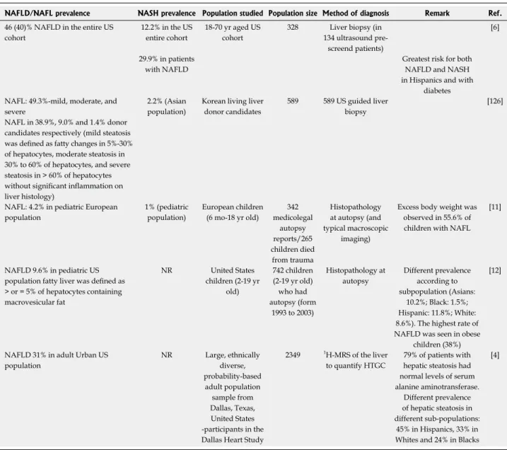
Table 1 Prevalence of non-alcoholic fatty liver disease, non-alcoholic fatty liver and non-alcoholic steatohepatitis
NAFLD/NAFL prevalence NASH prevalence Population studied Population size Method of diagnosis Remark Ref.
46 (40)% NAFLD in the entire US cohort
12.2% in the US entire cohort
18-70 yr aged US cohort
328 Liver biopsy (in 134 ultrasound pre-
screend patients)
[6]
29.9% in patients with NAFLD
Greatest risk for both NAFLD and NASH in Hispanics and with
diabetes NAFL: 49.3%-mild, moderate, and
severe
NAFL in 38.9%, 9.0% and 1.4% donor candidates respectively (mild steatosis was defined as fatty changes in 5%-30%
of hepatocytes, moderate steatosis in 30% to 60% of hepatocytes, and severe steatosis in > 60% of hepatocytes without significant inflammation on liver histology)
2.2% (Asian population)
Korean living liver donor candidates
589 589 US guided liver biopsy
[126]
NAFL: 4.2% in pediatric European population
1% (pediatric population)
European children (6 mo-18 yr old)
342 medicolegal
autopsy reports/265 children died
from trauma
Histopathology at autopsy (and typical macroscopic
imaging)
Excess body weight was observed in 55.6% of children with NAFL
[11]
NAFLD 9.6% in pediatric US population fatty liver was defined as
> or = 5% of hepatocytes containing macrovesicular fat
NR United States
children (2-19 yr old)
742 children (2-19 yr old) who had autopsy (form
1993 to 2003)
Histopathology at autopsy
Different prevalence according to subpopulation (Asians:
10.2%; Black: 1.5%;
Hispanic: 11.8%; White:
8.6%). The highest rate of NAFLD was seen in obese
children (38%)
[12]
NAFLD 31% in adult Urban US population
NR Large, ethnically diverse, probability-based adult population
sample from Dallas, Texas, United States -participants in the Dallas Heart Study
2349 1H-MRS of the liver to quantify HTGC
79% of patients with hepatic steatosis had normal levels of serum alanine aminotransferase.
Different prevalence of hepatic steatosis in different sub-populations:
45% in Hispanics, 33% in Whites and 24% in Blacks
[4]
Based on histological examination or proton magnetic resonance spectroscopy (1H-MRS) measurement in Hallmark Studies. HTGC: Hepatic triglyceride content; NAFLD: Non-alcoholic fatty liver disease; NAFL: Non-alcoholic fatty liver; NASH: Non-alcoholic steatohepatitis; NR: Not reported.
tance and pancreatic β-cell dysfunction.
The prevalence of ultrasonographic NAFLD was 69.4% in 180 patients with T2DM[30]. NAFLD was asso- ciated with obesity (abdominal), hypertriglyceridemia and high-normal ALT levels. The authors concluded that the progression of NAFLD is independent of the diabetes progression[30].
The ultrasonography results of 204 patients with T2DM showed fatty infiltration in 62.2% of the patients;
NAFLD was confirmed by liver biopsy with subsequent histology in 87% of the patients, indicating a 54.11%
histologically confirmed prevalence in T2DM. Steato- hepatitis and fibrosis were found in 38.9% and 23.2%, respectively, of Indian patients with T2DM[31].
Leite et al[32] found a 78% NASH prevalence at the histological examination in nearly 100 patients with T2DM and US evidence of NAFLD. The presence of high triglyceride, low HDL-cholesterol and increased ALT levels were independently associated with a higher risk of histologically confirmed NASH. The prevalence of advanced fibrosis (≥ stage 2) was found in 38% and 55% of the patients depending on the pathologist who conducted the histological examination. The presence of NASH was independently correlated with high serum γGT levels, older age and male gender[32].
Gestational diabetes mellitus
Recent novel findings have emerged to confirm the rela- tionship between diabetes mellitus and NAFLD: Women with a history of gestational diabetes mellitus (GDM) have an increased risk of developing T2DM decades later. Prikoszovich et al[33] recruited women with a history of GDM (pGDM) four to five years after delivery and assessed the glucose tolerance and oral glucose insulin sensitivity to measure the whole-body insulin sensitivity during a 75 g CH OGTT. The lipid storage in the muscle, liver and flux through the ATP synthase were measured using 1H/31P magnetic resonance spectroscopy. In a com- parison with women without any risk factor for T2DM, the hepatic content of lipids (HCL) was doubled in the insulin resistant pGDM women, despite they had normal glucose tolerance. HCL correlated positively with the body fat mass and inversely with insulin sensitivity. The authors concluded that increased hepatic lipid storage is an early and predominant abnormality in insulin resistant women with a history of GDM.
The fatty liver index (FLI) -measured using 1HMRS- in women with previous GDM predicted further meta- bolic deterioration and subjects with the highest FLI values showed significant alterations in FFA kinetics with a higher risk to develop T2DM in the future[34]. The re- sults of these studies should be taken into consideration when the role of NAFLD in determining the hepatic and whole body insulin sensitivity is under scrutiny in glucose tolerant individuals with insulin resistance.
In 2013, Brumbaugh et al[35] assessed the intrahepatic lipids in the neonatal offspring of obese women with gestational diabetes. The neonates born to obese women ease was 25% and 20%, respectively using the US based
method for identification[17]. The prevalence of NAFLD in Japan increased to 2.4-fold from the 12.6% preva- lence found in 1989 to the 30.3% prevalence observed in 1998[18]. A lower prevalence was reported in India, us- ing ultrasonography for the identification of NAFLD:
the prevalence of NAFLD was 18.9% in adults above 20 years of age, with a higher prevalence of NAFLD in males than females (24.6% vs 13.6%)[19].
A number of studies report the aminotransferase- based approach to diagnose; due to the observation that more than 75% of the patients with steatosis might have normal LT values, these studies are not discussed in detail here[4].
ASSESSmENT Of fIbROSIS ANd STEATOHEpATITIS
In most patients with NAFLD, a non-invasive score pro- posed by Angulo et al[20] could be applied to assess liver fi- brosis because advanced fibrosis could be diagnosed with accuracy in 90% of the cases. The authors analysed the clinical and liver biopsy data of more than 700 patients to construct this simple scoring system. Transient elastog- raphy (Fibroscan), which is a radiological modality used with high accuracy to non-invasively assess the degree of fibrosis based on the measurement of liver stiffness[21,22], might detect even low-grade steatosis because of a re- cently described novel ultrasonic controlled attenuation parameter of the machine[23].
Although there are promising biomarkers such as cytokeratin-18 fragments for NASH to be potentially ap- plied as diagnostic tools in the future[24], the accurate di- agnosis of NASH in some cases might require a liver bi- opsy. A biopsy is typically indicated in the cases in which the liver tests (LTs) are repeatedly elevated in a chronic manner (e.g., for six months) and are of unexplained ori- gin and when the patient must be carefully investigated for alternative diagnoses (e.g., to measure assess hepatic iron concentration in a C282Y HFE heterozygote indi- vidual; when the case is suggestive of autoimmune liver disease; rarely, when the diagnosis of Wilson disease may not be established without quantitation of the liver cop- per content; in other storage diseases; or in rare distinct forms of liver disease)[25-29].
WHY IS NAfld WORRISOmE?
Type 2 diabetes mellitus
Type 2 diabetes mellitus (T2DM) and NAFLD are par- ticularly closely related. This relationship among T2DM, insulin resistance (IR) and NAFLD is expected because insulin is subsequently delivered directly to the portal vein after secretion, taking the same route as the absorbed glucose, and the liver eliminates a large portion of portal insulin at the first pass.
Obesity in NAFLD is associated with dysfunctional adipose tissue, and lipotoxicity promotes insulin resis-
with GDM underwent MRS for intrahepatic lipid content determination at 1-3 wk of age and demonstrated a mean 68% increase in the IHCL compared with infants born to normal-weight mothers. The intrahepatic fat deposition in the neonates positively correlated with the maternal pre-pregnancy BMI and not with subcutaneous adiposity.
"Metabolic syndrome"
Although many authors agree that the “metabolic syn- drome” is a cluster of risk factors, but whether it may correctly be considered a syndrome is strongly ques- tioned[36]. Provided the term “metabolic syndrome” is accepted, insulin resistance should be at the core of the
“syndrome”, and many authors agree that NAFLD that is generally asymptomatic is frequently associated with obesity, type 2 diabetes and the “metabolic syndrome”[17]. Although a statement was published for synchronising the various definitions for metabolic syndrome[37], our experience confirmed that a large proportion of patients with NAFLD did not fulfill the “metabolic syndrome”
criteria (49%-46% depending on the NCEP in ATP-Ⅲ[38]
or the AHA and IDF joint criteria[37] were applied). The high proportion of NAFLD patients without “metabolic syndrome” was observed despite that the HOMA2-IR values of the NAFLD patients were even higher in this study population than those with T2DM, provided that the NAFLD patients were excluded from the latter study group. None of the definitions of “metabolic syndrome”
could be appraised as a consensus due to that the signifi- cant proportion of insulin-resistant NAFLD patients are excluded by these criteria[39].
bIOCHEmISTRY
The major biochemical event in NAFLD is the accu- mulation of triacylglycerols (TAG) in the hepatocytes.
Because of the strong associations described above and to understand the relation of NAFLD to whole-body metabolic status, Donnelly et al[40] conducted a study using gas chromatography/mass spectrometry. Hepatic TAG might accumulate from different sources in the hepato- cytes as follows: TAG as a nutrient after absorption from the intestine are delivered via chylomicrons to the liver, where they might subsequently be secreted as lipopro- teins. Hepatic TAG synthesis is possible, and this process requires fatty acids and glycerol in the liver The required fatty acids might be from the plasma non-esterified fatty acid pool (NEFA), and they might be produced in the liver as de novo hepatic lipogenesis. It has been reported that approximately 60% of the TAG accumulated in the liver is derived from the plasma NEFA pool, even in the fed state, and that adipose tissue is the largest contributor to the fatty acid content of the plasma NEFA pool (80%
in the fasted state). One-quarter of the TAG accumula- tion is derived from hepatic de novo lipogenesis that is elevated in the fasting state and demonstrates no diurnal variation, whereas approximately 15% is derived from the dietary intake. The situation is even worse when insulin
resistance - a hallmark of metabolic syndrome - is also present because of the lack of (insulin induced) down- regulation of the hormone sensitive lipase that eventually results in enhanced lipolysis and an increased efflux of free fatty acids to the plasma NEFA pool from the adi- pocytes. Hyperglycaemia (and hyperinsulinaemia) induces SREBP-1c and ChREBP in the liver, and these transcrip- tion factors subsequently activate genes that are required for lipogenesis, eventually resulting in increased hepatic de novo lipogenesis. Hepatic de novo lipogenesis is increased in insulin-resistant states and in NAFLD[41]. Beta-oxidation of fatty acids is increased in patients with NASH; the ox- idation might not overcome the increased hepatic TAG production, and the increased NEFA oxidation might result in increased oxidative stress, enhancing the transi- tion of NAFL to NASH[42,43]. This (patho)biochemical path provides the reason that the association is remark- ably strong between fatty liver and obesity-related insulin resistance[40,44].
NAfld CONCURRENTlY WITH VISCERAl ANd SUbCUTANEOUS AdIpOSITY ANd INSUlIN RESISTANCE
Is there accurate morphological evidence to support this biochemistry-driven hypothesis that the intrahepatic lipid content is hand-in-hand with different adipose tissue deposits, particularly the visceral adipose tissue accumula- tion that is strongly associated in impaired glucose me- tabolism?
Bosy-Westphal et al[45] assessed the fat volume includ- ing the visceral fat volumes (VAT), the pericardial adipose tissue (PAT, a well-known marker of visceral adiposity) and the abdominal subcutaneous adipose tissue using MRI and compared the results to the IHCL quantified by the highly sensitive 1H-MRS method in thirty overweight, not yet diabetic women. The participating individuals were restricted to a low calorie diet for three months;
at baseline, the visceral adipose tissue volume and PAT correlated with the IHCL as well as with the insulin resis- tance measured with the euglycaemic hyperinsulinaemic clamp and the homeostatic model assessment (HOMA)- IR. The strength of the relationship between the visceral fat volume and IHCL is shown by the finding that the reductions in IHCL induced by the dietary intervention and loss of body weight were only correlated with the decrease in VAT. The exceptional role of NAFLD in determining insulin resistance is confirmed by the im- provements in HOMA-IR and HOMA2-%B after three months of diet and weight loss that were only related to the decrease in IHCL[45].
The interpretation of HOMA-IR has been recently challenged. Traditionally, HOMA-IR reflects the degree of insulin resistance; however, these estimates based on the fasting plasma insulin and glucose concentrations[46]
do not take into account whether the secretion of insulin by the pancreatic β cells is altered or if an alteration is
in the insulin removal (clearance). The differentiation of total body insulin resistance, peripheral insulin resistance and hepatic insulin resistance merits research attention. A recent novel interpretation of HOMA-IR suggests that it is not a precise estimate of peripheral insulin action rather it might reflect the ability of insulin to suppress hepatic glucose production in the fasting state[47]. The clamp technique used in the previously mentioned study measures the peripheral insulin action in non-diabetic individuals resulting from hyperinsulinaemia during the measurement that is high enough to inhibit the hepatic glucose production completely[48]. This concept should be validated in studies using an insulin concentration that is lower than that regularly used during an euglycaemic- hyperinsulinaemic clamp measurement to avoid the abso- lute inhibition of hepatic glucose production[47]. Provided that this hypothesis regarding HOMA-IR is accurate, the 1H-MRS based follow-up observation that improve- ments in HOMA-IR and HOMA2-%B after dietary in- tervention were only related to the decrease in the IHCL indicates that a decrease in the intrahepatocellular lipid content increases the ability of insulin to suppress the hepatic glucose production in the fasting state, which is a major determinant of fasting plasma glucose levels[45].
In addition the contribution of the subcutaneous adipose tissue (SAT) to the whole-body adipose tissue dysfunction has been also recently confronted against the broadly accepted “innocent bystander to VAT” concept.
The results of an in vivo human study that functionally as- sessed SAT in patients with histology-confirmed NASH provided evidence that the abdominal SAT in NASH pa- tients is highly insulin resistant and required > 6 × more insulin to the gain similar degree of glycerol release sup- pression (referring to impaired suppression of lipolysis in SAT by insulin) than in healthy subjects. Authors there- fore suggested that abdominal SAT is dysfunctional in NASH and plays a profound role in NASH development and lipotoxicity[49].
lIpOTOXICITY
Free fatty acids (FFA) are directly hepatotoxic, and FFA levels are elevated in patients with NASH and correlate with disease severity[50]. Patients with severe fibrosis shown by liver biopsy had significantly greater serum concentration of free fatty acids than did the patients without severe fibrosis[50]. Saturated FFAs (e.g., palmitate) are apparently more hepatotoxic than unsaturated (mono- unsaturated) FFAs (e.g., palmitoleate); palmitoleate (known as a lipokine) was demonstrated to suppress hepatic steatosis[51,52]. Unsaturated fatty acids do not induce endo- plasmic reticulum (ER) stress or apoptosis and are able to rescue the palmitate-induced ER stress and apoptosis in liver cells. It has been proposed that the difference in tox- icity between saturated and unsaturated fatty acids is that unsaturated FFAs are more easily esterified into neutral triglycerides[53,54].
The impairment of liver cellular capacity in FFA utili-
sation, incorporation to TAGs and export contributes to the development of NASH, and hepatic injury is further accentuated by pathological FA oxidation and altered cell membrane composition[55]. Lipotoxicity induces hepatocel- lular apoptosis; Kupffer cell activation; impaired insulin signaling and hepatic insulin resistance; and hepatic stellate (Ito) cell activation with subsequent fibrosis. These patho- logical processes might eventually lead to cirrhosis[55].
dIACYlglYCEROl ACYlTRANSfERASE 2: dISSOCIATION Of STEATOSIS ANd INflAmmATION-fIbROSIS
The hepatotoxicity of saturated FFAs is further sup- ported by the genetic deletion of diacylglycerol acyltrans- ferase 2 (DGAT2) in mice models as follows: in parallel with the decreased TAG synthesis due to the the in- creased oxidative stress from the lack of intracellular FFA esterification, hepatocellular apoptosis with subsequent fibrosis occurs, resulting in the dissociation of hepatic steatosis and hepatic fibrosis in the NASH model[56,57]. Overexpression of DGAT2 in the experimental model causes hepatic steatosis without concomitant liver fibrosis or insulin resistance, providing evidence for the different roles that toxic saturated FFAs and TAGs have in the de- velopment of NAFLD[57,58].
The pathological effect of lipotoxicity is not limited to the liver cells, and it might affect the pancreatic β-cells, contributing to the β-cell dysfunction that is frequently observed in T2DM[59]. This pathology that affects both the liver and the pancreatic β-cells is highly important in determining the plasma glucose levels.
Lipotoxicity in the pancreatic β-cells
The intracellular signaling pathways altered because of lipotoxicity should partially overlap in the liver cells and in the pancreatic β-cells (Figure 1).
Saturated fatty acids, among other factors, induce endoplasmic reticulum stress that induce JNK activation via the IRE1α/ASK1, a signal that has been described in pancreatic β-cells as well as in liver cells[60,61]. Subsequent- ly, the activation of the JNK pathway induces the “sen- sitiser” BH3 proteins (DP5, Bad, Bik), which bind to the anti-apoptotic Bcl-2 proteins such as Bcl-2 and Bcl-XL;
these proteins designate the pro-apoptotic BH3-only pro- teins, BIM and PUMA (“activators”), eventually leading to their activation, which results in the apoptotic death of the pancreatic β and liver cells via Bak and Bax[61-63]. There is a substantial homology of this unsaturated FFA induced path and the path of cytokine induced β cell apoptosis. Palmitoleate (a mono-unsaturated fatty acid) could inhibit lipoapoptosis by blocking the endoplasmic reticulum stress-associated increases of the BH3-only proteins, Bim and PUMA, in hepatocytes[64]. The deterio- rating β cell function, in combination with the increasing hepatic IR and the decreasing suppression of hepatic glucose output, leads to hyperglycaemia that eventually
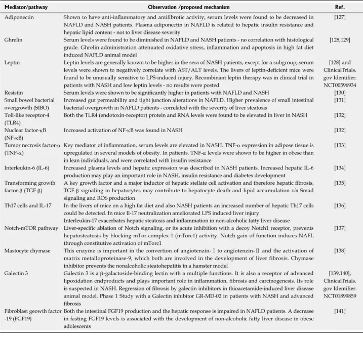
might directly (glucotoxicity) and by biochemical and metabolic consequences further promote this pathologic process. This cross-talk between the metabolic and cyto- kine induced pathways might facilitate the identification of novel drug targets (e.g., DP5, Bim) that would inhibit the unsaturated FFA induced ER stress mediated apop- totic liver cell death and possess protective properties against cytokine induced pancreatic β cell death[60-63]. The cytokines, growth factors and inflammatory mediators that are important in NAFLD are summarised in Table 2.
mITOCHONdRIAl dYSfUNCTION
A defective hepatic mitochondrial respiratory chain (MRC) was described in NASH[65]. The mitochondrial dysfunction, as measured by the activity of the MRC complexes in liver tissue, was correlated with the serum TNF-alpha levels and with the degree of insulin resis- tance that were higher in NASH and with the BMI[65]. In addition to the mitochondrial dysfunction, Sanyal et al[66]
described structural mitochondrial defects including the loss of the mitochondrial cristae and paracrystalline in- clusions, the presence of linear crystalline inclusions and
mitochondrial swelling in patients with NASH. Patients with T2DM of long duration might have decreased ATP production after fasting and after fructose administration.
The mitochondrial dysfunction in NAFLD, from lipo- toxicity, oxidative stress and as a result of inflammatory mediator effect, alters the hepatic energy metabolism, as recently reported by Koliaki and Roden[67].
ENTERO-INSUlAR AXIS (dpp-4 ANd INCRETINS) IN NAfld
A number of studies have assessed both the glucagon- like peptide-1 (GLP-1) and glucose dependent insuli- notropic peptide induced insulin secretion in T2DM patients and concluded that the response to the incretin hormone stimuli might be compromised as an early phenomenon in T2DM[68-71]. In the carriers of the tran- scription factor 7-like 2 gene polymorphism (TCF7L2- rs7903146, a widely known risk polymorphism in T2DM development) the GLP-1 induced insulin secretion, and not the GLP-1 secretion, is impaired[72]. Similar find- ings were presented for a common genetic variant in the
[sensitizers]*
DP5, Bad, Bik
BCL-2*
BCL-XL MCL-1
[Activators]
BIM, [binding] [Deliberation] PUMA
Pro-inflammatory cytokines
NEFA pool
Saturated FFA
Endoplasmatic reticulum stress
IRE1-α ATF6 PERK
ASK1 MLK3 CHOP
JNK [via
STAT1 and NF-
κB]
Bax Bak
Citochrome-c Apoptosis
Figure 1 Endoplasmatic reticulum stress caused by saturated free fatty acids. Endoplasmatic reticulum stress caused by saturated free fatty acids, via three main mediators [inositol-requiring endoplasmic reticulum-to-nucleus signal kinase 1α (IRE1α), activating transcription factor 6 (ATF6) and RNA-dependent protein kinase (PRK)-like endoplasmic roticulum (ER) kinase (PERK)], results in the activation of c-Jun N-terminal kinase (JNK) and C/ CCAAT/enhancer binding protein (EBP) homologous protein. The sequence of BH3 protein activation based on a sensitizer and an activator group was described by Gurzov and Eizirik in β-cells[62,63]. The sensitizers bind the anti-apoptotic proteins Bcl-2, Bcl-XL and Mcl-1 and release the activators from this bond. JNK both mediates the induction of the sensitizer and the activator BH3 proteins and also activates Bax. Upregulation of Bcl-2 interacting mediator of cell death (BIM) and p53 upregulated modulator of apoptosis (PUMA) was also demonstrated in liver cells as a ruslt of free fatty acid (FFA) induction. Proinflammatory cytokines also activate JNK. ER sterss also results in CHOP actvation and subsequently the activation of the activator BH3 proteins. This complex signaling pathway might link the metabolic (saturated FFAs) stress and the effect of pro- inflammatory cytokines both in the pancreatic β-cell as well as in liver cells.
WFS1 gene, and carriers of the polymorphism had im- paired GLP-1-induced insulin secretion independently of insulin sensitivity[73]. Variants of the KCNQ1 (rs151290, rs2237892 and rs2237895) gene altered the endogenous GLP-1 secretion[74,75].
Despite these well-designed, elegant studies that as- sessed the role of the entero-insular axis in T2DM and its association with the risk gene polymorphisms in T2DM, there is limited data regarding the role of the entero-in- sular axis in NAFLD. Recently, β cell connectedness has been reported to influence the incretin-induced insulin secretion in human islets, and lipotoxicity was demon- strated to be able to disrupt this incretin-regulated human β cell connectivity that might result in the loss of the co-
ordinated islet response to metabolic stimuli[76].
Increased serum DPP-4 activity (the soluble form of the enzyme in human sera) was described in patients with NAFLD in comparison to healthy controls and T2DM patients, provided that T2DM patients who pre- sented with clinically obvious (with an US and biochemi- cal based diagnosis) NAFLD were excluded from the study group[39]. We found a positive correlation among the γGT, ALAT and serum DPP-4 activities in NAFLD that supports the finding that the excess DPP-4 found in the serum of NAFLD patients is of hepatic origin. We concluded that it is the presence of (fatty) liver disease that primarily influenced the serum DPP-4 enzymatic activity and not hyperglycaemia alone[39]. Subsequently,
Table 2 Adipokine hormones, cytokines, growth factors and other mediators that play important role in non-alcoholic fatty liver disease pathology
Mediator/pathway Observation /proposed mechanism Ref.
Adiponectin Shown to have anti-inflammatory and antifibrotic activity, serum levels were found to be decreased in NAFLD and NASH patients. Plasma adiponectin in NAFLD is related to hepatic insulin resistance and hepatic lipid content - not to liver disease severity
[127]
Ghrelin Serum levels were found to be diminished in NAFLD and NASH patients - no correlation with histological grade. Ghrelin administration attenuated oxidative stress, inflammation and apoptosis in high fat diet induced NAFLD animal model
[128,129]
Leptin Leptin levels are generally known to be higher in the sera of NASH patients, except for a subgroup; serum levels were shown to negatively correlate with AST/ALT levels. The livers of leptin-deficient mice were found to be unusually sensitive to LPS-induced injury. Recombinant leptin therapy was in clinical trial in patients with NASH and low leptin levels - no results were posted
[128] and ClinicalTrials.
gov Identifier:
NCT00596934 Resistin Serum levels were shown to be significantly higher in patients with NAFLD and NASH [130]
Small bowel bacterial overgrowth (SIBO)
Increased gut permeability and tight junction alterations in NAFLD. Higher prevalence of small intestinal bacterial overgrowth in NAFLD patients - correlated with the severity of liver steatosis
[131]
Toll-like receptor-4 (TLR4)
Both the TLR4 (endotoxin-receptor) protein and RNA levels were found to be elevated in liver in NASH [132]
Nuclear factor-κB (NF-κB)
Increased activation of NF-κB was found in NASH [132]
Tumor necrosis factor-α (TNF-α)
Key mediator of inflammation, serum levels are elevated in NASH. TNF-α expression in adipose tissue is upregulated in several models of obesity. In patients, TNF-α levels were shown to be higher in obese than in lean individuals, and were correlated with insulin resistance
[133]
Interleukin-6 (IL-6) Increased plasma levels and hepatic expression was described in NASH patients. Increased hepatic IL-6 production may play an important role in NASH, insulin resistance and diabetes development
[134]
Transforming growth factor-β (TGF-β)
A key growth factor and a major inductor of hepatic stellate cell activation and therefore hepatic fibrosis, TGF-β signaling in hepatocytes may contribute to hepatocyte death and lipid accumulation via Smad signaling and ROS production
[135]
Th17 cells and IL-17 In the livers of mice on a high fat diet and also NASH patients an increased number of hepatic Th17 cells could be detected. In mice Il-17 neutralization ameliorated LPS induced liver injury
[136]
Interleukin-17 exacerbates hepatic steatosis and inflammation in non-alcoholic fatty liver disease Notch-mTOR pathway Liver-specific ablation of Notch signaling, or its acute inhibition with a decoy Notch1 receptor, prevents
hepatosteatosis by blocking mTor complex 1 (mTorc1) activity. Notch gain of function induces NAFL through constitutive activation of mTorc1
[137]
Mastocyte chymase This enzyme is important in the convertion of angiotenzin-Ⅰ to angiotenzin-Ⅱ and the activation of matrix metalloproteinase-9, which both are involved in the development of liver fibrosis. Chymase inhibitor prevents the nonalcoholic steatohepatitis in a hamster model
[138]
Galectin 3 Galectin 3 is a β-galactoside-binding lectin with a multiple functions. It is also a receptor of advanced lipoxidation endproducts and plays important role in inflammation, fibrosis and carcinogenesis. Its role is suspected in NASH. Regression of fibrosis by galectin inhibitors in thioacetamide-induced liver disease animal model. Phase 1 Study with a Galectin inhibitor GR-MD-02 in patients with NASH and advanced fibrosis
[139,140], ClinicalTrials.
gov Identifier:
NCT01899859 Fibroblast geowth factor
-19 (FGF19)
Both the intestinal FGF19 production and the hepatic response is impaired in NAFLD patients. A decrease in fasting FGF19 levels is associated with the development of non-alcoholic fatty liver disease in obese adolescents
[141]
A few of these molecules are therapeutic targets (e.g., galectin 3) in early phase clinical trials. NAFLD: Non-alcoholic fatty liver disease; NASH: Non-alco- holic steatohepatitis; AST: Aspartate aminotransferase; ALT: Alanine aminotransferase.
higher hepatic expression of DPP-4 at the mRNA level was described in NAFLD patients, and high glucose concentrations increased the DPP-4 expression in the HepG2 cells, in contrast to the insulin and FFAs that did not alter the DPP-4 expression[77]. The correlation among the serum levels of soluble CD26/DPP-4 activ- ity and other liver disease biomarkers was also confirmed in Asian patients[78-80]. A positive correlation between the serum DPP-4 activity and insulin resistance (HOMA2- IR) in NAFLD was also found that is not surprising provided that the serum DPP-4 activity is considered as a novel liver disease biomarker. The existence of such a correlation further supported the recent suggestion that the soluble form of DPP-4 is a novel adipokine hormone that could induce insulin resistance without the presence of incretin hormones in an experimental system[81]. In a meta-analysis, Fadini et al[82] found that the DPP-4 activ- ity was increased in T2D and was not lowered by glyce- mic control; the study confirmed that hyperglycaemia was not a direct determinant of DPP-4 activity and was lower in metformin-treated patients. A significant role of DPP-4 in hepatic glucose metabolism is supported by the study of Edgerton et al[83], who demonstrated that dur- ing DPP-4 inhibitor therapy (vildagliptin) and GLP-1 co- treatment, the net hepatic glucose uptake was three-fold greater in the DPP-4 inhibitor treated group than in the control group that was treated with portal vein GLP-1 infusion and not with the DPP-4 inhibitor; this effect was greater than that predicted by the change in insulin. This finding indicates that the DPP-4 inhibitor was able to in- crease the hepatic glucose disposal beyond the effects of GLP-1 on insulin and glucagon secretion.
The effect of DPP-4 inhibitor therapy on liver fibro- sis should be assessed, particularly because the fibroblast activation protein (FAP), which is a duplicate molecule of DPP-4 (FAP-DPP-4 shows 88% homology at the cDNA level), is present at the tissue remodelling interface on hepatic stellate cells (HSCs, ITO cells) that primarily produce the accumulating extracellular matrix proteins (including collagens) in chronic liver diseases, eventually leading to fibrosis and cirrhosis of the liver[84].
dpp-4 INHIbITORS ANd glp-1 mImETICS IN NAfld TREATmENT
DPP-4 inhibitors were reported to improve hepatic ste- atosis and adipose tissue inflammation in mice[85,86]. Hu- man treatments are also documented, in which the DPP-4 inhibitor, sitagliptin, was able to provide benefit for a refractory case of NAFLD[87]. Sitagliptin improved hepa- tocyte ballooning in a diabetic patient with NASH[88]. The available data on the use of GLP-1 mimetics is limited;
the following effects were reported in animal models:
improvement of the FA beta-oxidation; a decrease in the liver disease biomarker ALAT and hepatic TAGs; reduced ER-stress related hepatocyte cell death; and enhanced beneficial macroautophagy[89-92]. The GLP-1 derived non-
peptide (by the cleavage of the neutral endopeptidase), GLP-1 (28-36) amide, was shown to improve glucose disposal and decrease hepatic steatosis in high fat diet mice. The GLP-1 (28-36) amide suppressed the hepatic gluconeogenesis and improved the pyruvate tolerance in this model[93]. The large randomised controlled trials (RCTs) that would also assess safety issues of the DPP-4 inhibitors and the GLP-1 mimetics are missing in NAFL patients, as well as in NASH patients, in whom the indica- tion for (an auxiliary) drug treatment (in addition to diet, weight loss and exercise) might be more obvious.
OTHER THERApIES
There is consensus that the most effective treatment of NAFL in the overwhelming majority of cases is lifestyle change, including a supervised diet, exercise and weight loss. However doctors are not always successful in having patients reach this goal, and the potential pharmacologi- cal approaches that should be considered, predominantly in NASH patients, are briefly reviewed.
Metformin
Metformin is experiencing a booming renaissance in the treatment of T2DM; however, this drug has no signifi- cant effect on liver histology, probably in part because of the limited anti-steatogenic effect and failure to increase the adiponectin levels[94]. The guidelines do not recom- mend this biguanide specifically for the treatment of NASH (AASLD: Strength 1/Evidence: A)[26,95].
Thiazolidinediones
In contrast to metformin, thiazolidinediones (TZDs) are experiencing difficulty as pharmacological agents in hu- man medicine. In certain countries there are no remaining agents from this drug class (e.g., the French Agency for the Safety of Health Products requested a pharmaceutical company to suspend the use of pioglitazone containing products for the treatment of type 2 diabetes in France in 2011 because of an increased risk of urinary bladder can- cer). EASL and AASLD discussed pioglitazone, outlin- ing that, from the hepatologist point of view, glitazones consistently provided benefit for patients with NASH and could be used to treat biopsy proven steatohepatitis (AASLD: Strength: 1, Evidence: B)[26,95]. The associations emphasise that the long-term safety of pioglitazone in NASH is not established, and it is likely that no doctor is treating NASH isolated from other safety issues[26]. Vitamin E
The nature of the dilemma with vitamin E is somewhat similar to that with the TZDs. A meta-analyses (of over 100000 participants) reported increased all-cause mor- tality with a dose of vitamin E ≥ 400 IU/d, and one meta-analysis found a statistically significant relationship between vitamin E dosage and all-cause mortality in a dose-response analysis[96,97]. Although others have ques-
tioned the results obtained from these meta-analyses[98,99], recently vitamin E (400 IU/d) was shown to signifi- cantly increase the risk of prostate cancer among healthy
men[100]. According to the AASLD recommendation,
this vitamin E dose should be doubled (800 IU/d) to improve liver histology in non-diabetic adults with biopsy proven NASH (Strength: 1, Quality: B), and it is not recommended in NASH patients with T2DM (Strength:
1, Quality: C)[26]. Histological improvement in NASH in this context should be interpreted for all lesions, except for improvement in fibrosis, and vitamin E is not recom- mended for the treatment of NAFLD-cirrhosis or cryp- togenic cirrhosis[26,95].
Ursodeoxycholic acid-nor-ursodeoxycholic acid
The AASLD does not recommend ursodeoxycholic acid (UDCA) A for the treatment of NAFLD and NASH (Strength 1, Quality: B), and long term, high dose UDCA increased the rate of serious adverse events (the development of cirrhosis, varices, cholangiocarcinoma, liver transplantation or death) in patients with primary sclerosing cholangitis[26,101]. The pharmacological proper- ties of norUrsodeoxycholic acid (nor-UDCA) might be more attractive and might later be studied in patients with NASH[102].
Farnesoid X receptor agonists
The bile acids secreted upon feeding undergo enterohe- patic circulation and serve as endogenous ligands to a class of nuclear hormone receptors that function as li- gand-activated transcription factors. Farnesoid X receptor (FXR) belongs to this class and serves as a receptor for hydrophobic bile acids. In T2DM, the bile acid composi- tion is altered. The bile acid taurochenodeoxycholic acid increases insulin release via the FXR dependent inhibition of the KATP channels, and FXR has been described to improve insulin sensitivity and glucose uptake in adipose tissue, liver and skeletal muscle by regulating the genes that are important in metabolic control[103,104]. The FXR agonist WAY-362450 was able to decrease inflammation and fibrosis in a murine model of NASH[105]. The follow- ing results of a 6-wk double-blind, randomised, placebo- controlled clinical phase Ⅱ trial of a semi-synthetic bile acid derivative, obeticholic acid (OCA), in patients with T2DM and presumed NAFLD has recently been published: administration of 25 or 50 mg of OCA for six weeks increased insulin sensitivity and reduced the markers of liver inflammation and fibrosis. A longer trial is ongoing with OCA in biopsy-proven NASH patients (Clinical trials identifier: NCT01265498)[106].
Despite the primary enthusiasm regarding the FXR agonists, the controversy might not be ignored due to the recent finding that under high fat diet conditions, FXR knockout (FXR-KO) mice benefited from the receptor deficiency, and FXR-KO protected against the HFD- induced impairment of fasting plasma glucose levels and glucose tolerance[107]. In parallel with the clinical trials, a
better understanding of bile acid enterohepatic circula- tion and more research on the FXR-dependent and inde- pendent signaling pathways are warranted.
Pre and probiotics
The intestinal microbiome has an increasing role in the understanding of T2DM pathology. In a recent study, 20 patients with histology-proven NASH were randomised to receive probiotics or usual care for six months. The probiotic treatment decreased the ASAT, and the de- crease in IHCL was confirmed using 1H-MRS[108]. Other studies have suggested a potential role for prebiotic fibres (non-digestible carbohydrates modulating the human mi- crobiome) in NAFLD treatment, and larger RCTs might provide conclusive evidence[109].
Omega 3 polyunsaturated FA
Di Minno et al[110] recently summarised the potential of omega-3 fatty acids for the treatment of NAFLD re- ported in seven human trials, the largest of which was a 6-mo follow-up RCT of 144 patients with NAFLD; how- ever, the authors concluded that well-designed RCTs of adequate size and duration, with histological endpoints, are needed to assess the long-term safety and efficacy of such treatment.
Others
The phase 1 study to evaluate GR-MD-02, a Galectin-3 inhibitor in patients with NASH and advanced fibrosis (NCT01899859) could on one day be an excellent exam- ple of how a long term molecular research[139,140], may po- tentially pay out in benefiting certain patients. Galectin-3 protein (binding to terminal galactose residues in glyco- proteins) is implicated in the pathogenesis of liver fibro- sis and the results with compounds inhibiting the protein suggest a potential role of these drugs in human liver fibrosis and even in cirrhosis in the NAFLD spectrum as well (Table 2). Hypothetical approaches, including the development of a peripheral cannabinoid 1 receptor ago- nist without psychiatric side effects or the supplementa- tion of vitamin D3 to overcome vitamin D3 deficiency that is highly prevalent in NAFLD[92,111,112], might have an effect on future therapies; however, they have yet to be tested and even limited evidence from concept stud- ies is missing. There are ongoing randomised, controlled studies with potentially promising compounds such as resveratrol (500 mg three times daily for six months vs placebo-NCT01464801) in obese patients with NAFLD/
NASH; however, conclusions from these studies would be premature.
Surgical interventions
Surgical interventions including Roux-en-Y gastric bypass might have beneficial effects on NAFLD from calorie intake reduction that could increase hepatic insulin sensi- tivity and augment postprandial GLP-1 secretion with a subsequently improved β cell function[26,113].
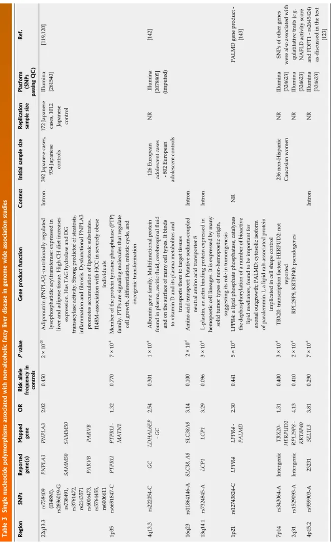
Table 3 Single nucleotide polymorphisms associated with non-alcoholic fatty liver disease in genome wide association studies RegionSNPs Reported gene(s)
Mapped geneOR
Risk allele frequency in controls
P valueGene product function ContextInitial sample size
Replication sample size Platform (SNPs
passing QC)
Ref. 22q13.3 rs738409 (I148M), rs2896019-G
PNPLA3PNPLA32.020.4502 × 10-20 Adiponutrin (PNPLA3)–nutritionally regulated lysophosphatidic acyltransferase: expressed in liver and adipose tissue. High CH diet increases expression. Has TAG hydrolase and DG transacylase activity. Strong predictor of steatosis, inflammation and fibrosis. Dysfunctional PNPLA3 promotes accumulation of lipotoxic substrates. I148M–association with HCC in severely obese individuals Intron392 Japanese cases, 934 Japanese controls
172 Japanese cases, 1012 Japanese control Illumina [261540][119,120] rs738491, rs3761472, rs2143571
SAMM50SAMM50 rs6006473, rs5764455, rs6006611
PARVBPARVB 1p35rs6691847-CPTPRUPTPRU - MATN11.320.7707 × 10-6Member of the protein tyrosine phosphatase (PTP) family. PTPs are signaling molecules that regulate cell growth, differentiation, mitotic cycle, and oncogenic transformation 4q13.3rs222054-CGCLDHAL6EP - GC2.540.3011 × 10-6 Albumin gene family. Multifunctional protein found in plasma, ascitic fluid, cerebrospinal fluid and on the surface of many cell types. It binds to vitamin D and its plasma metabolites and transports them to target tissues 126 European adolescent cases - 802 European adolescent controls NR Illumina [2078805] (imputed)
[142] 16q23 rs11864146-ASLC38, A8 SLC38A83.140.100 2 × 10⁻⁶Amino acid transport: Putative sodium-coupled neutral amino acid transporter 8Intron 13q14.1rs7324845-ALCP1LCP13.290.0963 × 10-6 L-plastin, an actin binding protein expressed in hemopoetic cell lineages. It is expressed by many solid tumor types of non-hemopoetic origin, suggesting its role in tumorigenesis
Intron 1p21rs12743824-CLPPR4LPPR4 - PALMD2.300.4415 × 10-6LPPR4: a lipid phosphate phosphatase, catalyzes the dephosphorylation of a number of bioactive lipid mediators, found to be important for axonal outgrowth; PALMD: cytosolic isoform of paralemmin-1, a lipid raft-associated protein implicated in cell shape control
NRPALMD gene product - [143] 7p14rs343064-AIntergenicTBX20- HERPUD21.310.4003 × 10-8TBX20: transcription factor; HERPUD2: not reported236 non-Hispanic Caucasian womenNRIllumina [324623]SNPs of other genes were also associated with qulatitative traits (e.g. NAFLD activity score and FDFT1 - rs2645424) as discussed in the text [123]
2q31rs1529093-AIntergenicRPL29P8 - KRT8P404.130.4102 × 10-6RPL29P8, KRT8P40: pseudogenesNRIllumina [324623] 4p15.2rs959903-A23231SEL1L33.810.2907 × 10-6 IntronNRIllumina [324623] Not all GWAS studies were replicated. Risk gene variants with OR higher than 1.31 are indicated in the table. Based on the National Human Genome Research. SNPs: Single nucleotide polymorphisms; NR: Not reported.
gENETICS
Familial clustering and prevalence differences according to ethnic origin
Although screening of family members is not recom- mended in NAFLD[26] based on a retrospective review of 90 cases, the authors concluded that familial clustering is common, and 18% of NASH patients had a first degree relative with a similar phenotype[114]. Schwimmer et al[115]
found that NAFLD was more common in siblings (59%) and parents (78%), using 1H-MRS based diagnostics of children with biopsy proven NAFLD in a familial ag- gregation study. After adjustment for age, sex, race and BMI, the study concluded that familial factors are a major determinant in NAFLD. Gene-environment interactions might have a role in the data e.g. the condition that fam- ily members are living in a common household might indicate common environmental risk factors (type of oil used in the diet etc.). The role of genetic risk factors is supported by the differences based on ethnic origin that were observed in the NAFLD prevalence in multi-ethnic cohorts (see the Prevalence chapter and Table 1), with a higher prevalence in Hispanics and a lower prevalence in African Americans compared to non-Hispanic whites[4,16]. Genome wide association studies
Five genome wide association scans (GWAS) are report- ed for NAFLD in the GWAS catalogue. Although these studies did not include case numbers that are typically employed in other GWA studies including T2DM, they provide remarkable evidence for genetic factors predis- posing to or protecting from NAFLD. The risk polymor- phisms with the highest ORs are summarised in Table 3, from the aspect of the NAFLD binary outcomes. Dis- cussion of all the candidate genes is beyond the scope of this review.
pATATIN-lIkE pHOSpHOlIpASE
dOmAIN CONTAININg 3 gENE (pNplA3- AdIpONUTRIN) IN NAfld SpECTRUm dISEASES -bINARY TRAITS
We should outline the single nucleotide polymorphism in the patatin-like phospholipase domain containing a 3-gene (PNPLA3) that is the most studied genetic risk variant in NAFLD. Adiponutrin is a nutritionally regulat- ed lysophosphatidic-acyltransferase possessing the TAG hydrolase and DG transacylase activity. This gene is expressed in liver and adipose tissues, and a high carbo- hydrate diet increases the gene expression at the mRNA level in the liver. Dysfunctional PNPLA3 promotes the accumulation of lipotoxic substrates[116,117]. After the description that in carriers of the rs738409 C/G poly- morphism of the PNPLA3 the hepatic lipid content was more than two-fold higher, PNPLA3 was subsequently confirmed in a Japanese GWAS as an NAFLD binary trait candidate gene[118,119] (Table 3). Polymorphisms in the SAMM50 and PARVB genes were associated with
the development and progression of NAFLD in this Japanese GWA study[119].
The quality trait data demonstrate the following find- ings: PNPLA3 I148M (rs738409) is a genetic marker of progressive liver disease that is characterised with steato- sis, inflammation and fibrosis; carriers are more insulin resistant and more susceptible to T2DM[120,121]; and, in a 15-year-follow-up, there is an association of this genetic variant and hepatocellular carcinoma incidence in se- verely obese individuals, with a hazard ratio of HCC of 5.9x for each PNPLA3 148M allele carried (reaching the HR- 16x in the PNPLA3 148M homozygotes even after adjustment for age, gender, BMI, type 2 diabetes status and ALAT)[121,122].
OTHER CANdIdATE gENES (SqUAlENE SYNTHASE ANd COllAgEN XIII A1)- qUAlITATIVE TRAITS
In the cases in which additional candidate genes were identified, qualitative traits were also assessed in GWAS.
The NAFLD activity score was associated with the rs2645424 polymorphism of farnesyl diphosphate farne- syl transferase 1 (FDFT1), the degree of fibrosis was as- sociated with the rs343062 SNP, and the lobular inflam- mation was associated with the rs1227756 polymorphism of the Collagen 13 A1 gene[123].
FDFT1 is a membrane-associated enzyme located at a branch point in the mevalonate pathway. The encoded protein is the first specific enzyme in cholesterol biosyn- thesis, catalysing the dimerisation of two molecules of farnesyl diphosphate in a two-step reaction to form squa- lene. In addition to the linkage of FDFT1 to the NAFLD activity score, a coding variant in the FDFT1 gene influ- ences the plasma cholesterol levels, likely via alteration of the intracellular production of cholesterol[123,124].
Collagen XIII is one of the nonfibrillar collagens and belongs to the transmembrane collagens, and a number of alternatively spliced transcript variants have been de- scribed; integrins mediate the cell adhesion to the type XIII collagen[125]. There is no validation of these associa- tions in larger studies.
In summary, we may conclude that genetic and molec- ular research might lead to the identification of additional risk and protective gene variants and this together with deeper understanding of gene-environment interactions might provide better insight into the molecular pathology and identification of molecular targets in NAFLD, which is the most common chronic liver disease affecting up to one-third of the adult population in industrialised coun- tries. In addition, little is known regarding the long-term effect of the increasing NAFLD prevalence in paediatric populations.
ACkNOWlEdgEmENTS
EFSD New Horizons grant facilitated the construction of this manuscript. I am grateful to my colleagues, espe-