Autophagy in major human diseases
Daniel J Klionsky
1, Giulia Petroni
2, Ravi K Amaravadi
3,4, Eric H Baehrecke
5, Andrea Ballabio
6,7,8, Patricia Boya
9, Jos e Manuel Bravo-San Pedro
10,11, Ken Cadwell
12,13,14, Francesco Cecconi
15,16,17, Augustine M K Choi
18,19, Mary E Choi
19,20, Charleen T Chu
21, Patrice Codogno
22,23,
Maria Isabel Colombo
24, Ana Maria Cuervo
25,26, Vojo Deretic
27,28, Ivan Dikic
29,30, Zvulun Elazar
31, Eeva-Liisa Eskelinen
32, Gian Maria Fimia
33,34, David A Gewirtz
35, Douglas R Green
36,
Malene Hansen
37, Marja J€ a€ attel€ a
38,39, Terje Johansen
40, G abor Juh asz
41,42, Vassiliki Karantza
43, Claudine Kraft
44,45, Guido Kroemer
46,47,48,49,50, Nicholas T Ktistakis
51, Sharad Kumar
52,53, Carlos Lopez-Otin
54,55, Kay F Macleod
56,57, Frank Madeo
58,59,60, Jennifer Martinez
61,
Alicia Mel endez
62,63, Noboru Mizushima
64, Christian M€ unz
65, Josef M Penninger
66,67,
Rushika M Perera
68,69,70, Mauro Piacentini
71,72, Fulvio Reggiori
73, David C Rubinsztein
74,75, Kevin M Ryan
76,77, Junichi Sadoshima
78, Laura Santambrogio
2,79,80, Luca Scorrano
81,82,
Hans-Uwe Simon
83,84,85, Anna Katharina Simon
86, Anne Simonsen
87,88,89, Alexandra Stolz
29,30, Nektarios Tavernarakis
90,91, Sharon A Tooze
92, Tamotsu Yoshimori
93,94,95, Junying Yuan
96,97, Zhenyu Yue
98, Qing Zhong
99, Lorenzo Galluzzi
2,79,80,100,101,*& Federico Pietrocola
102,**Abstract
Autophagy is a core molecular pathway for the preservation of cel- lular and organismal homeostasis. Pharmacological and genetic interventions impairing autophagy responses promote or aggra- vate disease in a plethora of experimental models. Consistently, mutations in autophagy-related processes cause severe human pathologies. Here, we review and discuss preclinical data linking autophagy dysfunction to the pathogenesis of major human disor- ders including cancer as well as cardiovascular, neurodegenerative, metabolic, pulmonary, renal, infectious, musculoskeletal, and ocular disorders.
Keywordsaging; cancer; inflammation; metabolic syndromes;
neurodegeneration
Subject Category Autophagy & Cell Death
DOI10.15252/embj.2021108863| Received1June2021| Revised7July2021| Accepted12July2021| Published online30August2021
The EMBO Journal (2021)40: e108863
See the Glossary for abbreviations used in this article.
Introduction
The staggering increase in life expectancy that has characterized the last century has progressively attenuated, until reaching an apparent plateau over the last decade. Conversely, aging increases the suscep- tibility to many chronic illnesses, a condition that poses a major threat to the socioeconomic stability of high- and low-income coun- tries (Kehler, 2019; Melzeret al, 2020). Consequently, the trajecto- ries of human lifespan and healthspan are estimated to diverge in the near future. During the last decade, investigators have endeav- ored to put forward a holistic view of the biological principles underlying the general concepts of “health” and “disease” at the cel- lular and organismal levels, by framing them into archetypical “hall- marks” (Lopez-Otinet al, 2013; Kennedyet al, 2014; Lopez-Otin &
Kroemer, 2021). On these bases, it has been possible to separate the quintessential processes that operate to maintain individual cells and multicellular entities in a “healthy” state, from those that per- turb thestatus quoof cells and tissues, thereby hastening the clini- cal onset of life-threatening diseases.
In this context, the process of autophagy can be considered as a bona fide health-modifying agent (Choiet al, 2013; Mizushima &
Levine, 2020). Indeed, a large body of evidence from the literature supports the view of autophagy as a pro-longevity mechanism (Morselliet al, 2009; Morselli et al, 2010; Rubinszteinet al, 2011;
1–102The list of affiliations appears at the end of this article
*Corresponding author. Tel:+1 646 962 2095; E-mail: deadoc80@gmail.com
**Corresponding author. Tel:+46 0 720327239; E-mail: federico.pietrocola@ki.se
Kaushik & Cuervo, 2015b; Madeoet al, 2015; Fernandezet al, 2018;
Hansenet al, 2018; Leidalet al, 2018; Markakiet al, 2018) and as a cardinal regulator of cellular and organismal fitness in response to multiple endogenous or exogenous sources of stress (Mizushima, 2018; Morishita & Mizushima, 2019). Conversely, time-dependent loss of autophagy proficiency is thought to critically contribute to the aged phenotype (Lopez-Otinet al, 2013; Kennedyet al, 2014;
Lopez-Otin & Kroemer, 2021). Furthermore, several of the lifestyle changes that have been attributed a positive role in the regulation of longevity (including calorie restriction and physical exercise) are commonly noted for their capacity to stimulate autophagy (Lopez- Otinet al, 2016).
Autophagy is also key in preventing stresses as one of the major quality control guardians in the cell (Mancias & Kimmelman, 2016;
Conwayet al, 2020). Noteworthy, the autophagy pathways acquire physiological relevance even under basal, non-stressful conditions.
In line with this notion, autophagy takes direct part in the regulation of developmental programs (Mizushima & Levine, 2010; Allen &
Baehrecke, 2020), maintenance of stem cell self-renewal potential (Chenet al, 2018c; Donget al, 2021a), cellular differentiation and plasticity (Boyaet al, 2018; Clarke & Simon, 2019). Concordant with this notion, the appearance of the “diseased” state associated with autophagy dysregulation may occur as a result of alterations in these central aspects of multicellular organism biology. Indeed, tissues that are mainly composed of cells that lay in a post-mitotic/quiescent state exhibit higher sensitivity to loss of autophagy competence.
The term “autophagy” refers to composite molecular pathways in which intracellular components are conveyed to the lysosomal compartment for degradation and recycling. To date, three major forms of autophagy have been described (Galluzzi et al, 2017a).
Macroautophagy (henceforth referred to as autophagy; Box 1) is a form of autophagy in which the cellular cargo becomes sequestered within a double-membraned vesicle, termed an autophagosome.
The choice of the autophagosomal content can proceed in a
relatively nonselective manner (known as “bulk autophagy”) or involve the tightly regulated elimination of individual cellular com- ponents (known as “selective autophagy”), depending on the induc- ing factor (He & Klionsky, 2009; Sica et al, 2015; Dikic & Elazar, 2018; Gohel et al, 2020). By contrast, chaperone-mediated autop- hagy (CMA) operates as a protein-exclusive type of autophagy in which KFERQ-like motif-bearing proteins are first recognized by the heat-shock cognate protein HSPA8/HSC70 and enter the lysosome for degradation, upon binding LAMP2A (lysosomal-associated mem- brane protein 2A) and translocation through a channel formed by oligomerization of this protein (Kaushik & Cuervo, 2018). Finally, microautophagy involves the sequestration of cellular material (in- cluding KFERQ-flagged proteins or bulk cytoplasmic content) directly via membranous invaginations formed at the surface of late endosomes or lysosomes (Sahu et al, 2011; Uytterhoeven et al, 2015; Mejlvang et al, 2018), in an ESCRT-dependent (Sahuet al, 2011) or ESCRT-independent (McNally & Brett, 2018) mode. Besides representing the terminal effector of the autophagy cascade, the lysosome operates as a primary regulator of the autophagy process, in light of its active role in nutrient sensing and signaling via the MTOR (mechanistic target of rapamycin kinase) complex 1 (MTORC1)-TFEB (transcription factor EB) axis (Ballabio & Bonifa- cino, 2020).
The complex molecular networks that underlie these distinct autophagic pathways, as well as other forms of canonical and non- canonical autophagy that will be mentioned in this review, have been the object of thorough investigation and extensive reviewing over recent years (Dupontet al, 2017; Galluzziet al, 2017a; Dikic &
Elazar, 2018; Kaushik & Cuervo, 2018; Chu, 2019; Kirkin & Rogov, 2019; Nakatogawa, 2020; Klionskyet al, 2021). Whereas autophagy proceeds at a basal (yet cell type dependent) rate in virtually all eukaryotic cells—inherent to its housekeeping function in the turnover of superfluous or damaged organelles and long-lived proteins—a prominent surge in the magnitude of the autophagic Glossary
AD Alzheimer disease
ALS amyotrophic lateral sclerosis ARMD age-related macular degeneration ATG autophagy related
ATZ mutant Z variant of SERPINA1/alpha-1antitrypsin CF cystic fibrosis
CMA chaperone-mediated autophagy CNS central nervous system
COPD chronic obstructive pulmonary disease CS cigarette smoke
CTLs cytotoxic T lymphocyte DC dendritic cell DKD diabetic kidney disease FA free fatty acid
FTD frontotemporal dementia
GEMM genetically engineered mouse model HD Huntington disease
HFD high-fat diet
IBD inflammatory bowel disease IFN interferon
IOP intraocular pressure IRI ischemia-reperfusion injury LANDO LC3-associated endocytosis
LAP LC3-associated phagocytosis LDs lipid droplets
LECs lens epithelial cells mtDNA mitochondrial DNA
NAFLD non-alcoholic fatty liver disease NK natural killer
NTG normal tension glaucoma OA osteoarthritis
PD Parkinson disease
PDAC pancreatic ductal carcinoma PDB Paget disease of bone polyQ polyglutamine
PtdIns3K class III phosphatidylinositol-3-kinase RGC retinal ganglion cell
ROS reactive oxygen species RPE retinal pigment epithelium T2D type2diabetes
TECs epithelial tubular cells TME tumor microenvironment TREG regulatory T cells
UUO unilateral ureteral obstruction WAT white adipose tissue
reaction occurs upon disturbance of the intracellular or environmen- tal homeostasis (He & Klionsky, 2009; Mizushima & Komatsu, 2011). From an evolutionary perspective, autophagy primarily equips cells with the ability to maintain viability under nutrient- restricted conditions, conferring autophagy-competent cells a sur- vival advantage over their autophagy-defective counterparts (Gal- luzziet al, 2014; Lahiriet al, 2019; Morishita & Mizushima, 2019).
This notion is fully supported by the finding that whole-body
autophagy-deficient mice undergo perinatal death due to their inability to withstand postnatal starvation (Kuma et al, 2004;
Komatsu et al, 2005; Kumaet al, 2017). Moreover, insightful evi- dence generated from preclinical models of partial or tissue-specific autophagy deficiency has contributed to broaden the physiological relevance of this pathway to several aspects of multicellular organ- ism biology (Kumaet al, 2017; Levine & Kroemer, 2019). As selec- tion pressure shifts from individual cell survival to reproductive Box1. Core regulation of canonical autophagy
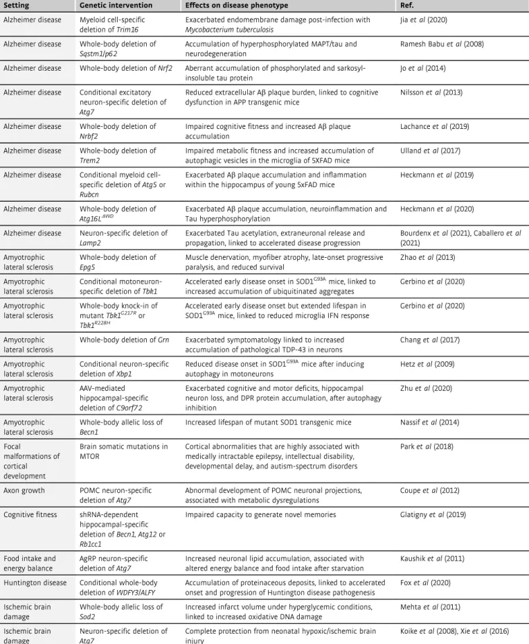
Canonical autophagy is a multiphasic process that involves the sequential and selective recruitment of ATG (autophagy related) proteins (Galluzziet al, 2017a). The initiation of the autophagic cascade is physiologically subjected to the repressive control of MTOR (mechanistic target of rapamycin kinase) complex1(MTORC1), which catalyzes the inactivating phosphorylation of ATG13and ULK1(unc-51-like autophagy-activating kinase1). ULK1and ATG13 are found in a supramolecular complex that also contains RB1CC1(RB1-inducible coiled-coil1) and ATG101, which cooperates with ATG9to promote autophagosome nucleation. The inhibitory action of MTORC1is counterbalanced by AMP-activated protein kinase (AMPK), which responds to dwindling ATP levels by phosphorylating ULK1and BECN1(Beclin1). ULK1favors the autophagic cascade by facilitating the phosphatidylinositol-3-kinase activity of a multiprotein complex formed by BECN1, PIK3C3/VPS34(phosphatidylinositol-3-kinase catalytic subunit type3), PIK3R4/VPS15(phosphoinositide-3- kinase regulatory subunit4), ATG14, and NRBF2(nuclear receptor binding factor2). Multiple regulatory interactors of the BECN1-PIK3C3/VPS34complex have been identified, including UVRAG (UV radiation resistance associated), SH3GLB1(SH3domain containing GRB2like, endophilin B1), and AMBRA1(au- tophagy and Beclin1regulator1), which facilitate the catalytic activity of PIK3C3/VPS34, as well as RUBCN (rubicon autophagy regulator) and BCL2 (BCL2 apoptosis regulator), which inhibit it. The production of phosphatidylinositol-3-phosphate (PtdIns3P), followed by the engagement of PtdIns3P- binding proteins of the WIPI (WD repeat domain, phosphoinositide interacting) family, is instrumental for the expansion of phagophores. This phase is promoted by two distinct ubiquitin-like conjugation modules. The first relies upon the activity of ATG7and ATG10and enables the buildup of a multipro- tein complex composed of ATG5, ATG12and ATG16L1(autophagy-related16-like1). The second one involves ATG3, ATG4, and ATG7 and is ultimately responsible for the cleavage of members of the Atg8-family proteins, including mammalian MAP1LC3/LC3(microtubule-associated protein1light chain3) and their conjugation to phosphatidylethanolamine (PE). Lipidated LC3(LC3-II; which is experimentally employed for quantifying autophagyin vitroand in vivo) serves as a receptor for LC3-interacting region (LIR)-containing proteins, including autophagy substrates and receptors such as SQSTM1/p62(se- questosome1). Upon closure of the phagophore, the resulting autophagosome fuses with a lysosome to form an autolysosome, culminating with the degradation of autophagic substrates by acidic lysosomal hydrolases. AKT1S1, AKT1substrate1; DEPTOR, DEP domain containing MTOR interacting pro- tein; MLST8, MTOR-associated protein, LST8homolog; RPTOR, regulatory-associated protein of MTOR complex1.
SQSTM1 LC3-I
Pre-LC3
ATG12
ATG16L1 ATG5 ATG12 ATG7
ATG10 ATG3 ATG4
ATG5 PE
Ub
WIPIs EXPANSION
Cargo
LC3-II LC3-II
NUCLEATION INITIATION
UVRAG BECN1 AMBRA1
BCL2 ATP
AMP
MTORC1 AMPK
ULK1
RPTOR
MLST8 MTOR
AKT1S1 DEPTOR
ATG101 RB1CC1 ATG13
ATG14
PIK3C3 PIK3R4
ATG9
LIR
© EMBO
fitness, however, autophagy regulation grows in complexity and the outcome of autophagy upregulation is less predictable (Cherra &
Chu, 2008). For example, autophagy can engage in cell death (Fair- lieet al, 2020; Milleret al, 2020), directly contributing to the patho- genesis of some human diseases (e.g., ischemia-reperfusion injury, neuronal, and muscle atrophy) (Galluzziet al, 2018b; Galluzziet al, 2018c; Patel & Karch, 2020; Pervaizet al, 2020).
The autophagy machinery participates in intercellular communi- cation, mediating processes of non-canonical protein secretion (an autophagy-independent function of autophagy proteins) (Ponpuak et al, 2015; Zahoor & Farhan, 2018), regulation of tissue-resident stem cells (Guanet al, 2013; Chang, 2020), modulation of immune cell functions (Deretic, 2021), and maintenance of tissue barrier integrity (Galluzzi & Green, 2019; Levine & Kroemer, 2019). As an example, in dendritic cells (DCs) autophagy and microautophagy serve the important role of feeding endogenous proteins to endoso- mal/lysosomal compartments for MHC class II molecule-mediated immunosurveillance (Balanet al, 2019; Kotsiaset al, 2019), and the biogenesis of endosomal microautophagy is tightly connected to exo- somal production (Sahu et al, 2011). As yet another example, in phagocytic cells several components of the autophagy machinery (including the phosphatidylinositol-3-kinase [PtdIns3K] complex, but not ULK1 [unc-51-like autophagy-activating kinase 1]) are recruited to the single-layered phagosomal membrane, following the engagement of cell surface receptors (e.g., TLRs [Toll-like receptors]) by pathogen-associated molecules (Martinezet al, 2015), immune complexes (Henaultet al, 2012), or phosphatidylserine exposed by apoptotic cells (Martinezet al, 2011). This process, defined as LC3- associated phagocytosis (LAP) (Heckmann & Green, 2019), exqui- sitely relies upon CYBB/NOX2 (cytochrome b-245, beta polypep- tide), RUBCN (rubicon autophagy regulator), and the WD domain of ATG16L1 (autophagy-related 16-like 1), which are dispensable for the execution of canonical autophagy (Martinezet al, 2015).
The multitiered repercussions of autophagy on organismal home- ostasis have spurred considerable efforts toward the identification of clinically actionable targets to modulate the autophagic pathway to prevent or treat diseases, in multiple pathological circumstances (Gal- luzziet al, 2017c). Our current understanding about the contribution of autophagy in human disorders mostly derives from (i) the imple- mentation of several mouse models of autophagy deficiency (Kuma et al, 2004), through which the role of autophagy can be interrogated at the whole body, or in a cell type-specific manner, and (ii) from the discovery that several components of the autophagic machinery have been found mutated in human diseases (van Beeket al, 2018; Levine
& Kroemer, 2019). Here, we discuss recent insights on the role of autophagy in the most penetrant human illnesses (Fig 1), placing par- ticular emphasis on preclinical findings obtained in murine models of diseases in which autophagy has been genetically dismantled. In this regard, the involvement of virtually all ATG (autophagy related) pro- teins in autophagy-independent tasks imposes a note of caution on the attribution of specific phenotypic effects to the mere inhibition of the autophagy process (Galluzzi & Green, 2019).
Neurodegenerative disorders
The autophagic process is essential in preserving the homeostatic requirements of post-mitotic neurons, both at the central and at the
peripheral nervous system levels (Menzieset al, 2017; Scrivoet al, 2018; Mallucci et al, 2020) (Table 1). Most neurodegenerative dis- eases are associated with the accumulation of aggregate-prone pro- teins. Studies performed in diseases with Mendelian-type inheritance suggest that these proteins are toxic drivers that are nec- essary and sufficient to cause pathology. A large body of evidence, supported by the demonstration thatATGgenes are found mutated in multiple human neurodegenerative illnesses, indicates that autop- hagy directly intervenes in the clearance of those proteins (Nixon, 2013). In addition, MTOR p.Cys1483Tyr somatic mutation resulted in impaired autophagy, caused aberrant accumulation of OFD1, and disrupted neuronal ciliogenesis, which accounted for cortical dys- lamination in Focal malformations of cortical development (Tang et al, 2013; Park et al, 2018). Furthermore, intact autophagy responses have been postulated to extinguish neuroinflammatory reactions, which directly contribute to the aetiopathogenesis of neu- rodegenerative disorders (Rubinsztein et al, 2015). For these rea- sons, upregulation of autophagy has attracted particular interest as a potential therapeutic strategy for various neurodegenerative condi- tions (Menzieset al, 2017; Thangarajet al, 2020).
The neuroprotective functions attributed to autophagy are esti- mated to transcend its well-defined roles as proteostasis keeper and organelle turnover regulator. Indeed, several findings have under- scored that the ATG machinery is functionally implicated in compartment-specific tasks along the soma-axon axis that include, among others, (i) the regulation of synaptic transmission (Kuijpers et al, 2021), (ii) the degradation of synaptic cargoes and vesicles, (iii) the anterograde/retrograde crosstalk between cell body and synaptic terminal, and (iv) myelination/demyelination events (Hill
& Colon-Ramos, 2020). With these compartment-specific physiologi- cal functions, it is no surprise that both insufficient and overactive nonselective or selective autophagy responses contribute to neu- rodegeneration (Chu, 2019).
Due to perinatal lethality related to ubiquitous inhibition of autophagy, our current degree of knowledge regarding the relevance of autophagy within the neural lineage mostly stems from fruit flies (Juhasz et al, 2007; Simonsen et al, 2008) and mouse models in which essential (i.e., Atg5, Atg7, Rb1cc1/Fip200 [RB1-inducible coiled-coil 1]) (Hara et al, 2006; Komatsuet al, 2006; Wanget al, 2013) or non-essential (i.e.,Wdr45/Wipi4[WD repeat domain 45], and Wdr45b/Wipi3) (Zhao et al, 2015; Ji et al, 2020; Yamaguchi et al, 2020) autophagic genes have been obliterated at the embry- onic stage by virtue ofNes(nestin)-driven Cre recombinase expres- sion. Compared to their wild-type littermates, mice that developmentally lack autophagy in the neuronal compartment dis- play shortened lifespan and early-onset neurodegenerative patholo- gies (whose severity varies depending on the targeted gene), associated with the pathological accumulation of proteinaceous aggregates in multiple neuronal populations (Hara et al, 2006;
Komatsu et al, 2006; Metcalf et al, 2012). Neuronal dysfunctions account for the lethality associated with systemic autophagic defi- ciency, as testified to by the fact that overexpression ofAtg5in the neuronal compartment rescues perinatal mortality ofatg5 / mice (Yoshii et al, 2016). Blunted expression of PIK3R4/VPS15 (phosphoinositide-3-kinase regulatory subunit 4) is associated with neurodevelopmental impairment and cortical atrophy, matching the phenotype of patients bearing loss-of-function mutations in this gene (Gstreinet al, 2018). Along similar lines,de novomutations in
the autophagy geneWDR45have been found in causal association with static encephalopathy of childhood with neurodegeneration in adulthood (also known as neurodegenerative disease b-propeller protein-associated neurodegeneration [BPAN]), a subtype of neu- rodegeneration with brain iron accumulation (NBIA) (Saitsuet al, 2013) and with human neurodegeneration (Suleimanet al, 2018).
Supporting the possible involvement of autophagy in this pathology, abnormal early autophagosomal structures have been identified in patient-derived lymphoblastoid cell lines (LCLs) (Saitsuet al, 2013).
In concordance with this result, CNS-specificwdr45knockout mice are defined by BPAN-like features, including cognitive defects and impaired axonal homeostasis, but not other ones like iron accumu- lation in basal ganglia (Zhaoet al, 2015). More recently, a mutation inWipi2 (WD-repeat protein interacting with phosphoinositide 2)
has been identified, linking defective autophagy to the appearance of complex neurodevelopmental defects (Jelani et al, 2019).
Impaired autophagosome–lysosome fusion, associated with loss-of- function mutations inEPG5(ectopic P-granule autophagy protein 5 homolog), causes autosomal recessive Vici syndrome (VICIS), pathologically defined by severe neurodevelopmental defects (Hori et al, 2017). The suppression of ATG5 expression during early brain development alters the differentiation trajectories and the rate of proliferation of neuronal progenitor cells, which eventually reflect into morphological defects in differentiated neurons. By analogy, a comparable phenotype has been described inAtg16l1hypomorphic mice (Lvet al, 2014; Wuet al, 2016; Menzieset al, 2017). Recently, a missense mutation inATG5has been found in causal association with the manifestation of ataxia, with neurodevelopmental delay in
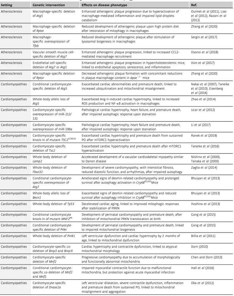
ORGAN-SPECIFIC ILLNESSES SYSTEMIC ILLNESSES OCULAR DISORDERS
• Glaucoma
• Age-related macular degeneration
MUSCULOSKELETAL DISEASES
• Degenerative myopathies
• PDB
• Osteoarthritis
• Osteoporosis NEURODEGENERATIVE DISORDERS
• Alzheimer disease
• Parkinson disease
• Neuropathies
• ALS
CARDIOVASCULAR DISEASES
• Ischemia/reperfusion injury
• Cardiomyopathie
• Atherosclerosis
PULMONARY DISORDERS
• COPD
• Cystic fibrosis
• Pulmonary fibrosis
RENAL DISEASES
• Acute kidney injury
• Chronic kidney disease
REPRODUCTIVE DYSFUNCTIONS
• Female infertility
• Male infertility
• Endometriosis HEPATIC DISORDERS
• Cirrhosis
• Cholestasis
• Hyperammonemia
CANCER
• Breast cancer
• Melanoma
• Pancreatic cancer
• Lung cancer
IMMUNITY TO PATHOGENS
• Bacterial infections
• Viral infections
AUTOIMMUNE DISORDERS
• Inflammatory bowel disease
• Systemic lupus erythematous
METABOLIC SYNDROMES
• Obesity
• Type 2 diabetes
• NAFLD
© EMBO
Figure1. Common human disorders linked to dysregulated autophagic activity.
Representation of the main organ-specific (red) and systemic (blue) human illnesses in which autophagy plays a critical role and that are discussed in this review. ALS, amyotrophic lateral sclerosis; COPD, chronic obstructive pulmonary disease; DKD, diabetic kidney disease; NAFLD, non-alcoholic fatty liver disease; PDB, Paget disease of bone.
Table1. Neurodegenerative disorders associated with genetic intervention of autophagy in mice.
Setting Genetic intervention Effects on disease phenotype Ref.
Alzheimer disease Myeloid cell-specific deletion ofTrim16
Exacerbated endomembrane damage post-infection with Mycobacterium tuberculosis
Jiaet al(2020)
Alzheimer disease Whole-body deletion of Sqstm1/p62
Accumulation of hyperphosphorylated MAPT/tau and neurodegeneration
Ramesh Babuet al(2008)
Alzheimer disease Whole-body deletion ofNrf2 Aberrant accumulation of phosphorylated and sarkosyl- insoluble tau protein
Joet al(2014)
Alzheimer disease Conditional excitatory neuron-specific deletion of Atg7
Reduced extracellular Abplaque burden, linked to cognitive dysfunction in APP transgenic mice
Nilssonet al(2013)
Alzheimer disease Whole-body deletion of Nrbf2
Impaired cognitive fitness and increased Abplaque accumulation
Lachanceet al(2019)
Alzheimer disease Whole-body deletion of Trem2
Impaired metabolic fitness and increased accumulation of autophagic vesicles in the microglia of5XFAD mice
Ullandet al(2017)
Alzheimer disease Conditional myeloid cell- specific deletion ofAtg5or Rubcn
Exacerbated Abplaque accumulation and inflammation within the hippocampus of young5xFAD mice
Heckmannet al(2019)
Alzheimer disease Whole-body deletion of Atg16LDWD
Exacerbated Abplaque accumulation, neuroinflammation and Tau hyperphosphorylation
Heckmannet al(2020)
Alzheimer disease Neuron-specific deletion of Lamp2
Exacerbated Tau acetylation, extraneuronal release and propagation, linked to accelerated disease progression
Bourdenxet al(2021), Caballeroet al (2021)
Amyotrophic lateral sclerosis
Whole-body deletion of Epg5
Muscle denervation, myofiber atrophy, late-onset progressive paralysis, and reduced survival
Zhaoet al(2013)
Amyotrophic lateral sclerosis
Conditional motoneuron- specific deletion ofTbk1
Accelerated early disease onset in SOD1G93Amice, linked to increased accumulation of ubiquitinated aggregates
Gerbinoet al(2020)
Amyotrophic lateral sclerosis
Whole-body knock-in of mutantTbk1G217Ror Tbk1R228H
Accelerated early disease onset but extended lifespan in SOD1G93Amice, linked to reduced microglia IFN response
Gerbinoet al(2020)
Amyotrophic lateral sclerosis
Whole-body deletion ofGrn Exacerbated symptomatology linked to increased accumulation of pathological TDP-43in neurons
Changet al(2017)
Amyotrophic lateral sclerosis
Conditional neuron-specific deletion ofXbp1
Reduced disease onset in SOD1G93Amice after inducing autophagy in motoneurons
Hetzet al(2009)
Amyotrophic lateral sclerosis
AAV-mediated hippocampal-specific deletion ofC9orf72
Exacerbated cognitive and motor deficits, hippocampal neuron loss, and DPR protein accumulation, after autophagy inhibition
Zhuet al(2020)
Amyotrophic lateral sclerosis
Whole-body allelic loss of Becn1
Increased lifespan of mutant SOD1transgenic mice Nassifet al(2014)
Focal
malformations of cortical development
Brain somatic mutations in MTOR
Cortical abnormalities that are highly associated with medically intractable epilepsy, intellectual disability, developmental delay, and autism-spectrum disorders
Parket al(2018)
Axon growth POMC neuron-specific deletion ofAtg7
Abnormal development of POMC neuronal projections, associated with metabolic dysregulations
Coupeet al(2012)
Cognitive fitness shRNA-dependent hippocampal-specific deletion ofBecn1, Atg12or Rb1cc1
Impaired capacity to generate novel memories Glatignyet al(2019)
Food intake and energy balance
AgRP neuron-specific deletion ofAtg7
Increased neuronal lipid accumulation, associated with altered energy balance and food intake after starvation
Kaushiket al(2011)
Huntington disease Conditional whole-body deletion ofWDFY3/ALFY
Accumulation of proteinaceous deposits, linked to accelerated onset and progression of Huntington disease pathogenesis
Foxet al(2020)
Ischemic brain damage
Whole-body allelic loss of Sod2
Increased infarct volume under hyperglycemic conditions, linked to increased oxidative DNA damage
Mehtaet al(2011)
Ischemic brain damage
Neuron-specific deletion of Atg7
Complete protection from neonatal hypoxic/ischemic brain injury
Koikeet al(2008), Xieet al(2016)
human patients. Notably, the introduction of human mutatedATG5 in flies is sufficient to recapitulate the clinical feature of the human disorders (Kimet al, 2016).
Disturbance in the autophagic process also has an impact on neu- rogenesis, which testifies to the central role of autophagy in the
maintenance of adult neural stem cell pools within the sub- ventricular zone (SVZ) of the lateral ventricle wall and subgranular zone (SGZ) of the dentate gyrus (Fleming & Rubinsztein, 2020).
Consistent with this finding, inhibition of autophagy elicited by Rb1cc1 ablation reduces differentiation potential and number of Table1 (continued)
Setting Genetic intervention Effects on disease phenotype Ref.
Nerve injury Schwann cell-specific deletion ofAtg7
Delayed myelin degradation and generation of repair cells after injury
Gomez-Sanchezet al(2015)
Neurodegeneration Neural cell-specific deletion ofAtg5
Development of progressive deficits in motor function linked to cytoplasmic inclusion body accumulation in neurons
Haraet al(2006)
Neurodegeneration Conditional CNS-specific deletion ofAtg7
Behavioral defects and premature death, linked to massive neuronal loss and cytoplasmic inclusion body accumulation
Komatsuet al(2006)
Neurodegeneration Conditional radial glial cell- specific deletion ofRb1cc1
Progressive loss of NSCs pool and impaired neuronal differentiation in the postnatal brain
Wanget al(2013)
Neurodegeneration Conditional CNS-specific deletion ofWdr45
Reduced motor coordination, impaired learning and memory, and extensive axon swelling
Zhaoet al(2015)
Neurodegeneration Conditional neuron-specific deletion ofWipi3
Behavioral defects and cerebellar neuronal loss after non- canonical autophagy inhibition
Yamaguchiet al(2020)
Neurodegeneration Conditional telencephalon- specific deletion ofVps15
Severe progressive cortical atrophy associated with caspase- induced apoptosis
Gstreinet al(2018)
Neurodegeneration Whole-body knock-in of hypomorphicAtg16l1
Developmental retention due to delayed differentiation of stem cells in the brain
Wuet al(2016)
Neurodegeneration Conditional NSC-specific co- deletion ofFoxO1, FoxO3 and FoxO4
Initial proliferation of neural progenitor cells in early postnatal life, followed by NSC pool decline in adult brains
Paiket al(2009)
Neurodegeneration Purkinje cell-specific deletion ofAtg7
Progressive cell autonomous dystrophy and degeneration of the axon terminals
Komatsuet al(2007)
Neurodegeneration Whole-body deletion of TAX1BP1
Aberrant accumulation of high molecular weight ubiquitin conjugates and lipofuscin
Sarrafet al(2020)
Neuropathies Whole-body deletion of Fam134b
Degeneration of sensory neurons after inhibition of ER-phagy Khaminetset al(2015)
Neuropathies Whole-body deletion of Tecpr2
Exacerbated age-dependent behavioral aberrations and neuroaxonal dystrophy, after accumulation of autophagosomes
Tamim-Yecheskelet al(2020)
Neurotransmission Post-mitotic excitatory neuron-specific deletion of Atg5
Increased accumulation of tubular ER in axons, linked to increased excitatory neurotransmission and premature death
Kuijperset al(2021)
Parkinson disease Microglia-specific deletion ofAtg7
Increaseda-synuclein accumulation and neurodegeneration Choiet al(2020)
Parkinson disease Whole-body deletion of Rubcn
Reduceda-synuclein accumulation in the brain, linked to reduced age-related interstitial fibrosis in kidney
Nakamuraet al(2019)
Parkinson disease Conditional SN neuron- specific deletion ofAtg7
Resistance to retrograde axonal degeneration Chenget al(2011)
Parkinson disease AAV-mediated SN-specific knock-in of dominant- negative Ulk1
Attenuated MPTP-induced axonal neurodegeneration Balkeet al(2020)
Parkinson disease Whole-body deletion ofPrkn Impaired striatal neural plasticity, linked to increased sensitivity to oxidative damage and mitochondrial
dysfunction (exacerbated in Mutator mice but rescued by loss of STING)
Goldberget al(2003), Palacinoet al (2004), Kitadaet al(2009), Pickrell et al(2015), Sliteret al(2018)
Parkinson disease Whole-body deletion of Pink1
Increased sensitivity to oxidative damage and mitochondrial dysfunction
Gautieret al(2008)
AAV, adeno-associated viral vector; AgRP, agouti-related protein; APP, amyloid precursor protein; CNS, central nervous system; DPR, dipeptide-repeated; MPTP, 1-methyl-4-phenyl-1,2,3,6-tetrahydropyridine; NSCS, neural stem cell; OGD, oxygen glucose deprivation; POMC, proopiomelanocortin; SN, substantia nigra;
TDP-43, transactive response DNA-binding protein of43kD.
adult neural stem cells (Wanget al, 2013). Likewise, combined con- ditional deletion of genes coding for FOXO (forkhead box, sub- group O;Foxo1, Foxo3,andFoxo4) in adult neural stem/progenitor cells correlates with abnormal morphological features of differenti- ated neurons (Paiket al, 2009).
Throughout the last decade, several mouse models of conditional autophagy disruption in specific populations of the CNS and periph- eral nervous system have been implemented, revealing the cell type-specific contribution of autophagy. These encompass Purkinje cells in the cerebellum (leading to progressive dystrophy) (Komatsu et al, 2007), hypothalamic AGRP (agouti-related neuropeptide) neu- rons (evoking altered energy balance and food intake after starva- tion) (Kaushiket al, 2011), POMC (proopiomelanocortin) neurons (perturbing axon growth and decreasing a-melanocyte-stimulating hormone [MSH] levels) (Coupe et al, 2012; Kaushik et al, 2012), and Schwann cells (delaying the process of demyelination after injury) (Gomez-Sanchezet al, 2015).
Functional autophagic responses are instrumental for preserving neuronal integrity upon circumstances of acute injury (Galluzzi et al, 2016). For example, it has been shown that a central role of autophagy is restraining the life-threatening effect tied to brain ischemic challenge. In mice in which cerebral stroke was induced by transient middle carotid occlusion (MCAO), genetic interventions that undermine autophagy, includingSod2(superoxide dismutase 2, mitochondrial) inactivation (Mehtaet al, 2011) or shRNA-mediated silencing ofTsc1(TSC complex subunit 1) (Papadakiset al, 2013), aggravate the neurological sequelae instigated by the stroke episode.
In apparent contrast with this finding, pharmacological inhibition of autophagy with 3-methyladenine or bafilomycin A1was observed to limit infarct size in a permanent MCAO, suggesting that autophagy may rather aggravate the ischemic injury (Zhanget al, 2013; Gal- luzziet al, 2016). Although the reduced specificity of these pharma- cological modulators limits the mechanistic interpretation of these results, it is nonetheless reasonable to propose that the actual con- tribution of autophagy in stroke-associated neurotoxicity would vary depending upon the cerebral compartment affected and the developmental stage in which the ischemic episode occurs (Galluzzi et al, 2016). In support of this concept, brain-specific deletion of Atg7confers protection against neonatal hypoxia–ischemia injury in mice (Koikeet al, 2008; Xieet al, 2016).
Intact hippocampal autophagy sustains the elevated degree of synaptic plasticity required to generate novel memories, as demon- strated by the fact that stereotactic delivery of shRNA targeting key autophagy genes (including Becn1 [Beclin 1, autophagy related], Rb1cc1, andAtg12) impairs cognitive fitness in mice (Glatignyet al, 2019). This effect, which can be phenocopied by pharmacological inhibition of autophagy (e.g., with spautin-1, leupeptin, or chloro- quine) and reversed by pharmacological activation of the ATG machinery with a Tat-Beclin 1 peptide, supports the essential role of autophagy in dendritic spine formation and long-term potentiation after stimuli (Glatignyet al, 2019). Of note, loss of autophagy per- formance may causally underlie the age-dependent decline in mem- ory tasks, as demonstrated by the fact that treatment of old mice with plasma derived from young donors improves cognitive fitness and restores normal levels of autophagy in the hippocampus (Glatignyet al, 2019). Further corroborating this result, dietary sup- plementation with spermidine, which also acts as an autophagy stimulator, mitigates age-dependent cognitive impairment in mouse
hippocampus andDrosophilaheads, contingent upon intact autop- hagy and mitophagy responses (Schroederet al, 2021).
In the recent past, autophagy has gained attention for its poten- tial involvement in the pathogenesis of late-onset neurodegenerative pathologies, owing to the historically rooted view of this pathway as a major determinant of long-lived/aggregation-prone protein dis- posal within the lysosome (Nixon, 2013; Menzieset al, 2017). Sup- porting this view, it has been demonstrated that the lack of the autophagic receptor TAX1BP1 (Tax1-binding protein 1) results in aberrant protein aggregation in the brain (Sarraf et al, 2020).
Although these disorders mainly follow a multifactorial pattern, evi- dence obtained from inherited variants of neurodegenerative ill- nesses has shed new light on the contribution of autophagy to the progressive loss of neural function.
Alzheimer disease
Alzheimer disease (AD) represents the most common form of dementia in humans, caused by the pathologically relevant accumu- lation of proteinaceous aggregates, i.e., intracellular MAPT/tau tan- gles and/or extracellular beta amyloid peptide [Ab] plaques, which progressively leads to neuronal cell death and decline in cognitive functions. Connections between autophagy and AD originate from the observation of expansion of autophagic compartments in AD brains (Nixonet al, 2005). As recently revealed by multilayer brain proteomics analysis performed at different stages of AD in humans, the autophagic substrate SQSTM1/p62 (sequestosome 1) accumu- lates in AD, suggestive of impaired autophagic flux (Baiet al, 2020) similar to the one reported in AD experimental models (Yuet al, 2005). In support of this notion, functional autophagy is required to degrade soluble and aggregated variants of MAPT/tau (Bergeret al, 2006; Silva et al, 2020). Lysosomal membrane lesions caused by MAPT/tau oligomers instigate an LGALS3 (galectin 3)-coordinated program, which leads to autophagy activation (Jia et al, 2020).
Genetic inactivation of SQSTM1/p62 in mice leads to accumulation of hyperphosphorylated MAPT/tau and neurodegeneration (Ramesh Babu et al, 2008). Supraphysiological accumulation of MAPT/tau tangles perturbs the retrograde axonal transport of autophagosomes by interfering with the dynein–DCTN (dynactin) complex, eventu- ally instigating the detrimental accumulation of MAPT/tau- containing autophagic vesicles (Butzlaffet al, 2015).
Notably, the NFE2L2/NRF2 (nuclear factor, erythroid-derived 2, like 2)-dependent transcription of the autophagy regulator CALCOCO2/NDP52 (calcium binding and coiled-coil domain 2) is instrumental in promoting the degradation of MAPT/tau in response to oxidative stress (Joet al, 2014). SQSTM1/p62 is also a target gene for NFE2L2/NRF2 (Jainet al, 2010), and it has been reported to medi- ate degradation of aggregated MAPT/tau (Xuet al, 2019b). In recent years, dysfunction of the endosomal-sorting complex, the retromer, has been linked to a number of neurodegenerative diseases, including AD. Reduced expression of the retromer proteins and variants of the core retromer component VPS35 (vacuolar protein sorting 35) are associated with neurodegenerative diseases, often overlapping with MAPT/tau aggregation in the brain (Carosi et al, 2021; Seaman, 2021). Recent data demonstrate that the autophagy–lysosomal axis is central for the clearance of aggregated MAPT/tau and depletion of VPS35 blocks autophagy, whereas VPS35 overexpression has the opposite effect (Carosi et al, 2020; Carosi et al, 2021). Thus, the retromer–autophagy axis may play a relevant function in preventing
multiple neurodegenerative diseases by ensuring that pathogenic pro- tein aggregates are cleared as they arise.
In addition, multitiered connections have been established between autophagy and Ab plaque formation. Ab is targeted for autophagy-dependent degradation within the lysosome, explaining why activation of autophagy reduces the burden of Abplaques in rodents (Bolandet al, 2008; Menzieset al, 2017; Menget al, 2019).
However, autophagy appears to be causally implicated in the PSEN1 (presenilin 1)-mediated conversion of APP (amyloid beta precursor protein) into Ab(Yuet al, 2005), as well as in the non-canonical secretion of Ab into the extracellular space (Nilsson et al, 2013;
Menzieset al, 2017). Mutations that alter PSEN1 function have been associated with defective autophagic vesicle clearance and early- onset AD, due to impaired autophagosome–lysosome fusion and defective lysosomal acidification (Lee et al, 2010b; Chong et al, 2018). Similarly, loss-of-function mutations affecting PICALM (phosphatidylinositol-binding clathrin assembly protein) impair autophagy dynamics, thus augmenting the risk for developing AD (Tianet al, 2013).
Additional autophagy modulators determine the cellular levels of Abprotein. As an example, NRBF2 (nuclear receptor-binding factor 2; a component of the PtdIns3K complex I) interacts with APP and favors its lysosomal disposal, as demonstrated by the fact that NRBF2 depletion leads to excessive levels of intracellular APP in cells (Yanget al, 2017b) and Abaccumulation in AD mouse models (Lachanceet al, 2019), whereas overexpression of NRBF2 reduces Ab levels and improves mouse memory (Lachance et al, 2019).
Recently, a possible link between autophagy activation in the micro- glial compartment and AD has been proposed. Importantly, ablation of the gene coding for TREM2 (triggering receptor expressed on myeloid cells 2), a surface receptor required for microglial responses to neurodegeneration, results in maladaptive accumulation of autophagosomes and disarray of microglia clustering around plaques (Ullandet al, 2017). This effect has been attributed to dys- regulated MTORC1 activation, in turn evoking metabolic abnormali- ties in microglial cells. Consistent with this notion, normalization of autophagic flux by cyclocreatine decreases neuronal dystrophy in murine models of AD (5XFAD mice) (Ulland et al, 2017). In this landscape, defective mitophagy appears to be a major determinant of the functional decay of neurons in AD, in that its pharmacological stimulation (through NAD+supplementation, urolithin A, and acti- nonin) is sufficient to retard memory impairment, while reducing the burden of amyloid aggregates upon stimulating microglial phagocytic capacity for extracellular Abplaques (Fanget al, 2019).
In addition, non-canonical functions of the ATG machinery in microglia contribute to alleviate the toxic effects associated with Ab plaque deposition in the 5XFAD mouse model. Notably, the genetic ablation ofAtg5orRubcn(but not that ofRb1cc1) in myeloid cells correlates with exacerbated Abplaque formation and aberrant pro- duction of inflammatory cytokines, while contributing to accelerate neuronal decay and cognitive impairment. Mechanistically, ATG5 and RUBCN take part in events of MAP1LC3/LC3 (microtubule- associated protein 1 light chain 3) conjugation to Ab–containing endosomal membranes positively marked by RAB5 and clathrin.
This process, named LC3-associated endocytosis (LANDO), appears to promote the recycling of putative Ab receptors (e.g., TLR4, TREM2 [triggering receptor expressed on myeloid cells 2]) from internalized endosomes to the plasma membrane of microglial cells.
While it remains to be clarified whether LANDO mediates Abrecep- tor degradation, its activation is instrumental to reduce Abburden and limit neuroinflammation in AD (Heckmannet al, 2019). Along similar lines, LANDO deficiency imposed on aged mice by deletion of the WD domain of ATG16L1 (which is dispensable for canonical autophagy), exacerbates the neuroinflammatory phenotype associ- ated with an AD-like symptomatology (Heckmannet al, 2020).
Chaperone-mediated autophagy also contributes to degradation of a large fraction of neuronal MAPT/tau under physiological condi- tions (Caballeroet al, 2018; Caballeroet al, 2021). However, muta- tions and posttranslational modifications of this protein, such as acetylation, not only prevent MAPT/tau degradation by CMA but also inhibit normal CMA functioning (Caballeroet al, 2018; Cabal- leroet al, 2021). Blockage of CMA leads to rerouting of some of the pathogenic forms of MAPT/tau toward endosomal microautophagy, as both pathways share the same chaperone, HSPA8, and this pro- motes fusion of late endosomes with the plasma membrane and subsequent extraneuronal release of the MAPT/tau variants, thus contributing to MAPT/tau propagation (Caballero et al, 2021).
Reduction in neuronal CMA activity has been recently shown in AD patient’s brains (Bourdenx et al, 2021; Caballeroet al, 2021), and pharmacological activation of CMA has been linked to ameliorated pathology in two different experimental models of tauopathies (Bourdenxet al, 2021).
Parkinson disease
Parkinson disease (PD) is pathologically defined by (i) the loss of dopaminergic neurons in the substantia nigra (SN) and (ii) the prevalence of proteinaceous Lewy bodies, mainly composed of SNCA/a-synuclein (synuclein alpha) and other polyubiquitinated proteins but also vesicular structures. PD symptomatology is charac- terized by prominent motor and autonomic dysfunction, sometimes accompanied by cognitive and psychological deficits. Early evidence suggested roles for CMA and macroautophagy in degrading SNCA/
a-synuclein (Webbet al, 2003; Cuervoet al, 2004). High expression of wild-type SNCA/a-synuclein, mutations or unwanted posttransla- tional modifications on this protein (such as formation of dopamine adducts) is toxic to CMA by preventing multimerization of LAMP2A and subsequent lysosomal internalization of cargo proteins (Cuervo et al, 2004; Martinez-Vicente et al, 2008). Recent evidence has demonstrated that selective autophagy clears neuron-released SNCA/a-synuclein through the autophagy receptor SQSTM1/p62 in microglia, offering protection of dopaminergic neurons (Choiet al, 2020). Consistent with this result, the activation of autophagy decreases the accumulation of SNCA/a-synuclein (Nakamuraet al, 2019). Conversely, uncontrolled expression of wild-type or mutated variants of SNCA/a-synuclein reduces autophagic flux or disturbs TFEB-mediated lysosomal biogenesis by preventing the nuclear translocation of TFEB (Decressacet al, 2013). Pathologically mean- ingful levels of SNCA/a-synuclein affect the intracellular localization of ATG9 via RAB1A (RAB1A, member RAS oncogene family), thereby perturbing autophagy dynamics in the brain of transgenic mice overexpressing SNCA/a-synuclein (Winslow et al, 2010).
Mutations in the geneGBA(glucosylceramidase beta) represent the most common genetic risk factor for PD. Of note, loss-of-function mutations inGBAdisrupt the autophagic flux and lead to the aggre- gation of SNCA/a-synuclein (Murphy et al, 2014). Likewise, an autosomal-dominant mutation affecting VPS35 curtails autophagy
by altering ATG9 localization (Zavodszky et al, 2014). A similar phenotype has also been described in the context of loss-of-function mutations in the P-type ATPase geneATP13A2, in which recessive, early-onset PD has been linked to defective acidification of lyso- somes and insufficient autophagy (Ramirezet al, 2006). Decreased autophagy in ATP13A2-deficient neurons in turn leads to accumula- tion of damaged mitochondria with increased leakage of reactive oxygen species (ROS) (Gusdonet al, 2012).
Dysregulated autophagy has also been associated with the expression of dominant mutants of LRRK2 (leucine-rich repeat kinase 2) (Ramonetet al, 2011), the most common cause of familial PD. While it remains controversial whether LRRK2G2019S elicits increased or decreased autophagic flux, these differences may reflect the compartment (soma vs. dendrites vs. axons) being studied.
Although autophagy upregulation may contribute to clearance of protein aggregates, the axo-dendritic arbor is susceptible to autophagy-mediated degeneration in cultured dopaminergic, sympa- thetic, and cortical neurons and in the axons of dopaminergic neu- ronsin vivo as evidenced by Atg7 knockdown/knockout (Plowey et al, 2008; Cheng et al, 2011), expression of dominant-negative ULK1 (Balkeet al, 2020), or expression of an autophagy-deficient LC3 phosphomimic, which protects against dendritic atrophy elicited by disease-linked LRRK2 mutations and the PD toxin MPP+ (Cherra et al, 2010). Increased mitophagy, due to post-synaptic mitochondrial calcium dysregulation, may contribute to dendritic degeneration (Verma et al, 2017). Emerging roles for LRRK2 in regulating RAB GTPases and other aspects of endolysosomal and vesicular transport may also complicate interpretation due to compensatory responses (Kuwahara & Iwatsubo, 2020).
A causal association has been established between autosomal recessive forms of PD and mutations affecting the mitophagy regula- tors PINK1 (PTEN-induced putative kinase 1) and PRKN/PARK2 (Parkin RBR E3 ubiquitin protein ligase) (Kitadaet al, 1998; Valente et al, 2004; Narendraet al, 2008; Matsudaet al, 2010). Mouse mod- els to monitor mitophagy show elevated basal mitophagy in dopaminergic neurons (McWilliams et al, 2018). Although PINK1 (McWilliamset al, 2018) and PRKN (Goldberget al, 2003; Perez &
Palmiter, 2005) deficiency do not elicit major defects under baseline conditions, defective striatal neural plasticity is observed inprkn / mice (Kitada et al, 2009). Importantly, mitophagy deficiency favored by ablation of Prkn (Palacino et al, 2004; Pickrell et al, 2015) or Pink1 (Gautier et al, 2008) sensitizes mice to oxidative stress, while worsening neural damage when combined with mito- chondrial dysfunction (mitochondrial DNA [mtDNA] mutator-prkn/
parkin-KO mice) (Pickrell et al, 2015). However, there are other pathways of mitophagy in neurons (Chuet al, 2013), and ablation ofPink1orPrknin mouse and fly mitophagy biosensor models sug- gests that neither protein is necessary to maintain normal basal levels of brain mitophagy (Lee et al, 2018a; McWilliams et al, 2018). Furthermore, serological markers of inflammation, which are also observed in individuals withPrknmutations, are reduced lead- ing to reversal of neuronal degeneration when these mice are crossed to STING1/STING (stimulator of interferon response cGAMP interactor 1)-deficient mice (Sliteret al, 2018). These results match the original observation indicating a close association between PD and serum or cerebrospinal fluid markers of inflammation, further reinforcing the concept that neuroinflammation directly contributes to the pathogenesis of PD (Dzamkoet al, 2015).
Polyglutamine diseases
Extensive experimental evidence has highlighted the role of autop- hagy in disorders caused by polyglutamine (polyQ) expansion, including Huntington disease (HD) and several forms of spinocere- bellar ataxias (Jimenez-Sanchezet al, 2012). The polyQ expansion in HTT (huntingtin) is the etiological driver of HD (Zheng et al, 2010), and the severity thereof is a direct function of polyQ length.
Importantly, a significant dichotomy has emerged between the func- tions of wild-type and mutated HTT toward the regulation of the autophagic process (Martin et al, 2015; Ashkenazi et al, 2017).
Wild-type HTT participates in the regulation of basal autophagy due to its role in the selection of the autophagic cargo (Ochabaet al, 2014; Ruiet al, 2015). However, expression of mutant HTT (i) nega- tively affects autophagosomal cargo recognition through dysregu- lated interaction with SQSTM1/p62 (Martinez-Vicente et al, 2010;
Ruiet al, 2015); (ii) sequesters the BECN1 interactor RASD2/RHES in the striatum (Mealer et al, 2014) and inhibits BECN1-PIK3C3/
VPS34 and ULK1 kinase activities (Lim et al, 2015; Wold et al, 2016); (iii) interferes with the regulatory interaction between ATXN3 (ataxin 3) and BECN1, compromising the response of neu- rons to starvation (Ashkenazi et al, 2017); (iv) disturbs axonal autophagosome transport (Wong & Holzbaur, 2014b); (v) drives a maladaptive unfolded protein response, which leads to ERN1/IRE1 (endoplasmic reticulum to nucleus signaling 1)-dependent inhibition of autophagy (Leeet al, 2012); and (vi) disrupts the ability of wild- type HTT to bind ULK1 and release it from the negative regulation of MTOR in order to activate autophagy (Ruiet al, 2015). Notably, overexpression of wild-type HTT in cells expressing its mutated variants restores autophagy and fosters the clearance of mutated HTT (Zhenget al, 2010). Of note, defective autophagy imposed by heterozygous depletion of the autophagy scaffold/adaptor WDFY3/
ALFY (WD repeat and FYVE domain containing 3) accelerates the onset (and worsens the sequelae) of HD in mice (Foxet al, 2020).
Interestingly, experimental rerouting of mutant HTT for degradation by CMA has proven effective in ameliorating disease phenotype in mice (Baueret al, 2010).
Neuropathies
Neuropathies are disorders caused by the progressive degeneration and death of peripheral sensory (e.g., hereditary sensory and auto- nomic neuropathy [HSAN]) and motor (hereditary spastic paraple- gia [HSP], Spastic paraplegia type 49 [SPG49]) neurons. Mutations in genes encoding several ER proteins involved in ER-remodeling have been associated with hereditary neuropathies (Hubner & Dikic, 2020). For example, loss-of-function mutations in the reticulon type ER membrane protein RETREG1/FAM134B (reticulophagy regulator 1) are associated with the development of HSAN type II (HSAN2) (Kurthet al, 2009; Murphyet al, 2012), whereas mutations in RTN2 (reticulon 2) are linked with HSP (SPG12) (Montenegroet al, 2012).
RETREG1 was identified as the first mammalian receptor for selec- tive ER autophagy (reticulophagy) implicated in the delivery of ER fragments via autophagosomes for lysosomal degradation (Khami- netset al, 2015). RETREG1 also plays a role in the clearance of ER- to-lysosome-associated degradation (ERAD)-resistant SERPINA1/
alpha-1 antitrypsin Z variant polymers (Fregnoet al, 2018) as well as endogenous procollagen (Forrester et al, 2019) within the ER.
Some patients with mutations in RETREG1 suffer from cardiac arrhythmia, an- or hypohydrosis and other symptoms of autonomic
malfunctions overlapping with amyotrophic lateral sclerosis (ALS) and myopathies (Eggermannet al, 2018). The HSAN-related ATL3 (atlastin GTPase 3) Y192C mutation has been connected to reduced complexity of the endoplasmic reticulon network, disturbed connec- tions between ER and mitochondria, and impaired mitochondrial function (Kornaket al, 2014; Behrendtet al, 2019; Krolset al, 2019;
Xu et al, 2019a). Mutations in ATL1 paralog can also result in autosomal-dominant spastic paraplegia (SPG3) (Zhaoet al, 2001) or in HSAN type I (HSAN1) (Guellyet al, 2011). Atlastins in general are thought to remodel the ER for efficient autophagosomal degrada- tion and functioning downstream of the reticulophagy receptor RETREG1 (Lianget al, 2018). As a caveat, it is worth mentioning that ATL1 and ATL3 are implicated in multiple ER-related pathways.
Therefore, additional studies are required to validate the hypothesis that dysfunctional autophagy primarily contributes to the pheno- typic aberrations associated with mutations affecting these genes.
Spastic paraplegia type 49 (SPG49) is a severe neurodegenerative disorder that starts in infancy and is caused by several mutations in the TECPR2 (tectonin beta-propeller repeat containing 2) gene.
Frame-shift mutations in exon 8 and exon 16 of TECPR2 (c.1319delT, c.3416delT) terminate in a premature stop codon (Oz- Leviet al, 2012; Heimeret al, 2016), and an initial link between this gene to defects in autophagy was reported (Behrendset al, 2010;
Oz-Leviet al, 2012). All SPG49 patients share unique dysmorphic features such as microcephaly, dental crowding, short chubby appearance and a short, broad neck, and suffer from evolving spas- ticity, moderate to severe intellectual disability, decreased pain sen- sitivity and infantile onset of chronic respiratory disease (Oz-Levi et al, 2012; Heimeret al, 2016). TECPR2 is a multi-domain protein comprised of three WD repeats at the N terminus, the mostly unstructured middle region and six TECPR2 repeats terminating with an LC3-interacting region (LIR) motif at its C terminus (Behrends et al, 2010; Stadelet al, 2015). TECPR2 was originally identified as an interactor of the Atg8-family proteins, a detailed interactome of TECPR2 validated its interaction with Atg8-family proteins through its functional LIR motif, and in addition identified its interaction with the biogenesis of lysosomal organelles complex 1 (BLOC1) and the homotypic fusion and protein sorting (HOPS) complex, two tethering protein complexes that mediate autophago- some–lysosome fusion (Stadelet al, 2015). A model for SPG49 was recently developed by creating a tecpr2 knockout mouse using CRISPR-Cas9 (Tamim-Yecheskel et al, 2020). This mouse exhibits behavioral aberrations accompanied by neuroaxonal dystrophy and autophagosome accumulation in the brainstem and spinal cord that is exacerbated in an age-dependent manner. The accumulation of autophagosomes upontecpr2knockout suggests compromised tar- geting to lysosomes. Consistently, SPG49-derived primary skin fibroblasts also exhibit accumulation of autophagosomes, strictly under basal growing conditions (Fraiberget al, 2020). This pheno- type is recovered by ectopically expressing the six carboxy-terminal TECPR2 repeats, the full length TECPR2 protein or by inhibition of MTOR (Fraiberg et al, 2020). Mechanistically, TECPR2 has been suggested to facilitate targeting of autophagosomes to lysosomes, a process that is dependent on its C-terminal LIR motif.
Recent studies of rare movement disorders have also provided links to autophagy.VPS13Dis a rare disease gene, with mutations inVPS13Dbeing associated with pediatric and young adult spastic ataxia or spastic paraplegia (Gauthier et al, 2018; Seong et al,
2018). Significantly, VPS13D is a regulator of autophagy, mitochon- drial size, and mitochondrial clearance (Andinget al, 2018). These cellular phenotypes appear to be caused by altered mitochondria and ER contact, a phenotype that is conserved between flies and patient-derived cells (Shenet al, 2021). Furthermore, a recent study indicated that mutations in VPS13D occur in 3 out of 64 children with Leigh syndrome features (Leeet al, 2020).
Further, a very recent study has identified a novel role for TRK- fused gene (TFG) in autophagy (Carinci et al, 2021). TFG is an essential protein in the regulation of vesicular trafficking between endoplasmic reticulum and Golgi, and severalTFGmutations have been associated with different neurological disorders, including hereditary motor and sensory neuropathy with proximal dominant involvement (HMSN-P), Charcot-–Marie–Tooth disease, and reces- sive hereditary spastic paraparesis (Yagiet al, 2016). Indeed, under starvation conditions, TFG controls proper ULK1 localization and steady-state levels by interacting with LC3C via a canonical LIR motif; this, in turn, regulates autophagy progression. These defects are also recapitulated in fibroblasts from a patient carrying an R106C TFG variant that has been previously associated with a com- plicated hereditary spastic paraplegia (HSP) phenotype (Beetzet al, 2013).
Amyotrophic lateral sclerosis
Amyotrophic lateral sclerosis is etiologically associated with the aber- rant amassing of misfolded proteins, including SOD1 (superoxide dis- mutase 1), TARDBP/TDP-43 (TAR DNA binding protein), or with the translation of dipeptide repeat proteins from the C9orf72 expanded repeat (the latter accounting for the most common variant of ALS) in motor neurons. ALS forms a genetic and pathological continuum with frontotemporal dementia (FTD). Interestingly, several FTD-ALS genes code for autophagy receptors, including SQSTM1/p62 and OPTN (optineurin), lowering the capacity of neural cells to clear protein aggregates, as do mutations in VCP (valosin containing protein). As an example, SQSTM1/p62 mutants fail to dispose of aggregation- prone SOD1 and TARDBP (Galet al, 2009; Bradyet al, 2011; Goode et al, 2016; Denget al, 2020). Likewise, defective OPTN, leading to impaired binding to MYO6 (myosin VI), compromises autophagoso- mal trafficking (Tumbarelloet al, 2012; Wong & Holzbaur, 2014a).
Further supporting the role of OPTN in ALS, mutations in TBK1 (TANK binding kinase 1), which phosphorylates OPTN and promotes mitophagy, lead to detrimental accumulation of damaged mitochon- dria (Moore & Holzbaur, 2016). Of note, loss of TBK1 activity in SOD1G93Amouse models of ALS curtails autophagy and accelerates the clinical manifestation of ALS (Gerbinoet al, 2020).
The strict nexus between ALS and autophagy is further strength- ened by experimental evidence indicating that genetic deletion of central (e.g., VCP) (Johnsonet al, 2010) or ancillary regulators of the autophagic cascade (e.g., GRN/progranulin, ALS2/alsin-2) pre- cipitate ALS symptomatology in mice and human patients (Yang et al, 2001; Changet al, 2017). VCP also cooperates with PINK1 in regulating mitophagy and promoting PINK1-dependent neuronal dendritogenesis through an independent mechanism (Kim et al, 2013b; Wang et al, 2018b). Mutations in the ESCRT-III subunit CHMP2B (charged multivesicular body protein 2B)—required to sort integral membrane proteins into intraluminal vesicles of the multi- vesicular body (MVB)—have been causally linked to frontotemporal dementia and ALS. Mechanistically, mutated CHMP2B undermines
autophagy-mediated degradation, resulting in an elevated burden of SQSTM1/p62- and WDFY3-containing protein aggregates in neu- rons. Further corroborating the central role of MVBs in the mainte- nance of neuronal proteostasis, MVBs are essential for the clearance of ubiquitinated TARDBP, which accumulates in ALS and fron- totemporal lobar degeneration (Filimonenkoet al, 2007). Mitophagy also appears to be defective in ALS (Wong & Holzbaur, 2014a). As result and in a non-mutually exclusive manner, an impairment of ESCRT-III function in phagophore sealing during mitophagy could contribute the ALS pathophysiology (Smithet al, 2019; Zhenet al, 2020). While these experimental observations suggest that defective autophagy may directly contribute to the phenotypic alterations linked to mutations in these genes, the fact that these proteins are involved in several autophagy-unrelated processes imposes a note of caution on the interpretation of these results.
Conversely, genetic interventions that promote autophagy, such as the inactivation of the transcription factor XBP1 (X-box binding protein 1) or restoration of HSPB8 expression in the nervous system, counteract ALS symptomatology by promoting the autophagy- dependent disposal of SOD1G93A (Hetz et al, 2009; Crippa et al, 2010). Mutated forms of C9orf72 lead to the clinical manifestation of ALS through a number of different mechanisms. Because wild- type C9orf72 is involved in central aspects of autophagosomes for- mation, maturation, and trafficking, it is likely that perturbations in autophagy contribute to the detrimental action of mutated C9orf72 in motor neuron dysfunction (Websteret al, 2016; Hoet al, 2019).
Supporting this notion, genetic ablation ofC9orf72 correlates with an increased burden of SQSTM1/p62 and TARDBP protein aggrega- tion and synergizes with polyQ ATXN2 to induce the demise of motor neurons (Sellier et al, 2016). Consistently, it has been recently observed that loss of wild-type C9orf72 function exacer- bates the neurotoxic effects of aC9orf72mutant allele, bearing hex- anucleotide expansions, by repressing autophagy (Zhuet al, 2020).
Conversely, the unexpected increase in lifespan elicited by BECN1 haploinsufficiency in the mutant SOD1 transgenic mouse model of ALS (Nassifet al, 2014) is difficult to reconcile. As for all the dis- eases discussed in this review, apparently conflicting, context- dependent conclusions indicate a nuanced relationship between autophagy dysregulation and neurodegeneration.
Cardiovascular diseases
Cardiovascular disorders represent the leading cause of death world- wide. Cardiomyocytes, the essential cellular constituents of the car- diovascular system, mostly lay in the post-mitotic state, implying that they are highly dependent upon intact autophagy and mito- phagy to preserve their physiological functions and cope with harm- ful insults (Lavanderoet al, 2015; Kaludercicet al, 2020) (Table 2).
In view of the reduced regenerative potential of the cardiovascular system, autophagy operates at the forefront to promote survival of quiescent cells in the cardiovascular compartment, while counter- acting events of apoptotic or necrotic cell death after injury (Hen- ning & Brundel, 2017; Sciarrettaet al, 2018).
Cardiomyopathies
As best illustrated by the genetic inhibition of essential or ancillary genes within the ATG machinery, autophagy deficiency renders
mice prone to develop early-onset cardiomyopathies, either under basal conditions or upon pre-pathological circumstances of stress (e.g., pressure overload) (Bravo-San Pedro et al, 2017). Consis- tently, mice with a cardiomyocyte-specific conditional inactivation ofAtg5, and challenged with transverse aortic constriction, display defects in sarcomere structure, aberrant aggregation of misfolded proteins, and altered mitochondrial dynamics, followed by promi- nent cardiac abnormalities (contractile dysfunction, maladaptive hypertrophy, left ventricular dilation) and early mortality (Nakai et al, 2007; Taneikeet al, 2010). Likewise, the deletion of a single copy of Atg5 worsens angiotensin II-induced cardiac hypertrophy (Zhaoet al, 2014; Bravo-San Pedroet al, 2017). Along similar lines, the cardiomyocyte-specific overexpression of miRNAs invalidating the transcriptional activity of FOXO3 (Ucaret al, 2012) or activating MTORC1 (Li et al, 2017) precipitates cardiac function, leading to heart failure. In addition, broad-spectrum autophagic defects tied to the systemic ablation of LAMP2 (causing Danon disease) account for the early development of hypertrophic cardiomyopathy (Nishino et al, 2000; Tanakaet al, 2000). In this scenario, the persistent acti- vation of MTORC1 lowers the capacity of cardiomyocytes to sustain pressure overload-induced stress, as testified to by the fact that mice bearing knock-in mutation in the MTORC1 inhibitorTsc2(TSC com- plex subunit 2) develop heart disease (Taneikeet al, 2016), while succumbing to pressure overload (Raneket al, 2019).
The detrimental effects associated with the inactivation of autop- hagy in cardiomyocytes are largely due to its involvement in the reg- ulation of proteostatic adaptations and in the maintenance of mitochondrial fitness. Thus, the genetic knockout of the muscle- specific ubiquitin ligase Fbxo32/atrogin-1 (F-box protein 32) pre- vents the proteasomal degradation of the autophagy regulator CHMP2B, possibly resulting in insufficient autophagic flux and aber- rant protein aggregation, which are etiologically associated with the development of severe cardiomyopathy (Zaglia et al, 2014). Simi- larly, the overexpression of ATG7 ameliorates signs of DES (desmin)-related cardiomyopathy in mice expressing the R120G mutant of CRYAB (crystallin, alpha B) (Bhuiyan et al, 2013), whereas the heterozygous loss of Becn1 accelerates heart failure under the same pathological setting (Tannous et al, 2008). How- ever, defective mitophagy calls for major cardiac abnormalities. In particular, Trp53(transformation-related protein 53, for simplicity referred to as TP53) whole-body deletion restrains the age- dependent decline in cardiac performance by promoting the stabi- lization of the central mitophagy regulator PRKN (Hoshino et al, 2013). Accordingly, (i) cardiomyocyte-restricted deletion ofPrknat birth (but not after weaning) hastens the manifestation of cardiac hypertrophy (Gonget al, 2015); (ii) whole-body knockout ofPink1, another modulator of mitophagy, links to left ventricular defects and compensatory cardiac hypertrophy (Billiaet al, 2011); and (iii) simultaneous deletion of genes coding for the mitophagy regulators BNIP3 (BCL2/adenovirus E1B interacting protein 3) and BNIP3L (BCL2/adenovirus E1B interacting protein 3-like) leads to cardiac hypertrophy and impaired contractile functions, tied to ultrastruc- tural mitochondrial alterations (Dorn, 2010).
Further highlighting the central role of proficient mitophagy in cardiac homeostasis, cardiomyocyte-specific ablation of the gene encoding the PRKN regulator MFN2 (mitofusin 2) phenotypically manifests as lethal cardiomyopathy associated with insufficient mitophagy (Chen & Dorn, 2013), and co-deletion ofMfn1andMfn2