Corresponding author:
Pal Maurovich-Horvat PhD, MPH
MTA-SE Cardiovascular Imaging Research Group Budapest, Hungary 68 Varosmajor St 1122 Budapest, Hungary Phone: +36 203879193 E-mail: maurovich.horvat@
gmail.com MTA-SE Cardiovascular Imaging Research Group, Budapest, Hungary
Submitted: 4 June 2016 Accepted: 23 August 2016 Arch Med Sci 2017; 13, 4: 864–874
DOI: https://doi.org/10.5114/aoms.2016.63259 Copyright © 2016 Termedia & Banach
Clinical importance of epicardial adipose tissue
Eszter Nagy, Adam L. Jermendy, Bela Merkely, Pal Maurovich-Horvat
A b s t r a c t
Different visceral fat compartments have several systemic effects and may play a role in the development of both insulin resistance and cardiovascu- lar diseases. In the last couple of years special attention has been paid to the epicardial adipose tissue (EAT), which can be quantified by non-invasive cardiac imaging techniques. The epicardial fat is a unique fat compartment between the myocardium and the visceral pericardium sharing a common embryologic origin with the visceral fat depot. Epicardial adipose tissue has several specific roles, and its local effects on cardiac function are incorpo- rated in the complex pathomechanism of coronary artery disease. Impor- tantly, EAT may produce several adipocytokines and chemokines that may influence – through paracrine and vasocrine effects – the development and progression of coronary atherosclerosis. Epicardial adipose tissue volume has a relatively strong genetic dependence, similarly to other visceral fat depots. In this article, the anatomical and physiological as well as patho- physiological characteristics of the epicardial fat compartment are reviewed.
Key words: coronary artery disease, epicardial adipose tissue, insulin resistance syndrome, visceral fat compartments, epicardial fat.
Introduction
Type 2 diabetes mellitus due to its prevalence rate and cardiovascular complications carries a serious burden for health care systems worldwide [1]. Insulin resistance syndrome with the dysfunction of the abdominal fat compartment plays an important role in the disease development [2, 3]. In the last couple of years it was documented that other fat compart- ments may also be involved in the insulin resistance syndrome and may contribute to the pathogenesis of atherosclerosis [4]. Recently, special attention has been paid to the epicardial fat compartment [5].
In the 19th century it was believed that fatty degeneration of the heart is the main cause of every heart disease [6]. Richard Quain was the most well-known proponent of this theory, recognizing the relationship of in- creased fat volume on the epicardial surface with coronary artery ob- struction. The diagnosis of fatty heart was very popular in the Victorian era but was later changed to fibrosus heart disease and chronic myo- carditis. All these diagnoses were replaced by the ischemia theory in the middle of the 20th century. Interestingly, it was recognized at that time that 70% of the fatty heart diagnoses in Quain’s pathological records corresponded with ischemic heart disease. Although the relationship be- tween increased epicardial fat and cardiac diseases was described near- ly 150 years ago, medicine did not dedicate too much attention to this
field. However, cardiovascular research has begun to explore the role of different fat compartments in line with the pandemic spread of obesity and the dynamic development of radiological imaging techniques [7]. In this regard, special attention was paid to the epicardial fat due to its anatomi- cal proximity with the coronary arteries [8]. While anatomical and biochemical characteristics of the epicardial fat compartment were described in early studies, its potential role in the pathomech- anism of coronary artery disease (CAD) and other cardiac dysfunctions has only been investigated recently.
In this article, the anatomical and physiologi- cal as well as pathophysiological characteristics of the epicardial fat compartment are reviewed.
Terminology
The terminology of fat compartments around the heart is not standardized; there are still many imprecise uses of these definitions in the litera- ture. Nevertheless, the most widely used and ac- cepted terms are summarized in Table I.
The epicardial fat as a part of the visceral fat is localized between the myocardial surface and the visceral layer of the pericardium. Pericardial fat in- volves adipose tissues between the two (visceral and parietal) pericardial layers and the fat depot on the external surface of the parietal pericardi- um. Paracardial fat contains fat deposits outside the parietal pericardium and therefore sometimes is called extra-pericardial intrathoracic fat. The
coronary arteries are surrounded by the perivas- cular/pericoronary fat, irrespective of location.
The term ectopic fat implies triglyceride deposits in non-adipose tissue of different organs such as myocardium, liver, pancreas, etc. [9].
The clear distinction of epicardial fat from peri- cardial fat is of great clinical importance [10]. In the embryological aspect they differ from each other. While the epicardial fat is similar to the vis- ceral fat and originates from mesodermal cells, the pericardial fat has an ectodermal origin, sim- ilar to subcutaneous fat. Moreover, there is also a difference in the blood supply between these two fat compartments; the epicardial fat is sup- plied by the small myocardial coronary arteries, while the circulation of pericardial fat is provided from the thoracic vessels. The amount of epicardi- al and pericardial fat compartments as a percent- age of total cardiac mass also differs (Table II).
Cardiac imaging of epicardial adipose tissue Epicardial fat tissue can be visualized and quantified non-invasively by echocardiography, magnetic resonance imaging (MRI) and cardiac computed tomography (CT).
Echocardiography provides a simple, cheap and readily available assessment which directly pic- tures the epicardial adipose tissue (EAT) thickness on the free wall of the right ventricle. Imaging by echocardiography provides the parasternal short- and long-axis view in three consecutive end-sys- tolic phases (Figure 1). Several studies have estab-
Table I. Terminology of fat compartments around the heart
Visceral fat Adipose tissue around the visceral organs
Epicardial fat Visceral fat between the myocardial surface and the visceral layer of the pericardium
Pericardial fat Adipose tissue between the two pericardial layers (visceral and parietal pericardium) and fat depot on the external surface of the parietal pericardium Paracardial fat Fat deposits outside the parietal pericardium (extra-pericardial thoracic fat) Perivascular (pericoronary) fat Adipose tissue around the vessels (coronary arteries) irrespective of location Ectopic fat Lipid (triglycerides) deposits in non-adipose tissue (i.e. myocardium, liver,
pancreas, etc.)
Table II. Differences between epicardial and pericardial fat compartments
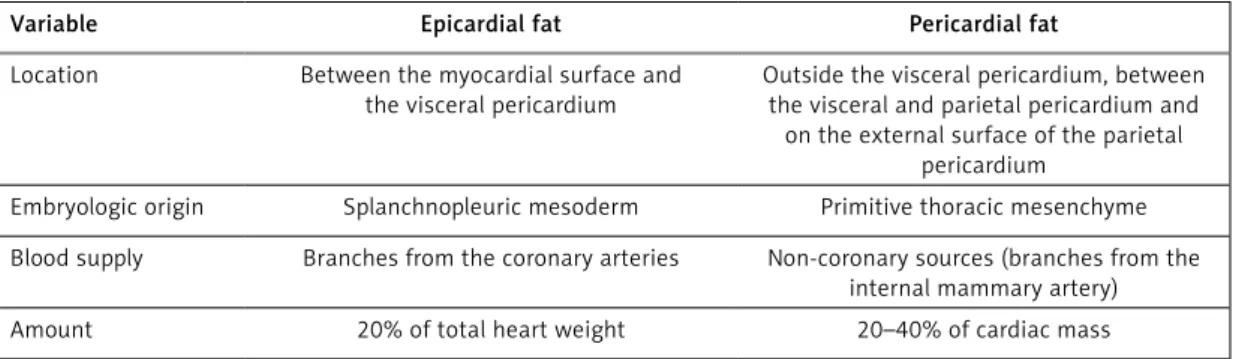
Variable Epicardial fat Pericardial fat
Location Between the myocardial surface and the visceral pericardium
Outside the visceral pericardium, between the visceral and parietal pericardium and
on the external surface of the parietal pericardium
Embryologic origin Splanchnopleuric mesoderm Primitive thoracic mesenchyme Blood supply Branches from the coronary arteries Non-coronary sources (branches from the
internal mammary artery)
Amount 20% of total heart weight 20–40% of cardiac mass
lished the general EAT thickness under 7 mm in the asymptomatic population [11]. Nevertheless, this method has several disadvantages includ- ing poor reproducibility and high dependence on the observer’s experience. In addition, it may not reflect accurately the whole quantity of the epi- cardial fat due to the two-dimensional nature of the measurement. In other words, the thickness rather than the entire quantity of the pericardial fat compartment can be assessed by echocardiog- raphy. Moreover, the method has poor intra- and interobserver variability, and its results may differ significantly from the measurements with CT [12].
In contrast to echocardiography, MRI provides accurate area measurements with enhanced spin- echo sequence and, in this way, EAT area mass and volume can be calculated (Figure 2). Area measurements with MRI correspond well with fat thickness determination with echocardiography, although the Bland-Altman analysis shows a sys- temic bias through overestimation of EAT with echocardiography [13]. Magnetic resonance imag-
ing is hardly available in routine clinical practice, is more expensive and has poorer spatial resolution compared to CT.
True volume assessment of EAT is feasible us- ing cardiac CT. The three dimensional (3D) image reconstruction with multidetector-row CT (MDCT) has the best spatial resolution among the im- aging modalities (Figure 3). It is of note that the specificity and sensitivity of measurements with MDCT are the best when compared to alternative imaging methods. The epicardial fat quantifica- tion is performed on prospectively ECG triggered non-contrast CT scans which extend from the pulmonary artery bifurcation to the diaphragm.
The identification of the EAT is based on thresh- olds of CT attenuation. Typically, lower thresh- olds of CT attenuation range from –250 to –190 Hounsfield units (HU), and upper thresholds are Figure 1. Quantification of epicardial adipose tis-
sue by echocardiography (parasternal view). The thickness of the area between the myocardium and the visceral layer of the pericardium is 0.85 cm, indicating epicardial adipose tissue
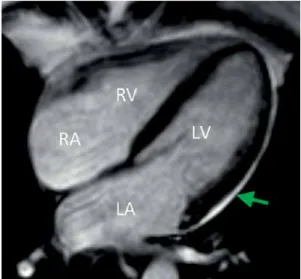
Figure 2. Epicardial adipose tissue (arrow) by using magnetic resonance imaging (MRI) technique RA – right atrium, LA – left atrium, RV – right ventricle, LV – left ventricle.
Figure 3. Measuring epicardial adipose tissue by cardiac computed tomography (CT). A – Axial section at the aortic root. Arrows indicate the visceral layer of the pericardium. Epicardial fat (E) is located inside and pericardial fat (P) outside the visceral layer. B – The visceral layer of the pericardium is traced manually (green). Epicardial adipose tissue (yellow) is marked automatically at the corresponding section. C – Three-dimensional reconstruction of the total epicardial fat compartment (yellow). The volume of epicardial adipose tissue was 112 cm3
C B
A
set between –50 and –30 HU. In contrast to area and thickness measurements, volume quantifica- tion provides the most accurate way for assess- ing the true epicardial fat quantity [14]. Using this method, coronary artery calcification may also be quantified, resulting in more reliable cardio- vascular risk assessment [15]. Importantly, native CT results in a very small (1 mSv) radiation dose.
Maurovich-Horvat et al. found in a collaborative study that the measurement of pericoronary adi- pose tissue was highly reproducible when using MDCT [16].
Anatomical characteristics of epicardial adipose tissue
In physiological circumstances the epicardial fat covers nearly 80% of the heart surface. Accord- ing to previous observations this fat compartment contributes 20% to the whole heart quantity [17].
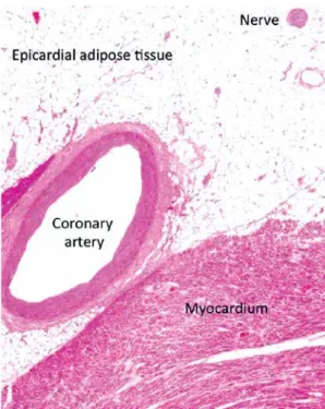
The EAT-covered heart region includes the heart base and the apex, the atrioventricular sulci, the entire surface of the right ventricle, and the great coronary vessels with their origins. The distribu- tion of EAT is mostly inhomogeneous; the biggest mass is localized on the lateral and anterior walls of the right atrium, but in normal circumstances it also covers the atrioventricular and the inter- ventricular sulci and the main coronary arteries as well. In the case of extremely enlarged EAT, it can also accumulate on the surface of the left atrium and along the vessel’s adventitia with spreading into the myocardium. It is of note that there is no separating fascia layer between the epicardial fat and the myocardium, providing close proximity of these two different tissues [18]. In histological investigations it has been previously established that adipocytes in the EAT are smaller than those in the abdominal or the subcutaneous fat com- partments [19]. Besides adipocytes, EAT includes nerves, ganglions, vessels, inflammatory cells and fibrocytes as well (Figure 4).
Age, gender, body weight and ethnicity should be taken into consideration among physiologi- cal determinants of EAT. Epicardial adipose tissue seems to increase with age [20]. The quantity of EAT depends on gender and body mass index (BMI). For example, pericardial fat was reported to be 137 ±54 cm3 among men and 108 ±41 cm3 among women of the Framingham offspring cohort [21]. In patients with a high BMI (> 27 kg/m2), EAT volume was more than two times higher compared to those with a BMI < 27 kg/m2 (155 ±15 cm3 vs.
67 ±12 cm3) [22]. Some ethnic differences in epicar- dial and pericardial fat thickness may also occur;
non-Hispanic White men have more epicardial and pericardial fat than do African Americans [23].
The biochemical features of small adipocytes in EAT may also differ from those of other fat
compartments. In experimental studies EAT had a higher rate of free fatty acid (FFA) release than adipose tissue elsewhere in the body, suggesting that EAT might play a role in local energy supply for the myocardium. In addition, a lower oxidative capacity and a lower rate of glucose utilization were also documented [24]. On the other hand, a 5-fold higher expression of uncoupled protein-1 (UCP-1) was found in EAT compared to other fat depots [25]. Uncoupled protein-1 is a specific pro- tein in brown fat which is necessary for its energy production, and does not appear in other types of fat tissues. This latter feature is in line with the fact that epicardial fat evolves from brown adi- pose tissue during embryogenesis.
Physiological function of epicardial adipose tissue
Several physiological functions of EAT are al- ready known from different studies or inferred from its biochemical or anatomical features. Unfor- tunately, experimental evidence supporting these observations are limited due to the very small amount of EAT in experimental animals (rodents).
It is suggested that functions of EAT may in- clude protection of the myocardium against hypo- thermia [25]. In addition, EAT can provide a me- chanical protective role for coronary circulation. It can attenuate the torsion developed by the myo- cardium contraction or the arterial pulse wave, but it has a permissive role as well in positive re- modeling of coronary arteries [26].
Figure 4. Microscopic view of the epicardial adi- pose tissue. It is of note that there is no separat- ing fascia layer between the epicardial fat and the myocardium
Also, EAT has a substantial role in energy supply to the myocardium and should be considered as a provider of energy during periods of high energy demand [27]. On the other hand, EAT may protect the myocardium from the cardiotoxic effect of a large amount of FFA due to its capacity for fast FFA utilization [28]. Taken together, EAT may serve as a unique energy buffering pool in the homeo- stasis of the myocardium.
In addition, adiponectin secretion from epicar- dial adipocytes may promote the coronary circu- lation. Adiponectin improves endothelial func- tion through stimulation of nitrogen monoxide synthase, reduces oxidative stress, and indirectly decreases the level of interleukin-6 (IL-6) and C-re- active protein (CRP) by reducing tumor necrosis factor-α (TNF-α) production [29, 30]. Adiponec- tin also has some extracardial effects such as in- creased glucose utilization in the hepatocytes and muscle cells which may result in improving insulin sensitivity [31].
Epicardial adipose tissue in the pathomechanism of atherosclerosis
Some years ago a hypothesis about the direct role of EAT in the development and progression of coronary atherosclerosis was raised, and para- crine and vasocrine effects of EAT due to close proximity of epicardial fat to coronary arteries were posited [32]. The hypothesis was indirectly supported by a pathological study in subjects with a myocardial bridge. Namely, no atherosclerosis was observed in coronary segments at the myo- cardial bridge where surrounding fat on the coro- nary arteries was lacking [33].
In a landmark study, Mazurek et al. analyzed epicardial and subcutaneous fat from the lower extremity in obese patients referred for coronary artery bypass grafting. They found increased lev-
els of inflammatory mediators (IL-6, TNF-α, inter- leukin-1β (IL-1β), monocyte chemoattractant pro- tein-1 (MCP-1)), macrophages, lymphocytes and basophils in epicardial fat as compared to subcu- taneous fat compartments [34]. Others found that epicardial and omental fat exhibited a broadly comparable pathogenic messenger ribonucleo- tide acid (mRNA) profile indicating macrophage infiltration into the epicardial fat [35]. In another study, mediators of the nuclear factor-kB (NF-kB) and c-Jun N-terminal kinase (JNK) pathways were suggested to be involved in the inflammatory profile of EAT, highlighting the role of the macro- phages in the inflammation within this tissue [36].
These studies indicate that chronic inflammation occurs locally as well as systemically, potentially contributing further to the pathogenesis of CAD.
It was documented that the epicardial adipo- cytes had impaired adiponectin secretion and in- creased leptin production in obese patients with hypertension, metabolic syndrome and CAD [37, 38]. This shift in the adiponectin/leptin ratio en- hances the development of atherosclerosis. Namely, the decreased adiponectin expression attenuates endothelial function and leads to increased TNF-α production, triggering systematic inflammation and oxidative stress. The altered leptin level promotes atherogenic changes in endothelial cells such as increased adhesion of monocytes, a higher level of macrophage-to-foam cell transformation, unfavor- able changes in lipid levels, and elevation of CRP and inflammatory cytokine levels. All these alter- ations may lead to development and destabilization of atherosclerotic plaques in coronary arteries [5].
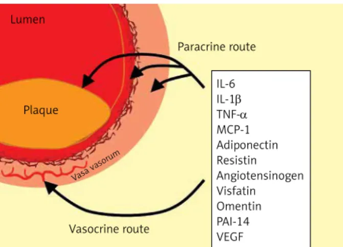
Based on several studies it became widely ac- cepted that EAT should be considered as a source of inflammatory mediators that might directly influence the myocardium and coronary arteries (Figure 5). Two mechanisms of influence (paracrine
Figure 5. Routes for paracrine and vasocrine effects of epicardial adipose tissue on coronary arteries and plaque formation
IL – interleukin, TNF-α – tumor necrosis factor-α, MCP-1 – monocyte chemoattractant protein-1, PAI-1 – plasminogen activator inhibitor-1, VEGF – vascular endothelial growth factor.
IL-6 IL-1β TNF-α MCP-1 Adiponectin Resistin Angiotensinogen Visfatin Omentin PAI-14 VEGF Paracrine route
Vasocrine route Plaque
Lumen
Vasa vasor um
and vasocrine) were suggested [39]. The paracrine way of influence means that adipokines released from pericoronary fat may diffuse across the arte- rial wall (adventitia, media, and intima) and final- ly can interact with endothelial cells in the intima and with vascular smooth cells in the media. The alternative vasocrine way of effect can be achieved by release of adipocytokines and FFAs from EAT di- rectly into the vasa vasorum of the coronary arte- rial wall [40]. It was suggested that the vasocrine way of influence may be predominant over the paracrine effect in the case of more advanced ath- erosclerotic lesions where inflammatory mediators may diffuse only with difficulties [35].
The association between EAT thickness and the metabolic syndrome was documented in a recent meta-analysis [41]. The relationship of EAT with CAD has been analyzed by several clinical studies [42, 43]. In the Framingham and the MESA (Mul- tiethnic Study of Atherosclerosis) epidemiologi- cal studies a significant association of epicardial fat with coronary artery calcification was found, which remained significant after adjustment for traditional cardiovascular risk factors [44, 45]. The increased epicardial fat proved to be associated with more advanced atherosclerosis in anoth- er study [46]. Epicardial fat was associated with non-calcified coronary plaques as well [47, 48].
A significant relationship of increased epicardial fat volume (> 130.7 cm3) with vulnerable plaques was also documented [49]. The relationship of morphological features of vulnerable plaques (positive remodeling, spotty calcifications, and low CT attenuation in the necrotic core) to the pericardial fat was also studied, and the volume of pericardial fat proved to be nearly twice as high in patients with vulnerable plaques as compared to those without CAD [50]. Pericardial fat was as- sociated with myocardial ischemia detected by single photon emission computed tomography (SPECT) in patients without known CAD [51]. Epi- cardial adipose tissue correlated with the degree of coronary atheromatosis, suggesting that its excessive accumulation might contribute to the development of acute coronary syndrome and cor- onary total occlusions [52]. In another study, EAT thickness was independently associated with the Thrombolysis in Myocardial Infarction (TIMI) risk score in patients with non-ST-elevation myocardi- al infarction (NSTEMI) and unstable angina pecto- ris [53]. In patients with the metabolic syndrome, increased EAT was associated with impaired coro- nary flow reserve [54]. In a different patient popu- lation (in women with chest pain and angiograph- ically normal coronary arteries), EAT thickness was correlated with reduced coronary flow reserve [55]. Different surrogate parameters of athero- sclerosis were also investigated by others, and an
association between EAT thickness and carotid in- tima-media thickness in type 2 diabetic patients as well as in children and adolescents with obe- sity was found [56, 57]. Moreover, EAT showed an independent association with arterial stiffness in an asymptomatic Korean cohort [58]. In a recent study, Maurovich-Horvat et al. investigated the re- lationship of different thoracic fat depots with cor- onary atherosclerosis and found an independent association between pericoronary fat and CAD. In addition, pericoronary fat correlated with inflam- matory biomarkers as well, suggesting that while systemic inflammation plays a role in the patho- genesis of CAD, additional local effects may exist [59]. In a systematic review and meta-analysis, an association between the elevated location-spe- cific thickness of EAT at the left atrioventricular groove and obstructive CAD was found [60].
Epicardial adipose tissue and other cardiac abnormalities
The relationship of EAT with atrial fibrillation was analyzed in several clinical studies. A strong association between EAT and atrial fibrillation (both paroxysmal and persistent) was document- ed by Al Chekakie et al.; the relationship proved to be independent of traditional risk factors and atri- al enlargement [61]. In another study, EAT thick- ness was verified as an independent predictor for post-ablative recurrence of atrial fibrillation [62].
In patients with peritoneal dialysis, increased EAT was associated with impaired left ventricle dia- stolic capacity independently of CRP level, a mark- er of systemic inflammation [63].
Epicardial adipose tissue necrosis: a benign cause of chest pain
Epicardial fat necrosis is a rare clinical con- dition; 26 cases were reported up to 2011 [64].
It should be considered in the differential diag- nosis of chest pain. The etiology is obscure, but the prognosis is good. In general, the presenting symptom is left-sided chest pain in a previously healthy individual with an associated juxtacardi- ac mass seen in chest radiography. The CT or MRI may confirm the correct diagnosis, resulting in the avoidance of surgical intervention.
Epicardial adipose tissue in type 2 diabetes, obesity and the insulin resistance syndrome (metabolic syndrome)
Typically, type 2 diabetes is preceded by pre- diabetes, but insulin resistance syndrome due to obesity may be the first pathological stage in the long-lasting asymptomatic period of diabetes. The insulin resistance syndrome (also called the met- abolic syndrome) includes insulin resistance and
different metabolic abnormalities (elevated serum triglycerides, lower HDL cholesterol, hyperglyce- mia) as well as elevated blood pressure. Obesity, especially abdominal visceral fat accumulation, plays a central role in this syndrome. Although the use of the term and the suggested pathomecha- nism of the metabolic syndrome became debat- able some years ago, the association between an enlarged abdominal visceral fat compartment and increased cardiovascular risk remained unques- tionable [65]. The enlarged visceral fat depot is characterized primarily by increased lipolysis lead- ing to hepatic steatosis. Non-alcoholic fatty liver disease (NAFLD) is often regarded as the hepat- ic manifestation of insulin resistance [66] and is considered as a novel predictor of cardiovascular disease [67, 68].
Several clinical investigations have aimed to assess the characteristics of EAT in the metabol- ic syndrome, prediabetes and type 2 diabetes. In a meta-analysis, EAT was 7.5 ±0.1 mm in thickness in the metabolic syndrome (n = 427) compared to 4.0 ±0.1 mm in controls (n = 301), and EAT cor- related significantly with the components of the metabolic syndrome [69]. Epicardial adipose tis- sue volume was significantly higher in patients with type 2 diabetes than in nondiabetic subjects, and EAT volume was significantly associated with components of the metabolic syndrome [46]. In as- ymptomatic type 2 diabetic patients the thickness of EAT proved to be an independent risk factor for significant coronary artery stenosis but not for si- lent myocardial ischemia [70]. A strong correlation was found between fasting plasma glucose and EAT measured with CT or echocardiography [71, 72]. Epicardial adipose tissue quantity was high- er in patients with type 2 diabetes mellitus com- pared to lean subjects or obese patients without diabetes. In addition, the difference in EAT volume between men and women was more pronounced in subjects with impaired fasting glucose or dia- betes mellitus [73]. A clear relationship of epicar- dial fat and serum alanine aminotransferase (ALT) and aspartate aminotransferase (AST) activity, surrogate markers of fatty liver, was documented in a cross-sectional, observational study [74]. Tak- en together, the insulin resistance syndrome (the metabolic syndrome), type 2 diabetes, NAFLD and CAD are associated with an increased amount of epicardial fat [75].
In the majority of studies an increase of EAT volume was associated with stenosis of the cor- onary arteries [76, 77]. Since these studies are cross-sectional, it is uncertain whether adipose tissue plays a causal role in the development of atherosclerosis. Importantly, two longitudinal studies have reported results that support the hy- pothesis of ‘outside to inside signaling’ as a cause
of atherosclerosis [45, 78]. In these studies, intra- thoracic and EAT volume were measured and an increase of the quantity of intrathoracic and EAT was associated with incident coronary heart dis- ease and with major adverse cardiac events. As- sociations were independent from BMI and other risk factors, suggesting that EAT is one of the fac- tors contributing to CAD.
Epicardial adipose tissue in type 1 diabetes Interestingly, higher epicardial fat and serum leptin levels were found in subjects with type 1 diabetes than in non-diabetic controls. The epicar- dial fat thickness and serum leptin levels proved to be the best independent correlates of each oth- er in patients with type 1 diabetes independently of BMI, glycemic control and daily insulin require- ment [79]. Recently, patients with type 1 diabetes (n = 100) from the Diabetes Control and Compli- cations Trial/Epidemiology of Diabetes Interven- tions and Complications (DCCT/EDIC) study were investigated. In this pilot study, the accumulation of adipose tissue in epicardial and intra-thoracic spaces was strongly associated with higher BMI, greater waist-to-hip ratio, higher weighted glycat- ed hemoglobin values, elevated triglycerides and a history of elevated albumin excretion rate or end-stage renal disease [80].
Role of genetic effects on epicardial adipose tissue
As EAT and other abdominal fat compartments (subcutaneous adipose tissue (SAT), visceral adi- pose tissue (VAT), hepatic lipid accumulation) car- ry different clinical significance [81] and epicardial fat differs from pericardial fat from an embryolog- ical point of view, the role of genetic effects on EAT and other fat compartments may differ. In a classical twin study with CT investigation our preliminary results indicated that EAT had a rela- tively strong genetic dependence, similarly to BMI and waist circumference [82, 83]. In contrast to abdominal fat compartment areas, a weak genet- ic and a stronger environmental dependence of hepatic lipid accumulation was found in our twin cohort [84].
Treatment options for modifying epicardial adipose tissue volume
Lifestyle changes, bariatric surgery and various drugs may be applied. Reduction in weight (BMI) by using a very-low calorie diet or exercise train- ing program in obese patients is associated with a decrease in EAT volume [85, 86]. Nevertheless, this was observed after bariatric surgery as well, although myocardial triglyceride content did not change significantly [87]. In a meta-analysis, diet
or bariatric surgery proved to be more beneficial than exercise training in reducing EAT volume [88]. The effect of drugs on EAT is controversial.
Atorvastatin resulted in a more pronounced de- crease of EAT than simvastatin/ezetimibe [89].
Pioglitazone compared with metformin increased pericardial fat volume in patients with type 2 di- abetes [90]. Short-term (3 months) use of gluca- gon-like-receptor agonists (exenatide, liraglutide) decreased the volume of EAT in type 2 diabetic patients [91]. In a longer (26 weeks) randomized controlled trial, exenatide twice daily (versus stan- dard antidiabetic treatment) proved to be effec- tive in reducing both epicardial and liver fat con- tent in obese patients with type 2 diabetes; the effects were mainly weight loss dependent [92].
In another study, sitagliptin, a dipeptidyl-pepti- dase-4 inhibitor, also decreased the volume of EAT in a 24-week long study with obese type 2 diabet- ic patients [93]. Clearly, EAT should be considered as a novel therapeutic target, and statins, pioglita- zone as well as incretin-based drugs are the best candidates so far [94, 95].
Conclusions
The epicardial fat is a unique fat compartment located between the myocardial surface and the visceral layer of the pericardium. The EAT can be quantified by non-invasive cardiac imaging tech- niques such as echocardiography, MRI or cardiac CT.
Among physiological determinants of EAT, age, gender, body weight and ethnicity should be con- sidered. The EAT volume has a relatively strong genetic dependence, similarly to other visceral fat depots. Physiological functions of EAT may include protection of the myocardium against hypother- mia and a mechanical protective role for coronary circulation. In addition, EAT may serve as a unique energy buffering pool in the homeostasis of the myocardium.
As for pathophysiological functions, it is wide- ly accepted that EAT should be considered as a source of inflammatory mediators that might directly influence the myocardium and coronary arteries. In line with these observations, clinical studies suggested that EAT – through paracrine and vasocrine effects – may have an impact on the development and progression of coronary ath- erosclerosis. In addition, an association between increased EAT and atrial fibrillation was also doc- umented. The insulin resistance syndrome (the metabolic syndrome), type 2 diabetes, NAFLD and CAD proved to be associated with an increased amount of epicardial fat. Interestingly, accumu- lation of EAT was also observed in patients with type 1 diabetes.
Treatment options for modifying EAT volume include lifestyle changes, bariatric surgery and
using different drugs. Weight reduction in obese subjects may lead to a decrease in EAT volume, while effects of different drugs on EAT are contro- versial. Nevertheless, EAT should be considered as a new cardiovascular therapeutic target.
Acknowledgments
A grant from the New Horizons Programme (European Foundation for the Study of Diabetes) is acknowledged. The microscopic imaging of hu- man myocardium and epicardial fat was provided by Zoltán Sápi MD, DSc, Semmelweis University, Institute of Pathology, Budapest.
Eszter Nagy and Adam L. Jermendy equally con- tributed to this manuscript.
Conflict of interest
The authors declare no conflict of interest.
R e f e r e n c e s
1. Zimmet P, Alberti KGMM, Shaw J. Global and societal implications of diabetes epidemic. Nature 2001; 414:
782-7.
2. Despres JP, Lemieux I. Abdominal obesity and metabolic syndrome. Nature 2006; 444: 881-7.
3. Poirier P, Despres JP. Waist circumference, visceral obe- sity, and cardiovascular risk. J Cardiopulmonary Rehabil 2003; 23: 161-9.
4. Lim S, Meigs JB. Links between ectopic fat and vascular disease in humans. Arterioscler Thromb Vasc Biol 2014;
34: 1820-6.
5. Iacobellis G, Malavazos AE, Corsi MM. Epicardial fat:
from biomolecular aspects to the clinical practice. Int J Biochem Cell Biol 2011; 43: 1651-4.
6. Bedford E. The story of fatty heart. A disease of Victori- an times. Br Heart J 1972; 34: 23-8.
7. Després JP, Cartier A, Côté M, Arsenault BJ. The concept of cardiometabolic risk: bridging the fields of diabetolo- gy and cardiology. Ann Med 2008; 40: 514-23.
8. Iacobellis G, Ribaudo MC, Assael F, et al. Echocardio- graphic epicardial adipose tissue is related to anthropo- metric and clinical parameters of metabolic syndrome:
a new indicator of cardiovascular risk. J Clin Endocrinol Metab 2003; 88: 5163-8.
9. Iacobellis G, Bianco AC. Epicardial adipose tissue:
emerging physiological, pathophysiological and clinical features. Trends Endocrinol Metab 2011; 22: 450-7.
10. Iacobellis G. Epicardial and pericardial fat: close, but very different. Obesity 2009; 17: 625.
11. Iacobellis G, Willens HJ. Echocardiographic epicardial fat: a review of research and clinical applications. J Am Soc Echocardiogr 2009; 22: 1311-9.
12. Saura D, Oliva MJ, Rodríguez D, et al. Reproducibility of echocardiographic measurements of epicardial fat thickness. Int J Cardiol 2010; 141: 311-3.
13. Sicari R, Sironi AM, Petz R, et al. Pericardial rather than epicardial fat is a cardiometabolic risk marker: an MRI vs echo study. J Am Soc Echocardiogr 2011; 24: 1156-62.
14. Gorter PM, van Lindert AS, de Vos AM, et al. Quantifi- cation of epicardial and peri-coronary fat using cardi- ac computed tomography; reproducibility and relation with obesity and metabolic syndrome in patients sus-
pected of coronary artery disease. Atherosclerosis 2008;
197: 896-903.
15. Madaj P, Budoff MJ. Risk stratification of non-contrast CT beyond the coronary calcium scan. J Cardiovasc Comput Tomogr 2012; 6: 301-7.
16. Maurovich-Horvat P, Kallianos K, Engel LC, et al. Influ- ence of pericoronary adipose tissue on local coronary atherosclerosis as assessed by a novel MDCT volumetric method. Atherosclerosis 2011; 219: 151-7.
17. Rabkin SW. Epicardial fat: properties, function and rela- tionship to obesity. Obes Rev 2007; 8: 253-61.
18. Iacobellis G, Corradi D, Sharma AM. Epicardial adipose tissue: anatomic, biomolecular and clinical relation- ships with the heart. Nat Clin Pract Cardiovasc Med 2005; 2: 536-43.
19. Bambace C, Telesca M, Zoico E, et al. Adiponectin gene expression and adipocyte diameter: a comparison be- tween epicardial and subcutaneous adipose tissue in men. Cardiovasc Pathol 2011; 20: e153-6.
20. Bertaso AG, Bertol D, Duncan BB, Foppa M. Epicardial fat: definition, measurements and systematic review of main outcomes. Arq Bras Cardiol 2013; 101: e18-28.
21. Fox CS, Gona P, Hoffmann U, et al. Pericardial fat, intra- thoracic fat, and measures of left ventricular structure and function: the Framingham Heart Study. Circulation 2009; 119: 1586-91.
22. Gorter PM, van Lindert AS, de Vos AM, et al. Quantifi- cation of epicardial and peri-coronary fat using cardi- ac computed tomography; reproducibility and relation with obesity and metabolic syndrome in patients sus- pected of coronary artery disease. Atherosclerosis 2008;
197: 896-903.
23. Willens HJ, Gómez-Marín O, Chirinos JA, Goldberg R, Lowery MH, Iacobellis G. Comparison of epicardial and pericardial fat thickness assessed by echocardiography in African American and non-Hispanic White men: a pi- lot study. Ethn Dis 2008; 18: 311-6.
24. Marchington JM, Mattacks CA, Pond CM. Adipose tissue in the mammalian heart and pericardium: structure, foetal development and biochemical properties. Comp Biochem Physiol B 1989; 94: 225-32.
25. Sacks HS, Fain JN, Holman B, et al. Uncoupling protein-1 and related messenger ribonucleic acids in human epi- cardial and other adipose tissues: epicardial fat func- tioning as brown fat. J Clin Endocrinol Metab 2009; 94:
3611-5.
26. Prati F, Arbustini E, Labellarte A, et al. Eccentric athero- sclerotic plaques with positive remodelling have a peri- cardial distribution: a permissive role of epicardial fat?
A three-dimensional intravascular ultrasound study of left anterior descending artery lesions. Eur Heart J 2003;
24: 329-36.
27. Iacobellis G, Bianco AC. Epicardial adipose tissue:
emerging physiological, pathophysiological and clinical features. Trends Endocrinol Metab 2011; 22: 450-7.
28. Iozzo P. Metabolic toxicity of the heart: insights from molecular imaging. Nutr Metab Cardiovasc Dis 2010;
20: 147-56.
29. Li R, Wang WQ, Zhang H, et al. Adiponectin improves endothelial function in hyperlipidemic rats by reducing oxidative/nitrative stress and differential regulation of eNOS/iNOS activity. Am J Physiol Endocrinol Metab 2007; 293: E1703-8.
30. Deng G, Long Y, Yu YR, Li MR. Adiponectin directly im- proves endothelial dysfunction in obese rats through the AMPK-eNOS pathway. Int J Obes (Lond) 2010; 34:
165-71.
31. Payne GA, Kohr MC, Tune JD. Epicardial perivascular adipose tissue as a therapeutic target in obesity-relat- ed coronary artery disease. Br J Pharmacol 2012; 165:
659-69.
32. Sacks HS, Fain JN. Human epicardial adipose tissue:
a review. Am Heart J 2007; 153: 907-17.
33. Ishii T, Asuwa N, Masuda S, Ishikawa Y. The effects of a myocardial bridge on coronary atherosclerosis and ischaemia. J Pathol 1998; 185: 4-9.
34. Mazurek T, Zhang L, Zalewski A, et al. Human epicardial adipose tissue is a source of inflammatory mediators.
Circulation 2003; 108: 2460-6.
35. Baker AR, Silva NF, Quinn DW, et al. Human epicardial adipose tissue expresses a pathogenic profile of adipo- cytokines in patients with cardiovascular disease. Car- diovasc Diabetol 2006; 5: 1.
36. Baker AR, Harte AL, Howell N, et al. Epicardial adipose tissue as a source of nuclear factor-kappaB and c-Jun N-terminal kinase mediated inflammation in patients with coronary artery disease. J Clin Endocrinol Metab 2009; 94: 261-7.
37. Eiras S, Teijeira-Fernández E, Shamagian LG, Fernan- dez AL, Vazquez-Boquete A, Gonzalez-Juanatey JR. Ex- tension of coronary artery disease is associated with in- creased IL-6 and decreased adiponectin gene expression in epicardial adipose tissue. Cytokine 2008; 43: 174-80.
38. Iacobellis G, Pistilli D, Gucciardo M, et al. Adiponectin expression in human epicardial adipose tissue in vivo is lower in patients with coronary artery disease. Cytokine 2005; 29: 251-5.
39. Sacks HS, Fain JN. Human epicardial adipose tissue:
a review. Am Heart J 2007; 153: 907-17.
40. Yudkin JS, Eringa E, Stehouwer CD. “Vasocrine” signalling from perivascular fat: a mechanism linking insulin resis- tance to vascular disease. Lancet 2005; 365: 1817-20.
41. Pierdomenico SD, Pierdomenico AM, Cuccurullo F, Iac- obellis G. Meta-analysis of the relation of echocardio- graphic epicardial adipose tissue thickness and the metabolic syndrome. Am J Cardiol 2013; 111: 73-8.
42. Yerramasu A, Dey D, Venuraju S, et al. Increased volume of epicardial fat is an independent risk factor for accel- erated progression of sub-clinical coronary atheroscle- rosis. Atherosclerosis 2012; 220: 223-30.
43. Wang TD, Lee WJ, Shih FY, et al. Association of epicar- dial adipose tissue with coronary atherosclerosis is region-specific and independent of conventional risk factors and intra-abdominal adiposity. Atherosclerosis 2010; 213: 279-87.
44. Rosito GA, Massaro JM, Hoffmann U, et al. Pericardial fat, visceral abdominal fat, cardiovascular disease risk factors, and vascular calcification in a community-based sample: the Framingham Heart Study. Circulation 2008;
117: 605-13.
45. Ding J, Hsu FC, Harris TB, et al. The association of peri- cardial fat with incident coronary heart disease: the Multi-Ethnic Study of Atherosclerosis (MESA). Am J Clin Nutr 2009; 90: 499-504.
46. Wang CP, Hsu HL, Hung WC, et al. Increased epicardial adipose tissue (EAT) volume in type 2 diabetes mellitus and association with metabolic syndrome and severity of coronary atherosclerosis. Clin Endocrinol (Oxf) 2009;
70: 876-82.
47. Konishi M, Sugiyama S, Sugamura K, et al. Association of pericardial fat accumulation rather than abdominal obesity with coronary atherosclerotic plaque formation in patients with suspected coronary artery disease. Ath- erosclerosis 2010; 209: 573-8.
48. Alexopoulos N, McLean DS, Janik M, Arepalli CD, Still- man AE, Raggi P. Epicardial adipose tissue and coronary artery plaque characteristics. Atherosclerosis 2010;
210: 150-4.
49. Ito T, Nasu K, Terashima M, et al. The impact of epicar- dial fat volume on coronary plaque vulnerability: insight from optical coherence tomography analysis. Eur Heart J Cardiovasc Imaging 2012; 13: 408-15.
50. Schlett CL, Ferencik M, Kriegel MF, et al. Association of pericardial fat and coronary high-risk lesions as deter- mined by cardiac CT. Atherosclerosis 2012; 222: 129-34.
51. Tamarappoo B, Dey D, Shmilovich H, et al. Increased pericardial fat volume measured from noncontrast CT predicts myocardial ischemia by SPECT. JACC Cardiovasc Imaging 2010; 3: 1104-12.
52. Ueno K, Anzai T, Jinzaki M, et al. Increased epicardial fat volume quantified by 64-multidetector computed tomography is associated with coronary atherosclerosis and totally occlusive lesions. Circ J 2009; 73: 1927-33.
53. Ozcan F, Turak O, Canpolat U, et al. Association of epicar- dial fat thickness with TIMI risk score in NSTEMI/USAP patients. Herz 2014; 39: 755-60.
54. Tok D, Çağli K, Kadife I, et al. Impaired coronary flow reserve is associated with increased echocardiographic epicardial fat thickness in metabolic syndrome patients.
Coron Artery Dis 2013; 24: 191-5.
55. Sade LE, Eroglu S, Bozbaş H, et al. Relation between epicardial fat thickness and coronary flow reserve in women with chest pain and angiographically normal coronary arteries. Atherosclerosis 2009; 204: 580-5.
56. Cetin M, Cakici M, Polat M, Suner A, Zencir C, Ardic I.
Relation of epicardial fat thickness with carotid intima- media thickness in patients with type 2 diabetes melli- tus. Int J Endocrinol 2013; 2013: 769175.
57. Cabrera-Rego JO, Iacobellis G, Castillo-Herrera JA, et al.
Epicardial fat thickness correlates with carotid inti- ma-media thickness, arterial stiffness, and cardiac geometry in children and adolescents. Pediatr Cardiol 2014; 35: 450-6.
58. Park HE, Choi SY, Kim HS, Kim MK, Cho SH, Oh BH. Epi- cardial fat reflects arterial stiffness: assessment using 256-slice multidetector coronary computed tomog- raphy and cardio-ankle vascular index. J Atheroscler Thromb 2012; 19: 570-6.
59. Maurovich-Horvat P, Kallianos K, Engel LC, et al. Rela- tionship of thoracic fat depots with coronary atheroscle- rosis and circulating inflammatory biomarkers. Obesity (Silver Spring) 2015; 23: 1178-84.
60. Wu FZ, Chou KJ, Huang YL, Wu MT. The relation of loca- tion-specific epicardial adipose tissue thickness and ob- structive coronary artery disease: systemic review and meta-analysis of observational studies. BMC Cardiovasc Disord 2014; 14: 62.
61. Al Chekakie MO, Welles CC, Metoyer R, et al. Pericardial fat is independently associated with human atrial fibril- lation. J Am Coll Cardiol 2010; 56: 784-8.
62. Chao TF, Hung CL, Tsao HM, et al. Epicardial adipose tis- sue thickness and ablation outcome of atrial fibrillation.
PLoS One 2013; 8: e74926.
63. Lin HH, Lee JK, Yang CY, Lien YC, Huang JW, Wu CK. Ac- cumulation of epicardial fat rather than visceral fat is an independent risk factor for left ventricular diastolic dysfunction in patients undergoing peritoneal dialysis.
Cardiovasc Diabetol 2013; 12: 127.
64. Baig A, Campbell B, Russell M, Singh J, Borra S. Epicar- dial fat necrosis: an uncommon etiology of chest pain.
Cardiol J 2012; 19: 424-8.
65. Borch-Johnsen K, Wareham N. The rise and fall of the metabolic syndrome. Diabetologia 2010; 53: 597-9.
66. Abdelmalek MF, Diehl AM. Nonalcoholic fatty liver dis- ease as a complication of insulin resistance. Med Clin North Am 2007; 91: 1125-49.
67. Hamaguchi M, Kojima T, Takeda N, et al. Nonalcoholic fatty liver disease is a novel predictor of cardiovascular disease. World J Gastroenterol 2007; 13: 1579-84.
68. Schindhelm RK, Diamant M, Heine RJ. Nonalcoholic fatty liver disease and cardiovascular disease risk. Curr Diab Rep 2007; 7: 181-7.
69. Rabkin SW. The relationship between epicardial fat and indices of obesity and the metabolic syndrome: a sys- tematic review and meta-analysis. Metab Syndr Relat Disord 2014; 12: 31-42.
70. Kim HM, Kim KJ, Lee HJ, et al. Epicardial adipose tissue thickness is an indicator for coronary artery stenosis in asymptomatic type 2 diabetic patients: its assessment by cardiac magnetic resonance. Cardiovasc Diabetol 2012; 11: 83.
71. Wang TD, Lee WJ, Shih FY, et al. Relations of epicardial adipose tissue measured by multidetector computed tomography to components of the metabolic syndrome are region-specific and independent of anthropometric indexes and intraabdominal visceral fat. J Clin Endocri- nol Metab 2009; 94: 662-9.
72. Iacobellis G, Barbaro G, Gerstein HC. Relationship of epi- cardial fat thickness and fasting glucose. Int J Cardiol 2008; 128: 424-6.
73. Iozzo P, Lautamaki R, Borra R, et al. Contribution of glu- cose tolerance and gender to cardiac adiposity. J Clin Endocrinol Metab 2009; 94: 4472-82.
74. Iacobellis G, Pellicelli AM, Grisorio B, et al. Relation of epicardial fat and alanine aminotransferase in subjects with increased visceral fat. Obesity (Silver Spring) 2008;
16: 179-83.
75. Iacobellis G. Local and systemic effects of the multifac- eted epicardial adipose tissue depot. Nat Rev Endocrinol 2015; 11: 363-71.
76. Picard FA, Gueret P, Laissy JP, et al. Epicardial adipose tissue thickness correlates with the presence and se- verity of angiographic coronary artery disease in stable patients with chest pain. PLoS One 2014; 9: e110005.
77. Sinha SK, Thakur R, Jha MJ, et al. Epicardial adipose tis- sue thickness and its association with the presence and severity of coronary artery disease in clinical setting:
a cross-sectional observational study. J Clin Med Res 2016; 8: 410-9.
78. Cheng VY, Dey D, Tamarappoo B, et al. Pericardial fat burden on ECG-gated noncontrast CT in asymptomat- ic patients who subsequently experience adverse car- diovascular events. JACC Cardiovasc Imaging 2010; 3:
352-60.
79. Iacobellis G, Diaz S, Mendez A, Goldberg R. Increased epicardial fat and plasma leptin in type 1 diabetes inde- pendently of obesity. Nutr Metab Cardiovasc Dis 2014;
24: 725-9.
80. Darabian S, Backlund JY, Cleary PA, et al. Significance of epicardial and intrathoracic adipose tissue volume among type 1 diabetes patients in the DCCT/EDIC: a pi- lot study. PLoS One 2016; 11: e0159958.
81. Lim S, Meigs JB. Links between ectopic fat and vascular disease in humans. Arterioscler Thromb Vasc Biol 2014;
34: 1820-6.
82. Maurovich-Horvat P, Tárnoki DL, Tárnoki ÁD, et al. Ratio- nale, design and methodological aspects of the BUDA- PEST-GLOBAL study (Burden of Atherosclerotic Plaques
Study in Twins – Genetic Loci and the Burden of Athero- sclerotic Lesions). Clin Cardiol 2015; 38: 699-707.
83. Maurovich-Horvat P, Jermendy AL, Horcsik DV, et al. Epi- cardial adipose tissue quantity shows a relatively strong genetic dependence: a classic twin study (meeting ab- stract). Diabetes 2015; 64 Suppl 1: A111.
84. Jermendy AL, Drobni Z, Horvath T, et al. Hepatic lipid ac- cumulation is mainly determined by environmental fac- tors: a classic twin study (meeting abstract). Diabetes 2015; 64 Suppl 1: A554.
85. Iacobellis G, Singh N, Wharton S, Sharma AM. Substan- tial changes in epicardial fat thickness after weight loss in severely obese subjects. Obesity (Silver Spring) 2008;
16: 1693-7.
86. Kim MK, Tomita T, Kim MJ, Sasai H, Maeda S, Tanaka K.
Aerobic exercise training reduces epicardial fat in obese men. J Appl Physiol 2009; 106: 5-11.
87. Gaborit B, Jacquier A, Kober F, et al. Effects of bariatric surgery on cardiac ectopic fat: lesser decrease in epicar- dial fat compared to visceral fat loss and no change in myocardial triglyceride content. J Am Coll Cardiol 2012;
60: 1381-9.
88. Rabkin SW, Campbell H. Comparison of reducing epicar- dial fat by exercise, diet or bariatric surgery weight loss strategies: a systematic review and meta-analysis. Obes Rev 2015; 16: 406-15.
89. Park JH, Park YS, Kim YJ, et al. Effects of statins on the epicardial fat thickness in patients with coronary artery stenosis underwent percutaneous coronary interven- tion: comparison of atorvastatin with simvastatin/eze- timibe. J Cardiovasc Ultrasound 2010; 18: 121-6.
90. Jonker JT, Lamb HJ, van der Meer RW, et al. Pioglitazone compared with metformin increases pericardial fat vol- ume in patients with type 2 diabetes mellitus. J Clin En- docrinol Metab 2010; 95: 456-60.
91. Morano S, Romagnoli E, Filardi T, et al. Short-term ef- fects of glucagon-like peptide 1 (GLP-1) receptor ago- nists on fat distribution in patients with type 2 diabetes mellitus: an ultrasonography study. Acta Diabetol 2015;
52: 727-32.
92. Dutour A, Abdesselam I, Ancel P, et al. Exenatide de- creases liver fat content and epicardial adipose tissue in patients with obesity and type 2 diabetes: a prospec- tive randomised clinical trial using magnetic resonance imaging and spectroscopy. Diabetes Obes Metab 2016;
18: 882-91.
93. Lima-Martínez MM, Paoli M, Rodney M, et al. Effect of sitagliptin on epicardial fat thickness in subjects with type 2 diabetes and obesity: a pilot study. Endocrine 2016; 51: 448-55.
94. Mazurek T, Opolski G. Pericoronary adipose tissue:
a novel therapeutic target in obesity-related coronary atherosclerosis. J Am Coll Nutr 2015; 34: 244-54.
95. Iacobellis G. Epicardial fat: a new cardiovascular thera- peutic target. Curr Opin Pharmacol 2016; 27: 13-8.