INTERACTION OF NANOPARTICLES WITH NEURAL STEM-AND TISSUE-TYPE CELLS
PhD thesis
Murali Kumarasamy
Neuroscience Doctoral School Semmelweis University
Supervisor: Dr. Emília Madarász PhD., D.Sc Official reviewers:
Dr. Jedlovszky-Hajdú Angéla, Ph.D Dr. Szabó Bálint, Ph.D
Dr. Hegedűs Tamás, Ph.D (Reserve member)
Head of the Final Examination Committee:
Dr. Benyó Zoltán, D.Sc
Members of the Final Examination Committee:
Dr. Czirják Gábor, Ph.D Dr. Eyre Mark, David, Ph.D
Budapest, 2015
1
“Anyone who doesn’t take truth seriously in small matters cannot be trusted in large ones either.”
- Albert Einstein
2 Table of Contents
LIST OF ABBREVIATIONS ... 5
LIST OF FIGURES ... 6
LIST OF TABLES ... 9
1.INTRODUCTION ... 10
1. Basic features of nanomaterials ... 10
2. Modification of nanoparticle features by surface functionalization ... 12
2.1 Poly-ethyleneglycol (PEG) ... 12
2.2 Polyvinylpyrrolidone (PVP) ... 13
3. Environmental and health risks of nanoparticle production and application ... 13
4. Possible routes of Nanoparticles to entering the human body ... 14
5. Interaction of nanoparticles with the central neural tissue ... 16
6. Nanoparticles used ... 20
6.1 Polystyrene (PS) nanoparticles ... 20
6.2 Silica (Si) nanoparticles ... 22
6.3 Silver (Ag) nanoparticles ... 24
7. Objectives of the thesis ... 27
2.MATERIALSANDMETHODS ... 28
1. Synthesis of nanoparticles ... 28
1.1 PS NPs ... 28
1.2 Preparation of Silica NPs ... 28
1.3 Preparation of Ag NPs ... 29
1.3.1. 50 nm bare and PVP-coated AgNPs ... 29
1.3.2. Synthesis of Ag nanocubes ... 29
1.3.3. Synthesis of Ag nanotriangles ... 30
1.3.4. Synthesis of Ag nanorods ... 30
2. Physico-chemical characterization of nanoparticles ... 31
2.1. Transmission electron microscopy (TEM) ... 31
2.2. Dynamic light scattering (DLS) and Z-Potential measurement ... 31
2.3. UV–visible spectrophotometry of Ag NPs ... 32
2.4. Nanoparticle tracking analysis (NTA)... 32
2.5. Differential centrifugal sedimentation (DCS) ... 32
3. Studies on material adsorption by NPs ... 33
3.1. Assays on protein adsorption at NP surfaces ... 33
3.1.1. Electrophoretic studies on protein adsorption onto PS NPs ... 33
3.1.2. Human blood proteins on spherical Ag PVP NPs ... 33
3.2. Assays on endotoxin adsorption at NP surfaces ... 34
3.2.1. LAL assay ... 34
3
3.2.2. SDS-PAGE ... 34
3.2.3. Spiking with LPS ... 35
3.2.4. Studies on interference of NPs with the LAL assay readout ... 35
3.2.5. Silver staining method ... 36
4. Cell cultures ... 36
4.1. NE-4C neuroectodermal stem cells ... 36
4.2. Primary brain cell cultures ... 37
4.3. Astroglial cultures ... 37
4.4. Microglial cultures ... 38
4.5. Mouse brain vascular endothelial cell cultures ... 38
5. Cellular assays ... 39
5.1. Exposing the cells to nanoparticles ... 39
5.2. Assays on cell viability and on cell membrane integrity ... 39
5.2.1. Assays on cell viability (MTT) ... 39
5.2.2 Assays on cell death (LDH leakage) ... 40
6. Immunocytochemical and uptake studies ... 41
7. Microscopic evaluation ... 42
7.1. Fluorescence spectrum analysis ... 42
7.2. TEM analysis of the cellular uptake of Ag NPs with different shape ... 42
8. In vivo experiments ... 43
8.1. Injection of PS NPs into mice ... 43
8.2. Microscopic evaluation of the tissues ... 43
3.RESULTS ... 44
1. Characterization of NPs ... 44
1.1. Physico-chemical properties of particles with non-toxic core material ... 44
1.1.1. Fluorescent silica NPs ... 44
1.1.2 The polystyrene nanoparticles (NPs) ... 45
1.2. Protein adsorption by Si- and PS NPs ... 46
1.2.1. Changes of physico-chemical characteristics of PS NPs after long-term storage ... 48
1.3. Synthesis and physicochemical characterization of Ag NPs with different shapes.39 1.3.1. Spherical shaped Ag NPs ... 50
1.3.2. Silver nanocubes ... 51
1.3.3. Silver nanotriangles ... 52
1.3.4. Silver nanorods ... 53
1.4. The protein adsorption by Ag NPs ... 53
1.4.1. In situ experiments with human plasma ... 53
2. Cellular responses to exposure to NPs with non-toxic core material ... 55
2.1. The experimental models ... 55
2.1.1. The cell models ... 55
2.1.2. Targeting the cells with nanoparticles ... 57
4
2.2. Cellular responses ... 58
2.2.1. Metabolic responses, cell membrane integrity and uptake reactions of neural cells in response to exposure to Si NPs with different chemical surface composition ... 58
2.2.2. Uptake of Si NPs by different neural cells ... 59
2.2.3. Cellular responses to exposure to PS NPs ... 61
2.2.4. Morphological effects and cellular uptake of PS NPs ... 65
3. Effects of particle aging on interactions of PS NPs with neural cells ... 68
3.1. Cellular effects of aged PS NP ... 68
3.2. Endotoxin contaminations on PS NP surfaces ... 70
3.3. Biological effects of LPS contaminated PS NPs ... 73
4. Interaction of PS NPs with physiological barriers protecting the central neural tissue 74 5. Cellular responses to silver NPs of different shapes ... 76
4. DISCUSSION ... 79
5.CONCLUSIONS...87
6.SUMMARY………88
7.REFERENCES……….90
8.LISTOFPUBLICATIONSRELATEDTOTHESIS………103
9.ACKNOWLEDGEMENTS……….104
5 I. List of Abbreviations
CX3CR1, fractalkine receptor1
DCS, Differential centrifugal sedimentation DLS, dynamic light scattering;
DMEM, Dulbecco’s modified Eagle’s medium;
FCS, foetal calf serum;
FITC, fluorescein isothiocyanate;
GFAP, anti-glial fibrillary acidic protein;
ITS, insulin-transferrin-selenite;
LAL, Limulus amebocyte lysate;
LDH, lactate dehydrogenase;
LPS, Lipopolysacchride
MEM, minimum essential medium
mPMS, 1-methoxy-methylphenazinium methyl sulfate;
MQ, Millipore water;
MTT, 3-(4,5-dimethylthiazol-2-yl)-2,5 diphenyltetrazolium bromide;
NE-4C, continuous mouse embryonic cell line of neuroectodermal origin NEDA, 3 N-(1-naphthyl)-ethylenediamine;
NMs, nanomaterial NPs, nanoparticles
NTA, Nanoparticle tracking analysis OsO4, Osmium tetroxide
PBS phosphate buffered saline;
PC, Protein corona;
PEG, polyethylene glycol;
PFA, Paraformaldehyde PS, polystyrene;
RA, Retinoic acid ROI, Region of interest;
SDS-PAGE, sodium dodecyl sulfate -polyacrylamide gel electrophoresis;
Si, Silica
TEM, transmission electron microscopy;
ZP, Zeta (ζ) Potential;
6 I. List of figures
Figure 1. Place of nanomaterials in the world of small objects,. 11 Figure 2. The chemical structure of polyvinylpyrrolidone (PVP). 13 Figure 3. Overview of the interdisciplinary science named nanotoxicology. 14
Figure 4. Possible adverse health effects of NPs. 15
Figure 5. Potential routes of nanoparticles to the brain and presumed consequences. 15 Figure 6. Schematic diagram of the neurovascular unit. 16 Figure 7. Nanoparticles injected into the maternal blood supply may enter the
embryo. 18
Figure 8. The styrene monomer: the structural unit of polystyrene. 20 Figure 9. Silica particle with various silanol groups. 23 Figure 10. PS NPs with different surfaces and fluorochrome labelling. 28 Figure 11. Differently functionalized FITC labelled silica nanoparticles. 28
Figure 12. Preparation of spherical Ag NPs. 29
Figure 13. Preparation of silver cubic nanoparticles. 30
Figure 14. Preparation of silver nanotriangles. 30
Figure 15. Preparation of silver nanorods. 30
Figure 16. Schematic diagram of DCS instrument. 33
Figure 17. The scheme of neural differentiation of NE-4C neural stem cells. 37
Figure 18. MTT reduction. 39
Figure 19. LDH release assay by using MTT reduction. 40 Figure 20. Scanning electron microscopic pictures and size distribution of Si NPs. 44
7
Figure 21. Electronmicroscopic images of fresh PS NPs. 45 Figure 22. Size distribution of fresh PS NPs in DCS analysis. 46
Figure 23. Adsorption of serum proteins by Si NPs. 47
Figure 24. Adsorption of serum proteins by carboxylated and PEGylated PS NPs. 47 Figure 25. Aggregation of PS NPs in PBS with or without 10 % FCS. 48 Figure 26. Electronmicroscopic images of aged (12 months) FITC labelled PS NPs. 49 Figure 27. Size distribution of aged PS NPs in DCS analysis. 49 Figure 28. Nanoparticle tracking analysis (NTA) of aged PS NPs. 50 Figure 29. Spherical shape and monodispersity of 50nm spherical Ag NPs. 51 Figure 30. UV/Vis absorption spectrum of the aqueous suspensions of Ag
nanocubes. 51
Figure 31. TEM images of Ag nanocubes obtained by a standard polyol synthesis. 52 Figure 32. UV/Vis absorption by aqueous suspensions of Ag nanotriangles. 52 Figure 33. TEM and HR-TEM images of Ag nanotriangles/plates. 52 Figure 34. UV/Vis absorption by aqueous suspensions of silver nanorods. 53
Figure 35. TEM and HR-TEM images of Ag nanorods. 53
Figure 36. DCS measurements of Ag NPs-corona complexes. 54 Figure 37. SDS-PAGE of the protein corona of different forms of Ag NPs. 54 Figure 38. Primary culture of mouse embryonic forebrain cells. 55 Figure 39. Primary cultures used for studying toxicity and uptake of NPs. 56 Figure 40. NE-4C neural stem cells and neuronal derivatives. 56
8
Figure 41. Metabolic activity and toxic reactions of different neural tissue-type
cells. 58
Figure 42. Cell damaging effects of silica NPs on primary microglia cells. 59 Figure 43. Confocal microscopic images of mouse forebrain neuronstreated with
SiNP 60
Figure 44. Uptake of SiO2 NPs by microglia cells. 60
Figure 45. Effects of PS NPs on MTT reduction. 61
Figure 46. Effects of PS NPs on LDH enzyme activity. 62 Figure 47. Effects of PS-COOH NPs on LDH release from primary forebrain cells. 62 Figure 48. Relative viability of cells after 24 hr exposure to PS NPs. 63 Figure 49. Cell decay responses of different types of neural cells. 64 Figure 50. Uptake of PS NPs by NE-4C stem cells and NE-4C-derived neurons. 65 Figure 51. Confocal microscopic images of primary neurons and astrocytesexposed
to PS-COOH particles. 65
Figure 52. In primary brain cell cultures, cells with microglial shape and location
accumulated FITC- labelled PS-COOH particles. 66
Figure 53. Different uptake of PS-COOH and PS-PEG NPs by primary microglia
cells. 66
Figure 54. Confocal microscopic and Z-stack images of glial cells incubated with
PS-COOH NPs. 67
Figure 55. Confocal microscopic pictures of brain microvessel endothelial cells
incubated with PS NPs. 68
Figure 56. Different responses of NE-4C.stem cells to fresh and aged PS-COOH
NPs. 68
Figure 57. Different responses of microglia cells to fresh and aged PS NPs. 68 Figure 58.Fluorescence microscopic pictures of NE-4C cells exposed to aged PS
NPs. 69
Figure 59. Fluorescence microscopic picture of microglia cells incubated with PS
NPs. 69
Figure 60. Confocal microscopic images and fluorescence spectral analysis of
microglia cells with or without loading with PS NPs. 70 Figure 61. Apparent LPS-contamination of three different batches of „fresh”
PS-NPs 70
9
II. List of Tables
Table 1. Classification of nanoparticles by origin………10
Table 2. Barrier function of placenta against nano-sized material ………..19
Table 3. Primary antibodies used for cell identification ………..42
Table 4. Physicochemical characteristics of core/shell Si NPs ………44
Table 5. Size and surface charge of PS NPs ………46
Table 6. The size-distribution of Si NPs after 48-hour incubation in cell culture media .47 Table 7. Changes in size and surface charge of PS NPs with ageing………49
Table 8. Applied doses of PS NPs for viability and toxicity assays ……….57 Figure 62. PS-PEG NPs did not interfere with the photometric read-out of the
LAL assay. 71
Figure 63. LAL-assays on bare and LPS-incubated „aged” particles. 72 Figure 64. SDS-PAGE of LPS-spiked and washed particles. 72 Figure 65. Apparent LPS-contamination of fresh and aged particles. 72 Figure 66. Microglial uptake of aged, non-spiked and aged and LPS spiked PS-
PEG NPs. 73
Figure 67. Confocal microscopic pictures and spectral analyses of sections made from the adult mouse forebrain and the placenta after 5 min of injection of PS NPs.
74
Figure 68. Confocal microscopic pictures and spectral analyses of sections made from the adult mouse forebrain and the placenta 4 days after the injection of PS NPs.
75
Figure 69. Confocal microscopic pictures and fluorescence spectra of embryonic
mouse brain cortex. 75
Figure 70. Shape-dependent effects of Ag-NPs on metabolic activity of NE-4C
stem cells. 76
Figure 71. The effects of particle-free supernatants of NP-suspensions on viability
of NE-4C cells. 77
Figure 72. TEM images of NE-4C cells exposed to Ag NPs. 78
10
1. Introduction
1. Basic features of nanomaterials
The prefix “nano” (Greek: nanos = dwarf) refers to nanometer (nm) which is one billionth of a meter (10-9 meter). Nanomaterials (NMs) are either nature-born, accidentally self-assembled or engineered structures with at least one dimension that ranges from 1 – 100 nm (Nel et al., 2006). The term “nanomaterials” (NMs) covers all nanosized materials including engineered nanomaterials (ENMs) and nano like objects existing in nature (Table 1., Figure.1). Nanoparticles (NPs) are discrete 3D particles with at least two dimensions in the nm range.
Table 1. Classification of nanoparticles by origin (Nowack and Bucheli, 2007)
Formation Examples
Natural
Organic
Biogenic
Organic colloids Humic, fulvic acids
Organisms Viruses
Geogenic Soot Fullerenes
Atmospheric Aerosols Organic acids
Pyrogenic Soot
carbon nanotubes (CNT) Fullerenes
Nanoglobules, onion-shaped nanospheres
Inorganic
Biogenic Oxides Magnetite
Geogenic
Metals Ag, Au
Oxides Fe-oxides
Clays Allophane
Atmospheric Aerosols Sea salt
Anthropogenic (manufactured, engineered)
Organic
By-product Combustion by-product carbon nanotubes (CNT)
Nanoglobules, onion-shaped nanospheres
Engineered Soot
Carbon Black Fullerenes
Functionalized CNT, fullerenes Polymeric NP Polyethyleneglycol (PEG) NP
Inorganic
By-product Combustion by-product Platinum group metals
Engineered
Oxides TiO2, SiO2
Metals Ag, iron
Salts Metal-phosphates
Aluminosilicates Zeolites, clays, ceramics
11
Materials with well-known properties and documented toxicity values can exhibit properly different features when moving from the bulk material to their nano state.
The unique characteristics of NPs are the high surface-to-volume ratio, and the size- dependent optical and magnetic properties. As 1 nm corresponds to a row of 3 to 5 metal atoms, many aspects of the altered „behaviour” of nanomaterials are rooted in the large number of surface-exposed atoms, which are different from the atoms of the same element embedded in the bulk material (Roduner, 2006). The surface atoms (with lower coordination number) have fewer neighbours and thus, less cohesive energy. The surface atoms are less stable than the inner ones: they display higher chemical activity, and the melting point of the surface layer is lower than that of the inside material. The relatively large proportion of less coordinated surface atoms results in a distortion of the electron orbits in small particles (Roduner, 2006). The energy-states of electrons are relatively easily modified in these particles, leading to some characteristic semiconductor, magnetic and optical behaviour.
In spherical NPs, all surface atoms have an approximately identical coordination number. In cubic, triangle or rod-shaped NPs, however, the number of bonds of a surface atom will depend on its surface position: at the corners and edges the coordination number will be smaller than on the planar surface areas. As less cohesive energy makes the atoms more reactive, the chemical surface activity of particles will depend on both, their size and shape. Smaller NPs have higher surface to volume ratio, and accordingly more surface-exposed atoms: the smaller a particle is the higher its surface chemical activity will be. Particles with cornered /edged shapes will display higher surface activity than spherical particles at the same size.
Figure 1.
Place of nanomaterials in the world of small objects, and indicating “top down” and
“bottom up” strategies of their productions.
12
2. Modification of nanoparticle features by surface functionalization
The high chemical surface activity of NPs results in steady adsorption of compounds from their environment resulting in molecular layers on particle surfaces with composition depending on the particle characteristics and on the available molecules in the actual surroundings. The interactions of NPs with the environment can be – at least partially – regulated by functionalizing them with specific moieties. NPs can be coated with a large variety of molecules including carboxylic acids, thiols, phosphines, amines, chemically inert polymers and active biomolecules (Roux et al., 2005, Woehrle and Hutchison, 2005, Zayats et al., 2005) using passive adsorption or covalent coupling. In NPs production, functionalisation has been used to protect NPs from aggregations/agglomerations (Grancharov et al., 2005).
With chemical functionalization, however, NPs can be designed for specific applications (Neouze and Schubert, 2008, Schulz-Dobrick et al., 2005).
Functionalized nanoparticles (FNPs) can react with specific organic or inorganic compounds, thus offer transport vehicles to targeting defined objects, surfaces, living structures (Panyam and Labhasetwar, 2003). Based on novel results, many NP- carried medicines have already been accepted for clinical use and numerous preparations are under clinical testing (Zhang et al., 2008). Conjugating NPs with biologically inert substances, on the other hand, can prevent invasion of particles into living tissues, thus keeping them in the blood circulation for longer time for improved bio-imaging purposes (Nune et al., 2009). For “passivating” NP surfaces, poly-ethylene glycol (PEG) and Polyvinylpyrrolidone (PVP) are used abundantly.
2.1 Poly-ethyleneglycol (PEG) has a general structure of HO- {(CH2CH2O)n}CH2CH2-OH encompassing a chemically inert polyether backbone.
The -OH groups of PEG can be used for conjugation of the polymer molecules to a variety of compounds (Roberts et al., 2002) including NP surfaces. The polyether chain can importantly reduce the chemical reactivity of NPs. Therefore PEG coating is regarded as a chemical tool to prevent absorption of NPs by living cells and thus extending the stay of particles in the blood circulation (van Vlerken et al., 2007).
PEG is gradually decomposing in vivo by enzymolysis and hydrolysis (Guiotto et al., 2004, Kawai, 2002), and in concentrations used for nano-purposes, it is not toxic.
13
2.2 Polyvinylpyrrolidone (PVP) (Figure 2) is a non-ionic, inert polymer which, if adsorbed on NP surfaces, can stabilize monodispersed particle suspensions. PVP is not toxic and depending on the grade of polymerization can be used to prepare large viscosity physiological solutions.
Figure 2. The chemical structure of polyvinylpyrrolidone (PVP).
PVP solutions act as blood plasm supplements and pharmaceutical vehicle material (Kadajji and Betageri, 2011).
3 Environmental and health risks of nanoparticle production and application Nanotechnology incorporates nanoscale particulates (i.e., 10-9 m) into the development of new products and applications. It is a rapidly developing, cross- disciplinary and industrial science. The concept of nanotechnology was first presented by the physicist Richard Feynman at the American Physical Society in 1959 (Feynman 1960). The term “nanotechnology” was used first by Norio Taniguchi at a conference titled on Precision Engineering in 1974, (Taniguchi 1974).
Since 2000, nanotechnology has grown from little more than a gleam in the eyes of researchers to a technology which is estimated to produce products between $1 trillion and $2.6 trillion by 2015 (U.S. GAO, 2010). The successful and sustainable use of the new technologies should depend on discovering safety prior to wide application (EEA 2001).
The increased production and industrial use of NPs result in an enhanced environmental nano-pollution and in an enhanced exposure of individuals either through inhalation or skin contact. The health impact of chronic exposure to nanoparticles and the potential accumulation of particles in the body need urgent exploration. Moreover, the increasing application of NPs as food supplement, cosmetic compounds, drug-delivery vehicles or contrast materials for medical
N O
n
14
imaging calls for urgent understanding of health risks of different types of NPs, and for a deeper understanding of the interactions of nanomaterials (NMs) with living organisms.
Nanotoxicology, a term coined by Donaldson and his co-workers (Donaldson et al., 2004), refers to the study of the potential toxic impacts of nanoparticles (NPs) on biological and ecological systems (Figure 3).
Figure 3. Overview of the interdisciplinary science named nanotoxicology.
(Fadeel and Garcia-Bennett, 2010)
Nanotoxicology covers many disciplines like material science, physics, chemistry, biology, ecology and medicine. Besides focusing on physicochemical properties and biological interaction of NPs, research in nanotoxicology focuses on methods to test the safety of particles. Because of the special nano-properties, methods developed for testing toxicity of bulk materials are not suitable to monitor the health risks of NMs.
Many important characteristics of NPs including size, shape, surface area, core- and surface chemical composition, solubility of core substances, and aggregation/agglomeration are relevant features for toxicity considerations (Nel et al., 2006)
4 Possible routes of Nanoparticles to entering the human body
Exposures to natural airborne nano-sized particles have been experienced by humans throughout the human history. Nano-sized materials can be generated by natural geological and biological events or from human created processes as NP production,
15
combustion or even by novel pharmaceutical/medical inventions. Several nanoparticles appear to accumulate in different organs, penetrate individual cells and trigger toxic responses. NPs can enter the body through various routes (Figure 4) including the respiratory system, digestive canal or body fluids. NPs if got into the blood circulation can be transported to various organs including the central nervous system (CNS).
Figure 4. Possible adverse health effects associated with ingestion, inhalation and contact with NPs.
Accumulating evidences show that NPs can reach the CNS also from the nasal cavity through the nasal epithelium (Win-Shwe and Fujimaki, 2011) (Figure 5).
.
Figure 5.
Potential routes of nanoparticles to the brain and presumed consequences (Adapted from Win-Shwe and Fujimaki, 2011)
16
In the brain, NPs may induce inflammation, oxidative stress and apoptosis of various types of neural tissue cells (Söderstjerna et al., 2014).
5. Interaction of nanoparticles with the central neural tissue
The central nervous system is protected from outside mechanical insults by the solid scaffold of the skull, and by the flexible pressure distributor cerebro-spinal fluid. The chemical/biological protection of the highly controlled intracerebral environment is provided by the blood-brain barrier (BBB). The BBB is a multicellular assembly of endothelial cells, astrocytes and pericytes (Zlokovic, 2008), which escort the entire brain / spinal capillary system (Figure 6).
Figure 6. Schematic diagram of the Neurovascular Unit (BBB) (Zlokovic, 2008).
Endothelial cells lining the brain capillaries represent a large part of brain cellular mass and display different functional features than the capillary endothel in the periphery. In the “healthy” brain capillaries, the transepithelial pynocytotic transport is highly reduced and the paracellular solute move is excluded. The material transport is established via specified transcellular pathways, controlled by selective transport proteins and specific receptor mediated-transcytoses (Abbott et al., 2006).
The specific feature of the brain microvessel endothelium exists only in coupling of endothelial cells with astrocytes and perycytes: the multicellular assembly can provide the controlled physical and chemical border between the circulating blood and the intracerebral extracellular space (Grammas et al., 2011).
Nanoparticles, however, were reported to cross the BBB (Ma et al., 2010). There are three main ways to penetrate NPs from the blood into the brain:
17
i) small (2-7 nm) metal or metal oxide particles can foul and/or damage the cell membrane, and passively penetrate the endothelial cells (Peters et al., 2006). Using similar mechanisms, they can get to the other side of the barrier, stochastically.
ii) larger (10-70 nm) particles may get through the BBB if molecules from the blood accumulate on NP surfaces and lead them to receptor-specific transcytosis paths. This route is considered for producing specifically functionalized NPs as BBB penetrating vehicles for drug delivery into the brain (Jones and Shusta, 2007)
iii) Large (>80 nm) particles can get through the barrier only if it is severely damaged. Transient damages of the BBB can be evoked by osmotic or local heat shock (Konofagou, 2012, Lindsberg et al., 1996, Wang et al., 2007); these routes are also considered for compound delivery to the brain.
Another route for invasion NPs into the brain is the translocation from the nasal epithelium to the olfactory neuronal pathway along the axons of the primary odor- sensing cells (Elder et al., 2006). While NPs can get directly to the forebrain on this way, the penetration area and therefore the capacity of this gate is much smaller than that of the BBB.
In early embryonic ages the developing brain has no separate barrier systems; the differentiating neural tissue is protected solely by the placenta, the interface between the fetal and maternal environments. Multiple lines (mouse) or syncytia (human) of the trophoblast cells provide the main structural and functional components which bring the fetal and maternal blood systems into close contact. The fetal vascular compartment of the placenta arises from the allantoic mesoderm of the embryo, and the maternal components derive from
the maternal vasculature and uterine decidual cells (Figure 7). Nanoparticles injected into the maternal blood supply, however, may pass through and damage the trophoblast layers and can enter the embryonic circulation.
18
Despite of extended studies on the maternal-fetal transfer of nano-scale substances (Alexis et al., 2008) (Peer et al., 2007) and the associated risk of growth and developmental defects in the fetus (Ema et al., 2010); (Bar-Ilan et al., 2009), contradictory data are available on the barrier function of placenta against nano-sized material (Table 2) .
Behind the BBB or placenta, the adult or developing CNS tissue comprises multiple cell types including neurons, astrocytes, oligodendrocytes, microglia, as well as a number of different neural stem/progenitor cells (Madarsz, 2013). According to the fairly different physiological characteristics of these cells, vulnerability and reactions to NPs are expected to show high cell-type dependency. Neurons, the most vulnerable cells, do not display high endocytotic activity, thus will not take up large amount of medium or large-size (>20 nm ) particles but might be invaded passively by small NPs. In contrast, astrocytes and especially microglia cells can phagocytose larger particles and give cell-specific responses to internalized foreign bodies. To understand cell-type specific responses, we compared the effects of nanoparticles on in vitro model systems including primary cultures of mouse forebrain neurons (Madarasz et al., 1984), astrocytes (Környei et al., 2005), brain microvessel endothelial cells (Nakagawa et al., 2009) and microglia cells (Saura et al., 2003)., as well as embryonic mouse neural stem cells (Schlett, Madarasz 1997) and their neuron-derivatives.
Figure 7. Nanoparticles (shown in red) injected into the maternal blood supply (top) may pass through three layers of trophoblast cells to enter the fetal blood. NPs may damage placental trophoblast layers and enter the embryo. (Yamashita et al., 2011)
19
Table 2. Barrier function of placenta against nano-sized material (Buerki-Thurnherr et al., 2012)
6. Nanoparticles used
20
As the characteristics of NPs including size, shape, core- and surface chemical composition, solubility of core substances, and aggregation/agglomeration influence their biological interactions, each sort of particles need separate nanotoxicological / nanosafety considerations. The extension of the thesis does not allow listing the huge amount of data published on reactions of neural cells to various NPs fairly differing in size, surface charge, functionalization and core material. In this project, we used nanoparticles with equal size-range (45-70 nm), but different core materials:
polystyrene (PS) silica (Si) and silver (Ag). The overview of the cellular responses to NPs has been restricted to these particles.
6.1. Polystyrene (PS) nanoparticles
PS is a commonly used and well characterised polymer, with many applications in the everyday life. It is an inexpensive hydrophobic polymer which allows physical adsorption of proteins, and can be functionalized with reactive molecule groups which enables covalent binding of various substances. Styrene (Figure 8) oligo- and polymers are present naturally in vegetables, nuts, beverages, and meats (ATSDR 2007) and are widely used in a number of products including fibreglass, automobile parts, plastic pipes, drinking cups, food containers wound dressings, implantable medical devices (Ahmad and Bajahlan, 2007). It can be used also as a hydrophobic encapsulation material in biomedical applications, (Singer et al., 1987).
Figure 8. The styrene monomer: the structural unit of polystyrene
The bulk form of PS is non-toxic, not carcinogenic to humans (Snyder 2009).
Clinical laboratory reports revealed that the small amount of styrene leaching to food from styrene-based packaging material has low acute toxicity, while its uptake is rapid and the elimintion is slow (t½ 2-4 days) (Cohen et al., 2002). Styrene can be bio-transformed into styrene-7,8-oxide in the liver and 90% of an oral dose is excreted as catabolites. The excretion rate, however, was shown to be species dependent. While polystyrene and even styrene, in their bulk form may be non-toxic, it is imperative for a full toxicological characterization of its nano form before it can
21
be considered for biological applications. Our choice of nanopolystyrene was influenced by the explicit requirement for investigating the health and environmental risks of PS NPs (OECD 2008).
Polystyrene nanoparticles are commercially available in different sizes, with various surface modifications and fluorescent labels. Such particles are used as immunofluorescent reagents, microinjectable cell tracers as well as calibration standards for microscopy and flow cytometry.
Despite of the non-toxic nature of PS bulk material, recent data indicated mild toxicity of PS NPs. Mahler and co-workers reported that 50 nm polystyrene nanoparticles could interfere with iron adsorption by the gut epithelium (Mahler et al., 2012). Lunov and co-workers showed that PS NPs modulated human macrophage inflammosomes (Lunov et al., 2011). ROS generation by macrophages was detected upon exposure to PS NPs (Xia et al., 2006) with an indication of NP- induced mitochondrial injury leading to oxidative stress. Bexiga and co-workers (Bexiga et al., 2011) demonstrated morphological changes of the mitochondria in a human brain astrocyte cell line resulting in increased ROS production and consequent apoptotic cell death. Size-dependent uptake (Varela et al., 2012) and lysosome-damaging actions of carboxylated polystyrene nanoparticles (Frohlich et al., 2012) were also detected and showed that only small PS NPs (20 nm) induced apoptosis and necrosis in human endothelial cell lines. Frohlich and co-workers (Frohlich et al., 2010) found that while NPs are “attacking” at the first place the endosomes, lysosomes and mitochondria, the drug-metabolizing cytochrome P450 (CYP) enzyme activity is also influenced by small/medium (< 60 nm) PS NPs. Clift and co-workers demonstrated (Clift et al., 2010) that carboxylated PS NPs could cause hemolysis, thrombocyte and granulocyte activation upon in vitro exposure of blood samples to small (< 50 nm) particles. Detailed studies (McGuinnes et al., 2011), however, indicated that platelet aggregation was induced by aminated or carboxylated PS NPs; therefore the cytotoxicity seemed to depend on the surface composition (and not on the PS core material) of the particles. Negatively charged PS NPs induced an up-regulation of adhesion receptors, while positively charged particles caused perturbation of the cell membrane (Liu et al., 2011). In general, cationic (amine group functionalized, positively charged) NPs seem to exert higher cytotoxicity. High cytotoxicity of 60 nm amine-functionalised PS NPs was shown on macrophages and also on epithelial cells (Xia et al., 2008).
22
In vivo studies revealed large variations in the body distribution of PS NPs depending on the size of the particles and on the route of body penetration (Sarlo et al., 2009) While about 90 % of particles were settled in the lung after minutes of inhalation, the clearance from the lung and the accumulation in other organs were completely different for 20nm, 100 nm and 1000 nm size PS NPS. Small particles were rapidly cleared from the lung and also from the circulation. As particles larger than 10 nm are not excreted by the kidney (Soo Choi et al., 2007), they are cleared from the circulation by penetration into various tissues. Accumulation of PS NPs in the liver had been known for a long time (Moghimi et al., 1991) It is thought that particles can be partially cleared from the body by bile excretion (Cho et al., 2009) The potential penetration of PS-NPs through the bovine nasal epithelia (Sundaram et al., 2009) highlights the importance for studies on their potential toxic effect on neural tissue cells.
6.2. Silica nanoparticles
Silicas are some of the most abundant compounds found naturally in the earth’s crust and can be divided into crystalline or non-crystalline (amorphous) silicas, all having the same basic molecular formula (almost 100% SiO2) (Arts et al., 2007). SiO2 is widely used in many industrial fields including production and application of glass, microelectronics, insulation material etc. Despite of the large body of studies, the role of SiO2 as a chemical compound in the mammalian body are far from clear.
While SiO2 is regarded generally as a non-toxic chemical, silica dust (containing micron and nano-sized silica particles) is known to cause silicosis, inflammatory reactions and respiratory system cancers.
In stable cristalline form of silica, 4 oxygen atoms surround a central Si atom providing a tetrahedral coordination and giving a final molecular ratio of SiO2. Engineered amorphous silica nanoparticles (SiO2 NPs) are built up by a random packing of [SiO4]n units with the same general molecular formula SiO2 (Bergna and Roberts, 2006). The molecular structure at the surfaces, however, May consists of siloxane groups (≡Si‐O‐Si≡) or silanol groups (≡Si‐OH). Different forms of silanols and siloxane are presented in Figure 9.
23
Figure 9. Silica particle with various silanol groups
The surface-exposed oxygen or OH groups result in a net negative charge of the SiO2
particles and provide reactive sites for spontaneous or intentional chemical modification of particle surfaces.
Silica NPs (SiO2 NPs) are produced in industrial scale. They are used as additives to cosmetics, drugs, foods and have wide applications in biotechnology and biomedicine as drug delivery systems (Venkatesan et al., 2005), vehicles for anti- cancer therapeutics (Hirsch et al., 2003) or DNA transfecting agents (Bharali et al., 2005). SiO2 has also found extensive usage as additive in paints and varnishes, anticaking agents in various powders including salt or spices, as coating material in confectionery products and in improved packaging materials, serving as gas barrier to prolong the product shelf life (Chaudhry et al., 2008).
The safety or toxicity of SiO2 NPs has been studied extensively. Arts et al demonstrated that SiO2 NP induced respiratory fibrogenesis in Wistar rats (Arts et al.
2007), while other tudies indicated that SiO2-coated cerium (CeO₂) NPs induced only minimal lung injury and inflammation (Demokritou et al., 2013). Toxic effects of SiO2 NPs with diameters of 10-15 nm were reported in various mouse tissues (Hassankhani et al., 2014).Some in vivo studies reported that silica nanoparticles induced autophagy in endothelial cells and influenced angiogenesis (Duan et al., 2014). Intravenously administrated amorphous silica nanoparticles (SNPs) were found mainly in the macrophages of the liver and spleen in mice (Yu et al., 2013).
Silica particles evoked systemic Th2 response and exacerbations of Atopic dermatitis (AD)-like skin lesions by enhanced IL-18 and thymic stromal lymphopoietin (TSLP) production (Hirai et al., 2012).
24
In vitro studies reported that SiO2 NPs evoked pro-inflammatory reactions in rat endothelial cells (Peters et al., 2004), showed dose-dependent cytotoxicity on human bronchoalveolar cells (A549), embryonic kidney cells (HEK293) and mouse macrophages (RAW264.7), and could induce oxidative stress and glutathione depletion (Park and Park, 2009, Wang et al., 2009). Napierska et al. demonstrated size-dependent cytotoxic effects of amorphous silica in vitro and concluded that the surface area of amorphous silica is an important determinant of cytotoxicity (Napierska et al., 2009). The exposure of Calu-3 cells to 10nm SiO₂-NPs showed time- and concentration-dependent cell death and increased the expression of interleukin (IL)-6, IL-8 and matrix metalloproteinase-9 coding genes, while 150 or 500 nm SiO₂-NP did not exert toxic effect (McCarthy et al., 2012). Studies on 59 nm and 174 nm SiO2-NPs showed clear increase in microtubule (MT) dynamics and reduced cell motility in A549 human lung carcinoma cells (Gonzalez et al., 2014).
50nm silica-coated magnetic nanoparticles were shown to penetrate the blood brain barrier (BBB) (Kim et al., 2006). Wu and co-workers showed that SiO2-NPs could enter the brain also upon intranasal loading (Wu et al., 2011). Inside the brain, SiO2
particles were found in the striatum, where they induced oxidative damage and evoked inflammatory responses. (Wu et al., 2011). These data together with the intended medical use of SiO2 NPs raise important questions concerning the potential neurotoxicity of SiO2-NPs.
6.3. Silver NPs
Since the earliest times, silver has been used in daily life as well as in medicine. In ancient Italy and Greece silver was used for storage vessels to keep water fresh.
Silver has been used in consumer’s products for centuries, particularly as jewellery, silverware and photographic material (Wijnhoven et al., 2009). The antibacterial effect of silver, however, was not scientifically described until the late 19th century (Russell and Hugo, 1994). Subsequently, silver has been used in a wide range of medical devices and surgical textiles (Lansdown, 2006). Silver salts have been used to treat a variety of diseases even today to prevent infections (Lansdown, 2006).
Silver can be absorbed orally, by inhalation and through damaged skin (Drake and Hazelwood, 2005). Soluble silver compounds are more readily absorbed than metallic or insoluble silver and are thus more likely to cause adverse health effects (Drake and Hazelwood, 2005). The most common adverse health effect associated
25
with prolonged exposure to silver compounds is the development of a characteristic, irreversible pigmentation of the skin (argyria) and/or the eyes (argyrosis) (ATSDR (1990)) in the ophthalmic mucosal membranes (Jonas et al., 2007).
Silver nanoparticles (Ag NPs) are synthesized using various techniques resulting in different shapes and sizes for use in numerous applications. The most common technique involves the dissolution of silver salt into a solvent and the subsequent addition of a reducing agent supplemented with stabilizing agents to prevent agglomeration of NPs. Some of the most commonly used stabilizing agents are sodium citrate and polyvinylpyrrolidone (PVP) which yield particles with a negative surface charge at physiological pH. The solvents and reducing agents used in the synthesis process affect the physical and morphological characteristics of the resulting Ag NPs.
In contact with living material and/or physiological solutions, Ag+ ions dissolve from Ag NPs. Moreover, the particles serve as a store for Ag+ ions resulting in a prolonged, long-term Ag+ release. Therefore, argyria easily develops in response to direct oral or skin exposure to suspensions of Ag NPs (Kim et al., 2009), or through inhalation of AgNP from room disinfectant spray. Regardless of whether exposure is dermal, oral or respiratory, rodent studies show that silver ions (Ag+) released from AgNPs enter the systemic circulation and accumulate in a number of tissues, including the brain (Johnston et al., 2010). Silver nanoparticles invade the rat brain after subcutaneous injection (Tang et al., 2008), and maternal exposure to Ag NPs induce oxidative stress and apoptosis in the developing brain (Fatemi et al., 2013).
(Lee et al., 2013) reported that silver content in brain and testes were not cleared even after 4 months.
A study on global gene expression analysis (Kyoto Encyclopedia of Genes and Genomes (KEGG) demonstrated that a total of 279 mouse genes were up-regulated and 389 genes were down-regulated in response to silver-NP suspension, while only 3 genes were up-regulated and 41 genes were down-regulated due to silver ion exposure. A pathway analysis on different cells (KEGG) showed that 23 signal transduction pathway-elements were affected after exposure to Ag NP suspension, not silver ion (Ag+) alone.
Several in vitro studies have been focused on revealing the cellular mechanisms of Ag+ / AgNP toxicity. On primary rat brain microvessel endothelial cells Ag NPs were shown to increase the membrane permeability mainly by activating
26
proinflammatory mediators (Trickler et al., 2010). By inducing interleukin-6 (IL-6) mRNA expression, 20 nm Ag NPs were found to activate rat lung epithelial (RLE) and rat aortic endothelial (RAEC) cells (Shannahan et al., 2014). Most significant report indicates that the inflammatory signal pathways were induced by exposure to Ag NPs but not to solutions of Ag+ ions (Xu et al., 2014).
Clathrin mediated endocytotic uptake and cytoplasmic and nuclear accumulation of Ag NPs were revealed in human glioblastoma cells (U251) (Asharani et al., 2009).
Also, a concentration-dependent accumulation of Ag NPs was demonstrated in primary astrocytes (Luther et al., 2011). Silver NPs of 20 and 80 nm sizes affected the growth of human embryonic neural precursor cells (Soderstjerna et al., 2013).
Recent studies on the same 20 and 80 nm Ag NPs showed that all neuronal layers of the retina took up particles and displayed neural tissue damages (Soderstjerna et al., 2014). Ag NPs of 20nm size were shown to affect the neurite outgrowth and to reduce the viability of premature neurons and glial cells (Xu et al., 2013)
Despite of the large body of literature data, the nano-size caused effects of silver are far from clear.
27 7. Objectives
The main objective of the studies was to explore the reactions of different neural tissue cells to defined types of nanoparticles. The study focused on
• the roles of chemical surface composition of otherwise identical nanoparticles. To avoid variations by size and dissolution of biologically active compounds, particles with uniform size and non-toxic, (polystyrene, silica) core material but with different surface groups were probed in vitro on neural stem cells, stem cell-derived and primary neurons, astrocytes, microglia and brain microvessel endothelial cells.
• the barrier function of the placenta against the invasion of differently functionalized NPs. The distribution of negatively charged and PEG- passivated PS NPs in the placenta and embryonic brain was investigated 5 minutes and 4 days after a single intravenous injection of particles.
• the roles of aging of nanoparticles in biological interactions. The cellular uptake and viability effects of fresh and aged (shelf-life > 6 months) NPs were compared and were related to the physico-chemical changes of NPs during ageing.
• the roles of shape of Ag NPs in neurotoxicity. Ag NPs with different (sperical, cubic triangle, rod) shapes were synthezised, characterized and probed on neural stem cells.
28
2. Materials and Methods
1. Synthesis of nanoparticles
In the studies, 40-70nm PS NPs, 50 nm silica NPs and silver nanoparticles of different geometries with at least one dimension about 50 nm were used.
1.1. PS NPs of 45-70 nm diameter, core-labelled with NileRed, Yellow or FITC fluorochrome, and with carboxylated or PEGylated surfaces were purchased from Spherotech, Inc. (Lake Forest, IL, USA, IL) and from Kisker Biotechnology Gmbh (Steinfurt, Germany). The PEG chains on the NP surfaces were 600 Da or 2 kDa.
Figure 10. PS NPs with different surfaces and fluorochrome labelling 1.2. Preparation of silica nanoparticles
The silica particles were synthesized, functionalized and characterized by Emilia Izak-Nau at Bayer (Izak-Nau et all, 2014). Technological services, GmBH, Germany.
Spherical core-shell 50 nm SiO2 NPs encapsulating fluorescein-isothiocyanate (FITC, ≥90%, Fluka) were synthesized with modified Stöber method (Stöber et al., 1968). The NPs surface was either coated with polyvinypyrrolidone (PVP K-15, Sigma) or modified to generate amino and mercapto functionalities by addition of 3- aminopropyltriethoxysilane (APTES, 98 %, Alfa Aesar) and 3-mercapto-propyl- trimethoxysilane (MPTMS, Sigma-Aldrich) organosilanes, respectively (Cassidy and Yager, 1971).
Figure 11. Differently functionalized FITC labelled silica nanoparticles
29 1.3. Preparation of Silver (Ag) NPs
1.3.1. 50nm bare and PVP-coated AgNPs were synthesized by Murali Kumarasamy, at ICN Barcelona according to Bastús and co-workers (Bastús et al., 2014).
1ml of 0.5M sodium citrate and 1ml of 25mM tannic acid were mixed with 97mL H2O in a three-neck round bottom flask. The mixture was heated to boiling under vigorous stirring followed by a fast injection of 1ml 50mM AgNO3. The growth of nanoparticles was achieved by consecutive additions of 50mM AgNO3 (1 ml per addition). After 1ml of 50mM AgNO3 each injection, the solution was kept under reflux to complete the reaction for 30 mins. 50nm spherical Ag NPs were obtained at the 10th injection. The as-prepared nanoparticles were centrifuged at 8000g for 15mins prior to conjugation with PVP
Figure 12. Preparation of spherical Ag NPs
Conjugation of Silver Nanoparticles with Polyvinylpyrrolidone.
Synthesized Ag NPs (∼50 nm, 7.5 × 10¹¹ NPs/mL) was redispersed in a fresh solution of 5 mM polyvinylpyrrolidone (PVP, MW = 55,000 kDa) and left during 72 h under vigorous stirring. Then, the Ag NPs were washed again in order to eliminate the excess of PVP.
1.3.2. Synthesis of Ag nanocubes (Zhang et al., 2010)
Ethylene glycol (5 ml; EG) was heated with magnetic stirring in a 100 ml round bottom flask in oil bath preset to 150o C. Sodium hydrosulfide (NaSH; 0.06 ml; 3 mM in EG) was quickly injected into the solution after its temperature reached 150o C. After 2 min incubation, 0.5 ml aliquot of 3 mM HCl in EG, then 1.25 ml PVP (20 mg/ml in EG, MW 360,000) were injected into the reaction solution. After another 2 min incubation, silver trifluoroacetate (CF3COOAg; 0.4 ml, 282 mM in EG) was added into the mixture. During the entire process, the flask was capped with a glass stopper except the addition of reagents. After addition of CF3COOAg, the transparent
30
solution took a whitish color and became slightly yellow in 1 min, indicating the formation of the Ag seeds and then nanocubes.
Figure 13. Preparation of silver cubic nanoparticles
1.3.3. Synthesis of PVP coated Ag nanotriangles (Zhang et al., 2011)
A 24.04 mL aqueous solution containing AgNO3 (0.05 M, 50µL), trisodium citrate (75 mM, 0.5 mL), PVP (40K, 17.5mM, 0.1mL) and hydrogen peroxide (H2O2) 30 wt
%, 60µL) was vigorously stirred at room temperature in air. Sodium borohydride (NaBH4, 100 mM, 250µL) was rapidly injected into this mixture to initiate the reduction. The solution gradually turned from light yellow to dark blue in color within 60 mins.
Figure 14. Preparation of silver nanotriangles
1.3.4. Synthesis of Ag Nanorods
0.5 ml of FeCl3 solution (0.6 mM, in EG) was added to 6 ml EG in a round-bottom flask and was heated to 150±4 °C, then 6 ml EG solution containing 0.052 M AgNO3
and 0.067 M PVP (average molecular weight 360 kDa) was added. The reaction mixture was kept at 150±2 °C with stirring at 250 rpm, until AgNO3 was completely reduced (about 70-90 minutes).
Figure 15. Preparation of silver nanorods
31
In order to examine the yield and morphology of Ag nanorods, 1 ml of the resulted suspension was diluted with 8 ml acetone and 8 ml of ethanol, and centrifuged at 2000 rpm for 10 min for two times. At every stage, the supernatant solution was measured with a UV-spectrometer to confirm the relative amount of silver nanoparticles.
All the synthesized Ag NPs were washed several times with water and then Stored at 2-8°C and protected from light. In the specified conditions the colloidal silver is stable for at least one year.
2. Physico-chemical characterization of nanoparticles
PS and Ag NPs were fully characterised by different techniques including Dynamic Light Scattering (DLS), Zeta Potential (Z-Potential), Nanoparticle Tracking Analysis (NTA), Transmission Electron Microscopy (TEM), differential centrifugal sedimentation (DCS) and UV–Visible spectrophotometry.
Si NPs were thoroughly characterized by Emilia Izak-Nau, using also X-ray photoelectron spectroscopy (XPS) and time-of-flight secondary ion mass spectrometry (ToF-SIMS, ION-TOF) (Izak-Nau et al., 2014).
2.1. Transmission electron microscopy (TEM)
TEM images were obtained with a (TEM; JEOL JEM 1010, JEOL Ltd., Tokyo, Japan and Phillips CM20; Philips, Amsterdam, Netherlands) at 200 keV and by using carbon grids (S162, Plano GmbH, Wetzlar, Germany). Carbon grids were dried at Room temperature (RT) and the areas of the grid were observed at different magnifications. TEM pictures were computer-analysed on spot, and the size distribution and average size of particles were determined.
2.2. Dynamic light scattering (DLS) and Z-Potential measurement
Nanoparticles suspended in water, phosphate buffered saline (PBS), 10% fetal calf serum in PBS and culture media were characterised by dynamic light scattering (DLS) and by zeta potential determination (Malvern Zetasizer Nano ZS90; Malvern, UK). Particles were sonicated for approximately 20 seconds before being dispersed in the appropriate dispersants. All DLS measurements were done with a Malvern Zetasizer Nano ZS90 (Malvern, UK) operating at a light source wavelength of 532 nm and a fixed scattering angle of 173o, on 1 ml aliquots of the NP suspensions. Zeta
32
potential and DLS assays were performed at 25°C and 37°C and are presented as averages and standard deviations of data obtained from 3 to 5 assays in each solution.
2.3. UV–visible spectrophotometry of Ag NPs
UV–Visible spectra of 1ml aliquots of the NP suspensions were assayed with a Shimadzu UV-2400 spectrophotometer, in the 300–800 nm wavelength range. This technique provides characteristic absorbance maximum for metallic NPs (due to their surface plasmon resonance), which changes with the size, morphology and surface alterations of the NPs.
UV-vis extinction spectra were taken at room temperature using a 1cm optical path quartz cuvette by diluting 0.1mL of sample solutions into 1mL.
2.4. Nanoparticle tracking analyses (NTA)
Nanoparticle tracking analyses (NTA) were performed using a Nanosight instrument model LM10 (NanoSight Ltd., Salisbury, UK) equipped with red laser (630 nm) and a CCD camera. The samples were dispersed in milli-Q water and the experiments were performed at 220 C. The brownian motion of the particles were analysed on 60- second records by NTA software.
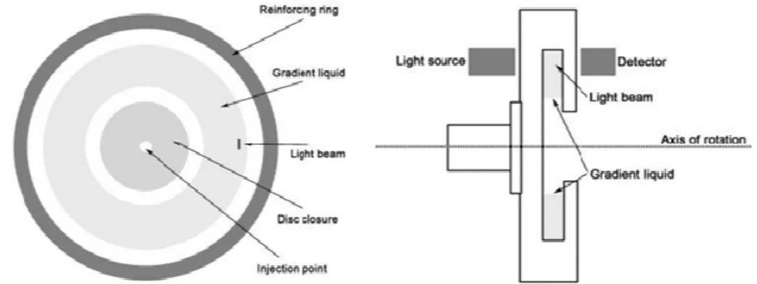
2.5. Differential Centrifugal Sedimentation (DCS)
Differential centrifugal sedimentation experiments were performed with a disc centrifuge (Model DC 24000; CPS Instruments Europe, Oosterhout, The Netherlands). A gradient of 2%–8% sucrose equilibrated with spinning at 22000 rpm for 30 minutes was established and calibrated by running standard polystyrene beads.
After establishment of the gradient, 100µl aliquots of particles dispersed in water were injected. Samples were spinned for approximately 2 hours in case of PS NPs and 5-10 minutes for Ag spherical NPs. The position of particles in the gradient was analysed with CPS Instrumental software. The tallest peak (the most frequent size value) was regarded as the ‘base’ peak (100%) and all other particle size peaks were normalized against this base peak (relative size distribution).
33
Figure 16. Schematic diagram of DCS Instrument
3. Studies on material adsorption of NPs
3.1. Assays on protein adsorption at NP surfaces
3.1.1. Electrophoretic studies on protein adsorption onto PS NPs
PS NPs were dispersed in MEM supplemented with 10% fetal calf serum (FCS).
After 1 h incubation at 37°C, the NPs were centrifuged for 45 min at 20000 x g.
Sedimented NPs were washed with PBS to remove non-bound proteins. Washed NPs were resuspended in Laemmli buffer containing 1% (w/v) sodium dodecyl sulfate (SDS), and loaded onto 10% polyacrylamide gel. The protein components of the corona complexes were separated from the NPs and were denatured by boiling at 100oC for 5 minutes in the loading buffer (62.5 mM Tris-HCl (pH 6.8), 2% (w/v) SDS, 10% glycerol, 0.01% (w/v) bromophenol blue, 40 mM DTT). The denatured corona proteins coated with SDS surfactant (which gives them a negative net charge) were separated by size on 10% polyacrylamide gel (SDS-PAGE). The electrophoresis was run under constant voltage of 130 V for about 45 minutes using a Mini-Protean Tetra electrophoresis system (Bio-Rad). All gels were run in duplicates – one subjected for commassie blue (50% methanol, 10% acetic acid, 2.5% (w/v) brilliant blue) staining for 3 hours and de-stained overnight in 50% methanol, and 10% acetic acid. The other gel was stained with silver staining (Ohsawa and Ebata, 1983) kit (Cosmobio Ltd., Tokyo, Japan ) (see below).
3.1.2. Human blood proteins on spherical Ag PVP NPs
In situ protein coronas on spherical Ag PVP NPs were prepared by incubating 0.1 mg/ml NPs in 10%, 80% and 100% human plasma solution (total protein content 34–
47 mg/ml) at room temperature for 1 hour.
34
The human plasma was obtained from Centre for BioNano Interactions (CBNI), School of Chemistry and Chemical Biology, University College Dublin, Dublin, Ireland. The blood donation procedure was approved by the Human Research Ethics committee at University College Dublin. The blood plasma was prepared following the HUPO BBB SOP guidelines (Rai et al., 2005). In brief, after the blood collection, the blood was mixed with the 2 mM EDTA and centrifuged for ten minutes at 1300 g at 4oC. Plasma from each donor were collected into a 50 ml falcon tube and then centrifuged at 2400 g for 15 minutes at 4oC. Supernatant was collected, aliquoted into 1 ml cryo vials and stored at -80oC until use. Following this procedure, the plasma protein concentration was estimated to be 80 g/l. Before the experiments, the plasma sample was thawed at RT and centrifuged for 3 min at 16200 RCF.
After incubation with human plasma, the NP-samples were directly injected into the DCS instrument without spinning down and washing.
3.2. Assays on endotoxin adsorption at NP surfaces 3.2.1. LAL assay
The endotoxin contamination of different NPs was tested by chromogenic Limulus amoebocyte lysate (LAL) assay (Lindsay et al., 1989) (Associates of Cape Cod, Inc., East Falmouth, MA, USA) according to the manufacturer’s instructions. Two-fold dilution series were prepared with endotoxin-free water (LAL Reagent Water; LRW) from each NP preparation in duplicates, and fresh endotoxin standard was prepared for each test using 0.1, 0.25, 0.5, 1.0 and 2.0 EU/ml LPS. Interference of NPs with the assay readout was investigated.
3.2.2. SDS PAGE
For SDS_PAGE assays on endotoxin adsorption, NPs were dispersed in LRW containing 1 mg/ml endotoxin (LPS from E.coli 055:B5; Sigma-Aldrich) and incubated for 1 h at 37°C. After incubation, the particles were collected by centrifugation (12000 g, 45 min ) and washed 3x with LRW. Washed particles in LRW and consecutive washing solutions were treated for 5 min at 100°C in Tris- hydrochloride buffer (pH 6.9), 10% w/v SDS, 0.01% bromo phenol blue, and loaded onto SDS-polyacrylamide gel (10 cm by 10 cm by 1 mm) containing 5% and 15%
acrylamide in the stacking and separating gels, respectively. Electrophoresis was done at 130 V until the tracking dye had run about 10 cm.
35
LPS components were visualised by staining the gels with a silver staining kit (Cosmobio Ltd. Tokyo, Japan) according to the method of (Tsai and Frasch, 1982) Briefly, gels were fixed in a 40% ethanol 5% acetic acid solution overnight, then oxidized with 0.7% periodic acid for 20 min. The gels were then washed 3x for 30 min in deionised water, and stained for 15 min with the staining solution of the kit, containing AgNO3, NH4OH and NaOH. The gels were washed again 3x for 30 min in deionised water, and placed in a developing solution containing citric acid, formaldehyde and sodium thiosulphate until optimal staining had taken place. The gels were rinsed with water and subjected to gel scanning.
3.2.3. Spiking with LPS
‘‘Spiking’’ controls, were made to identify the whether the samples to inhibit/enhance the detection of the endotoxin in the assay, consisted of NP dilutions to which a known amount (0.5 EU/ml) of standard endotoxin was included. Two-fold dilution series were prepared with endotoxin-free water (LAL Reagent Water; LRW) from each NP preparation in duplicates, and fresh endotoxin standard was prepared for each test. NP samples were serially diluted from the stock suspensions with LRW and distributed 50µL/well in endotoxin-free microplates for the endotoxin assay. The traditional chromogenic LAL assay is based on the detection of the endotoxin- stimulated LAL end-product 4-nitroaniline (pNA) at 405 nm but the new chromogenic assay was used in new version, with readout shifted from 405 to 540 nm. Briefly, after sample incubation with enzyme and substrate, the diazo reagents (provided by the kit) were added sequentially: 6mM sodium nitrite in 0.48N HCl (reagent 1), 26.3mM ammonium sulfamate in water (reagent 2) and 3.76mM N-(1- naphthyl)ethylenediamine dihydrochloride in water (reagent 3). The reagents modify pNA to turn from yellow to deep purple, thus allowing detection of the azo dye product at a wavelength of 540 nm.
3.2.4. Studies on interference of NPs with the assay readout
Twofold dilutions of p-nitroaniline (pNA, Sigma-Aldrich) were distributed in flat- bottomed 96-well plates in a volume of 50 ml/ well. For each pNA dilution, different concentrations of NPs and corresponding solvents were added in 50 ml aliquots in triplicate wells and mixed. For measuring interference at 405 nm, another 100 µl of water (in place of the substrate) and 100 µl of stop solution (sodium dodecylsulfate
36
solution) were added to bring the final volume to 300 µl, i.e. the same volume as in the QCL-1000 LAL assay. Optical density was measured with a microplate reader at 405 nm. For measuring interference at 540 nm, diazo reagents were added rapidly into the wells containing pNA and NPs and the optical density was immediately measured with a microplate reader at 540 nm.
3.2.5. Silver staining method
After gel electrophoreses, proteins and LPS components were visualised by staining the gels with a silver staining (Ohsawa and Ebata, 1983) kit (Cosmobio Ltd.) according to the method of Tsai and Frasch. Briefly, gels were fixed in 40% ethanol and 5% acetic acid solution, overnight, and then oxidized with 0.7% periodic acid for 20 min. The gels were then washed 3-times for 30 min in deionised water, and stained for 15 min with the staining solution of the kit, containing AgNO3, NH4OH and NaOH. The gels were washed again 3-times for 30 min in deionised water, and placed in a developing solution containing citric acid, formaldehyde and sodium thiosulphate until optimal staining occured. The gels were rinsed with water and subjected to gel scanning.
4. Cell cultures
4.1. NE-4C neuroectodermal stem cells
NE-4C neuroectodermal stem cells (ATTC CRL-2925; (Schlett and Madarasz, 1997)) cells were cloned from primary brain cell cultures prepared from the fore- and midbrain vesicles of 9-day-old transgenic mouse embryos lacking functional p53 tumor suppressor protein. NE-4C neuroectodermal stem cells were maintained in poly-L-lysine coated culture dishes, in minimum essential medium (MEM; Sigma- Aldrich, Hungary) supplemented with 4 mM glutamine and 10% fetal calf serum (FCS; Sigma-Aldrich) (MEM-FCS).
NE-4C cells were differentiated into neurons and astrocytes by adding 10-6 M all- trans retinoic acid (RA; Sigma-Aldrich, Hungary) to confluent cultures for 48 hours ((Schlett and Madarasz, 1997); Varga et al. 2009.; Madarász 2013). After 48-hour treatment with RA, the culture medium was changed to serum-free neural differentiation medium (MEM-ITS: MEM:F12= 1:1 supplemented with 1 % N2 neuronal supplement (Sigma-Aldrich)) containing insulin, transferrin and selenite. In MEM-ITS medium, RA-primed NE-4C cells differentiate into neurons and