INTERACTION OF BAICALIN WITH TRANSPORTERS
PhD thesis
Bernadett Kalaposné Kovács
Doctoral School of Pharmaceutical Sciences Semmelweis University
Supervisor: Dr. Imre Klebovich, D.Sc Official reviewers:
Dr. Éva Szökő, D.Sc
Dr. Györgyi Horváth, Ph.D
Head of the Final Examination Committee:
Dr. Tamás Török, D.Sc
Members of the Final Examination Committee:
Dr. László Tóthfalusi, Ph.D Dr. Gábor Halmos, Ph.D
Budapest, 2016
1 Table of contents
1. ABBREVIATIONS ... 4
2. INTRODUCTION ... 6
2.1. Baicalin ... 7
2.2. Efflux transporters ... 14
2.3. Uptake transporters ... 17
3. OBJECTIVES ... 20
4. MATERIALS AND METHODS ... 21
4.1. Materials ... 21
4.2. Vesicular transport inhibition studies ... 22
4.2.1. MDR1, MRP2, MRP3, MRP4 and BCRP ... 23
4.2.2. MRP1 ... 26
4.2.3. Follow-up experiments ... 27
4.3. Vesicular transport substrate study ... 29
4.4. Uptake transport studies ... 31
4.4.1. Cell culture and preparation... 31
4.4.2. Incubations ... 31
4.4.3. Sample preparation and analysis ... 31
4.4.4. Protein quantitation... 32
4.4.5. Data analysis ... 32
4.5. Analytics ... 33
2
5. RESULTS ... 35
5.1. Inhibition of efflux transporters by baicalin ... 35
5.1.1. Dose-response curves ... 35
5.1.2. IC50 values ... 41
5.2. Efflux of baicalin by selected transporters ... 44
5.2.1. MDR1 ... 45
5.2.2. MRP1 ... 46
5.2.3. MRP2 ... 47
5.2.4. MRP3 ... 48
5.2.5. MRP4 ... 48
5.2.6. BCRP ... 50
5.2.7. Summary of efflux transport experiments ... 51
5.3. Uptake of baicalin by transporters ... 53
5.3.1. Feasibility results ... 53
5.3.2. Time dependence of OATP2B1-mediated BG uptake ... 56
5.3.3. Concentration dependence of OATP2B1-mediated BG uptake... 57
6. DISCUSSION ... 58
6.1. Inhibition of efflux transporters by baicalin ... 60
6.2. Several ABC transporters efflux baicalin ... 64
6.3. Baicalin is a substrate of OATP2B1 ... 67
7. CONCLUSION ... 71
8. SUMMARY... 73
3
9. ÖSSZEFOGLALÁS ... 74
10. BIBLIOGRAPHY ... 75
11. BIOBLIOGRAPHY OF THE CANDIDATE’S PUBLICATIONS ... 88
12. ACKNOWLEDGEMENT ... 89
4 1. Abbreviations
ABC transporters: ATP-binding cassette transporters ADME: absorption, distribution, metabolism, excretion ATP: adenosine triphosphate
AMP: adenosine monophosphate B: baicalein
BCRP: Breast cancer resistant protein
BCS: Biopharmaceutical Classification System BG: baicalin
B-GS: bimane-glutathion conjugate CHO cells: chinese hamster ovary cells Cmax: maximal concentration
Clint: intrinsic clearance
ctrl K: control membrane for K562 cells ctrl M: control membrane for mammalian cells defMRP: functionally defective mutant MRP DHEAS: dehydroepiandrosterone sulfate DMSO: dimethyl sulfoxide
E217βG:estradiol-17-β-D-glucuronide EMA: European Medicines Agency E3S: estrone-3-O-sulfate
FDA: Food and Drug Administration GFJ: grapefruit juice
GSH: glutathione
HEK293: human embryonic kidney cells
HEK ctrl: functionally defective control membrane for HEK293 cells K562: human myelogenous leukemia cells
Km: substrate concentration at half Vmax
LC-MS: liquid chromatography–mass spectrometry LLOQ: lower limit of quantification
M: Michigan Cancer Foundation-7 (Breast cancer cell line) cells
5
MCF7: Michigan Cancer Foundation-7 (Breast cancer cell line) MDCK: Madin-Darby canine kidney cells
MDR: multidrug resistance
MRP: multidrug resistance associated protein NMQ: N-methyl-quinidine
OATP: organic anion-transporting polypeptide OCT: organic cation transporter
OCTN: carnitine/organic cation transporter Papp: apparent permeability coefficient QC: quality control
RS: Radix Scutellariae
±SD: standard deviation
Sf9: Spodoptera frugiperda ovarian cells SLC: solute carrier transporter
SULT: sulfotransferase
UGT: uridine 5'-diphospho-glucuronosyltransferase Vmax: maximal reaction velocity
VT: vesicular transport
6 2. Introduction
In the last decade, interest in studying the pharmacologic effects of phytomedicines has grown extensively. This is largely a result of the higher tendency among the general population to use complementary and alternative medicine.
Flavonoids, the main bioactive components found in herbs, are a group of polyphenolic compounds, diverse in chemical structure and characteristics, found ubiquitously in plants. Until now, more than 9000 different flavonoids have been studied and described.
There has been increasing interest in their research due to growing evidence of their versatile health benefits including anti-inflammatory, antioxidant, ant proliferative and anticancer activity, free radical scavenging capacity, antihypertensive effects, coronary heart disease prevention and anti-human immunodeficiency virus functions.
Additionally, flavonoids are safe and associated with low toxicity, making them excellent candidates for chemo preventive agents (Conseil et al., 1998; Nabekura et al., 2005). However, to achieve successful therapeutic efficacy, flavonoids must be absorbed adequately and consistently after oral administration; this behavior depends heavily on the drug delivery system (Tian et al., 2009).
Clinical studies and case reports have identified a number of herb-drug interactions potentiated by the concurrent use of herbal medicines with prescription drugs, raising concerns by health professionals regarding the potential for flavonoids to affect pharmacokinetics and pharmacodynamics of drugs (Bailey et al., 1993; Fasinu et al., 2012; Kantamreddi et al., 2009; Rajnarayana et al., 2004).The clinical consequences of herb-drug interactions varies, from being well-tolerated to moderate or serious adverse reactions, or possibly life-threatening events. Undoubtedly, the early and timely identification of herb-drug interactions is imperative to prevent potentially dangerous clinical outcomes (Chen et al., 2011; Hu et al., 2005). The potential of pharmacokinetic interactions occurring between phytomedicines and conventional drugs is therefore increasingly being recognized (Kennedy et al., 2010; Laki et al., 2013; Mohamed et al., 2011; Nguyen et al., 2015).
7 2.1. Baicalin
Radix Scutellariae (RS), officially listed in the Chinese Pharmacopoeia, is the dried root of the medicinal plant Scutellariae baicalensis, known as Huang Qin in Chinese traditional medicine (Figure 1) (CP, 2005).
RS is widely used for the prevention and treatment of various ailments including cardiovascular diseases, hypertension, bacterial infection, inflammation, and cancer (Blach-Olszewska et al., 2008; Gao et al., 2011b; Jung et al., 2012; Tseng et al., 2010;
Zhang et al., 2011c). More than 50 flavonoids have been purified and identified from RS (Chen et al., 2014). The major components are baicalin (baicalein-7-O- glucuronide, BG), and its aglycone baicalein (5, 6, 7 -trihydroxyfavone, B) (Li-Weber, 2009; Li et al., 2009). Due to their relatively low toxicity and high abundance in RS, BG Figure 1:Scutellaria baicalensis (https://en.wikipedia.org/wiki/scutellaria_baicalensis;
2016. September 26.)
8
Figure 2: (A) The roots of Scutellaria baicalensis; (B) the powder of BG; (C) chemical structures of BG and B(Zhang et al., 2011c).
and B became the most widely researched components in recent years (Figure 2) (Li- Weber, 2009).
Numerous in vivo and in vitro studies carried out in the last decade demonstrated that BG and its aglycone B were important medical agents with a variety of pharmacological activities such as chemopreventive, hepatoprotective, anti-aging, antioxidant, anti- fibrotic, anti-allergic, anti-depressant, anti-microbial, anti-inflammatory, antimutagenic, neuroprotective, memory improving, endotoxin, as well as anxiolytic effects (Dou et al., 2007; Gao et al., 2016; Hu et al., 2009; Kim et al., 2012; Kumagai et al., 2007; Oga et al., 2012; Sahebkar, 2012; Shang et al., 2010; Takahashi et al., 2011; Waisundara et al., 2011; Wang et al., 2015; Woo et al., 2005; Xu et al., 2011; Yu et al., 2016b). The clinical applications of BG include the treatment of pneumonia, hepatitis and cardiovascular diseases. BG might serve as a novel approach for the treatment of patients with Parkinson’s disease (Xue et al., 2014). BG can exert anti-H1N1 and H5N1
9
effects (Chu et al., 2015; Sithisarn et al., 2013) and antiviral activity against dengue virus (Moghaddam et al., 2014).
Treatment with BG showed to be a potential therapeutic strategy for acute lung injury (Ding et al., 2016). BG pre-treatment attenuated brain ischemia reperfusion injury by suppressing cellular apoptosis (Zhou et al., 2016). Other in vivo findings demonstrated that BG had significant potential as a novel anti-inflammatory agent for therapy of autoimmune diseases such as multiple sclerosis (Zhang et al., 2015). B exhibited anti- tumor effects in several types of cancers by inducing cancer cell apoptosis and suppressing metastasis. B might also be used in the treatment of pancreatic cancer, bladder cancer, lung cancer, hepatoma, breast cancer and skin carcinoma (Chao et al., 2007; Chen et al., 2000; Chiu et al., 2011; Du et al., 2010; Jiang et al., 2010; Li-Weber, 2009; Mu et al., 2016; Takahashi et al., 2011; Wu et al., 2011b; Yang et al., 2011; Yu et al., 2015). BG exerted anti-aging effects likely through attenuating oxidative stress (Gao et al., 2016). B has also shown to affect xenobiotic and carcinogen metabolism by inhibiting several metabolizing enzymes’ activity (Moon et al., 2006).
Moreover, BG extracts are easily accessible over-the-counter herbal remedies, purchasable online and in numerous stores in liquid or bulk powder form.
Recommended daily dosage of BG powder is 60-500 mg meaning a 540-4480 µM dose (in 0.25 liters).
Since BG and B have such enormous therapeutic potentials, a better understanding of their pharmacokinetics and bioavailability is necessary to specify clinical effects, developing clinical regimens and elucidating potential drug interactions.
Oral administration is a popular drug delivery route because it is usually convenient for both doctors and patients. To achieve successful therapeutic efficacy, BG must be absorbed adequately and consistently after oral administration (Tian et al., 2009).
Glucuronidation is a significant metabolic pathway that facilitates efficient elimination and detoxification of numerous endogenous substances (e.g., bilirubin and estradiol) and xenobiotics (e.g., SN-38 and indinavir) (Wu et al., 2011a).
10
In recent decades, numerous studies have accumulated evidence indicating that natural polyphenols are rapidly and extensively metabolized to glucuronides and sulfates after oral ingestion. Based on a previous pharmacokinetic study of RS, the glucuronides and/or sulfates of B were the major molecules in the bloodstream after dosing a RS decoction to rats (Hou et al., 2011).
Several animal studies showed that BG, instead of B, was the predominant form in the general blood circulation after oral administration of B or BG (Akao et al., 2000; Akao et al., 2013; Cai et al., 2016; Fong et al., 2015; Gao et al., 2011a; Gao et al., 2012; Lai et al., 2003; Taiming et al., 2006; Xing et al., 2005).
Upon oral intake, BG is either directly absorbed from the upper intestinal tract (Akao et al., 2000; Lu et al., 2007; Zhang et al., 2005a) or undergoes hydrolysis by intestinal glucuronidase or intestinal microflora to release its aglycone B, which will then be absorbed via passive diffusion (Abe et al., 1990; Kang et al., 2014; Lu et al., 2007; Noh et al., 2016; Wang et al., 2012; Xing et al., 2014; Zhang et al., 2005a).
Concomitantly upon oral intake of B, B is absorbed via passive diffusion. Absorbed B undergoes extensive first-pass intestinal Phase II metabolism in enterocytes, including glucuronidation (>90%), catalyzed by the enzyme UDP-glucuronosyltransferase (UGT) and less significant sulfation, catalyzed by sulfotransferase (SULT) resulting in its conjugated metabolites, BG and baicalein-7-O-sulfate (Akao et al., 2000; Zhang et al., 2007a).
Although B demonstrates good permeability due to its good lipophilicity, its metabolite BG formed inside the intestinal epithelial cells is too polar to cross the lipid bilayer by passive diffusion (Dai et al., 2008).
Because of low oral bioavailability, various formulations have been developed to improve the gastrointestinal absorption of BG. Solid dispersions, microcapsules, cyclodextrins, emulsions, phospholipid complex, liposomes and nanoparticles have been described (Gabrielska et al., 1997; Li et al., 2011b; Liu et al., 2011; Luo et al., 2010; Wu et al., 2014). BG belongs to Class IV of Biopharmaceutical Classification System (BCS) due to its extremely low hydrophilicity (solubility 0.052 mg/mL in water)
11
and lipophilicity (Papp = 0.037 × 10⁻⁶ cm/s) (Wu et al., 2014). B is highly permeable (Papp = 1.7 × 10⁻⁵ cm/s) but poorly water soluble, which is classified as a Class II compound according to BCS (Zhang et al., 2007b; Zhang et al., 2014).
Accordingly, after administration of a single ascending dose of B (100-2800 mg) chewable tablets to healthy subjects, the Cmax values of BG were about ten-fold higher than Cmax values of B (Li et al., 2014). Another study using rat intestine perfusion model and Caco-2 monolayer model uncovered that B was rapidly converted to BG, before being transported to the mesenteric system (Zhang et al., 2005a).In addition, significant biliary as well as sinusoidal transport of BG from hepatocytes was shown (Akao et al., 2009).
There have been many studies reporting that the glucuronides and sulfates of xenobiotics are the substrates of multidrug resistance–associated proteins (MRPs) or breast cancer resistance protein (BCRP). The involvement of efflux transporters such as BCRP is necessitated by the fact that a glucuronide is too polar to passively diffuse out of cells. Therefore, in addition to UDP-glucuronosyltransferases (UGTs) that catalyze glucuronidation reaction (i.e., glucuronide formation), efflux transporter is another element that enables glucuronide clearance (Xu et al., 2009; Zamek-Gliszczynski et al., 2006). In recent years, numerous studies have shown that BCRP is involved in intestinal and/or biliary excretion of glucuronides of a diverse group of compounds including flavonoids (Xu et al., 2009). Thus, the effective transport of intracellularly formed glucuronides of B from enterocytes likely depends on a carrier-mediated transport.
Owing to the various mechanisms involved in the absorption, reconversion dynamics between BG and B, and metabolism of BG, the attainment of peak concentrations in plasma for BG appeared to be prolonged, suggesting a significant role of the enterohepatic recycling of BG. The extensive enterohepatic recycling distributive phase has been confirmed after both oral and intravenous routes of dosing in rats (Xing et al., 2005). Biliary excretion plays a major role in bringing the glucuronide and sulfate conjugates of B back to the small intestine where it undergoes hydrolytic cleavage through intestinal beta-glucuronidase (Liu et al., 2010).
12
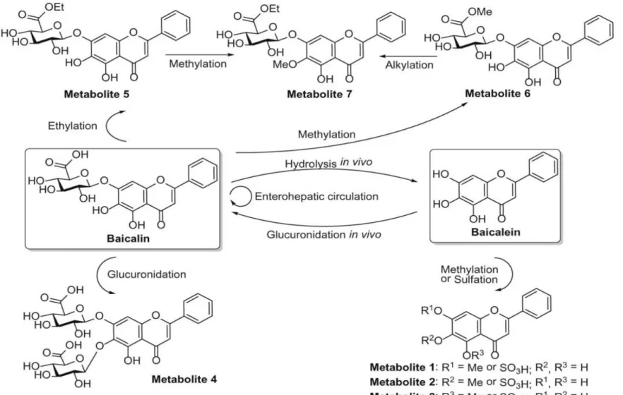
The role of hepatic biliary excretion in the modulation of pharmacokinetics of BG has been recently clarified. In that study, the pharmacokinetics of BG were evaluated in wild type rats and Mrp2-deficient rats. Following oral administration of B to Mrp2- deficient rats, the peak concentration and AUC value for BG were five-fold and eight- fold higher than the relative values obtained in normal rats (Akao et al. 2009). When B was dosed into the portal vein of Mrp2-deficient rats, a four-fold reduction in the biliary excretion and a 30-fold elevation in systemic exposure was observed as compared with a similar B dose administration into the portal vein of regular rats. Therefore, this work not only clarified the biliary excretory pathway for BG, but also indicated the propensity of the sinusoidal efflux mechanism (Akao et al., 2009; Srinivas, 2010). The potential biotransformation pathway of BG and B can be seen on Figure 3 (Chen et al., 2014).
The liver is regarded as the most important organ for the disposition of various endogenous and exogenous substances in the body. As for the hepatic disposition of conjugates already existing in the circulation, the hepatic uptake of metabolites is critical due to their difficulty to traverse the basolateral cell membrane. Before hepatic metabolism and biliary excretion, drugs need to enter the hepatocytes first, either through passive diffusion or mediated by transporters. Uptake transporters are membrane proteins that modulate the cellular influx of numerous substances including clinically important agents such as antibiotics, anti-cancer agents, and non-steroidal antiinflammatory drugs.
Because of its poor passive permeability, the hepatic uptake of BG mediated by an uptake transporters could be a key determinant in hepatobiliary excretion of BG.
13
Figure 3: Biotransformation pathway of BG and B (Chen et al., 2014).
14 2.2. Efflux transporters
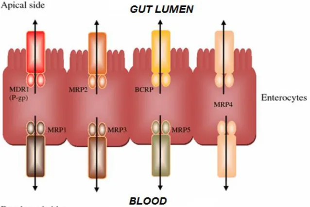
Drug transporters are multispecific transmembrane proteins that facilitate the membrane transport of a large number of drugs. Drug transporters have a distinct expression pattern in the human body lining pharmacological barrier tissues, most importantly the small intestinal epithelium (Figure 4), the endothelial cells in the blood–brain barrier, the epithelium of the proximal tubule cells in the kidney, and hepatocytes in the liver (Chandra et al., 2004; Feng et al., 2010; Klukovits et al., 2015; Muller et al., 2011;
Suzuki et al., 2000; Tamai et al., 2000).
Figure 4: ABC transporter localisation in gut epithelial cells. P-glycoprotein (Pgp/MDR1), MRP2 and BCRP are localised in the apical membrane, effluxing compounds back in to the gut lumen; whereas MRP1, 3 and 5 are localised in the basolateral membranes pumping substrates in to the blood stream. MRP4 is present in both the apical and basolateral membranes of gut epithelia (Brand et al., 2006).
15
The superfamily of human ATP-binding cassette (ABC) proteins comprises 49 members divided into 7 subfamilies (ABCA – ABCG) (Dean et al., 2001; Vasiliou et al., 2009). The first ABC transporter involved in drug trafficking (ABCB1) was described in 1976 (Jani et al., 2014b). ABC transporters transport a wide variety of substrates across plasma- and intracellular membranes, including metabolic products, lipids and sterols, and drugs (Figure 4) (Brand et al., 2006).
The members of multidrug resistance-associated proteins (MRPs), especially MRP2(ABCC2), and MRP3(ABCC3), possess similar substrate selectivity and prefer to transport organic anion and phase II metabolites, including glutathione, glucuronide, and sulfate conjugates (Borst et al., 2000). It also seems likely that MRP2 and/or BCRP (ABCG2) contribute to the efflux of flavonoid conjugates across the intestinal apical membrane (Sesink et al., 2005).
In a recent study performed on MDCKII-MRP2 and MDCKII-BCRP cell lines, RS inhibited transport by the selected transporters. Consistently, the cell study further confirmed that BG inhibited the efflux transport mediated by MRP2 (Yu et al., 2016a).
Other study results indicated that, in rat, a large proportion of B absorbed is retained, transformed into BG within the intestinal mucosal cells, and coordinately excreted through MRP2 into the intestinal lumen (Akao et al., 2004; Cao et al., 2008). In other studies, BG was shown to inhibit very efficiently BCRP-, MRP2- and MRP3-mediated vesicular transport and to activate the ATPase activity of BCRP, MRP2 and MRP3 (Gao et al., 2012). Another inhibitory study on MDCKII-MRP2 and MDCKII-BCRP cell lines indicated that BG inhibited both the BCRP- and MRP2-mediated efflux transports (Yu et al., 2016a). Moreover experiments performed on Caco-2 cells showed that BG may be a P-gp inhibitor (Miao et al., 2016). An increase of the sinusoidal transport of BG was seen in Mrp2-deficient rats (Akao et al., 2009).
These findings indicate that MRP3 could likely play a role in the basolateral transport of BG from intestinal cells while MRP2 and BCRP might be the transporters effluxing BG on the apical side of enterocytes and hepatocytes (Akao et al., 2007; Akao et al., 2009;
Li et al., 2012; Zhang et al., 2007a). The ATPase assay, however, is not a functional transport assay hence the substrate or inhibitory potential needs to be confirmed by a
16
functional vesicular transport assay, which is optimal for testing low passive permeability substrates.
On the other hand, by inhibiting the activity of ABC transporters, BG could modulate absorption and disposition of drugs, increasing the risk of therapeutic failure, adverse effects and toxicity (Li et al., 2013; Zamek-Gliszczynski et al., 2006). Impaired transporter activity could result in reduced (intestinal/hepatic) glucuronide clearance and elevated glucuronide accumulation in systemic circulation (Xu et al., 2009).
Therefore elucidating the modulation of intestinal and hepatic efflux transport by BG is of crucial importance for the evaluation of flavonoid–drug interactions, since the majority of drug products and food supplements are given orally at large quantities.
Driven mainly by the needs of the industry, in vitro methods that assess transporter interactions have matured into routine tools in the past two decades and are widely applied to predict in vivo and clinical phenomena as well as to characterize interactions on a molecular level (Sjogren et al., 2014).
The first membrane vesicle applications on ABCB1 were demonstrated in 1986 using selected mammalian and transduced Sf9 cells (Doige et al., 1992; Horio et al., 1988;
Sarkadi et al., 1992). Since then the methods have been further refined and extended on other ABC transporters and are presently widely used in the study of interactions between drugs and ABC transporters (Stieger et al., 2000). Vesicular transport is very efficient in the characterization of inhibitors, where the studied compound modulates transport rate of a reporter probe (Glavinas et al., 2008).
The vesicular transport assay relies on membrane preparations enriched in inside-out vesicles (Heredi-Szabo et al., 2012). In this orientation, the binding sites of the transporter are facing the solvent; thus concentrations and other conditions affecting the transport process can be precisely defined. Substrate molecules will be pumped into the membrane vesicles in an ATP dependent manner, and typically incubations containing AMP provide the baseline (Doige et al., 1992). Transported and non-transported substrate molecules are separated by filtration through glass fiber or nitrocellulose membranes. Vesicles with trapped molecules will be retained on the filters, and
17
substrate molecules can be quantified via common analytical methods after elution with a suitable agent, such as methanol.
It has been demonstrated that the vesicular transport assay is a useful tool to investigate the contribution of transporters in the permeability of flavonoids (Cooray et al., 2004;
Dreiseitel et al., 2009; Tan et al., 2014; Tan et al., 2013; Valdameri et al., 2012; Zhang et al., 2004). Vesicular transport is also very efficient in the characterization of inhibitors, where the studied compound modulates transport rate of a reporter probe (Glavinas et al., 2004; Heredi-Szabo et al., 2012; Heredi-Szabo et al., 2013; Jani et al., 2014b; Klukovits et al., 2015). In the drug transporter area, the potential for inhibition is commonly assessed via the determination of an in vitro IC50 value. Current FDA and EMA guidance for drug transporter interactions is dependent on IC50 measurements as these are utilized in determining whether a clinical interaction study is warranted (FDA, 2012). These guidances contain decision trees on whether a clinical drug-drug interaction study is warranted which are based on the IC50 value in combination with clinical drug concentrations (Ellens et al., 2013; FDA, 2012).
2.3. Uptake transporters
The organic anion transporting polypeptides (OATPs) are essential solute carrier (SLC) transporters expressed in key human organs / tissues, in particular the intestine, kidney, and liver. OATPs mediate the sodium-independent transport of a diverse range of amphiphilic organic compounds. These include bile acids, steroid conjugates, thyroid hormones, anionic peptides, numerous drugs and other xenobiotic substances (Giacomini et al., 2010). OATP1A2, OATP2B1, OCT1, OCTN1, and OCTN2 transporters may assist in the intestinal absorption of many clinically important and frequently prescribed drugs at the lumen facing apical membrane of enterocytes, whereas OCT1 and OCT2 may mediate the drug uptake from blood at the basolateral membrane of enterocytes. OATP1B1, OATP1B3, OATP2B1, OAT2, OCT1, and OCT3 are responsible for the uptake of drugs into the liver at the basolateral membrane of
18
hepatocytes, which is the first step of the subsequent biliary excretion and/or drug metabolism (Roth et al., 2012).
It was already shown that BG impacted on the pharmacokinetic performance of rosuvastatin, a substrate of several hepatic SLC transporters, including OATP1B1, OATP1B3, OATP2B1 and OATP1A2 (Fan et al., 2008).
In another inhibitory study performed on CHO-OATP1B1, MDCKII-OATP2B1 and CHO-OATP1B3 transfected cell lines, BG has shown to inhibit transport by OATP2B1 and OATP1B3, uptake transporters expressed in the apical membranes of enterocytes and sinusoidal membranes of hepatocytes. However, BG did not affect transport by OATP1B1 (Zhang et al., 2011b). In addition, another study performed on HEK293 cells exhibited that BG did not inhibit transport by OATP1A2 and OATP1B1, but affected transport by OATP1B3 and OATP2B1 (Xu et al., 2013).
Moreover, OATP2B1 was found to be primarily responsible for the hepatic uptake of Scutellarin-6-G, a structural analog of BG (Gao et al., 2012).
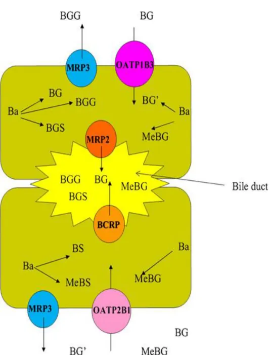
Therefore, OATP2B1 and OATP1B3 might be ideal candidates for the role of hepatic uptake of BG (Figure 5).
19
Figure 5: Proposed diagram of hepatic metabolism and disposition of B (Zhang et al., 2011a). Ba: Baicalein, BGG: baicalein-O-diglucuronide, BGS baicalein-O-glucuronide-O- sulfate, BGGlu:baicalein-O-glucose-O-glucuronoide, BG: baicalein-7-O-glucuronide, MeBG: methyl-O-baicalein-O-glucuronide, BG′ : baicalein-6-O-glucuronide,
BS: baicalein-O-sulfate, MeBS: methyl-O-baicalein-O-sulfate.
20 3. Objectives
As explained in the introduction, in recent years, a wealth of evidence has been generated from in vitro and in vivo studies showing that BG could interact extensively with drug transporters and might play critical roles in multidrug resistance reversal and drug disposition. Altered drug disposition due to pharmacokinetic interactions may result in clinically relevant changes in drug ADME properties and therefore drug efficacy or toxicity. Moreover, since BG and B have such enormous therapeutic potentials, a better understanding of their pharmacokinetics and bioavailability is necessary for extrapolating the data from pharmacological assays to clinical effects and developing clinical regimens.
The objectives of this thesis were therefore:
to investigate the inhibitory effect of BG on ABC transporters
to identify the transporters responsible for the efflux of BG from enterocytes and hepatocytes
to identify transporters responsible for the uptake of BG into hepatocytes and enterocytes
The information obtained from these studies will help us better understand and predict the potential in vivo BG-drug interactions mediated by these drug transporters and to elucidate pharmacokinetics of BG.
21 4. Materials and methods
4.1. Materials
3H-Estradiol-17-β-D-glucuronide (3H-E217βG), 3H-Estrone-3-O-sulfate (3H-E3S) and
3H-Dehydroepiandrosterone sulfate (3H-DHEAS) were purchased from Perkin Elmer Inc. (Waltham, MA, USA) and 3H-N-Methyl-quinidine (3H-NMQ) from BRC Radio- Lab Ltd. (Szeged, Hungary).
Membranes isolated from BCRP-, MDR1-, MRP1-, MRP2-, MRP3- and MRP4- overexpressing cells and bimane-glutathion conjugate (B-GS) were provided by Solvo Biotechnology Ltd. (Budaörs, Hungary). HEK293-OATP1B3 and MDCKII-OATP2B1 cells stably overexpressing the human transporters of interest and control cells (HEK293-Mock and wild type MDCKII) were obtained from Solvo Biotechnology Ltd.
(Budaörs, Hungary).
All other chemicals were of analyitical grade and were purchased from Sigma–Aldrich Ltd. (Budapest, Hungary).
22
Figure 6: Vesicular transport inhibition study principles 4.2. Vesicular transport inhibition studies
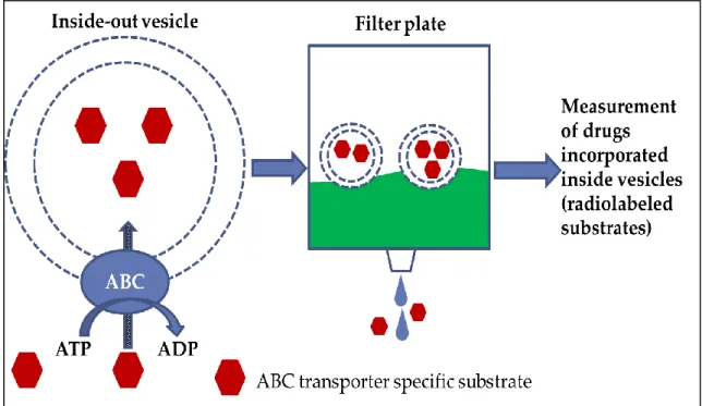
The interaction of BG with the transporter can be detected as the modulation of the initial rate of a labeled radioactive substrate transport by the transporter into membrane vesicles purified from Sf9 or MCF7 or HEK293 cells expressing the transporter (Figure 6).
Inhibitory effects of BG on transport of NMQ in membrane vesicles from K562 cells overexpressing MDR1, on transport of BGS in membrane vesicles from Sf9 cells overexpressing MRP1, on transport of E217βG in membrane vesicles from Sf9, then HEK293 cells overexpressing human MRP2, on transport of E217βG in membrane vesicles from Sf9, then HEK293 cells overexpressing human MRP3, on transport of DHEAS in membrane vesicles from HEK 293 cells overexpressing human MRP4 and on transport of E3S in vesicles from MCF7 cells expressing high levels of BCRP were investigated with rapid filtration techniques as described previously (Bodo et al., 2003;
Pal et al., 2007).
23
The baculovirus insect cell system (Sf9) is easy-to-use and gives high expression of the transduced gene. Obviously, no other mammalian transporters are present in the insect cells. In case of membrane preparations from insect cells, the baseline activity is very high, so most interacting compounds actually inhibit this baseline activity. In case of membranes prepared from mammalian cells (HEK293, MCF7, K562), the baseline activity is lower, and some interacting compounds inhibit, while known transported substrates activate the baseline activity. Experiments were performed in Sf9 and mammalian cell system as well, if available, to compare the systems.
4.2.1. MDR1, MRP2, MRP3, MRP4 and BCRP
Experiments were performed on MDR1-K562, MRP1-Sf9, MRP2-Sf9, MRP2-HEK293, MRP3-Sf9, MRP3-HEK293, MRP4-HEK293 and BCRP-MCF7 membranes.
A 30 mM BG stock solution and a 3-fold serial dilution was prepared in dimethyl sulfoxide (DMSO).
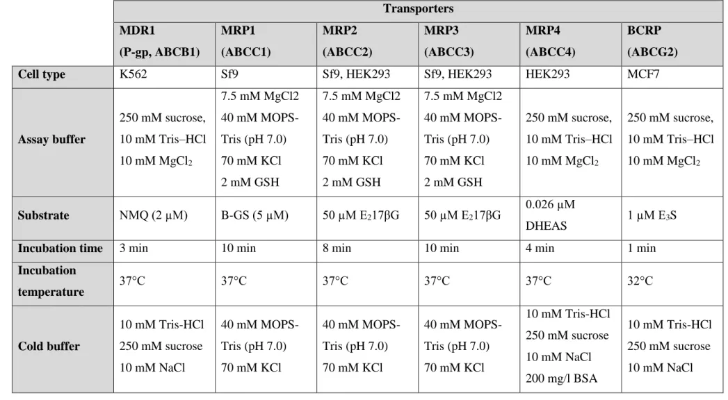
Inside-out membrane vesicles were preincubated. The 3H labeled transporter specific substrate was added to the mixture (Table 1). The reaction volume was 75 µl in each well, with 50 µg protein/well.
0.75 µl of the BG dilution series was added to each well. The dilution series was diluted 150-fold when added to the wells. Transport was initiated with the addition of 4 mM ATP or AMP in the appropriate well. Transport was carried out on specific cell lines, under specific buffer solution, incubation temperature and time conditions for each transporter (Table 1).
The transport was stopped by the addition of cold wash buffer, after specific incubation time. The samples were transferred to class B glass fiber filters, 1-µm pore size (Millipore, Billerica, MA, USA). Filters were washed with 5 x 200 µL of ice-cold wash buffer and dried. After adding 100 µL of scintillation cocktail to each well, radioactivity retained on the filter was measured by liquid scintillation counting (Perkin Elmer 1450 LSC, Luminescence counter, Microbeta Trilux). Results were obtained in cpm.
24
Table 1: Experiment parameters for inhibition type vesicular transport experiments
Transporters MDR1
(P-gp, ABCB1)
MRP1 (ABCC1)
MRP2 (ABCC2)
MRP3 (ABCC3)
MRP4 (ABCC4)
BCRP (ABCG2)
Cell type K562 Sf9 Sf9, HEK293 Sf9, HEK293 HEK293 MCF7
Assay buffer
250 mM sucrose, 10 mM Tris–HCl 10 mM MgCl2
7.5 mM MgCl2 40 mM MOPS- Tris (pH 7.0) 70 mM KCl 2 mM GSH
7.5 mM MgCl2 40 mM MOPS- Tris (pH 7.0) 70 mM KCl 2 mM GSH
7.5 mM MgCl2 40 mM MOPS- Tris (pH 7.0) 70 mM KCl 2 mM GSH
250 mM sucrose, 10 mM Tris–HCl 10 mM MgCl2
250 mM sucrose, 10 mM Tris–HCl 10 mM MgCl2
Substrate NMQ (2 µM) B-GS (5 µM) 50 µM E217βG 50 µM E217βG 0.026 µM
DHEAS 1 µM E3S
Incubation time 3 min 10 min 8 min 10 min 4 min 1 min
Incubation
temperature 37°C 37°C 37°C 37°C 37°C 32°C
Cold buffer
10 mM Tris-HCl 250 mM sucrose 10 mM NaCl
40 mM MOPS- Tris (pH 7.0) 70 mM KCl
40 mM MOPS- Tris (pH 7.0) 70 mM KCl
40 mM MOPS- Tris (pH 7.0) 70 mM KCl
10 mM Tris-HCl 250 mM sucrose 10 mM NaCl 200 mg/l BSA
10 mM Tris-HCl 250 mM sucrose 10 mM NaCl
25 Calculations:
ATP dependent transport (cpm): the mean cpm values measured in the absence of ATP were substracted from the mean cpm values measured in the presence of ATP.
ATP dependent transport (pmol/mg/min): Total activity (cpm) was calculated by multiplying the cpms measured in the designated well. The rate of transport in pmol/mg membrane protein/min was calculated using the following formula:
(min) e tim mg protein membrane
ml Volume nM
ion concentrat Substrate
cpm activity Total
cpm transport dependent
ATP
* ) (
) (
* )
* ( )
(
) (
ATP dependent transport (%): the percent activation or inhibition of the test drug. In this representation the ATP dependent transport determined in the drug fee control were taken as 100% and all other values were represented on this relative scale, using the following formula:
All experiments were performed 3 times. All concentrations were tested in duplicates.
BG concentration-relative transport (%) “dose-response curve” was generated for each transporter.
The IC50 is defined as the concentration needed to inhibit transport of the reporter substrate by 50%. The IC50 parameters were derived from the equation of a one-binding site, dose-response curve fitted onto the relative activity against the concentration of BG, plotted by non-linear regression using GraphPad (San Diego, CA) Prism version 5.
100 (cpm) *
control free
drug in transport dependent
ATP
(cpm) drug test of presence the
in transport dependent
ATP
26 4.2.2. MRP1
Inhibitory effects of BG on transport of B-GS in membrane vesicles from Sf9 cells overexpressing MRP1 were investigated with rapid filtration techniques. The interaction was detected as the modulation of the initial rate of bimane-glutathion conjugate (B-GS) transport of MRP1 into membrane vesicles purified from Sf9 cells expressing the transporter.
Same inhibition vesicular transport experiments as described before were conducted in the presence of BG concentrations in duplicates in MRP1-Sf9 vesicles. The BG stock solution and the dilution series were prepared in dimethyl sulfoxide (DMSO) and were diluted 100-fold when added to the wells
The transport was stopped by the addition of cold wash. The samples were transferred to class B glass fiber filters, 1-µm pore size (Millipore, Billerica, MA). Filters were washed with 5 x 200 µL of ice-cold wash buffer. After adding 100 µl of the detector solution (0.01 M HCl), fluorescence was measured at Ex: 430 nm, Em: 538 nm (BMG Fluostar optima). Data was analyzed following the preparation of a B-GS calibration curve.
Calculations:
ATP dependent transport (fluorescence): we took the average of the duplicates.
Fluorescence values measured in the absence of ATP were substracted from the fluorescence values measured in the presence of ATP for control and samples.
ATP dependent transport (%): percent activation or inhibition of the test drug. In this representation the ATP dependent transport determined in the drug free control was taken as 100% and all other values were represented on this relative scale, using the following formula:
100 control *
free drug in transport dependent
ATP
drug test of presence the
in transport dependent
ATP
27
ATP dependent transport (pmol/mg/min): after setting up a calibration curve with the help of the measured fluorescence values and the B-GS concentrations used, we substituted the fluorescence values into the equation of the calibration curve and calculated the amount of B-GS / well (pmol). After that, we divided this value by the amount of protein per well (0.05 mg) and by the time (10 min).
Relative transport values (%): This curve shows the effect of the test drug on B-GS transport by MRP1 in percentages. 100% represent B-GS transport by MRP1 in the absence of test drug ,while 0% is the transport in the absence of ATP (non-specific binding of B-GS).
If the test drug interacts with the B-GS transport, then a dose-dependent decrease in transport is observed. The IC50 value for the test drug is the concentration where the B- GS transport is inhibited by 50%. In case of a non-interactor, the transport of the reporter substrate typically does not change.
4.2.3. Follow-up experiments
After the first set of experiments, starting concentration of BG was optimized for each transporter according to the first obtained inhibition curves, if needed (Table 2). The inhibition curve for each transporter has to contain at least 2 data points on the inflection and on both plateaus. The same experiments were then performed.
The IC50 can be used for ranking a series of compounds based on their inhibition potential. Compounds inhibiting the transport can be either substrates or inhibitors of the transporter protein investigated. In order to determine which is the case, further experiments (named as substrate accumulation assay, direct vesicular assay or feasibility assay) were performed to reveal the mechanism of inhibition.
28
Table 2: BG concentrations in mixture for follow-up experiments in inhibition type vesicular transport experiments
MDR1- K562
MRP2- Sf9
MRP2- HEK293
MRP3- Sf9
MRP3- HEK293
MRP4- HEK293
BCRP- MCF7
MRP1- Sf9 BG concentrations (µM) 0.41-300 0.41-300 0.41-300 0.41-300 0.41-300 0.41-300 0.41-300 0.41-300 Follow-up experiments BG
concentrations (µM) 0.41-300 3.7-2700 3.7-2700 0.41-300 0.41-300 0.41-300 0.05-33.33 3.7-2700
29 4.3. Vesicular transport substrate study
Vesicular transport substrate experiments, also known as feasibility assay, were performed at 2 concentrations and at 2 reaction times, to evaluate whether ATP- dependent accumulation of BG could be detected directly in transporter-overexpressing membrane vesicles, with rapid filtration techniques (Heredi-Szabo et al., 2012).
Accumulation of BG in membrane vesicles from K562 cells overexpressing MDR1, in membrane vesicles from Sf9 cells overexpressing MRP1, in membrane vesicles from HEK293 cells overexpressing human MRP2, in membrane vesicles from HEK293 cells overexpressing human MRP3, in membrane vesicles from HEK293 cells overexpressing human MRP4 and in vesicles from MCF7 cells expressing high levels of BCRP were investigated with rapid filtration techniques as described previously (Bodo et al., 2003), (Pal et al., 2007) (Figure 7).
Vesicular transport of BG was also tested on control membranes (control K562 for K562 vesicles, defMRP for Sf9 vesicles, control M for M vesicles, HEK293 control for
Figure 7: Vesicular transport experiment principle
30
HEK293 vesicles) with no, or significantly lower transporter activity, in order to elucidate transporter dependent accumulation inside the vesicles.
1 mM BG stock solution was prepared in dimethyl sulfoxide (DMSO) and were diluted 100-fold when added to the wells. The reaction was performed at one or two BG concentrations (low and high) - adjusted to the previous IC50 data for each transporter, and one or two time points. Experiments were repeated 2 more times with criteria were the accumulation rate was maximal.
Inside-out membrane vesicles were preincubated. BG was added to the mixture. The inside-out membrane vesicles were incubated in the presence or absence of 4 mM ATP in the appropriate assay buffer and temperature (Table 1). The reaction volume was 75 µl in each well, with 50 µg protein/well.
The transport was stopped by the addition of the appropriate cold wash buffer. The samples were transferred to class B glass fiber filters, 1-µm pore size (Millipore, Billerica, MA, USA). Filters were washed with 5 x 200 µL of ice-cold wash buffer.The vesicles were lysed with 2 x 150 µl methanol and the eluted volumes, containing BG, were collected, the organic solvent dried with a speedvac concentrator (Thermo DNA 120) and the BG on the plates were subjected to bioanalysis.
The amount of transported BG was determined by LC-MS/MS analysis (Magda et al., 2015).
Experiments were performed 3 times, at the concentration and incubation time giving the most adequate result. All concentrations were tested in triplicates. The effectiveness of the membranes was controlled with a specific substrate for each transporter.
Statistical significance was calculated using ANOVA (one-way analysis of variance).
31 4.4. Uptake transport studies
4.4.1. Cell culture and preparation
OATP2B1-MDCKII, OATP1B3-HEK293, wild type MDCKII and HEK293 mock cells were plated in 24-well tissue culture plates at a density of 4 x 10⁵ cells/ well. For HEK293 cells, plates were precoated with poly-D-lysine. Feasibility studies were performed 24 hours after seeding. Before experiments, cell culture medium was removed and the reaction was initiated by adding transport buffer (Henseleit–Krebs buffer: KCl 4.83 mM, KH2PO4 0.96 mM, NaHCO3 23.8 mM, NaCl 142 mM, MgSO4
1.2 mM, CaCl2 1.53 mM, 4-(2-hydroxyethyl)- 1-piperazineethanesulfonic acid (HEPES) 12.5 mM, D-glucose 5 mM, and pH 7.4).
4.4.2. Incubations
For feasibility screening, experiments were performed by adding BG at two different concentrations (1 µM and 10 µM) and the cells were incubated at 37ºC for 2 or 20 minutes. Experiments were carried out 3 times in triplicates.
For time course, cells were incubated at 37ºC for the indicated periods of time (1-45 minutes) after adding 5 µM BG to the transport medium in transfected and control cells.
Experiments were carried out twice in triplicates.
For concentration dependence, experiments were performed by adding different concentrations of BG (1 µM - 100 µM) to the transport medium in transfected and control cells. The cells were incubated at 37ºC for 3 minutes. Experiments were carried out three times in triplicates.
4.4.3. Sample preparation and analysis
The uptake was terminated by the addition of ice-cold transport medium and immediate rinsing of cells twice with ice-cold transport medium. Cells were lysed with methanol- water (2:1) solution, and the plates were centrifuged at 5000 g for 10 min, 4°C.
32
Supernatants were transferred into a U-bottom plate and vacuum dried. Samples were dissolved in eluent. The amount of accumulated BG was determined by LC-MS/MS analysis (Magda et al., 2015).
4.4.4. Protein quantitation
Bicinchoninic acid kit (Sigma-Aldrich, St Louis, MO, USA) was used to check the total protein concentrations in cells. Positive control experiments with specific substrates of the transporters were performed to control the activity of the cells.
4.4.5. Data analysis
GraphPad Prism 5 (GraphPad Software Inc., San Diego, CA, USA) was used for curve fitting and calculation of kinetic parameters. Data shown in the figures are arithmetic means with standard deviation (± SD). Statistical significance was calculated using ANOVA (one-way analysis of variance).
33 4.5. Analytics
LC-MS/MS method was developed to quantify BG in membrane vesicles (Figure 8).
The analytical method was developed in collaboration with the Institute of Organic Chemistry, Research Centre for Natural Sciences, Hungarian Academy of Sciences.
The chromatographic separation used was an "inverse gradient elution" on a reversed phase column.
A QTRAP 6500 triple quadruple - linear iontrap mass spectrometer, equipped with a Turbo V Source in electrospray mode (AB Sciex, Redwood City, CA, USA) and a Perkin Elmer Series 200 micro LC system (Waltham, MA, USA) was used for LC- MS/MS analysis of BG.
Chromatographic separation was achieved by an Agilent Zorbax SB-C8 column (250 mm × 4.6 mm, i.d.: 5 μm) (Waldbronn, Germany). Sample was eluted with a gradient of solvent A (0,1% formic acid in water) and solvent B (0,1% formic acid in MeOH). The MS/MS system was operated under positive mode and multiple reaction monitoring mode.
To each well containing the transported BG, 200 µl water in methanol (2:8, v/v) containing 0.1% formic acid was added and kept at room temperature for one hour. The samples were transferred to 200 µl vials before injection into the LC-MS system. The calibration curve was linear (r=0.9987) from 1-1000 nM over the 3 concentration range.
The coefficient of variation and relative error of BG for 4 intra- and inter-assay at three quality control (QC) levels was 2.0-10.2 % and -6.1-6.7 %, respectively. The lower limit of quantification (LLOQ) for BG was 1 nM (0.446 ng/ml), without preconcentration of the sample (Magda et al., 2015).
34
Figure 8: Graphical abstract for the quantification of BG in membrane vesicles (Magda et al., 2015)
35 5. Results
Phase II metabolism of flavonoids in the intestinal cells and in hepatocytes as well as transport by active transporters greatly affect the disposition and bioavailability of flavonoids (Li et al., 2012). The purpose of the thesis was to provide data on the interaction of BG with efflux and uptake transporters playing a role in intestinal and hepatic transport and reported to interact with BG (Akao et al., 2009; Li et al., 2012;
Zhang et al., 2007a).
5.1. Inhibition of efflux transporters by baicalin
Inhibitory vesicular transport assays were performed to determine wether BG is an inhibitor of the selected transporter. These assays are ideal for testing low permeability compounds. Therefore, inhibitory effect of BG on the efflux of transporter specific substrates was studied using membrane vesicles from cells overexpressing the transporter of interest (MDR1, MRP1, MRP2, MRP3, MRP4 and BCRP).
In case of MRP2 and MRP3, the experiments were performed in 2 cell systems, HEK293 and Sf9, to determine whether there is a difference in inhibition potential.
5.1.1. Dose-response curves
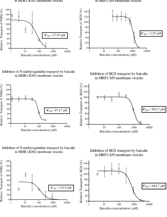
BG concentration-relative transport (%) curve was generated for each transporter (Figures 9a-d). From this curve, the IC50 value was calculated using GraphPad Prism 5.
The IC50 is defined as the concentration needed to inhibit transport of the reporter substrate by 50%. The IC50 parameters were derived from the equation of a one-binding site model, dose-response curve fitted onto the relative activity against the concentration of BG, plotted by log inhibitor-vs response-variable slope using GraphPad (San Diego, CA) Prism 5.
36
Concentration-dependent percent inhibition by BG of transport of specific substrates by MDR1, MRP1 (Figure 9a), MRP2 (Figure 9b), MRP3 (Figure 9c), MRP4 and BCRP (Figure 9d), was observed, with 2 plateaus.
When comparing the two cell systems in case of MRP2 and MRP3, the depicted curves showed similar inhibitory tendency (Figures 9b and 9c).
37
Inhibition of BGS transport by baicalin in MRP1-Sf9 membrane vesicles
1 10 100 1000 10000
0 20 40 60 80 100 120 140
IC50= 1129 µM
Baicalin concentration ( µM)
Relative Transport of BGS (%)
Inhibition of BGS transport by baicalin in MRP1-Sf9 membrane vesicles
1 10 100 1000 10000
0 20 40 60 80 100 120 140
IC50= 963.5 µM
Baicalin concentration ( µM)
Relative Transport of BGS (%)
Inhibition of BGS transport by baicalin in MRP1-Sf9 membrane vesicles
1 10 100 1000 10000
0 20 40 60 80 100 120 140
IC50= 694.7 µM
Baicalin concentration ( µM)
Relative Transport of BGS (%)
Figure 9 a: Dose-response curves: inhibition of transporter-specific substrate transport by BG in membrane vesicles.
Inhibition of N-methyl-quinidine transport by baicalin in MDR1-K562 membrane vesicles
1 10 100 1000 10000
0 20 40 60 80 100 120 140 160
IC50= 67.95 µM
Baicalin concentration ( µM)
Relative Transport of NMQ (%)
Inhibition of N-methyl-quinidine transport by baicalin in MDR1-K562 membrane vesicles
1 10 100 1000 10000
0 20 40 60 80 100 120 140 160
IC50= 87.67 µM
Baicalin concentration ( µM)
Relative Transport of NMQ (%)
Inhibition of N-methyl-quinidine transport by baicalin in MDR1-K562 membrane vesicles
1 10 100 1000 10000
0 20 40 60 80 100 120 140 160
IC50= 128.9 µM
Baicalin concentration ( µM)
Relative Transport of NMQ (%)
38
Inhibition of E217ßDG transport by baicalin in MRP2-Sf9 membrane vesicles
1 10 100 1000 10000
0 20 40 60 80 100 120
IC50= 276.6 µM
Baicalin concentration (µM) Relative Transport of E217ßDG(%)
Inhibition of E217ßDG transport by baicalin in MRP2-Sf9 membrane vesicles
1 10 100 1000 10000
0 20 40 60 80 100 120
IC50= 271.8 µM
Baicalin concentration (µM) Relative Transport of E217ßDG(%)
Inhibition of E217ßDG transport by baicalin in MRP2-Sf9 membrane vesicles
1 10 100 1000 10000
0 20 40 60 80 100 120
IC50= 242.9 µM
Baicalin concentration ( µM) Relative Transport of E217ßDG(%)
Inhibition of E217ßDG transport by baicalin in MRP2-HEK293 membrane vesicles
1 10 100 1000 10000
0 20 40 60 80 100 120 140 160
IC50= 279.3 µM
Baicalin concentration (µM) Relative Transport of E217ßDG(%)
Inhibition of E217ßDG transport by baicalin in MRP2-HEK293 membrane vesicles
1 10 100 1000 10000
0 20 40 60 80 100 120 140 160
IC50= 268.4 µM
Baicalin concentration ( µM) Relative Transport of E217ßDG(%)
Inhibition of E217ßDG transport by baicalin in MRP2-HEK293 membrane vesicles
1 10 100 1000 10000
0 20 40 60 80 100 120 140 160
IC50= 82.70µM
Baicalin concentration (µM) Relative Transport of E217ßDG(%)
Figure 9 b: Dose-response curves: inhibition of transporter-specific substrate transport by BG in membrane vesicles.
39
Inhibition of E217ßDG transport by baicalin in MRP3-Sf9 membrane vesicles
0.1 1 10 100 1000
0 20 40 60 80 100
IC50= 38.99 µM
Baicalin concentration ( µM) Relative Transport of E217ßDG(%)
Inhibition of E217ßDG transport by baicalin in MRP3-Sf9 membrane vesicles
0.1 1 10 100 1000
0 20 40 60 80 100
IC50= 24.87 µM
Baicalin concentration (µM) Relative Transport of E217ßDG(%)
Inhibition of E217ßDG transport by baicalin in MRP3-Sf9 membrane vesicles
0.1 1 10 100 1000
0 20 40 60 80 100
IC50= 14.16 µM
Baicalin concentration (µM) Relative Transport of E217ßDG(%)
Inhibition of E217ßDG transport by baicalin in MRP3-HEK293 membrane vesicles
0.1 1 10 100 1000
0 20 40 60 80 100
IC50= 16.91 µM
Baicalin concentration ( µM) Relative Transport of E217ßDG(%)
Inhibition of E217ßDG transport by baicalin in MRP3-HEK293 membrane vesicles
0.1 1 10 100 1000
0 20 40 60 80 100
IC50= 12.52 µM
Baicalin concentration ( µM)
Relative Transport of E217ßDG(%)
Inhibition of E217ßDG transport by baicalin in MRP3-HEK293 membrane vesicles
0.1 1 10 100 1000
0 20 40 60 80 100
IC50= 12.61 µM
Baicalin concentration ( µM) Relative Transport of E217ßDG(%)
Figure 9 c: Dose-response curves: inhibition of transporter-specific substrate transport by BG in membrane vesicles.
