Development of new therapeutic strategies targeting cancer associated fibroblasts (CAFs) in pancreatic ductal
adenocarcinoma
PhD thesis
Magdolna Djurec
Pathological Sciences Doctoral School Semmelweis University
Consultant: László Kopper, MD, D.Sc, Official reviewers: Krisztina Buzás, PhD
Zoltán Wiener, PhD
Head of the Final Examination Committee: Zsuzsa Schaff, MD, D.Sc, Full Member of the Hungarian Academy of Science Members of the Final Examination Committee: Erika Tóth, MD, PhD
Attila Patócs, MD, PhD
Budapest
2018
2 TABLE OF CONTENTS
1 INTRODUCTION ... 12
1.1 PANCREATIC DUCTAL ADENOCARCINOMA ... 12
1.1.1 Epidemiology and biological features ... 12
1.1.2 Histopathology and molecular characterization of PDAC ... 12
1.1.2.1 Histopathology ... 12
1.1.2.2 Molecular genetics of pancreatic ductal adenocarcinoma ... 14
1.1.2.3 Signaling pathways activated in PDAC ... 15
1.1.2.3.1 RAS – MAPK ... 15
1.1.2.3.2 Growth factor signaling... 15
1.1.2.4 Stroma modulating pathways in PDAC ... 16
1.1.2.4.1 Hedgehog signaling ... 16
TGF-ß signaling ... 16
1.1.2.4.3 IL – 6/JAK-STAT pathway ... 17
1.1.2.4.4 NF-B pathway ... 17
1.1.2.5 Molecular subtypes of PDAC ... 18
1.1.2.6 Molecular subtypes of PDAC stroma ... 19
1.1.3 Therapeutic approaches to treat pancreatic cancer ... 20
1.1.3.1 Conventional chemotherapies and standard of care treatments ... 20
1.1.3.2 Molecular targeted therapies – clinical trials ... 20
1.1.3.3 Immune therapies – clinical trials ... 21
1.1.4 Mouse models to study pancreatic cancer ... 21
1.1.4.1 K-Ras induced mouse models ... 21
1.2 TUMOR MICROENVIRONMENT ... 24
1.2.1 Distinct cell types of the tumor microenvironment ... 24
1.2.1.1 Immune cells - immunosurveillance ... 25
1.2.1.1.1 Innate immune cells ... 25
1.2.1.1.2 Adaptive immune cells ... 25
1.2.1.2 Endothelial cells – angiogenesis ... 26
1.2.1.3 Extracellular Matrix ... 26
1.2.1.4 Cancer Associated Fibroblasts (CAFs) ... 27
1.2.1.4.1 Origins of CAFs ... 27
1.2.1.4.2 Molecular markers ... 28
3
1.2.1.4.1 Functional properties of CAFs ... 28
1.2.2 Tumor microenvironment in PDAC... 29
1.2.3 Cancer Associated Fibroblasts (CAFs) in PDAC ... 30
1.2.3.1 Preclinical studies targeting CAFs in PDAC ... 32
1.3 SERUM AMYLOID A PROTEIN FAMILY ... 35
1.3.1 SAA functions in normal homeostasis ... 35
1.3.1.1 SAA family members ... 35
1.3.2 SAAs in disease and cancer ... 36
1.3.2.1 SAAs as major acute phase protein and inflammatory cytokine ... 36
1.3.2.2 Regulation, receptors and signaling ... 36
1.3.2.3 Dual role of SAA in cancer... 37
1.3.2.4 Functional studies of Saa3 in mice ... 37
2 OBJECTIVES ... 39
3 MATERIALS AND METHODS ... 40
3.1 MOUSE MODELS ... 40
3.1.1 KPeCY mouse model ... 40
3.1.2 Saa3 germline KO ... 40
3.1.3 Therapeutic strain ... 40
3.1.4 Maintenance of mice... 41
3.1.5 Generation of mouse models by CRISPR ... 41
3.1.5.1 sgRNA design and validation ... 41
3.1.5.2 sgRNA preparation and Microinjection ... 42
3.1.5.3 Genotyping strategy ... 42
3.1.5.3.1 T7 endonuclease assay ... 42
3.1.5.3.2 Subcloning and sequencing ... 43
3.1.6 Subcutaneous and orthotopic allograft models ... 43
3.2 ISOLATION OF CELLS – TISSUE CULTURE ... 44
3.2.1 Generation of fibroblasts and tumor cells by outgrowth ... 44
3.2.2 Isolation by cell sorting ... 44
3.2.3 Tumor organoid – fibroblast co-cultures ... 45
3.2.4 Wound healing and migration assay ... 45
3.2.5 Lentiviral infection of cells ... 45
3.3 GENE EXPRESSION PROFILING ... 46
4
3.3.1 RNA extraction from sorted cells ... 46
3.3.2 RNA sequencing, gene expression profiling and GSEA analysis ... 46
3.4 IMAGING ... 47
3.4.1 Tumor monitoring by micro-ultrasound ... 47
3.4.2 Imaging by contrast agent ... 47
3.5 TREATMENTS ... 47
3.6 PROCESSING OF MOUSE TISSUES ... 47
3.6.1 Necropsies ... 47
3.6.2 Histology – Immunohistochemistry ... 48
3.6.3 Immunofluorescence ... 48
3.7 FACS ANALYSIS ... 48
3.8 WESTERN BLOT ... 49
3.8.1 Protein extraction ... 49
3.8.2 Blotting ... 49
3.9 GENOTYPING ... 50
3.9.1 DNA extraction ... 50
3.9.2 General PCR reaction ... 51
3.10 QUANTITATIVE REAL TIME –PCR(QRT-PCR) ... 51
3.10.1 RNA isolation... 51
3.10.2 cDNA synthesis and q – RT – PCR reaction ... 51
3.11 HUMAN SAMPLES ... 53
3.12 STATISTICAL ANALYSIS ... 53
4 RESULTS ... 54
4.1 TARGETING CAFS IN PDAC ... 54
4.1.1 Characterization of stromal cell populations in PDAC mouse model ... 54
4.1.2 Isolation of CAFs from PDAC and NPFs from normal pancreas ... 55
4.1.2.1 Fibroblast isolation by “outgrowth” ... 55
4.1.2.2 Fibroblast isolation by cell sorting using PDGFR ... 56
4.1.2.3 Tumor promoting PDGFRα+ CAFs and tumor suppressing PDGFRα+ NPFs ... 58
4.1.2.4 Comparative transcriptional profiling of CAFs versus NPFs ... 59
4.1.2.4.1 Gene expression profiling of fibroblasts from “outgrowth” ... 59
5
4.1.2.4.2 Gene expression profiling of PDGFRα+ CAFs and NPFs isolated by
cell sorting ... 61
4.1.2.4.3 Target selection and validation ... 63
4.1.2.4.4 Validation of RNAseq results by quantitative RT-PCR ... 65
4.1.3 In vivo functional validation – mouse models ... 66
4.1.3.1 Subcutaneous allograft models ... 66
4.1.3.2 Generation of mouse models to study the role of the targets in PDAC ... 68
4.1.3.3 Generation of mouse models by CRISPR targeting the stroma ... 69
4.1.3.3.1 Single guided RNA design and validation in CAF cell lines ... 69
4.1.3.3.2 Direct microinjection of sgRNA into zytgotes to generate single mutant mice ... 71
4.1.3.3.3 One-step generation of triple mutant mice by zygote injection ... 72
4.1.3.4 Generation of Saa3 null mice by conventional targeted deletion ... 74
4.1.4 Saa3 in mouse PDAC development ... 76
4.1.4.1 PanIN formation and survival ... 76
4.1.4.2 Stroma reorganization in Saa3 tumors ... 76
4.1.4.2.1 Stroma remodeling has low impact on treatment efficiency ... 79
4.1.4.3 Undifferentiated tumor phenotype ... 81
4.1.4.3.1 Stem cell-like tumor cells... 81
4.1.4.3.2 Migratory properties – metastasis ... 82
4.1.4.4 Anti-tumorigenic properties of Saa3 null CAFs ... 86
4.1.4.4.1 Organoid co-culture of Saa3 competent and null CAFs and tumor cells ... 86
4.1.4.4.2 Orthotopic allografts of Saa3 competent and null CAFs and tumor cells ... 87
4.1.4.5 Transcriptional profiling of Saa3 null cells ... 90
4.1.4.5.1 Comparative expression profile of Saa3 null and competent CAFs ... 90
4.1.4.5.2 Comparative expression profile of Saa3 null and competent tumor cells ... 91
4.1.4.5.3 Cytokine profiles ... 92
4.1.4.5.4 Mpp6 – Saa3 axis ... 93
4.1.5 SAA1 in human PDAC ... 96
4.1.5.1 Gene expression profiling of human CAFs ... 96
4.1.5.2 Analysis of SAA1 in the DKFZ human PDACdata set ... 97
6
4.1.5.3 Analysis of the Moffitt human PDAC data set ... 98
5 DISCUSSION ... 100
5.1 TARGETING THE STROMA IN PDAC BY REPROGRAMMING CAFS ... 100
5.1.1 Isolation and gene expression profiling of CAFs ... 100
5.1.2 PDGFRα+ CAFs are protumorigenic ... 101
5.1.3 Target selection validation in CAFs ... 102
5.1.3.1 Functional validation of targets by RNAi silencing ... 102
5.1.3.2 Generation of knockout mouse models by CRISPR in PDAC stroma ... 102
5.2 SAA3 IS PROTUMORIGENIC IN CAFS BUT NOT IN TUMOR CELLS... 103
5.2.1 Complete elimination of Saa3 did not affect overall PDAC development .... 104
5.2.2 Saa3 null tumor cells have increased migratory but not homing properties 105 5.2.3 Saa3 is required for the pro-tumorigenic properties of CAFs but not for tumor cells ... 105
5.3 SAA1 IN HUMAN PDAC ... 107
6 CONCLUSIONS ... 109
7 SUMMARY ... 111
8 REFERENCES... 113
PUBLICATIONS ... 139
ACKNOWLEDGEMENTS ... 140
7
Abbreviations
A2m: Alpha-2-macroglobulin
ADEX: Aberrantly Differentiated Endocrine exocrine tumor ADM: Acinar to Ductal Metaplasia
Aldh2: Aldehyde dehydrogenese 2 ApoA-1: Apolipoprotein A1
Apo-SAA: Serum Amyloid A Apolipoprotein Arg1: Arginase 1
SMA: alpha-Smooth Muscle Actin ATP: Adenosine Tri-Phosphate
BRCA: Breast Cancer susceptibility protein BSA: Bovine Serum Albumin
CAFs: Cancer Associated Fibroblasts CCL: Chemokine (C-C motif) ligand CDK: Cyclin-Dependent Kinase
CDKN2A: Cyclin-Dependent Kinase Inhibitor 2A cDNA: Complementary DNA
Cfh: Complement Factor H Chi3l: Chitinase 3 like 1
Clec3b: C-type Lectin Domain Family 3 Member B Col11a1: Collagen type 11 Alpha 1 chain
Cre: Cre recombinase
CRISPR: Clustered Regularly Interspaced Short Palindromic Repeats Ct: Comparative cycle Threshold
CTGF: Connective Tissue Growth Factor
CTLA-4: cytotoxic T-lymphocyte-associated protein 4 CXCL: Chemokine (C-X-C motif) ligand
Dcn: Decorin
DMEM: Dulbecco ́s Modified Eagle ́s Medium DMSO: Dimethyl Sulfoxide
DNA: Deoxyribonucleic acid DNase: Deoxyribonuclease
8 dNTP: Deoxynucleotide
DRS: Dual Recombinase System DTT: Dithiothrietol
ECM: Extracellular Matrix
EDTA: Ethylene Diamine Tetraacetic Acid EGFR: Epidermal Growth Factor Receptor ERK: Extracellular signal Regulated Kinases EYFP: Enhanced Yellow Fluorescent Protein FAP: Fibroblast activation protein
FBS: Fetal Bovine Serum
FDA: Food and Drug Administration FDR: False Discovery Rate
FGF: Fibroblast growth factor Flp: Flipase
FPR2: formyl peptide receptor 2 FSP: Fibroblast specific protein Fzd1: Frizzled class receptor 1 GAG: Glycosamino Glycans
GAPDH: Glyceraldehyde 3-Phosphate Dehydrogenase GEMM: Genetically Engineered Mouse Model
GFAP: Glial Fibrillary Acidic Protein GFP: Green Fluorescent Protein GLI: Glioma – associated oncogene
GM-CSF: Granulocyte-macrophage colony stimulating factor GPCR: G-Protein Coupled Receptors
GSEA: Gene Set Enrichment Analysis GTP: Guanosine Triphosphate
HA: Hyaluronic acid
HAS1: Hyaluronic Acid Synthase HABP: Hyaluronic acid binding protein HBSS: Hank ́s Balanced Salt Solution H&E: Hematoxylin and Eosin
HDL: High Density Lipoprotein
9 HP: Haptoglobin
Hpse: Heparanase
HRP: Horseradish Peroxidase HSC: Hepatic Stellate Cell hUBC: Human Ubiquitin C iCAFs: inflammatory CAFs IGF: Insulin Growth Factor
IGFBP: Insulin Growth Factor Binding Protein IGFR: Insulin Growth Factor Receptor
IHC: Immunohistochemistry IL: Interleukin
IPMN: Intraductal Papillary Mucinous Neoplasms ITGA11: Integrin Subunit Alpha 11
JAK: Janus Kinase
JNK: c-Jun N-terminal kinases KPC: K-Ras; P53; Cre
KPeCY: K-Ras; P53; Elastase-Cre; EYFP Lgals1: Galectin 1
LSL: Lox-STOP-Lox
Lrg1: Leucine Rich Alpha-2-Glycoprotein1 Lum: Lumican
Lyz2: Lysozime 2
MAPK: Mitogen Activated Protein Kinase
MAGUK: Membrane-Associated Guanylate Kinases MDM2: Mouse double minute 2 homologue
MDSC: Myeloid Derived Suppressor Cells MEK: Mitogen Activating Protein Kinase MMP: Matrix Metalloproteinase
MPO: Myeloperoxidase
MPP6: Membrane Palmitoylated Protein 6 MSC: Mesenchymal Stem Cells
Msln: Mesothelin
MyCAFs: Myofibroblast CAFs
10
nAb-Paclitaxel: nanoparticle albumin-bound Paclitaxel Neo: Neomycin
NF-B: Nuclear Factor Kappa B NK cells: Natural Killer Cells
NPFs: Normal Pancreatic Fibroblasts
OCT: Optimum Cutting Temperature compound OS: overall survival
PanIN: Pancreatic Intraepithelial Neoplasia PBS: Phosphate Buffered Saline
PCR: Polymerase Chain Reaction PD-1: Programmed Death Receptor 1
PD-1L: Programmed Death Receptor 1 Ligand PDAC: Pancreatic Ductal Adenocarcinoma PDGF: Platelet Derived Growth Factor
PDGFR: Platelet Derived Growth Factor Receptor PDX: Patient-Derived Xenografts
PDX1: Pancreatic and Duodenal Homeobox 1 PI3K: Phosphoinositol-3-kinase
PIK3CA: Phosphatidylinositol-4,5-Bisphosphate 3-Kinase Catalytic Subunit Alpha PSCs: Pancreatic Stellate Cells
Ptger3: Prostaglandin E Receptor 3
PTEN: Phosphatase and Tensin homolog
qRT-PCR: Quantitative Reverse Transcription Polymerase Chain Reaction RAF: Rapidly Accelerated Fibrosarcoma
RAGE: Receptor for Advanced Glycation End Products RIN: RNA Integrity Number
ROS: Reactive Oxygen Species RNA: Ribonucleic acid
RT: Room Temperature
RT-PCR: Reverse Transcription Polymerase Chain Reaction SAA: Serum Amyloid A
SDS: Sodium Dodecyl Sulfate
11 Sfrp: Secreted Frizzled Protein 1
sgRNA: single guide RNA Shh: Sonic hedgehog shRNA: short hairpin RNA SMO: Smoothened
SPARC: Secreted Protein Acidic and Rich in Cysteine Spon1 Spondin 1:
SR-BI: scavenger receptor class B type I
STAT: Signal Transducer and Activator of Transcription TAE: Tris Acetate EDTA
TAMs: Tumor Associated Macrophages
Taq-Polymerase: Thermus aquaticus DNA Polymerase TBS: Tris Buffered Saline
TBST: Tris Buffered Saline with Tween TGF-ß: Transforming Growth Factor β Thbd: Trombomodulin
TNF-α: Tumor Necrosis Factor α Tnfsf8: TNF Superfamily Member 8 TLR: Toll-like receptors
TKI: Tyrosine Kinase Inhibitors
Trp53/P53: Transformation Related Protein 53 TME: Tumor Microenvironment
VEGF: Vascular Endothelial Growth Factor
VEGFR: Vascular Endothelial Growth Factor Receptor WT: Wild Type
12
1 Introduction
1.1 Pancreatic Ductal Adenocarcinoma
Pancreatic ductal adenocarcinoma (PDAC) is the most frequent type of pancreatic tumors. It is an extremely aggressive disease with diagnosis at advanced stages and highly refractory to most treatments. By 2030 it will become the second leading cause of cancer death in developed countries (1).
1.1.1 Epidemiology and biological features
Pancreatic cancer is a disease of the elderly, the median age at diagnosis is 71 years and it is more frequent in men (1). PDAC is associated with a very poor prognosis, with a 5-year survival rate of only 6% and a median survival of less than 6 months (2). This low survival rate is the result of its aggressive features and its late diagnosis due to the lack of early symptoms and early biomarkers. Therefore, at the time of detection 80% of patients have locally advanced or metastatic PDAC and less than 20% of the patients are eligible for resection. Moreover, PDAC biology contributes to early recurrence, distant metastasis and resistance to chemotherapy and radiotherapy (3). A very important player in this poor prognosis is the extensive stromal reaction (desmoplasia) resulting in a hypovascular and hypoxic microenvironment and evasion of tumor immunity (4).
1.1.2 Histopathology and molecular characterization of PDAC 1.1.2.1 Histopathology
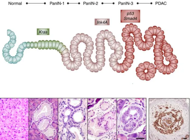
PDAC commonly arises and progresses through a multistep process of well-defined and non-invasive precursor lesions that gradually accumulate mutations in oncogenes and tumor suppressors (5) (Fig 1). Histological and clinical studies identified distinct types of precursor lesions, being the most frequent, the pancreatic intraepithelial neoplasia (PanIN) (6). PanIN lesions can be classified into four grades based on the degree of dysplasia: PanIN1A (flat) and PanIN1B (papillary type) are low-grade lesions with minimal cytological and architectural atypia; PanIN2, a high-grade lesion with frequent papillary structure and nuclear pleomorphia, crowding, and hyperchromasia; and PanIN3 (or in situ carcinoma) characterized by severe cytological and architectural atypia with papillary morphology. All grade PanINs are non-invasive lesions that do not invade the basement membrane (5, 7).
13
Figure 1. Multistep pancreatic carcinogenesis. (Upper) Scheme of step-wise genetic alterations followed by histological transformation. Mutation in the K-Ras oncogene initiating ductal reprogramming and transformation into PanIN1A/B low grade lesions.
Accumulation of subsequent mutations in tumor suppressors, such as INK4A (PanIN2);
and P53 and SMAD4 (PanIN3) results in high grade lesions and finally to PDAC formation. (Lower) H&E staining of histological alterations during pancreatic tumor development and CK19 IHC of PDAC (adapted from Neesse et al., 2015; and Barbacid, 2013).
PanIN lesions develop through a reprogramming process (transdifferentiation), known as acinar-to-ductal metaplasia (ADM), which is frequently associated with atrophy of acinar parenchyma and activation of the K-RAS oncogene as initiating steps (6). Molecular analyses have demonstrated that PanINs harbor many of the genetic alterations found in PDACs (8). Early events include mutations in K-RAS and CDKN2A as well as telomere shortening providing an environment that is permissive to acquisition of chromosomal rearrangements and preceding mutations in P53 and SMAD4, tumor suppressors that are found lost in high grade lesions (PanIN3) (Fig 1) (8).
14
1.1.2.2 Molecular genetics of pancreatic ductal adenocarcinoma
More than 90% of PDACs harbor mutations in the K-RAS oncogene, detected already in early PanIN lesions (8, 9). Therefore, it is considered the initial driver mutation.
The K-RAS oncogene: K-RAS is a member of the RAS family of GTP-binding proteins that mediate different cellular functions: proliferation, differentiation, and survival (10). In pancreatic cancer, oncogenic mutations in the K-RAS locus cluster in hot spots, more frequently in codon 12 (11, 12). This results in inhibition of GTP hydrolysis activity that leads to constant activation of the protein and finally aberrant cell proliferation (13). The most common mutations are G12D (44%), followed by G12V (30%) and G12R (20%), respectively. The latter was present with high prevalence in samples with multiple K-RAS mutations suggesting distinct signaling properties of this allele (9). In addition, PDACs with wild-type K-RAS have activating mutations in members of the MAPK pathway (9). Oncogenic mutations in BRAF, such as V600E, can be found in 3% of PDAC samples (14) and are mutually exclusive with K-RAS mutations (15). Interestingly, a subset of K-RAS wild-type PDACs display elevated activation of MTOR pathway, converting it into a therapeutic target for these patients (9).
CDKN2A: The most frequent allelic losses in PDAC affects the locus that encodes the cyclin-dependent kinase CDKN2A/p16 tumor suppressor that inhibits cell cycle progression (16). Deletion, mutation or promoter hypermethylation of 9q21 locus occurs in 80–95% of low-grade PanINs (17, 18). This locus encodes two overlapping tumor suppressors - INK4A and ARF, and their respective protein products P16INK4A and P14ARF (19). INK4A inhibits CDK4/6-mediated phosphorylation of RB, thereby blocking the cell cycle; ARF stabilizes P53 by inhibiting its MDM2-dependent proteolysis. Cooperation of K-RAS and INK4A mutation was postulated in several studies (20) given their mutual genetic alteration during early steps of pancreatic carcinogenesis.
P53: Inactivating missense mutations in the DNA binding domain of P53 are frequently found in PDAC patients (50-70%) (17). These genetic alterations appear in later stages of PanIN formation, in an established environment of genomic instability and ROS induced DNA damage response (20). Interestingly, loss of function of ARF and P53 coexist in 40% of human PDAC (18).
SMAD4: sporadic loss of function of SMAD4/DPC transcriptional regulator by deletion or intragenic point mutations takes place in 50% of human PDACs (21). As a central component of TGF-ß signaling, SMAD4 mutations appear at late stages of pancreatic tumorigenesis (22).
15 1.1.2.3 Signaling pathways activated in PDAC
1.1.2.3.1 RAS – MAPK
In addition to the role of K-RAS mutations in PDAC initiation, constitutive RAS signaling has been described to be required for PDAC maintenance (2, 23). Three RAS genes encode four RAS isoforms (H-RAS, N-RAS and K-RAS4A and K-RAS4B), with the splice variant K-RAS4B being the main isoform expressed in human cells (24) and tumors (25). Upon extracellular stimuli RAS proteins activate numerous downstream signaling pathways including the MAPK signaling, a key pathway that controls essential cellular processes, such as proliferation, survival and differentiation (10). This pathway includes a family of serine/threonine kinases (RAF/MEK/ERK), that through phosphorylation events result in a proliferative phenotype in many cells (26). RAS-GTP binds to RAF proteins (A-RAF, B-RAF, C-RAF), initiating a signaling cascade and activates MEK1/2, which phosphorylates ERK1/2 kinases. The latter has more than 150 substrates in the cytosol and in the nucleus, most of them involved in cell proliferation (27).
RAS can also activate the PI3K signaling pathway, which is an essential regulator of cell survival (28) via AKT, p70-S6K, and PDK-1 downstream effectors (29). This pathway is constitutively active in most pancreatic cancers. Moreover, mutations in the catalytic subunit of PI3K (p110α, encoded by PI3KCA) and amplification of AKT are commonly found in human PDACs, however mutations in its endogenous inhibitor, PTEN, are uncommon (15). The importance of this pathway in pancreatic tumorigenesis was shown by activating the p110α subunit of PI3K in K-Ras driven mouse model resulting in PDAC promotion (30, 31), whilst PDK-1 ablation abrogated tumor development (31).
1.1.2.3.2 Growth factor signaling
PDAC shows increased expression of Epidermal Growth Factor Receptors (EGFR and ERBB2) and their ligands (TGF and EGF), consistent with the presence of an autocrine loop (32). Importantly, EGFR inhibitors decrease PDAC cell growth and tumorigenesis in vitro (33), as well as inhibit growth of orthotopic tumors in combination with cytotoxic chemotherapy (34). In 2007, a clinical trial in phase III showed a limited benefit in survival of PDAC patients with the combination of gemcitabine and the EGFR inhibitor Erlotinib, compared with the treatment with gemcitabine only (35).
16
Other important growth factors, such as the Insulin Growth Factor (IGF), regulate survival, invasion, and angiogenesis of many human cancers. PDACs show elevated expression of IGF-I in both the tumor and stromal compartment, as well as aberrant activation of the IGF-I receptor (IGF-IR) in tumor cells (36). In PDAC patients, IGF1R overexpression was associated with decreased survival (37). Increased levels of IGF binding proteins (IGFBPs) are found in PDAC (38), however, low expression of IGFBP3 and IGFBP7 has been correlated with poor clinical outcome (39). IGFBP-s are reservoirs of circulating IGFs but also regulate cell growth and survival (40), although their complete function is not well understood.
Fibroblast Growth Factor (FGF) and Vascular Endothelial Growth Factor (VEGF) signaling appears to contribute to mitogenesis and angiogenesis of PDAC (41).
Overexpression of FGF receptors has been detected in pancreatic tumors (42), where elevated bFGF levels contributed to the PDAC desmoplasia (43). VEGF promotes endothelial cell proliferation and survival by binding to the VEGFR-1 and VEGFR-2 transmembrane receptors (44). VEGF is overexpressed by PDAC cells (45), whereas disruption of VEGF signaling strongly suppresses tumor growth of pancreatic cancer xenografts (46).
1.1.2.4 Stroma modulating pathways in PDAC 1.1.2.4.1 Hedgehog signaling
The Sonic Hedgehog family is comprised of secreted signaling proteins that regulate the growth of many organs, including the pancreas during embryogenesis (47).
Hedgehog ligands, such as SHH, disrupt inhibition of SMO and activate the GLI transcription factor. SHH is activated in PanINs and neoplastic cells (48), yet, the activity of GLI in PDAC is restricted to the stromal compartment (49). Pharmacological inhibition of Shh signaling in PDAC GEMMs resulted in reduced stroma, increased vessel density and enhanced drug delivery (50). However, genetic deletion of Shh in mouse tumor cells recapitulated stroma reduction but resulted in aggressive, undifferentiated tumors (51).
This is in line with another study demonstrating that Shh activation provokes stromal hyperplasia and reduced growth of epithelial compartment (52).
TGF-ß signaling
TGF-ß belongs to a superfamily of secreted proteins, whose other members include growth factors (BMPs, Activins) that activate SMAD proteins and regulate proliferation,
17
differentiation, as well as migration. More importantly, TGF-ß promotes transformation and proliferation of fibroblasts and controls the process of epithelial mesenchymal transition (EMT) in tumors (53).
In pancreatic cancer, more than 50% of the tumors show inactivation of SMAD4 (6). In tumor cells TGFß regulates EMT process downregulating the activity of Snail and Zeb-1 transcription factors (54). Moreover, TGFß regulates PDAC stroma as a major factor of fibrosis via the secretion of several pro-tumorigenic growth factors including VEGF and CTGF, as well as MMP2 and MMP9 (55). In addition, it suppresses inflammatory processes through inhibition of cytotoxic T-cells, macrophages and NK cells (56).
1.1.2.4.3 IL – 6/JAK-STAT pathway
The Signal Transducer and Activator of Transcription (STAT) family of transcription factors are phosphorylated by Janus Kinases (JAK) tyrosine kinases (57, 58). They regulate numerous cellular processes including self-renewal, proliferation and inflammatory pathways (59).
In human PDAC, frequency of STAT3 alteration ranges between 30-100% (60) and correlates with decreased survival (61). Moreover, STAT3 is not essential for normal pancreatic homeostasis (62) but is involved in all stages of pancreatic tumorigenesis (63, 64) Recent studies have demonstrated that IL6 cytokine-induced activation of STAT3 is responsible for remodeling the desmoplastic PDAC stroma and for immune surveillance (61, 65). In addition, IL6 secreted by stromal cells also induced Stat3/Socs3 expression via IL6 trans-signaling and accelerated PDAC progression in mouse models (66).
1.1.2.4.4 NF-B pathway
Nuclear factor kappa B (NF-B) signaling might be another downstream mediator of the mutated RAS pathway in pancreatic cancer. Its activation occurs in response to cellular stress through pro-inflammatory cytokines and growth factors resulting in regulation of immune response and apoptosis (67, 68). A link between K-RAS and NF-
B signaling through pP62 was reported to drive tumor initiation and progression in PDAC (69). Moreover, NF-B connects inflammation and cancer by recruitment of inflammatory cells and activation of cytokines. Cross activation of TGF-ß and IL-1ß signaling via NFB further contributes to the generation of complex inflammatory PDAC stroma (70).
18 1.1.2.5 Molecular subtypes of PDAC
In 2011, an analysis based on gene expression data of primary tumors and tumor cell lines correlated with clinical outcome and response treatment identified three molecular subtypes of PDAC: classical, quasimesenchymal (QM) and exocrine-like tumors. Classical subtype was characterized by overexpression of adhesion-related and epithelial genes, such as GATA6. QM tumors had high expression of mesenchymal- associated genes and these patients showed the worst median survival. Exocrine-like PDAC genes were enriched in digestive enzyme genes (71). Interestingly, QM PDAC cells were more sensitive to gemcitabine than the Classical subtype. Conversely, the latter responded better to Erlotinib suggesting treatment specificity.
In 2015, virtual microdissection of gene expression in PDAC samples by non- negative matrix factorization identified two tumor-specific subtypes: classical (more differentiated tumors associated with GATA6 expression and characterized by significantly higher SMAD4 expression) and basal [tumors with significantly worse median survival and faster growth rate in PDX (Patient Derived Xenografts) (72)].
In 2016, a study based on integrated genomic analysis of 456 PDACs defined 4 molecular subtypes: squamous, pancreatic progenitor, aberrantly differentiated endocrine exocrine (ADEX) and immunogenic (73). Squamous tumors, presented poor prognosis and were associated with mutations in P53. EGF signaling and upregulated TP63DN transcriptional network were found activated, whereas genes involved in pancreatic cell differentiation (ie: Pancreatic and Duodenal Homeobox 1 (PDX1), GATA6) were downregulated (73). Pancreatic progenitor tumors overexpressed genes involved in early pancreatic development like FOXA2, FOXA 3 and PDX1 among others (73). Immunogenic tumors appeared similar to the pancreatic progenitor subtype, but with a significant increase in immune cell infiltrates. These tumors exhibited overexpression of immune network pathways, including CD4+ and CD8+ T cell and Toll-like receptor signaling (73). ADEX tumors showed deregulation of pathways involved in late stages of pancreatic development. They were characterized by upregulating genes associated with K-RAS activation and transcriptional networks related to acinar and endocrine differentiation (73).
Finally, recent findings showed integrated molecular analysis and classification of 150 primary tumor samples from the TCGA database. Whole exome sequencing, mRNA and protein profiling provided a complex molecular landscape of PDACs. The above classifications were applied on the TCGA PDAC data set and the analysis found an
19
overlapping between basal – like (Moffitt et al.) and squamous tumor (Bailey et al.) subtypes, enriched in P53 mutations. Likewise, classical and pancreatic progenitor subtypes of PDAC were confirmed across platforms and were associated with increased GNAS mutations (9).
1.1.2.6 Molecular subtypes of PDAC stroma
Interestingly, the study of Moffitt et al. described PDACs with two distinct stroma subtypes: ‘activated and normal’ (Fig 2). Additionally, this study identified a cluster of samples with low or missing stroma (‘low stroma’). Patients with ‘activated’ stroma had significantly worse median survival than patients with ‘normal’ stroma (Fig 2) (72).
Normal stroma was characterized by high expression of the well-established myofibroblast marker SMA (alpha-smooth muscle actin), Vimentin and Desmin.
Activated stroma was characterized by a diverse inflammatory signature of macrophage related chemokines (CCLs) and integrins, as well as other tumor promoting factors like SPARC, members of the Wnt pathway, collagens and matrix metalloproteinases (MMPs) (72).
Figure 2. Stroma subtypes of PDAC in the virtual microdissection study of Moffitt et al. (Left) Heat map of primary tumor samples separated based on transcriptome profiles and matrix factorization. Samples clustered into three groups, describing samples with activated stroma, samples with normal stroma and samples with low or absent stromal gene expression. (Right) Kaplan-Meier survival analysis of patients with resected PDAC from the activated and normal stromal clusters shows that samples in the activated stroma group have worse prognosis (P = 0.019) (adapted from Moffitt et al., 2015).
20
Taken together, identification of tumor and stroma specific molecular signatures suggests an important interaction between tumor compartments to be considered in the future for tumor characterization and for stroma and immune modulating therapies.
1.1.3 Therapeutic approaches to treat pancreatic cancer
1.1.3.1 Conventional chemotherapies and standard of care treatments
One of the reasons of the poor overall survival (OS) rates of pancreatic cancer is the lack of efficient therapies. Despite of the progress already achieved among gastrointestinal malignancies, there have been modest advances in the treatment of pancreatic ductal adenocarcinoma (74, 75). In 1996, Gemcitabine was approved by the FDA for treatment of pancreatic cancer. This was further confirmed by a randomized clinical trial showing a significant improvement in the OS of gemcitabine versus 5- fluorouracil (76). A decade later Erlotinib (EGFR inhibitor) in combination with Gemcitabine was approved for treating metastastic PDAC. However, the survival improvement was marginal (0.4 month) (35). Other combinations of cytotoxic agents or targeted therapies failed to achieve survival benefit or presented increased toxicity.
Indeed, first-line therapy FOLFIRINOX, a combination of leucovorin, 5-fluorouracil, irinotecan and oxaliplatin, showed significant advantage versus Gemcitabine (11.1 vs. 6.8 months), but only for patients with good performance (77). The other important advance was a clinical trial in 2013 with the combination of gemcitabine plus nab-paclitaxel (the nanoparticle albumin-bound formulation of paclitaxel) and since then, it became the standard of care treatment. Yet, the improvement in survival remains low compared to Gemcitabine alone (8.5 months vs. 6.7 months, respectively) (76), which highlights the urgency for developing new and more effective therapeutic strategies.
1.1.3.2 Molecular targeted therapies – clinical trials
In addition to the aforementioned novel front-line treatments, many others possibilities are under investigation. It is expected that molecular targeted therapies, monoclonal antibodies and immunologic activation will add survival benefit and will increase life quality of PDAC patients.
Direct pharmacologic inhibition of K-RAS has been unsuccessful in the past decades due to its high binding affinity to GTP and the inability to identify an easily accessible active site. Studies are ongoing to find alternative approaches, still with limited success (78). Targeting of the downstream RAF/MEK/ERK signaling pathway by MEK inhibition resulted in PI3K mediated reactivation of EGFR (79). PDAC cells also
21
overexpress IGF-1R, although its inhibition with ganitumab combined with gemcitabine in a Phase III randomized controlled trial did not show improvement and the study finished earlier (80).
Targeting JAK/STAT signaling by ruxolitinib in combination with capecitabine (precursor of the 5-fluorouracil) showed improved OS in patients with high C reactive protein levels in a Phase II trial (81). Phase II trial is currently ongoing to evaluate ruxolitinib in metastatic PDAC (ClinicalTrials.gov identifiers: NCT02119663 and NCT02117479). In addition, inhibitors of TGF-ß, WNT and NOTCH signaling pathways based on preclinical studies are under clinical testing (82).
1.1.3.3 Immune therapies – clinical trials
Different strategies have been addressed to harness the host’s immune system against PDAC (82). For instance, by using vaccines such as the Mesothelin specific CD8+
T cell that improved overall survival (83). Immunotherapy by immune checkpoint inhibitors (i.e., CTLA-4, PD-1, PD-L1 and others) inhibitors offers encouraging results in preclinical models but often fails to show clear benefits in clinical trials for PDAC.
The monoclonal antibody anti-CTLA4 Ipilimumab was ineffective in PDAC (84).
However, its combination with a GM-CSF secreting PDA vaccine (GVAX) resulted in synergistic effects and raised OS (85). Clinical trial of PD-L1 inhibition with the monoclonal antibody BMS-936559 was sadly unsuccessful in PDAC (86). Yet, in preclinical studies combination with the CXCR4 inhibitor, AMD3100, the treatment induced tumor regression (87, 88). Currently, combination of Ulocuplumab (anti- CXCR4) and Nivolumab (anti-PD1) is in a phaseⅠstudy (NCT02472977) (82).
1.1.4 Mouse models to study pancreatic cancer
Genetically engineered mouse models (GEMMs) that recapitulate the human disease are important tools to understand PDAC biology and to design novel therapeutic approaches (89). Homologous recombinant technology on ES cells and the use of Cre and Flp recombinases allows a fine-tuned control of genetic alterations in a time- and tissue- specific manner.
1.1.4.1 K-Ras induced mouse models
Remarkable efforts have been made to generate GEMMs that recapitulate the full spectrum of histological alterations found in human patients. Since transformation of K-
22
RAS is considered to be the initiating genetic event in PDAC tumorigenesis, most of the models involve endogenous expression the K-RAS oncogene. The first model that fulfilled the above criteria included the conditional expression of a mutant K-RasLSLG12D allele controlled by the expression of the Cre recombinase in early embryonic development under the Pdx1 or P48 pancreatic lineage specific promoters (Pdx1-Cre; K- RasLSLG12D, referred as “KC”) (90).
Our laboratory generated a bitransgenic strain (Elas-tTA/tetO-Cre) that allows the control of the K-Ras oncogene expression by a tet-off strategy: the expression of the Cre recombinase is controlled by the acinar cell specific Elastase promoter that controls the expression of a tetracycline trans-activator and the expression of the Cre recombinase is under the control of a Tet operon. These mice were crossed with the K-RasLSLG12Vgeo conditional knock-in mice (91). In the absence of doxycycline, this compound strain (K- RasLSLG12Vgeo; Elas-tTA/tetO-Cre) expresses the K-Ras oncogene and the - Galactosidase reporter in a 20-30% of acinar cells from E16.5 of embryo development (92). These mice recapitulate the human disease, develop the full spectrum of PanIN lesions and a small proportion develop PDAC. Surprisingly, when mice are treated with doxycycline until the age of 8 weeks and the K-Ras oncogene is expressed in adult acinar cells no neoplastic growth occurs in the pancreas unless these mice undergo chronic pancreatitis (92).
The low frequency of malignant transformation suggested the need of additional genetic events that occur in later stages of PDAC development, such as mutations in tumor suppressors (p16Ink4a/p19Arf and p53) (6, 93). p53 inactivation, either by a conditional knock-in mutant (p53R172H) (K-RasLSLG12D;p53R172H
;
Pdx1-Cre, referred as“KPC”) or by p53 conditional null alleles (K-RasLSLG12Vgeo;p53lox/lox;Elas-tTA/tetO- Cre, in this study referred as “KPeC”) results in accelerated tumor progression and generation of invasive lesions with complete penetrance (94). In addition, a percentage of these mice also develop metastatic tumors (89). Inactivation of p16Ink4a/p19Arf results in 100% penetrance of PDAC and decreases tumor latency in mice.
Numerous mouse models were generated and characterized with additional genetic alterations known to play a role in PDAC development. Modifications in Smad4, Ink4/Arf, or elimination of Lkb1 and Tgfbr2, Notch1 acted as tumor suppressors and accelerated PDAC formation. On the other hand, Egfr was shown to be essential for pancreatic tumor development by two independent groups including our laboratory (95,
23
96). Many of them target all cell types of the pancreas (acinar, ductal and endocrine), and are expressed at early embryonic stages. Therefore, these studies only represent preventive strategies.
To perform real therapeutic trials new mouse models have been developed that allow the elimination of the target in established lesions. These models utilize a dual recombinase system (DRS) (Cre-LoxP, Flp-FRT), where tumors are induced by K-Ras in cells expressing Flp recombinase driven by Pdx1, along with the ablation of the p53 tumor suppressor gene (p53Frt) during embryonic development. When the tumor is developed, secondary modifications can be obtained in targets flanked by loxP sites by tamoxifen induced Cre recombination. On the other hand, expression of the Cre recombinase, can be also controlled by a stromal lineage specific promoter (i.e. fibroblasts) (97).
In parallel, our laboratory has generated a “therapeutic strain” using the same approach. These animals express the Flp recombinase in Elastase positive cells during late embryonic development leading to the expression of the resident K-RasG12V oncogene and to the ablation of the p53 tumor suppressor gene. When the tumor is developed, tamoxifen induced elimination of Egfr or C-Raf targets occur ubiquitously in cells expressing the Cre-recombinase driven by the human Ubiquitin C promoter (Blasco et al.
unpublished).
These new models will help not only to target tumor cells, but also other cell types in the tumor microenvironment. This will greatly contribute to better understanding of tumor – stroma interactions and to develop novel combinatory therapeutic approaches.
24 1.2 Tumor microenvironment
Cancers are heterogeneous cellular entities, whose growth not only depends on tumor cells that harbor driver mutations of oncogenes and loss of tumor suppressors, but also on interactions with the dynamic microenvironment (stroma) co-evolved during tumor development (98).
1.2.1 Distinct cell types of the tumor microenvironment
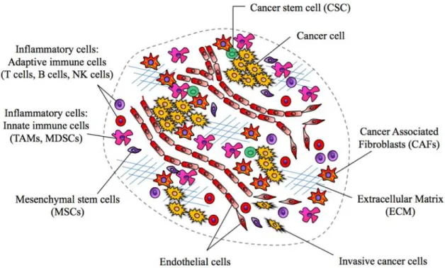
The tumor microenvironment (TME) is constituted by a diverse population of activated and/or recruited cell types by cancer cell and cancer stem cells (CSCs), such as cancer associated fibroblasts (CAFs), innate and adaptive immune cells, endothelial and other cell types that form blood and lymphatic vessels. Interaction between cancer cells and the closed normal tissue, as well as the components of the stroma regulates and define the aspect of tumorigenesis (Fig 3) (99).
Figure 3. Distinct cells types of the tumor microenvironment (TME) in solid tumors.
Subtypes of the stromal cells, such as inflammatory cells can include both tumor- promoting as well as tumor-killing subclasses either they belong to adaptive (T cells, B cells, natural killer (NK) cells) or innate immune (tumor associated macrophages (TAMs), myeloid derived suppressor cells (MDSCs)) response. Cell types including cancer associated fibroblasts (CAFs), endothelial cells, mesenchymal stem cells (MSCs)
25
are depicted. Cancer cells and cancer stem cells (CSCs) orchestrating the recruitment of the TME, while invasive cancer cells break away from primary tumor sites.
1.2.1.1 Immune cells - immunosurveillance
A functional link between inflammation and cancer is well accepted. Patients suffering from chronic inflammation are more prone to develop tumors due to the pro- growth environment of the inflammatory cells (98). However, the immune system plays dual role in tumor development by either tumor inhibition or support (100).
1.2.1.1.1 Innate immune cells
Components of the innate immunity, including macrophages, dendritic cells, mast cells, granulocytes or myeloid-derived suppressor cells (MDSCs) are recruited by growth factors, such as TGF-ß, VEGF or colony-stimulating factor-1 (CSF-1) and chemokines (CCL2, CCL5, etc.). These inflammatory cells release mediators that contribute to tumor growth, invasion and metastasis (101).
Tumor associated macrophages (TAMs), with similar characteristics as of M2 polarized (anti-inflammatory) macrophages, produce factors (101), that can directly affect cancer growth and metastatic dissemination by establishing pre-metastatic niches (102, 103).
Furthermore, TAMs are also responsible for therapeutic resistance by antagonizing antitumor activity of treatments or by regulating T-cell activation (104).
MDSCs are a heterogeneous population of immature myeloid cells recruited from bone- marrow (105), and have strong immunosuppressive activities such as the regulation of T and NK cells anti-tumor activity and stimulation of regulatory T cells (106).
1.2.1.1.2 Adaptive immune cells
A typical solid tumor will contain all adaptive immune cell-types (natural killer (NK) cells, B and T cells), mainly located in the surrounding layer. Mature T cells are divided into two major groups based on the T cell receptors (TCRs) and are further classified according to the effector functions as CD8+ cytotoxic T cells (CTLs) and CD4+
helper T (Th) cells, which include Th1, Th2, Th17, and T regulatory (Treg) cells, as well as natural killer T (NKT) cells (107). The process of activating cytotoxic CD8+ T cells and/or DC4+ T helper cells can be skewed in different ways, e.g. by cancer cells reprogramming the protective immune response, termed immunosurveillance (108).
Increased numbers of T cells usually are correlated with better prognosis in several cancer types, including melanoma, colon and pancreatic cancer (108). The ratio of CD8+
26
CTLs and Treg cells indicates the balance between host defense or tumor promotion (109). Treg cells mostly suppress antitumor immune responses (110), whilst NK cells and CTLs perform cytotoxic immunity (111). Recently, programmed death-ligand 1 (PD-L1) overexpressed by various tumor cell types, and its receptor (PD-1) on T cells became an important target. In several tumors refractory to conventional chemotherapy anti-PD- 1/PD-L1 succeeded, such as in melanoma (112). Yet, a group of solid cancers remain unresponsive (86).
1.2.1.2 Endothelial cells – angiogenesis
For the rapid expansion of a primary tumor, oxygen and nutrition supplies are needed. This requires the generation of new blood vasculature by activation of quiescent vessels (angiogenesis) (113). However, tumors develop irregular and dysfunctional new vessels (114), very often via overexpression of VEGF growth factor.
Endothelial cells can be activated by cytokines (bFGF, TNF-α, TGF-ß, PDGFs, PIGF and Neuropilin-1), chemokines (CXCL12, IL8/CXCL8), matrix metalloproteinases (MMPs), ROS and bioactive mediators, such as nitric oxide (NO) (115). Angiogenesis can be regulated by tumor associated macrophages (TAMs) through direct VEGF-A production (116) or via MMP9 secretion, which releases VEGF-A from the extracellular matrix (ECM) (117). Blockade of TAM secreted CSF-1 resulted in vascular normalization and improved therapeutic response (118). In addition, neutrophils were also reported to promote angiogenesis by MMP9 production (119), as well as cancer associated fibroblasts (CAFs) through pro-angiogenic signaling factors (120).
1.2.1.3 Extracellular Matrix
The tridimensional organization of the TME is highly dynamic and is dependent of the extracellular matrix (ECM) surrounding the cells. The ECM contains a mixture of fibrillar proteins, glycoproteins, proteoglycans, cytokines and growth factors (121), which supports cell adhesion via binding cell surface adhesion receptors and integrin signaling (122). Physical features of the ECM include its porosity and rigidity, spatial arrangement and orientation of insoluble components, as well as other features that together determine its role supporting tissue architecture.
Abnormal ECM and increase in collagen deposition can result in tumor stiffness and upregulation of integrin signaling, thus promoting cell survival and proliferation (123). Additional components, such as Hyaluronic acid also defines the structure and physical properties of the stroma (124). In addition, aberrant regulation of the ECM may
27
convert a normal stem cell niche into a cancer stem cell niche, disrupt tissue polarity and integrity to promote invasion (125). Importantly, in the periphery of benign tumors, enhanced collagen synthesis results in tight encapsulation of the tumor (126), suggesting that initial stromal responses may retain neoplastic expansion. However, reprogramming of the stroma by cancer cell directs them towards malignant progression (98).
1.2.1.4 Cancer Associated Fibroblasts (CAFs)
Fibroblasts are important and abundant cells in any context. They survive severe stress that is usually lethal to all other cells and are essential in tissue homeostasis, wound healing and repair processes in response to exposure to chemicals or carcinogens (127).
Indeed, there is an increasing body of evidence of their role in tumor development, in agreement with the hypothesis of Dvorak stating “cancer is a wound that never heals”
(128).
1.2.1.4.1 Origins of CAFs
In tissue repair, fibroblasts proliferate and differentiate into myofibroblasts, along with the expression alpha-smooth muscle actin (α-SMA), collagen, fibronectin, and other fibrillar proteins resulting in a reactive desmoplastic stroma (129). Aberrant regulation of the constitutive wound healing process leads to the generation of malignant stromal tissue and diverse fibroblast populations. In the process of tumorigenesis, they are collectively designated as cancer associated fibroblasts (CAFs).
CAFs are a heterogeneous cell population (Fig 4) derived from multiple origins, such as bone marrow, adipose tissue, mesenchymal stem cells (MSCs), epithelial and cancer cells through EMT process, endothelial cells via endothelial mesenchymal transition (EndMT) or mainly from adjacent normal tissue fibroblasts (130). They are defined by elongated, spindle-like morphology and by expression of distinct markers, characterizing each subtype (127). They are found in many solid cancers, however, abundance of CAFs is a typical feature of prostate, breast and pancreatic cancer (131).
28
Figure 4. Origins of CAFs. Bone marrow derived cells (BMDCs) including fibrocyte precursors and mesenchymal stem cells (MSCs) contribute to the diverse CAF population, as well as epithelial, cancer and endothelial cells via EMT or EndMT process.
The majority of CAFs is derived from tissue resident fibroblasts.
1.2.1.4.2 Molecular markers
The molecular characterization of CAFs has illustrated that there is no unique marker to label all CAFs and that most markers are not even specific to CAFs or fibroblasts. While αSMA is used as a robust CAF marker, which usually identifies CAFs with myofibroblast morphology (132), it is also expressed by normal fibroblasts (133) and in some cases at comparable or even higher level (134, 135). FSP1 or S1004A is another marker of CAFs, even though it seems to have a differing role in cancer (136).
Another well described marker is the cell surface serine protease fibroblast activation protein (FAP) (137). Further overexpression among cell surface proteins include the neural marker, NG2 and PDGFRß, that is also found on vascular cells (138). Interestingly, PDGFRß activation was also reported in invasive pancreatic tumor cells (139).
Finally, it was reported in different cancer types, such as skin and pancreatic tumors that PDGFRα is a marker of a CAF population characterized by pro-inflammatory gene signature (140). However, it also labels immune, adipose and mesenchymal stem cells;
and drives adipose tissue derived fibrosis (141, 142). Of note, PDGFRα could be considered as EMT marker in tumor cells (143).
1.2.1.4.1 Functional properties of CAFs
Each of CAF subtypes can contribute to a variety of tumor-promoting functions in different organ-specific TMEs (Fig 5). For example, CAFs are a source of paracrine
29
signaling molecules that include mitogenic epithelial growth factors, hepatocyte growth factor (HGF), EGF family members, insulin-like growth factor-1 (IGF-1), stromal cell- derived factor-1 (SDF-1/CXCL12), and a variety of FGFs and VEGFs, with the capability to stimulate cancer cell proliferation, angiogenesis, invasion and metastasis (88, 127, 144, 145). CAFs can also orchestrate functional attributes associated with EMT via secretion of TGF-ß (146). In addition, they can express a wide range of ‘‘proinflammatory’’
cytokines (140, 147), thereby recruiting and activating inflammatory cells, that in turn provide proliferative signals. Importantly, CAFs also undergo metabolic reprogramming by switching from oxidative phosphorylation to glycolysis via IDH3 downregulation, resembling a Warburg-like effect that leads to tumor growth support (148).
Nevertheless, evidence suggests that normal connective tissue fibroblasts (but not CAFs) from various organs can inhibit tumor growth through a process that requires contact of the normal fibroblasts with cancer cells, in governing epithelial homeostasis and proliferative quiescence (149, 150). Therefore, normal fibroblasts could act as tumor suppressors, a function that is lost upon reprogramming to become CAFs.
1.2.2 Tumor microenvironment in PDAC
Among many epithelial tumors, pancreatic cancer displays the most extensive stromal reaction accounting for up to 90% of the tumor volume. This profuse desmoplastic stroma is characterized by CAFs and inflammatory infiltrates, as well as huge amount of ECM generating a rigid, impenetrable tumor tissue with high interstitial fluid pressure and compression of vessels (124). This reactive environment acts as a physical and a chemical barrier against treatments (151, 152).
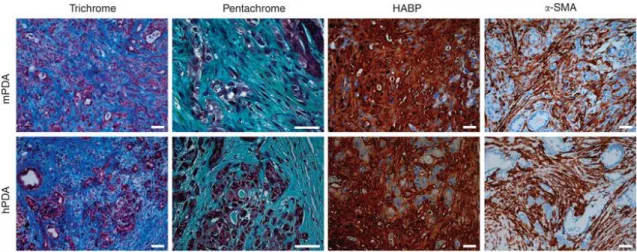
CAFs are the most abundant cell type in PDAC stroma that produce ECM components, such as collagens, fibronectin, laminins and hyaluronic acid, glycosamino glycans (GAGs) (Fig 5) (124). They also stimulate tumor cell growth by paracrine signaling, support migration and invasion, as well as acquired resistance mechanisms.
30
Figure 5. Histology of human and mouse PDAC. Masson’s trichrome histochemistry shows robust collagen deposition (blue). Pentachrome staining reveals collagen, (GAGs) and mucins (turquoise/green). Hyaluronic acid binding protein hybridization probe displayes intense HA content. αSMA immunohistochamistry shows abundant expression in the stroma but not in tumor cells. (Adapted from Provenzano et al. 2012.)
The reactive fibrotic environment contributes to hypoxia and immune cell infiltrates. While immune cells are also plentiful within the stroma, they mostly belong to immunosuppressive subsets, such as regulatory T cells (Tregs), T-helper (Th cells) cells, TAMs and multiple subsets of immature MDSCs. TAMs support tumor progression and invasion by producing pro-tumorigenic factors and induce resistance to gemcitabine treatment by upregulating the levels of the drug metabolism related enzyme, cytidine deaminase, in PDAC cells (153). In a recent study, Zhu et al. identified TAMs of distinct origins in PDAC: tissue resident macrophages display pro-tumorigenic functions and pro- fibrotic transcriptional signature, whilst monocyte-derived TAMs appear to play a role in antigen presenting (154). In contrast, CD8+ T cells are not frequent in the tumor stroma and when present they are located in the surrounding tumor tissue (152).
Therefore, these findings suggest that stroma elimination could deplete the physical barrier and enhance drug delivery to the cancer cells located inside of the tumor mass, while also disrupting deleterious stroma – cancer cell interactions. Studying tumor – stroma interactions by GEMMs can shed light on important cellular processes and therapeutically targetable pathways.
1.2.3 Cancer Associated Fibroblasts (CAFs) in PDAC
In pancreatic cancer, a specific cell type, pancreatic stellate cells (PSC) are the major source of CAFs. PSCs is a specific cell type (155), that can be found in pancreas,
31
liver, kidney, lung and intestine (156) and that share many characteristics of fibroblasts.
In normal pancreas, PSCs are in a quiescent state with a specific stellate morphology located in the peri-acinar space and contain lipid droplets that serve as Vitamin A storage (Fig 6). Various markers have been described to identify quiescent PSCs, including desmin, nestin, GFAP, vimentin (155, 157, 158). Upon activation, they start to express αSMA, change their cytoskeleton, and acquire elongated shape and myofibroblast phenotype. They control the ECM turnover by producing MMPs and collagens. They can be activated by PDGFs TGF-ß, TNF-α, and interleukins, such as IL1, IL6 and IL10.
Indeed, PSCs associated to PDAC express receptors of these cytokines (159). This activation could be reversed in vitro by retinoic acid (Fig 6) (160).
Figure 6. Activation of Pancreatic Stellate Cells (PSCs). Quiescent PSCs express Desmin, Vitamin A and GFAP; and contain lipid droplets. Upon activation by inflammatory cytokines, tissue injury or oxidative stress (ROS) they transform into myfibroblasts, change their morphology and start to express markers, such as αSMA, Vimentin, PDGFRs, etc. Activated PSCs can be reverted by retinoic acid.
As described above, CAFs are key players in the TME of pancreatic cancer. Being the most frequent cell type in PDAC stroma (80%) they are also responsible for the extreme stiffness of the tumor tissue by producing insoluble fibrillary matrix components.
This results in hypoxia and a reactive microenvironment, rich in infiltrating, suppressive immune cell populations, ideal for tumor progression and therapy resistance (124).
Subpopulations of CAFs in PDAC
CAF subpopulations can be defined by their origins or/and molecular profile (127, 130). CAF subtypes are not well characterized, indeed, there is only minor description of these subpopulations in PDAC. For instance, a CD10-positive subpopulation of CAFs was identified in human PDAC specimens. These cells were localized juxtatumorally, in
32
the vicinity of tumor cells in patients with shorter survival (161). In a recent study, Öhlund at el. defined two distinct populations of CAFs showing molecular divergence. MyCAFs (myofibroblast CAFs), with increased expression of the well-known myofibroblast marker SMA, located in close proximity of neoplastic cells. iCAFs (inflammatory CAFs), on the other hand were localized more distant from tumor cells and displayed inflammatory-secretory expression profile with elevated expression levels of IL-6 but lacking SMA in a mutually exclusive but reversible fashion (162).
1.2.3.1 Preclinical studies targeting CAFs in PDAC
Targeting the pro-tumorigenic effects of CAFs in pancreatic cancer offers many possibilities due to their functional diversity. Indeed, several strategies have been suggested to obtain therapeutic benefits in PDAC (Fig 7).
Targeting the stroma and ECM as physical or chemical barrier. The desmoplatic stroma in PDAC has been considered a barrier to drug delivery. Targeting the production of the ECM or its degradation are both feasible strategies to loosen the stroma and to induce expansion of blood vessels. In 2009, inhibition of the Hedgehog pathway by the Smo inhibitor IPI-926, was shown to be efficient to reduce stromal content, induce angiogenesis and improve intratumoral Gemcitabine content (151). In contrast, when Hh was genetically deleted in a pancreatic cancer mouse model, despite the attenuated stroma and an increased vascularization, these tumors appeared undifferentiated and more aggressive leading to reduced survival in mice (51, 52).
High interstitial fluid pressure can be reduced by enzymatic digestion of hyaluronic acid, a major component of the ECM (124). Degradation of hyaluronan by the peglylated form of hyaluronidase (PEGPH20) normalized the hydrostatic pressure, lead to increased delivery of chemotherapy and prolonged survival in KPC mice (124, 163). The matrix protein SPARC is overexpressed in the ECM of many tumor types. nAb-Paclitaxel, an albumin-bound Paclitaxel, was postulated to bind to SPARC and thereby induce stromal depletion. This hypothesis was supported by the analysis of PDAC samples and patient- derived xenografts (PDX) (164). However, in KPC mice stromal loss occurred rather due to implicated drug–drug interactions via reduction of cytidine deaminase levels (165).
Moreover, tumor-bearing KPC mice lacking SPARC did not respond differently to nAb- paclitaxel compared to control mice, showing the mechanism of action is independent of SPARC expression (166).
33
It was recently illustrated in KPC mice that CAFs can act as a chemical barrier by retaining gemcitabine metabolites through reduced levels of key inactivating metabolic enzymes compared to tumor cells. By this mechanism, CAFs limited the drug uptake of cancer cells (167).
Targeting secreted factors of CAF-tumor cell interactions. Blocking the CAF- secreted connective tissue growth factor (CTGF) resulted in a synergistic effect with gemcitabine without increasing the intratumoral gemcitabine concentration via deregulation of the apoptosis modulating protein XIAP (168).
On the other hand, Cxcl12 secreted from FAP-positive cells was shown to be important for immune suppression, explaining why immune checkpoint inhibitors, such as anti-PD- L1, have failed in pancreatic cancer (84). Notably, in KPC mice, inhibition of Cxcr4, the Cxcl12 receptor, promoted the intratumoral T cell recruitment and strongly cooperated with anti-PD-1L (137). Likewise, inhibition of Cxcr2, a receptor activated by CAF produced ligand Cxcl1/2 in KPC mice prolonged survival and improved T cell entry.
Interestingly, germline elimination of Cxcr2 completely abrogated metastasis but had no effect on tumor development suggesting cellular context and tumor stage dependent action of this receptor (169).
Targeting CAFs by reprogramming. Depletion of CAFs in a pancreatic cancer model led to more aggressive and less differentiated tumors (170). Thus, the protective role of certain stromal elements should be taken into consideration and reprogramming, rather than eliminating CAFs, should be considered. Reprogramming of CAF behavior to change their properties to a more “normal” phenotype can be achieved by multiple mechanisms. Since CAFs undergo metabolic changes, normalization of the metabolic phenotype and inhibition of metabolic pathways have also been suggested as a possible way to target tumors (171).
Another approach postulated to dedifferentiate them into a quiescent state is based on Vitamin D since Vitamin D Receptor (VDR) ligands promoted the dedifferentiation of liver stellate cells and abrogated fibrosis (172). In PDAC, Vitamin-D mediated stromal reprogramming markedly reduced inflammation and returned PSCs into a quiescent state, thereby decreasing tumor volume and increasing chemotherapy efficacy (173).
Finally, inhibition of PDGF signaling, an important pathway in the activation of CAFs, can reverse CAFs into normal fibroblasts (174). In PDAC, metastatic potential was significantly reduced upon Imatinib treatment of tumor-bearing KPC mice, while showing no effect on primary tumor development (139).
34
Figure 7. CAF targeting approaches. 1. Targeting the stroma as a physycal barrier :by Hyaluronidase and Hedgehog inhibitors to decrease ECM and to improve drug delivery.
2. Targeting CAF secreted factors: by inhibitors of Ctgf, Cxcl12 to block interactions with tumor cells. Targeting CAFs – secreted ECM mediated pro-tumorigenic effect by Cxcr4 inhibition. Blocking Cxcl1/2 improves intratumoral T cell entry. 3.
Reprogramming CAFs into “normal” fibroblasts by deactivation using PDGFR inhibitors or VDR ligands.
In conclusion, there is emerging evidence from preclinical studies that pro- tumorigenic properties of CAFs represent an attractive and promising therapeutic target in PDAC that deserve further studies.
35 1.3 Serum Amyloid A protein family
Serum Amyloid A (SAA) is an acute phase high-density apolipoprotein family, whose levels are highly increased upon inflammatory stimuli, tissue injury, trauma or cancer (175). Although, different pro- and anti-inflammatory properties have been recently connected to distinct isoforms of SAAs, their role in defense mechanisms and cancer development is not entirely understood.
1.3.1 SAA functions in normal homeostasis
SAA is a highly conserved protein family (176) suggesting evolutionary significance and physiological importance (177). They are considered apolipoproteins (proteins that bind lipids and transport them through the circulatory systems) since in the circulation they associate with high-density lipoproteins (HDL), such as cholesterol, thereby playing an important role in lipid metabolism (178). During inflammatory events, acute phase response is initiated to eliminate pathogens and restore normal homeostasis.
This involves HDL remodeling, where SAA1 and SAA2 displace ApoA-1 and become apolipoprotein of HDL (179). It is unknown if the role of SAAs during acute inflammation is to raise cholesterol removal from tissue damage sites or to deliver cholesterol esters to cells involved in tissue repair (180). In addition, it was recently reported that SAAs could have functional role in retinol (Vitamin A) binding and transport during infection and inflammatory response (181).
1.3.1.1 SAA family members
In humans, four SAA encoding genes are clustered on chromosome 11 in a segment of 150 Kb (182). SAA1 and SAA2 are located in close proximity and contain several allelic variants. The third gene, SAA3 is situated further downstream of SAA4 and was identified as pseudogene in humans (hSAA3P) with a defective promoter generating a translational stop signal (183). However, mRNA transcripts were reported in mammary gland epithelial cells (184) and in cancer. SAA1 and SAA2 are designated as ‘acute phase SAA’ (A-SAA) since their serum concentration could strike to 1000-fold during inflammatory response, whilst SAA4 is referred as ‘constitutive SAA’ (C-SAA) secreted into the blood circulation constitutively by hepatocytes in ‘normal’ physiological conditions (185).
In mice, as well as in humans, Saa1 and Saa2 are major acute phase proteins along with constitutive expression of Saa4 in liver. Nevertheless, Saa3 is secreted