CHAPTE R EIGH T
Growth and Morphogenesis in Tissue and Free Cell Cultures
F . C. STEWARD,1 WITH M . O . MAPES AND P . V . AMMIRATO
I. Introduction: Concepts of Free Cells and Their Growth 329
II. The Biochemistry of Growth Induction 331 A. The Nutrient Requirements for Growth of Cells and Tissue Explants . 331
B. Accessory, Nonnutrient, Growth Factors for Angiosperm Cells . . . 335 C. Factors Which May Limit the Growth of Tissue Explants . . . . 335
D. Growth Factors for Cell Division 336 E. Components of Growth-Promoting Systems: Synergisms 341
F. Some Limitations to the Growth of Tissue Explants 346
G. Growth Induction: A Summary 349
III. Free Cells of Angiosperms 350
A. Techniques 350 B. Cell Growth and Cell Division in Small Tissue Explants or Free Cells . 351
C. The Behavior of Free Cells of Carrot 353 D. The Growth of Free Cells and Embryogenesis 355
E. Cell Multiplication and Morphogenesis: The Sequential Effect . . . 360 F. Stimuli to Cell Growth and Cell Division in Relation to Development
and Morphogenesis 367
References 370
I. Introduction : Concept s o f Fre e Cell s an d Thei r Growt h
This section is concerned with what has been learned about the requirements for growth and morphogenesis by studying the conversion of isolated tissues of higher plants into free cell cultures. Its general purpose is to explore the factors that cause cells of angiosperms to grow, to divide, and to develop into organized structures. Although this subject has developed rapidly in recent years in work from various laboratories, the account here given is in large part based on the
1 This work developed during a program of research which has been supported by grants from the National Institutes of Health, Bethesda, Maryland, latterly GM 09609.
During this work, and for some years, the responsibility for the aseptic cultures has been taken by Mrs. Marion O. Mapes, and in this account the illustrations are largely her work. From time to time other research assistants and graduate students (e.g., L. M. Blakely and Ann E. Kent), working under the direction of the senior author, have also made their contribution and, currently, P. V. Ammirato has contributed substantially to the account presented here in Section III. Section II is based on work done in this laboratory with Dr. Ε. M. Shantz.
329
3 3 0 F . C . STEWARD, M . O . MAPES, AND P. V . AMMIRATO
work of this laboratory with sufficient citation to relate it to other similar investigations.
In all angiosperms there are two crises in development at which the organism is reduced to a free-celled condition: this occurs at spore formation and in the formation of gametes. Spores, which originate at meiosis, initiate the gametophytic phase of the angiosperm life cycle, which in due course culminates in the formation of male and female gametes in anthers (pollen grains) and ovules (embryo sacs), respec- tively. But the spores and the gametes differ fundamentally, in that the former can separately and individually grow, whereas the gametes commonly grow only after syngamy, the fusion of the male and female sex cells. This contrast poses the still essentially unanswerable question of what it is that predisposes cells to divide.
Much may now be said about how cells multiply and about the factors that control their division, but it is difficult to stipulate why cells divide. It is as though the zygote has some mysterious "growth energy," a "built-in capacity to grow," which is not inherent in the gametes but which predisposes the fertilized egg to divide. At each subsequent division, new form and order is created out of random molecules, so entropy is reduced. One may, therefore, see the pro- pensity of cells to multiply as a function of their ability to reduce entropy, and one might visualize a "negative entropy scale" of num- bers which would express the cell's propensity to grow ( 8 9 ) . How- ever, there seems to be no known way of making measurements on cells which will express why one cell of the plant body may be able to divide, while another may not. Nor can one easily see what feature of the daughter cells during interphase "winds them up" for an en- suing division, for this is only in part a function of the new substances they create.
Although angiosperms, as organisms, are typically autotrophic, their constituent cells are often heterotrophic. In the division of labor of the plant body, the cells of growing regions rely on other organs for much of their nutrients and upon the exogenous stimuli for their division.
This is especially true of the fertilized egg, which in its normal environ- ment is commonly nourished by a complex array of substances in endosperm—substances which are, in turn, derived from the parent sporophyte via the nucellus. Significantly, therefore, it has been shown ( 1 0 3 ) that the vicinity of immature embryos is a potent source of substances that stimulate and regulate growth and the effects of these substances may be made apparent even when they are applied to more adult tissue cells.
In microorganisms it is, of course, the rule that cultures may be
8. GROWTH, MORPHOGENESIS IN TISSUE, F R E E C E L L CULTURES 331 derived from single cells or single spores. However, it was Haberlandt who, in 1902 ( 2 5 ) , had the insight to see that one day the growth of higher plants from their constituent living cells should also become possible. But, remarkably enough, Haberlandt went further, for, as recently recognized ( 9 0 ) , he also visualized that one should be able to make "artificial embryos" out of free, living angiosperm cells ( 2 5 ) . This remarkable prophecy, without foundation in anything that Haber- landt could achieve with isolated cells in 1902, has been recently verified, but over sixty years later (97-100, and references there cited).
But one may safely assume that Haberlandt made his prophecy secure in the knowledge of many cases of apomictic development, in which distinctive cells of the plant body do give rise to embryos without the necessity of the complex apparatus of meiosis and syngamy, for he devoted much of his subsequent writing to studies of these events
(27, 27a, 2 8 ) .
Like many subsequent workers, Haberlandt commenced with those mature cells (like those of potato tuber, cf. Chapter 4, Volume I I ) that have some residual capacity to divide which is displayed during the phenomenon of wound healing. In fact, Haberlandt postulated a "wound-hormone," which supposedly "triggered" this division and believed it emanated from phloem cells in the tissue mass ( 2 6 ) . Haberlandt also implicated the hypothetical wound hormone in the early stages of the morphogenetic events by which cells so stimulated to divide may lead to parthenocarpy (27a) and to adventive embry- ogeny (27, 2 8 ) . From these origins derives the philosophy that under- lies all the subsequent work with explanted tissue and later that with free cells: this is that a complete knowledge of the nutritional require- ments of cells and of their responses to stimuli should permit one to cause them to recapitulate, in isolation, their behavior in situ in the plant body.
II. Th e Biochemistr y o f Growt h Inductio n
A . T H E NUTRIENT REQUIREMENTS FOR GROWTH OF CELLS AND TISSUE EXPLANTS
There is nothing intrinsically unique about the inorganic nutrients that nourish explants of angiosperms or the free cells that may be derived from them. One may turn here to Volume III of this treatise, which is devoted to the problems of mineral nutrition, or to the chapters in this volume. Just as there is no "best" nutrient solution for any given angiosperm under all conditions (for requirements surely
3 3 2 F . C . STEWARD, M . O . MAPES, AND P. V . AMMIRATO
vary with the environment and the stage of development), it is equally to be questioned whether there is any "best" solution for the culture of all tissue explants or all free cells. Thus, the practice of identifying supposedly distinctive nutrient solutions by name is not really to be encouraged, for it is evident that the various solutions so distinguished are rarely unique for the responses that they elicit.
One may, however, make the following general statements about such basal nutrient solutions.
Prior to modern knowledge of trace elements (especially boron, manganese, cooper, zinc, and molybdenum), a chief preoccupation of those who considered the mineral nutrients for cultured tissues or cells was to supply the then known major nutrient elements (nitrogen, phosphorus, sulfur, potassium, calcium, magnesium, iron) in suitable form. All the problems of concentration, the mutual relationships of the individual nutrient elements, the suitable salts by which they may be supplied, the purity of the water in which they are dissolved, and the presumed insolubility of their containers, which historically vexed those who worked with plants in water culture, have applied equally to those who have essayed the separate culture of the organs, tissues and now free cells of angiosperms. Thus at any point in time the mineral content of solutions for the isolated culture of tissue explants and of cells has reflected the then current status of knowl- edge of mineral nutrition generally. Early preoccupation with pH, the need to keep iron and later manganese both soluble and available at near neutral reactions, which is affected by the possible role of organic matter and later of the chelating agents, have also engaged attention in the tissue culture field. The alternative uses of nitrate, ammonia, and organic nitrogen (whether in the form of glutamine or casein hydrol- yzate) with the now added prospect that the needs for molybdenum and manganese may be affected by the form in which nitrogen is supplied, all raise questions in particular cases, but not more than in other nutritional situations (Volume III; and this volume, Chapter 6 ) . These and other similar questions could, and perhaps later should, be entirely reinvestigated for the growth of isolated free cells of angiosperms.
However, the point of departure for the present chapter must be as follows. It is to be presumed that there is an ultimate requirement of isolated cells and small tissue explants for any and all of the essential nutrient elements, and the smaller the inoculum, down even to a few free cells, the more critical is the exogenous nutrient balance at the outset of their growth. The need for simultaneous supply of all the essential nutrients in the same proportions throughout the growth
8. GROWTH, MORPHOGENESIS IN TISSUE, F R E E C E L L CULTURES 333
is commonly presumed, but there is no valid proof—perhaps there are even some disturbing indications to the contrary, for the optimal nutrient requirements may change sequentially with the developmental status of the material being cultured. [For example, carrot (Daucus carota var. sativus) phloem explants will tolerate low calcium levels that certainly would not suffice for their roots.] Nevertheless, the presumption now is that the organic requirements for the essentially heterotrophic growth of tissue explants and cells could be added to almost any complete, dilute mineral nutrient solution, if this is done without secondary consequences to the solubility or availability of its essential constitutents. Thus the basal nutrient solutions of White
(112-116), Heller (31, 3 2 ) , or Gautheret (18-20) or their many named successors have, consciously or accidentally (as sometimes occurred in the cases of molybdenum) often supplied the essential minerals in ways that supported growth. This is not to say that some plant tissue and cells will not profit by higher potassium, or higher total salt concentrations, or ammonium versus nitrate, etc. The point is rather that the chief preoccupation in this chapter must be the organic requirements for heterotrophic growth and principally with those accessory, regulatory substances which, singly or collectively, catalyze the growth of otherwise mature or quiescent cells. There is every rea- son to believe that when these organic, accessory, nutrient substances are fully known they will involve, and interact with, the essential mineral elements. In fact, the cell division factors of coconut milk, early known to act at sites vulnerable to cyanide and carbon monoxide (108) are also now known to interact strongly and critically with iron [work of Neumann (62a, 103a) in this laboratory]. The supplements re- quired to render a basal nutrient medium (such as that of White) able to support the most rapid growth of angiosperm tissue explants should now be summarized, although no attempt will be made to deal exhaustively with all the relevant, or even all the conflicting, evidence.
The major organic nutrients are all dealt with more fully by Street (Chapter 6 ) . The preferred exogenous source of carbon seems to be sucrose, probably because it contains fructose in the furanose form.
In general, therefore, the nutritional requirements for carbon may be met by sucrose and those for nitrogen by nitrate. Even so it is often found that enzymatic casein hydrolyzate (or even glutamine) may stimulate growth by furnishing reduced nitrogen, or even catalyze growth because it furnishes special products (certain amino acids) or other substances (e.g., those derivable from trytophan when it is autoclaved).
It is axiomatic that the cells and tissues of angiosperms grow more
334 F . C . STEWARD, M . O. MAPES, AND P. V . AMMIRATO
rapidly on agar or in liquid cultures if they do so heterotrophically.
If the development of chloroplasts in callus tissue, or free cells, is poor or lacking even when the cultured tissue originated from shoots and is grown in the light, fully autotrophic growth is, of course, im- possible. Nevertheless, many angiosperm cells which are green, will (like Chlorella), grow even better in the light as heterotrophs than as autotrophs.
There are, therefore, two distinct questions. One concerns the efficiency of green plant photosynthesis to furnish cultured cells or tissue explants with carbon compounds for their growth, in comparison with the exogenous supply of sugars; the answer here is usually, and overwhelmingly, in favor of exogenous sugars and a heterotrophic mode of nutrition. The other question, however, is whether green chloroplasts in the light contribute anything which favors the growth and nutrition of the cells, which cannot easily be duplicated by the exogenous supply of sugar. This question is not as easy to answer. Suffice it to say, how- ever, that in general those treatments which cause tissues or free cells to turn green invariably enhance their growth, even in the presence of sugar, and they also seem especially conducive to morphogenesis.
In fact, one of the special features of the coconut milk growth factors in the culture of carrot explants and cells is the facility with which normal green chloroplasts develop (33, 34), so that one does not believe that an artificial, or isolated, source of the growth stimulus duplicates that of coconut milk until it also encourages the tissue to turn green in the light. All this may merely mean that green plastids in the light either furnish catalytically active carbon com- pounds, not easily derived from exogenous sugar, or may act by supply- ing readily usable energy as ATP from photosynthetic phosphorylation.
Having recognized the probably nutritional, as well as photomorpho- genetic, role of light (mediated by plastids when they are present in cultured cells and tissues), other external factors become relevant.
Commonly cells and tissue explants are cultured under relatively constant conditions; these may range from continuous darkness to continuous light at constant temperature, or perhaps a fixed diurnal light-dark cycle. The more nearly the external medium becomes fully competent to support the full range of activity of the cells, the more important it may become to utilize their responsiveness to prescribed fluctuations of light and dark with corresponding "day" and "night"
temperatures. Eventually these fully programmed sequences may be expected to be as effective in controlling both metabolism and morpho- genesis of cultured tissue and cells as they are now proving to be in the interpretation of the behavior of whole plants (cf. Went and Sheps, Chapter 5, Volume VA). Before this stage can be usefully
8. GROWTH, MORPHOGENESIS IN TISSUE, F R E E C E L L CULTURES 335
reached, however, the main limitations to growth which can be met by exogenous substances which function as accessory, nonnutrient, growth- regulating substances need to be known.
B. ACCESSORY, NONNUTRIENT, GROWTH FACTORS FOR ANGIOSPERM CELLS
There have been many indications, over the years, that organic accessory growth factors and stimulants regulate the ability of angio- sperm cells to engage their intrinsic abilities for growth and cell division. These range from the stimuli for the growth of orchid gynaecia, which was well known in 1909 to Fitting ( 1 7 ) , and the wound hormone of Haberlandt (26) to many examples in which fer- tilization, or pollination, also stimulate the growth of somatic tissue in fruits (cf. 13, 2 4 ) . Bottomley's ill-fated concept of auximones ( 6 ) probably anticipated much later knowledge of vitamins, for it became well recognized in the 1930's that such substances could limit the heterotrophic growth of explanted angiosperm cells and organs as well as that of microorganisms. Thus vitamin supplements, made either specifically or generally in the form of yeast extract, have become standard features of cell and tissue culture media (cf. Street, Chapter 6 ) . But the concept of a specific growth substance or substances, over and above salts, sugars, vitamins, etc., which have a strictly nutritional role, really dates from the recognition of auxins as a class, and of indoleacetic acid in particular. At the present time, however, plant physiologists recognize an array of growth-regulating substances, both natural (cf. Thimann, Volume VI) and synthetic (cf. Wain and Fawcett, Chapter 4, Volume VA), and their relationship to the problems of growth of isolated tissues and cells arises.
The classes of accessory growth-regulating substances commonly recognized are the auxins, the gibberellins, the inhibitory substances which may work antagonistically against them (such as antiauxins, like coumarin, irans-cinnamic acid or p-chlorophenoxyisobutyric acid, and the abscisins, or the dormancy-inducing factors, like dormin or abscisic acid as it is now called), and also the multiplicity of sub- stances, or combinations of substances, which collectively induce cell division and for which such terms as kinins, cytokinins, or phytokinins have been recommended and are now being widely used.
C. FACTORS WHICH M A Y L I M I T THE GROWTH OF TISSUE EXPLANTS
The growth of tissue explants and cells involves many distinctive steps or processes, any one of which may be rendered limiting and therefore make regulation possible; this regulation could be mediated
3 3 6 F . C . STEWARD, M . O . MAPES, AND P . V . AMMIRATO
by the exogenous supply of a missing factor, or by the endogenous presence, or disappearance, of an inhibitor. All can agree that chemical regulatory control of the powers of growth that are inherent in all cells is the crux of the problem. However, the multiplicity of terms to describe the substances by which this chemical control is regulated may not always be illuminating. Too often the terminology of growth factors has been linked to a type of observed response, and not to a known mechanism of action, and rarely to the chemical configuration of the active substances which has become known only long afterward.
Ultimately, it should be recognized that even an essential element, whether potassium or iron, may discharge the functions of a cytokinin if it alone is the key limiting factor in a given situation. Likewise, there are also many examples of the catalytic role of oxygen (no doubt mediated by a variety of oxygen carriers) in the induction of growth, wholly or partially, as in the phenomena of wound healing.
If the roles of auxins, or gibberellins, are often predominantly (but in their respective ways) to stimulate cell enlargement, it is equally obvious that, having done so, they may set in train events which may lead to cell division, so that they trespass on the preserves of cyto- kinins. And there are abundant examples of substances [like 2,4-di- chlorophenoxyacetic acid ( 2 , 4 - D ) or naphthaleneacetic acid (NAA)]
which are commonly recognized as synthetic auxins and which un- doubtedly can stimulate cell enlargement; in the context of situations in which they act synergistically these may be very potent substances that "trigger" cell division ( 3 , 9 1 ) .
In short, in this difficult field, in which one needs to consider the growth of cells as intact organizations, performing all the functions of growth, metabolism, and development, the terminology should remain flexible; it should not set up rigid categories which often tell more about the limitations of certain test systems than about the role in growing plants of the substances tested. This idea develops from the stimuli which induce growth and development in explanted angiosperm cells and tissues. The salient point is that no single substance 4 unlocks the door of cell division," for the growth and development of cells is controlled by many categories of exogenous growth-regulating sub- stances which act both synergistically and sequentially to "tell" the cells how to harness their nutrients so as to express their intrinsic, genetically determined, powers of growth ( 9 1 ) .
D . GROWTH FACTORS FOR C E L L DIVISION
The reality of supplementary, nonnutrient factors for cell division was evident after the observation by Caplin and Steward ( 8 ) of the
8. GROWTH, MORPHOGENESIS IN TISSUE, F R E E C E L L CULTURES 337
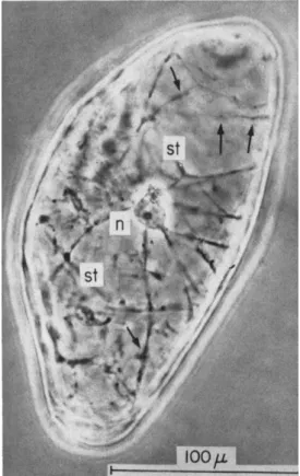
effect of coconut milk (or coconut water, i.e., the liquid endosperm of the coconut) when it was added to an otherwise complete nutrient medium (White's basal medium) for tissue cultures. The tissue tested consisted of small (2.0-2.5 mg) explants of the secondary phloem of carrot root, which were cut at a distance ( 2 - 3 mm) from the cambium so that the cells they contained would not normally have divided again.
In the absence of the coconut milk, or casein hydrolyzate, the carrot tissue only made sluggish increase of fresh weight even under the optimal conditions for growth. The addition of the coconut milk ( 5 - 10% by volume) to the basal medium resulted in an eightyfold increase of fresh weight in almost 20 days, and an even greater relative increase in the number of cells (8, 104). The effect of the coconut milk was increased by, but not dependent upon, the presence of casein hydrolyzate (as a source of reduced nitrogen compounds) in the me
dium. The role of the coconut milk, when discovered, was shown to be over and above anything which auxin (i.e., IAA) alone could accomplish ( 8 ) . Later observations showed that the normal environ
ment of immature embryos in the ovule commonly contained such stimuli to cell division which were assayable in the carrot assay system.
Such activity was detected (93) in extracts of immature corn grains (Zea mays) (less than 2 weeks after pollination), in the liquid from the vesiculate embryo sacs of walnut (Juglans regia), and of a species of horse chestnut (Aesculus woerlitzensis) as well as in a comparable situation in a gymnosperm (Ginkgo biloba) represented by an extract of the female gametophyte. The formative layer in the parthenocarpic fruit of banana (109), the genetic tumors on tobacco hybrids (Nico
tiana glauca Χ IV. langsdorffii), the crown gall tumors of Kalanchoe, all yielded extracts capable of stimulating cell division and so causing a recrudescence of growth in otherwise quiescent carrot secondary phloem cells ( 9 4 ) .
Thus a variety of extracts which fostered active growth furnished the evidence for growth-stimulating mechanisms; and others, in which it was repressed (extracts of dormant tubers, bulbs, and buds) fur
nished equally dramatic evidence of inhibitors that could counteract the effect of the coconut milk ( 9 2 ) . In retrospect, the use of carrot root phloem, under the standardized conditions described, was fortu
nate in that this tissue seems to be more free of growth inhibitors than the tissue of many other storage organs that might have been used, and it also responded to the stimulus of coconut milk without requiring a synergist like 2,4-D and NAA. Much knowledge has been gained by exploiting this carrot assay system and by fractionating the materials which coconut milk and other effective extracts contain.
The substances in question whether isolated from whole coconut
3 3 8 F . C . STEWARD, M . O. MAPES, AND P. V . AMMIRATO
milk, from an alcoholic extract of immature corn grains, or from the content of vesicular embryo sacs of Aesculus, are both heat stable and water soluble. Although they may, or may not, contain nitrogen, they have ring configurations so that modes of action seem linked to the molecular architecture into which they fit.
Compounds A, B, and C (as well as a later one designated F ) emerged from large-scale mercuric acetate precipitations of the active cell division components of coconut milk ( 7 7 ) . Only compound A, sym- 1,3-diphenylurea was fully identified ( 7 8 ) . Although these crystalline compounds, with distinctive recorded analyses and properties, were, together with casein hydrolyzate, unquestionably able to cause cell division in carrot explants, their activity varied somewhat with the carrot strain or root in question. Even if these substances existed as such in the original coconut milk and had not been, in part, modified during the long course of isolation, their content would not account for more than a part of the total activity.
All the endosperm extracts examined are rich in the phenolic bodies known as leucoanthocyanins which, by chemical change, may give rise to the anthocyanin pigments of plants ( 7 9 , 1 0 4 ) . The importance of this class of substances in the induction of cell division became obtru
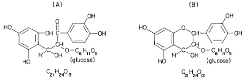
sive when liquid-liquid separations were made of the contents of Aesculus fruits. Although difficult to purify, and more difficult to synthesize, active substances isolated had chemical properties consistent with the formula shown (Fig. 1 ) , and the sugar and cyanidin moieties which arise on hydrolysis were also isolated and identified. The recog
nition of this kind of substance as a component part of a growth regulating complex led to interest in, and tests of, a large number of phenolic substances and flavonoids, and many of these which occur naturally were found to be more or less active ( 7 9 , 1 0 4 , cf. p. 1 8 3 , 1 0 5 ) . These ranged from protocatechuic acid, to catechins, to naturally oc-
(A ) (B )
OH
H ^ O H
C H — \ - 0 H
(glucose)
w ,
2FIG. 1. Molecular formulas of leueoanthocyanin ( A ) based on Robinson and Robinson (72A); and with ring closure (Β) after Bauer et al. ( l b ) and Swain
(109a). The materials isolated from Aesculus were consistent with these formula
tions (104, 106).
8. GROWTH, MORPHOGENESIS IN TISSUE, F R E E CELL CULTURES 339 curring leucoanthocyanins from various other sources. The presence in these molecules of catechol groups suggests that they may play a role as hydrogen donors, or acceptors. Prominent and specific claims have also been made that the amino acid tyrosine may have a distinc
tive role to play in such responses (72, 8 5 ) . In fact, some active substances isolated from Aesculus were so similar to, if not identical with, chlorogenic acid that this again directed attention to both the ring configuration and phenolic character of these active substances
(108).
Although many substances which would not induce cell division when used in lieu of coconut milk in the carrot assay were isolated from immature corn grains, an active substance was obtained which proved to be a compound of one molecule of indoleacetic acid with one molecule of arabinose (80, 108). Although the constitution of the substance is to this extent known, it has not yet been possible to con
firm it by synthesis. Alongside the activity of this naturally occurring complex of indoleacetic acid, one may place the frequently observed activity of the synthetic 2-benzthiazolyloxyacetic acid, which has a remarkable ability to induce cell division in carrot and many other systems, in some of which it works as a synergist (104). It is now known ( 7 ) , however, that the substance originally thought to have the constitution shown at Fig. 2A really contained the substance shown at Fig. 2B and that the original active substance designated BTOA was probably active because it contained the substance shown at Fig. 2B.
Ν > | N - C H 2- COO H
II L II J L
(A)(m.p . 16 1 °-162°) (B ) (m.p.l76°-177°)
FIG. 2. Structure of (A) 2-benzthiazolyloxyacetic acid, and (B) 2-oxobenzo- thiazolin-3-ylacetic acid, after Brookes and Leafe ( 7 ) .
The later work of Letham (49, 5 0 ) , Shaw and Wilson ( 8 4 ) , and Miller (56) on zeatin (Fig. 3 ) provides evidence of an adenyl com-
C H3
I 3
"1ST
FIG. 3. Structural formula for zeatin as isolated by Letham et al. (50) and as synthesized by Shaw and Wilson ( 8 4 ) .
3 4 0 F . C . STEWARD, M . O . MAPES, AND P . V . AMMIRATO
pound, isolated from corn grains, which induces cell division in carrot cells in the presence of indoleacetic acid, and which is also active on cells of other plants. Zeatin may also occur as its riboside. Although kinetin itself (6-furfurylaminopurine) may not be a frequent, naturally occurring, constituent of fluids that induce cell division, it is neverthe
less the prototype of the class of active substances to which zeatin so obviously belongs. As mentioned later (p. 344 et seq.), these substances seem to work synergistically with indoleacetic acid.
The isolation of a still incompletely identified substance from Aescu
lus which comprises indoleacetic acid and a sugar moiety (containing rhamnose and glucose) adds yet another to the list of complex com
pounds of indoleacetic acid which induce cell division in quiescent cells ( 8 3 ) . So far as is known, however, the increasing number of known amino acid-indoleacetic acid complexes in plants (la, 76) are as yet inactive in this respect. Great interest, however, attaches to indoleacetic acid-inositol complexes, which have been recognized and which are even now being tested (45).
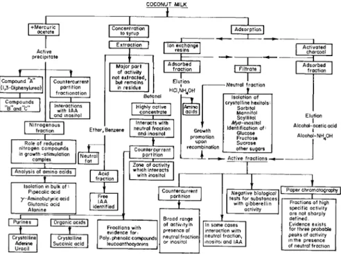
The conference on naturally occurring growth substances held at Gif in 1963 (82, 107) summarized the then known evidence with respect to coconut milk (Fig. 4 ) , corn extract (Fig. 5 ) , and Aesculus (Fig.
COCONUT MILK
Γ+Mercuric
I ocetate Concentration
to syrup 1
I Adsorption I
Active precipitate
Compound A (l,3-Diphenylurea)|
Compounds
"B"and"c"
Countercurrent partition fractionation | Interactions I
with IAA and inositol Nitrogenous
fraction
I
Ion exchange I resins |Major part of activity not extracted,
but remains in residue
Adsorbed fraction
— ι —
Elution ι HCI,NH40H
Highly active concentrate
Ether, Benzene
Role of reduced nitrogen compounds in growth-stimulation
complex
I
I Analysis of amino acids |
Interacts with neutral fraction
and inositol
Countercurrent partition
Acid fraction Isolation in bulk of ··
Pipecolic acid y-Aminobutyric acid
Glutamic acid Alanine
Purines I
Free IAA identified
I Organic acids Crystalline
Adenine Uracil
Crystalline Succinic acid Crystalline
Adenine Uracil
Zone of activity which interacts with inositol
[Amino]
acids I
Growth promotion
upon recombination
Neutral fraction Isolation of JL
crystalline hexitols Sorbitol Mannitol Scyllitol Myo- inositol Identification of:
Glucose Fructose Sucrose other sugars - Active fractions -
Countercurrent partition
Fractions with evidence for Poly-phenolic compounds|
leucoanthocyanins
Broad range of activity in presence of neutral fraction]
or inositol
Negative biological tests for substances with gibberellin
activity
In some cases interaction with neutral fraction, inositol and IAA
Activated charcoal
Adsorbed fraction
Elution Alcohol-acetic acid I
I Alcohol-NH.OH
Paper chromatography Fractions of high 1
specific activity are not sharply defined.
Evidence exists for three probable peaks of activity in the presence of neutral fraction
FIG. 4 . Scheme for fractionation of coconut milk. After Shantz and Steward ( 8 2 )
8. GROWTH, MORPHOGENESIS IN TISSUE, F R E E CELL CULTURES 341
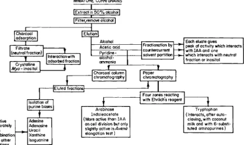
IMMATURE CORN GRAINS I Extract in 5 0 % alcohol I Filter,remove alcohol]
Charcoal adsorption Filtrate X (neutral fraction)
Crystalline I
Myo- inositol
Interaction with 1
adsorbed fraction
ZZJ—
Alcohol Each eluate gives
peak of activity which interacts with IAA and one which interacts with neutral fraction or inositol Acetic acid Fractionation by
countercurrent solvent partition
Each eluate gives peak of activity which interacts with IAA and one which interacts with neutral fraction or inositol Pyridine -
Fractionation by countercurrent solvent partition
Each eluate gives peak of activity which interacts with IAA and one which interacts with neutral fraction or inositol alcohol-
ammonia
Fractionation by countercurrent solvent partition
Each eluate gives peak of activity which interacts with IAA and one which interacts with neutral fraction or inositol
J
Charcoal column
chromatography Paper chromatography 1
Isolation of purine bases
JEluted fractions|-
Four zones reacting with Ehrlich's reagent
Inactive Adenine
separately Adenosine
or in Uracil
combination Xanthine with other Isoguanine fractions Isoguanine
Arabinose Indoleacetate (More active than IAA
on cell division but only slightly active in Avena elongation test)
Tryptophan (Interacts, after auto-
claving, with coconut milk and with 6-substi- tuted aminopurines)
FIG. 5. Scheme for fractionation of extracts of immature grains of Zea. After Shantz and Steward ( 8 2 ) .
6 ) . A later review (76) supplemented this information, and a still later conference in Ottawa (83) added to the existing stock of knowl- edge. Thus, it should be clear that the exogenous control over growth by cell division that is exerted by nonnutrient regulatory substances, whether seen in terms of the induction of growth in otherwise quies- cent cells, or in its suppression in otherwise growing cells, is extremely complicated. However, one should not attribute, unequivocally, all the regulatory activity to a single master molecule, or even class of molecules.
A more rational approach recognizes the diversity of effects involved in growth, the variety of salient points at which the control of growth may be exercised, and the very great diversity of substances, combina- tions of substances, and even sequences of substances that may exer- cise growth-controlling effects, because they, or reactions triggered by them, are endogenously limiting in a given situation.
E . COMPONENTS OF GROWTH-PROMOTING SYSTEMS: SYNERGISMS
All the materials mentioned from the environment of young embryos (coconut milk, corn extract, fluid from Aesculus fruits) owe their effectiveness to combinations of substances, not to single substances.
In addition to the nonspecific, but nutritionally valuable, materials that they contain (potassium, magnesium, phosphorus compounds,
3 4 2 F . C . STEWARD, M . O. MAPES, AND P. V . AMMIRATO
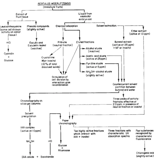
AESCULU S W0ERL1TZENSI S (Immature fruits )
Extract o f fruit tissu e
1
Liquid fro m vesicular embryo sa c
Leucoanthocyanins (some cel l division activity o n carro t
(tissue)
Cyanidin + Glucose
Phenolic compound s (slightly active )
Charcoal adsorption Solvent extraction -
Ether extrac t (active a t 1 0 ppm )
Esculinand Filtrat e Ε luted fractions
Esculetin teste d (neutra l fraction)
(inactive) -Alcohol eluat e
(inactive) - Aceti c aci d eluat e -
(active at25ppm )
Butanol extrac t (active a t 2 5 ppm )
+ N F o r inosito l
Stimulation o f cell divisio n b y interaction upo n recombination
- Pyr i dine eluate (active a t 5 ppm ) - ΝH40H-alcohol eluat e
(slightly active )
Countercurrent solven t partition betwee n butanol an d wate r
Chromatography o n silica ge l column s
Solvent precipitation
IAA comple x (active a t I Oppm )
Three peak s o f activity;
fractions effective a t - 2 0 pp m i n presenc e o f
neutral fraction o r inosito l
1 tr
Paper chromatography
Tw o highly activ e fractions which interac t wit h IAA + inosito l
IAA amid e + Saccharid e
1 1
Three fractions wit h Fou r substances characteristic U V recognize d b y absorption spectr a characteristi c fluorescence
Chlorogenic aci d (slightly active )
FIG. 6. Scheme for fractionation of extracts of immature fruits of Aesculus. After Shantz and Steward ( 8 2 ) and later modified ( 8 3 ) .
carbohydrates, reduced nitrogen compounds, etc.), their effectiveness is due to:
1. The composite fraction termed in this laboratory the active frac
tion ( A F ) which contains the several substances which stimulate cell division per se.
2. A composite fraction, here termed the neutral fraction ( N F ) , which provides substances which synergistically respond with the A F to promote growth and cell division. The neutral fraction (free of the obvious carbohydrates which furnish carbon in bulk) owes its effec
tiveness to hexitols, of which four have been isolated and identified:
myo-inositol, scyllitol, sorbitol, and mannitol, but which are replace
able by mj/o-inositol ( 6 6 ) .
8. GROWTH, MORPHOGENESIS IN TISSUE, FREE CELL CULTURES 343 3. Having combined the effectiveness of an isolated active fraction ( A F ) with an isolated neutral fraction ( N F ) , a further response to enzymatic casein hydrolyzate may often be obtained ( 8 1 ) ; the casein hydrolyzate here acts mainly as a source of reduced nitrogen, but possibly also as a source of specific amino acids, e.g., possibly tyro- sine, phenylalanine, or substances derivable from tryptophan.
4. When the active fractions and neutral fractions are represented by their more purified constituents, it becomes evident that there are two categories of active cell division stimulants in the active fraction, and these are distinguished by different degrees of synergistic response to indoleacetic acid or to inositol.
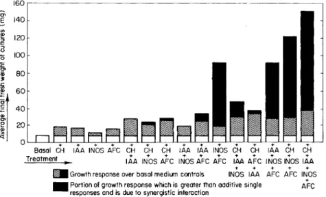
The interactions of the categories of substances mentioned to pro- duce highly significant synergisms, over and above their additive effects, is conveniently illustrated by Fig. 7 for the subfractions ob- tained from Aesculus fruits. Similarly, the evidence for two categories of cell division substances, distinguished by their dependence upon exogenous indoleacetic acid or inositol, is to be seen in Fig. 8 and in Table I. Even when the entire coconut milk system is present, the proliferative growth of some tissue, such as that of potato (Solarium tuberosum) tuber, may require the further action of other synergists, e.g., 2,4-D or NAA (Fig. 9 ) , whose structure is sensitively related to the growth response elicited (88, 91).
160
Portion o f growt h respons e whic h i s greate r tha n additive singl e AF C responses an d i s du e t o synergistic interactio n
FIG. 7. Growth-promoting effects and interactions of casein hydrolyzate, indole- acetic acid, rayo-inositol, and Aesculus active fraction concentrate on carrot phloem explants. After Shantz and Steward ( 8 2 ) .
344 F . C . STEWARD, M . O . MAPES, AND P . V . AMMIRATO
0 5 1 0
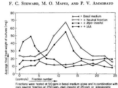
(controls) Fractio n numbe r
Fractions wer e teste d a t 5. 0 pp m i n basa l mediu m alon e an d i n combination wit h corn neutra l fractio n a t 2 5 0 p p m , / ^ - i n o s i t ol a t 25pp m o r indoleaceti c acid a t 0. 5 pp m
Original weigh t o f explant s =3.0m g Growt h perio d = 1 8 day s
FIG. 8. Growth-promoting effects on carrot root phloem explants of fractions obtained from the alcoholic ammonia eluate of charcoal-adsorbed corn extract by countercurrent partition between butanol and water. After Shantz and Steward ( 8 2 ) . The consequences for growth of the interactions between otherwise quite different categories of growth-regulating substances complicates the task of isolation, discourages the overemphasis upon any one sub- stance or category of substances, and also complicates the nomenclature of growth-regulating substances. Nevertheless, it draws attention to the degree to which the growth and behavior of angiosperm cells may be regulated exogenously by applied chemical substances; these must have their counterpart in the endogenous substances which regulate the growth and behavior of the cells in situ.
The most difficult and still incomplete task is the final identification of all the exogenous substances which, especially in natural extracts, may contribute to the growth of the test tissue.
The substances that induce growth in carrot tissue explants and which do so in synergistic interaction with indoleacetic acid includes a category of substances which are adenyl compounds. These include the prototype, kinetin, which arose in the work of Skoog and Miller from aged or autoclaved nucleic acids (57, 58, 85), and its naturally occurring counterpart, zeatin, isolated from corn grains ( 5 0 ) . Another category of substances includes those which act in synergistic asso- ciation with inositol, and these include at least two which are glyco- sides having combined indoleacetic acid (e.g., an indoleacetic acid- arabinoside from corn; a rhamnoglucose-indoleacetic acid compound
8. GROWTH, MORPHOGENESIS IN TISSUE, F R E E C E L L CULTURES 3 4 5
T A B L E I
COMPARISON OF E F F E C T S OF ZEATIN AND K I N E T I N WITH E F F E C T S OF ACTIVE C O N C E N TRATES D E R I V E D FROM COCONUT M I L K AND Aesculus LIQUID, IN P R E S E N C E
AND A B S E N C E OF I A A AND/OR INOSITOL IN THE MEDIUM**
Treatment Expt. 292Α Expt. 292B
Basal medium 15.3 12.3
+ Zeatin 13.2 15.8
+ Kinetin 9.6 11.5
+ C M factor 19.1 20.0
-f- Aesculus factor 37.7 36.9
Basal medium + I A A 31.9 42.4
+ Zeatin 101.0 85.2
+ Kinetin 19.3 67.2
+ C M factor 72.1 101.2
+ Aesculus factor 63.2 84.8
Basal medium + inositol 31.5 26.5
+ Zeatin 37.4 36.6
+ Kinetin 24.0 21.7
+ C M factor 84.2 59.9
+ Aesculus factor 87.6 66.1
Basal medium + I A A + inositol 63.4 70.5
+ Zeatin 138.3 98.3
+ Kinetin 89.0 68.6
+ CM factor 157.4 105.0
+ Aesculus factor 164.8 118.7
Basal medium + 10% C M 240.6 143.8
α After Shantz and Steward (83).
from Aesculus). The activity of such fluids or extracts as those from the coconut, from corn, or from Aesculus fruits is, therefore, the resultant of the action of these different systems. Indeed, even other substances, like the phenolic compounds to which activity has been ascribed (e.g., the leucoanthocyanins which are abundant in endo
sperms, and some simpler phenolic compounds which resemble chloro- genic acid) also play a part. It is still too soon to state simply how all these different parts of the growth-promoting system act, separately or in combination. Parenthetically, it may be noted here that the best combinations of the adenyl growth substance zeatin (as isolated from corn grains, and later synthesized) or of kinetin with IAA, will not fully equal the growth that whole coconut milk stimulates ( 1 4 ) . That part of the coconut milk system (Α ¥τ) which interacts with inositol is now referred to in this laboratory as System I; that part ( A F2) which interacts with indoleacetic acid (IAA) is being referred to as System II.
But even the combination of System I ( A F i X inositol) and System II
346 F . C . STEWARD, M . O . MAPES, AND P. V . AMMIRATO
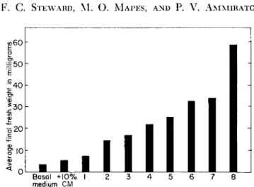
Basal + 1 0 % I 2 3 4 5 6 7
medium C M 8
FIG. 9. Effectiveness of various synthetic growth-regulating compounds as syner- gists with coconut milk in promoting the growth of potato tuber explants. Average growth response of 5 replicate cultures for each treatment grown for 28 days. Treat- ments: basal medium -f coconut m i l k - f - ( l ) a-(2-naphthoxy)phenylacetic acid,
1.0 ppm; (2) indoleacetic acid, 1.0 ppm; (3) a-(2-naphthoxy)propionic acid, 6.0 ppm; (4) a-(2,4,5-trichlorophenoxy)propionic acid, 6.0 ppm; (5) 2,4-dichloro- phenoxyacetic acid, 6.0 ppm; (6) 1,2,3,4-tetrahydronaphthoic acid, 30 ppm; (7) indolebutyric acid, 1.0 ppm; (8) naphthaleneacetic acid, 1.0 ppm. From work of Shantz and Steward; for data see Shantz et al (83a) and Steward ( 8 8 ) . ( A F2 X IAA) in association with casein hydrolyzate ( C H ) will not fully equal the effect of whole coconut milk.
F. SOME LIMITATIONS TO THE GROWTH OF TISSUE EXPLANTS
Tissue explants that are relatively inactive in growth in the basal medium may be limited by their endogenous production or utilization of: ( a ) One or other members of the groups of active cell division substances (AFX and A F2) that interact with inositol on the one hand or with indoleacetic acid on the other, ( b ) Indoleacetic acid or inositol or both, ( c ) Some constituent or constituents of casein hydrolyzate.
While explants from all carrots tested respond to whole coconut milk, nevertheless, explants from given roots may display different degrees of responsiveness to the different categories, and combinations, of sub- stances mentioned above. The ubiquitous response of carrot explants to whole coconut milk, and to this plus casein hydrolyzate, may conceal the fact that explants from different carrots of the same stock have a
"built-in" responsiveness to indoleacetic acid or to inositol, or to both which, when alleviated, allows them to achieve varying amounts of growth; this response will be in accord with the extent to which the cells in situ in the roots were limited endogenously by the counterparts
8. GROWTH, MORPHOGENESIS IN TISSUE, F R E E CELL CULTURES 347
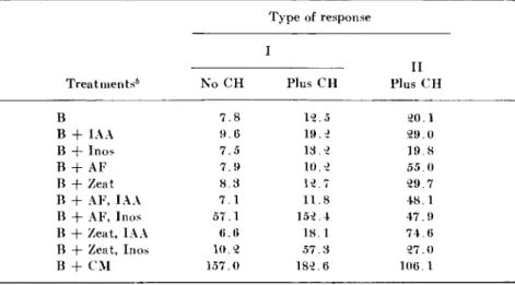
T H E GROWTH ( M I L L I G R A M S , F R E S H W E I G H T ) OF CARROT E X P L A N T S ( I N I T I A L L Y 2 . 5 MG) DURING 1 8 DAYS IN A B A S A L M E D I U M WITH VARIOUS S U P P L E M E N T S "
Treatments6 No C H
Type of response I
Plus C H
II Plus C H
Β 7 . 8 1 2 . 5 2 0 . 1
Β + IAA 9 . 6 1 9 . 2 2 9 . 0
Β + Inos 7 . 5 1 3 . 2 1 9 . 8
Β + AF 7 . 9 1 0 . 2 5 5 . 0
Β + Zeat 8 . 3 1 2 . 7 2 9 . 7
Β + AF, IAA 7 . 1 1 1 . 8 4 8 . 1
Β + AF, Inos 5 7 . 1 1 5 2 . 4 4 7 . 9
Β + Zeat, IAA 6 . 6 1 8 . 1 7 4 . 6
Β + Zeat, Inos 1 0 . 2 5 7 . 3 2 7 . 0
Β + C M 1 5 7 . 0 1 8 2 . 6 1 0 6 . 1
° From Degani and Steward ( 1 4 ) .
b Key: Β = basal medium; C H = casein hydrolyzate; IAA = indoleacetic acid ( 0 . 5 ppm); Inos = wi/o-inositol ( 2 5 ppm); AF = cell division factor from Aesculus fruits ( 0 . 1 ppm); Zeat = synthetic zeatin ( 0 . 1 ppm); C M = coconut milk ( 1 0 % by volume)
These represent extremes of behavior in which the growth induction of the phloem explants from the carrot root is sharply distinguished by their responses to Systems I and II, respectively. Explants from the carrot root selected of type I, and in the absence of casein hydrolyzate, show that their clear response is to the interaction between the Aesculus cell division factor and inositol (i.e., to System I ) and there was no observed response to System II (i.e., zeatin + IAA). By contrast, the explants from the same carrot root again responded strikingly to System I in the presence of casein hydrolyzate, producing 8 3 % of the growth induced by coconut milk, but in addition they were also enabled to respond to some extent to zeatin. Thus casein hydrolyzate broadened of these growth substances. One can in fact distinguish individual carrot roots according to their lack of either indoleacetic acid, or of inositol, which should, therefore, be supplied in the medium.
On the basis of work compiled by Degani (14) and later trends in this work, carrot roots may be identified in terms of their respective responses to inositol and to IAA. This in turn reflects the relative im
portance of Systems I and II in the determination of their growth responses. Typical examples from the records of this laboratory are given in Table II.
TABLE II
3 4 8 F . C . STEWARD, M . O . MAPES, AND P . V . AMMIRATO
the base of response to these cell division factors and their interactions with inositol. The carrot selected for type II showed the clearest re- sponse to zeatin and IAA, (i.e., System I I ) in the presence of casein hydrolyzate though with some lesser response to System I. Thus, the responses of these carrot explants to growth-inducing substances is not clear cut. Even in the presence of casein hydrolyzate a major response to a growth-promoting system (e.g., System I or I I ) may well be appar- ent but, in addition, the casein hydrolyzate also brings into play some interactions which otherwise might have been ineffective. Some con- stituent of the casein hydrolyzate may therefore act as a link between the two otherwise distinctive growth-inducing systems.
The baffling array of substances that "trigger' cell division to some degree in carrot tissue explants, exposed to an otherwise complete nutrient medium, has thus been recognized. These substances are at present more distinguished by the range of their chemical configuration than by any singly recognizable functional group to which their activity may be ascribed. In this respect the problem resembles that of carcino- gens. In short, there are so many possible endogenous limitations to growth, and so many different tasks that a universal exogenous growth- promoting system needs to perform to cover all contingencies, that chemical uniformity and simplicity is neither encountered, nor is it to be expected. This is compatible with the many distinctive ways in which the activated cells respond when observed at the level of the electron microscope (33, 3 4 ) . What can be said, however, is that, in angiosperms, zygotes are commonly highly heterotrophic cells and, by the time their cotyledons have emerged in the light, the whole com- plicated course of embryogeny and seedling development has been run.
The mineral requirements for this growth must have been furnished exogenously via the parent sporophyte, while the organic requirements for the growing regions are furnished in different ways at different stages of development, at which different degrees of specialization become possible. At the level of the zygote (i.e., of a single free dividing totipotent cell) there is maximum dependence for growth on the content of the bathing medium both for organic nutrients and for cell division and morphogenetic stimuli. It is here that the role of liquid endosperms
—the contents of vesicular embryo sacs—the specialized environment of the developing embryos is paramount and indisputable. (Thus, the more effectively one can subsequently reduce an angiosperm growing system to a similar free cell state, the more probable it is that it will again display similar limitations and requirements.) Later, as specialized deposits are stored in cotyledons, in endosperm, in hypocotyls, etc., they provide in distinctive ways for the heterotrophic requirements of
8. GROWTH, MORPHOGENESIS IN TISSUE, F R E E C E L L CULTURES 349
the cells of the growing regions they nourish which, by then, have moved a long way from the unrestricted totipotent requirements of the proembryonic cells from which they were derived. However, the larger the tissue piece, or even the whole organ that is cultured, the more the dividing cells will have had access to stored deposits from previous autotrophic growth. Hence it is the more likely that wholly, or in part, their requirements will be met and that the exogenous requirements may seem, in consequence, to be simplified. It was a fortunate circum- stance that the carrot root phloem system proved so capable of growth in the form of small (2.0 mg) explants and, later, as the free totipotent cells which simulate the zygote itself. Thus, specifically, one needs to know more concerning the biochemical consequences of growth induc- tion, for these release the essential totipotency of otherwise quiescent cells.
G . GROWTH INDUCTION: A SUMMARY
This subject has been dealt with to some extent elsewhere in this treatise—in Chapter 4, Volume IVA in connection with nitrogen metabo- lism, and in Chapter 4, Volume II in connection with cell physiology.
For the purposes of this chapter, the stimuli that induce growth in otherwise resting cells act primarily as follows. They cause protein to be synthesized at the expense of endogenous reserves of nonprotein nitrogen-rich compounds. They reactivate cells in terms of water and solute uptake. They increase respiration and invoke respiratory path- ways that are strongly aerobic and, in submerged cells, cause them to be oxygen saturated only at partial pressures far greater than those needed to saturate the cytochrome system. The agents that cause growth induction also stimulate protein synthesis and turnover. In the outcome carbon moves over a cycle of protein metabolism, into which it is drawn from sugar, and from which it eventually emerges as carbon dioxide.
Meanwhile the need of growing cells for usable and coupled energy as ATP exercises a regulatory role over respiratory metabolism which, to this extent, like protein metabolism "follows the lead of growth." Cells which are metabolically activated by exposure to external nutrients, to appropriate temperatures, and to oxygen, but which lack the stimulus to divide, can nevertheless synthesize nucleic acid in the form of ribo- somes, although they obviously lack the means to make these effective in protein synthesis and growth (100). But the agents that put all this metabolic machinery into gear, so that growth ensues, simultaneously cause the formation of a hydroxyproline-rich, alcohol-insoluble, struc- tural nonmetabolizable moiety which, when inhibited, also suppresses
3 5 0 F . C . STEWARD, M . O . MAPES, AND P . V . AMMIRATO
all growth and morphogenesis. While the nature of this essentially structural moiety is not yet fully known, it seems to reside elsewhere than in the cell wall [cf. recent work of Steward, Israel, and Salpeter (96) on the location of tritiated proline-labeled compounds in cultured carrot cells]. The proteins synthesized in growing cells seem to be different in other ways (e.g., in their behavior under acrylamide gel electrophoresis) from those of mature resting cells. And throughout the cells there are evident signs of cytoplasmic activity, signs which extend from intensified protoplasmic streaming to changes in all the cytoplasmic organelles as seen under the electron microscope ( 3 3 ) .
It is against this background that the growth and development of free cells of angiosperms now needs to be evaluated.
III. Fre e Cell s o f Angiosperm s
A. TECHNIQUES
From Haberlandt's original prophecy (25) many years were to elapse before useful work was done with free cells of angiosperms. Many observers had noted that cells sloughed off from root caps could survive.
In the period when micromanipulations were in vogue, surviving cells dissected from such fruits as Symphoricarpos or Ligustrum or various hair cells were popular subjects for such investigations (10, 11, 74, 7 5 ) . Curiously, such work as that of de Ropp (16) on free carrot cells, which might conceivably have anticipated the later work on the growth and embryogenesis of such cells, reached the diametrically opposite conclu- sion, namely that the free, isolated, vacuolated cells never divide. Even surviving protoplasts in isotonic solutions were successfully manipulated by Plowe (65) and later by others, who even stripped off the outer cytoplasm to leave freely suspended, osmotically responsive, vacuoles.
Such work anticipated the later observations of Cocking (12) on free- living plant protoplasts. Although the earlier work established the isolated protoplasts as osmotically stable systems, remarkably little has been accomplished by the use of isolated protoplasts, for as yet they do not grow. These naked angiosperm protoplasts do not fuse. However, there is a reported case of fusion of naked moss protoplasts ( 2 ) . This brings to mind the observation of fusion of animal cells in culture, which, lacking a cell wall, can occur more readily than in plant cul- tures. Remarkably, cultured mouse and human cells have been seen to fuse ( 3 0 ) .
However, for the study of isolated plant protoplasts, the various members of the Siphonales, which normally undergo what Boegesen
8 . GROWTH, MORPHOGENESIS IN TISSUE, F R E E C E L L CULTURES 3 5 1
( 5 ) called segregative cell division to give naked protoplasmic masses that readily form plants, are far more versatile systems for investigation
( 8 6 ) .
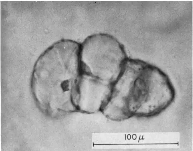
Although some use pectinases to digest middle lamellae, or low calcium concentrations to avoid the formation of rigid cementing mate- rials, by far the most effective way of obtaining active free cell suspen- sions is as follows. The first step is to obtain very rapid growth at the surface of small tissue explants. In this laboratory, this is best achieved by the use of a basal tissue culture medium supplemented with coconut milk, with or without an auxin (e.g., naphthaleneacetic acid) synergist according to the material in question. The very gentle abrasion of the cultured tissues as they grow in slowly rotated ( 1 rpm) culture tubes or flasks ( 1 0 4 ) permits some of the actively growing surface cells to float off into the ambient liquid in which they continue to grow and from which they can be subcultured. In fact, violent horizontal or rotary shakers are not to be recommended if angiosperms cells are to be grown as free as possible from cellular debris and fragments. In fact, such techniques may lead to unusual, even anomalous effects. By hori- zontal motion, a clone of long filaments was cultivated in which subse- quent growth was predominantly by transverse division ( 4 ) .
B. C E L L GROWTH AND C E L L DIVISION IN SMALL TISSUE EXPLANTS OR F R E E CELLS
One might have expected that Sachs' law of equal masses, or Errera's law of minimal surface of new walls, would have led to readily predict- able forms when it became possible to observe the growth of free cells into cultured masses. The form of such cultured masses might even have been anticipated from the multiplication of liquid systems with their boundary surfaces, like soap bubbles, in equilibrium. Or one might have expected that the cell shapes which uniformly and most efficiently partition space might have predetermined their arrangement in tissue cultured masses. Before the observed results are described, one may give the essential conclusion.
When cells grow within the intact plant body, attached to each other, they are subjected to certain constraints due to their position in a given organ; thus their responses are dictated (in large part from without) and their innate potentialities are, in these circumstances, to a large extent restricted. The laws of Sachs and Errera apply to cells which are more or less in equilibrium with their surroundings, and which are not subjected to markedly asymmetric stimuli. These laws are especially adapted to cells in which the partitioning walls may be