CULTURE VIRUS PRODUCTION LABORATORY
ROGER W. JOHNSON CHARLES V. BENTON
ALBERT PERRY JOHN HATGI GEORGE P. SHIBLEY
National Cancer Institute Frederick Cancer Research Center
Frederick, Maryland
I. INTRODUCTION
Those involved in the use of tissue culture on a large scale basis are well aware of the consequences of contamination. The growth of bacteria in several hundred roller bottle cultures rep- resents not only an economic loss, but a loss of time and effort.
The failure of cells to grow in culture because of endotoxin con- tamination of the medium or detergent not rinsed from glassware results in similar losses. It is the task of our laboratory to provide virus for investigators within the National Cancer In-
stituted Virus Cancer Program for biochemical, immunological and genetic studies. Therefore, it is essential that a steady supply of virus, free of contaminants be produced and be made available
571
572 ROGER W. JOHNSON et ai
to these investigators at all times. To inadvertantly distribute a contaminated product results in the loss of respect and reputa- tion within the scientific community, and leads to wasted research efforts by the investigators receiving the product.
II. TYPES OF CONTAMINATION ENCOUNTERED IN A LARGE SCALE VIRUS PRODUCTION LABORATORY
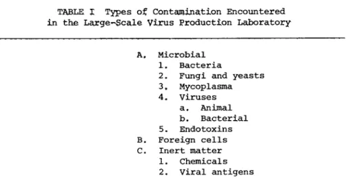
Contaminants which one may encounter in a tissue culture vi- rus production operation are shown in Table I.
A. Microbial
The type of contamination encountered most frequently in the tissue culture laboratory is microbial.
1. Bacteria, Fungi, and Yeasts
Bacteria are the most common offenders in this group, followed by fungi and yeast. While antibiotics may completely supress some contaminants, they will only mask others and still other organisms will proliferate at a remarkable rate in the tissue culture medi- um. In our experience gram negative rods, generally of the genus Flavobacter, are encountered most frequently. Tumilowicz (1974) recently described the contamination of tissue cultures with lep- tospira; the source was possibly a deionized water system used to prepare the tissue culture media.
2. Mycoplasma
Several years ago mycoplasma were described as the crab-grass of tissue culture. Ten years later the description still holds.
Although there are numerous descriptions in the literature of methods for freeing cell cultures of mycoplasma contamination
(Barile, 19751, it is evident that these organisms are merely be^
ing supressed rather than eliminated.
TABLE I Types of Contamination Encountered in the Large-Scale Virus Production Laboratory
A. Microbial 1. Bacteria
2. Fungi and yeasts 3. Mycoplasma 4. Viruses
a. Animal b. Bacterial 5. Endotoxins B. Foreign cells C. Inert matter
1. Chemicals 2. Viral antigens
Although mycoplasma contamination of cultures is difficult to detect (Hayflick, 1965, Barile, 1975), it is easier to prevent then other microbial contaminants, since the sources of potential mycoplasma contamination are limited. The serum used in tissue
culture media, or cell stocks obtained from other laboratories represent the primary sources of contamination. Another common source is mouth pipetting, still practiced in some laboratories.
This practice results in the introduction of mycoplasma into cul- ture from the oropharynx of the technician (Crawford and Kray- bill, 1967, Barile, 1975).
3. Viruses
Adventitious viruses represent one of the least recognized forms of tissue culture contamination. Bovine viral diarrhea virus, bovine herpes virus, and para-influenza type-3 virus (Mo- lander et al,, 19721 all may be isolated from serum. If murine tissues are used, contamination can occur with a variety of ad- ventitious murine viruses (Collins and Parker, 1972). When sev- eral viruses are under investigation in a laboratory in which com^
mon reagents, equipment and technicians are used, contamination of cell lines with virus may and often does occur. Merrill et al.
574 ROGER W. JOHNSON et al.
C19721 and others (Petriccian et al., 1973? Cho et al., 1973 and Moody et al,, 1975Î have demonstrated that serum may be contami- nated with bacteriophages. Although the bacteriophage will not replicate in tissue culture, their presence none the less, con- stitutes a foreign antigen in concentrated culture fluids,
4. Endotoxins
Endotoxin contamination of serum (Moody et al., 1975) or medi- um, detectected by the Limulus amebocyte lysate procedure (Wild- feuer et al., 1974; Jorgenson and Smith, 1973) occurs when gram negative bacteria were allowed to proliferate prior to filtration of these reagents.
5. Viral Antigens
Autoclaved or chemically decontaminated equipment which con- tained virus material must be adequately washed and rinsed to re- move all traces of coagulated protein and virus. Residual virus,
although inactivated, represents a potential source of foreign an- tigen. Certain pieces of equipment used in the harvesting and centrifugation process, e.g., rubber and tygon tubing, continuous flow rotors, present special problems of cleaning.
B. Foreign Cells
Not every cell line one receives from another laboratory is necessarily what it is labelled to be, or entirely of one species of origin (Nelson-Rees et al., 1974). Only by karyotyping and fluorescent staining of cells (Stulberg and Simpson, 1973) is it possible to determine correctly the species of origin of a cell line.
C. chemicals
Improperly washed and rinsed glassware or equipment may be contaminated with detergents or disinfectants used in the glass-
ware preparation area or elsewhere in the laboratory. This re- sults in failure of cells to grow well, or failure to grow over portions of the glass surface of the culture bottle. Phenol and aldehyde groups are often involved, as are the detergents used in washing the glassware.
III. SOURCES OF CONTAMINATION
Potential sources of contamination are summarized in Table II.
A. Cell Seed Stocks
Cell stocks received from other laboratories or even developed within one1 s own laboratory are potential sources of contamina- tion with mycoplasma (Giradi, et al., 1965, Macpherson, 1966), viruses or foreign cells (Nelson-Rees, 1974). Mycoplasma may be spread by aerosols from infected to uninfected cultures (O'Connel et al., 1964). In a similar manner, cells from one line may be introduced into cultures of another line as a result of improperly labelled flasks or by carry over through the use of common bottles of reagent. We encountered a first-hand example of this problem recently when a putative human breast tumor cell line was sent to us for study. Upon investigation, by karyotyping and fluorescent staining testing, this line was found to be a mixture of 95% rat cells and 5% HeLa cells.
B. Tissue Culture Reagents
Tissue culture reagents are an obvious source of microbial contamination, albeit a controllable one. Bacterial or fungal contamination of medium usually can be detected with no testing other than observation. However, only by testing can endotoxin contamination be detected.
576 R O G E R W . J O H N S O N et al.
A. Cell seed stocks
B. Tissue culture reagents
C. Failure to maintain aseptic technique D. Environmental overload
E. Inadequate washing, rinsing and sterilization of equipment
Low (1974) reported the isolation of mycoplasma from a com- mercial lot of serum-free medium. This is an isolated and probab-
ly unusual instance of such contamination. Low suggested possible aerosol exposure of the medium to contaminated bovine serum during manufacture.
Serum is a potential source of more contaminants than any other reagent (Fedoroff et al., 1972; Molender et al., 1972;
Boone et al., 1972; and Merrill et al., 1972). Serum can be con- taminated with bacteria or fungi, as well as with mycoplasma, ani- mal and bacterial viruses and endotoxins.
Trypsin, although a likely source of mycoplasma contamination has apparently never been proven to be an actual source (Barile, 1973).
C. Failure to Maintain Aseptic Technique
Failure to exercise proper aseptic technique while carrying out tissue culture procedures is a common source of contamination.
Proper aseptic technique is developed only through training and repeated stressing of the importance of care being exercised dur- ing culture manipulations and the importance of each individual's participation in the program.
TABLE II Sources of Contaminants
D, Environmental Overload
Under certain conditions, the laboratory environment may be- come overloaded with microorganisms. Dr. Lloyd Herman, Environ- mental Serivices, NXH, Bethesda, Md. (Personal Communication), has described this situation to have occurred when floor drains are left uncovered or when large amounts of moisture are allowed to collect in the tissue culture areas. Environmental overload also occurs when technicians fail to exercise personal hygiene
(Voxakis et al,, 1974), or when the integrity of laminar flow hoods is repeatedly broken with surface contaminated items.
Breakdown in aseptic technique, e.g., allowing gloved hands to come in contact with the lips of culture vessels and environ- mental over-load, account for most of the microbial contamination we experience in our laboratory,
E. Inadequate Washing, Rinsing and Sterilization of Equipment Inadequately washed, rinsed or sterilized glassware and other equipment are obvious sources of inert or microbial contamination.
Unfortunately, this contamination is recognized only after the fact. Strict adherance to glassware processing protocols is the only way such contamination can be prevented.
IV. SCALE-UP AND PRODUCTION
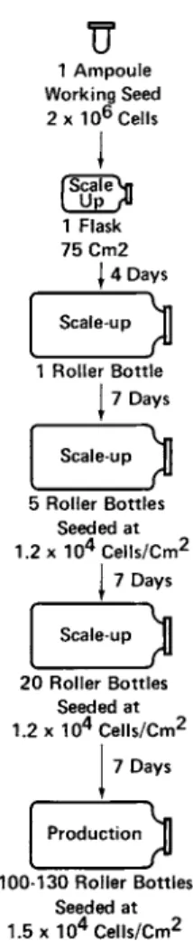
In our laboratory each production lot of virus is initiated from an ampoule of tested and proven working cell seed. These cells, which are chronically infected with the desired RNA C- or B-type tumor viruses, are scaled up to production level within 4 to 5 transfers (Fig. 1Ï, At each one of these transfers, and at each intermediate feeding of the cultures with, fresh medium, sarn- ies of culture fluid are taken for sterility tests. These steril- ity checks are examined before the next step in the scale-up se- quence is carried out.
578 ROGER W. JOHNSON et al.
U
1 A m p o u l e Working Seed 2 ÷ 1 06 Cells
I
ί Scale Vii
I
U P />1 Flask 75 Cm2
J 4 Days Scale 1 Roller Bottle
j 7 Days
Scale-up jj 5 Roller Bottles
Seeded at 1.2 ÷ Ί 04 C e l l s / C m2
j 7 Days
Scale-up 20 Roller Bottles
Seeded at 1.2 ÷ 1 04 C e l l s / C m2
7 Days
Production 100-130 Roller Bottles
Seeded at 1.5 ÷ 1 04 C e l l s / C m2
FIGURE 1 Typical scale-up procedure for large-scale virus production.
V. PRODUCTION PROCEDURE
Each, week the Virus Production Laboratory seeds from 300 to 800 production roller bottle cultures. Currently, we are pro- ducing 300 1/week of oncogenic RNA virus, This material is ob- tained from chronically infected cells by multiple harvests of the cultures at 24- to 72^hour intervals, A single production lot of
cultures may be at production level for as long as 10 to 14 days, depending on the cell line, and as many as seven harvests may be obtained. For example, in the production of Gross leukemia virus, six harvests are obtained from 400 production-level cultures.
Therefore, 2800 manipulations are made during the production se- quence, from the seeding of the production level cultures through the sixth harvest. At the end of the production period, cells from working seed stock are phased into a new production lot.
All culture procedures are carried out in laminar flow bio- logical safety cabinets. Virus containing fluids are harvested from the cultures by vacuum. The fluids are pumped through stain- less steel coils imbedded in ice, into plastic 20 liter carboys, then through a continuous flow centrifuge rotor to remove cellular debris. The fluids are stored at 4C until the virus can be con- centrated and purified by continuous flow zonal centrifugation in a sucrose gradient in a K-II ultracentrifuge.
VI MASTER AND WORKING CELL SEED STOCK DEVELOPMENT AND TESTING
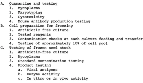
The first step in our contamination control system is the preparation and testing of master and working seed stocks (Table III) .
A. Quarantine and Testing
Upon receipt, starter cultures or cells are placed in quaran- tine. All culture manipulation's are carried out in a laminar flow biological safety cabinet in a laboratory isolated both phys- ically and by air balance from the remainder of the production area. Portions of the cells and culture supernatants from the starter cultures are submitted for mycoplasma testing, karyotyp- ing and fluorescent antibody staining, In the case of cells which are murine in origin, a culture is submitted for mouse antibody
580 R O G E R W . J O H N S O N et al.
A. Quarantine and testing 1. Mycoplasma
2. Karyotyping 3. Cytotoxicity
4. Mouse antibody production testing B. Cell preparation for freezing
1. Antibiotic free culture 2. Tested reagents
3. Contamination checks at each culture feeding and transfer 4. Testing of approximately 10% of cell pool
C Testing of frozen seed stock 1. Antibiotic-free culture 2. Mycoplasma
3. Standard contamination testing 4. Product testing
a. Viral antigens b. Enzyme activity
c. In vitro or in vivo activity
production testing (Collins and Parker, 1972) to assure freedom from a number of murine virus contaminants.
When the described testing indicates that the cultures are free of mycoplasma and are of the appropriate karyotype, the cells are amplified in antibiotic-free, pretested medium to yield enough cells to make a frozen master seed stock. The fluids removed from these cultures, when they are transferred or when intermedi- ately fed fresh medium are filtered thru 0.22 μ membrane filters, and the filters incubated on blood agar at 37 C. Portions of the cells at transfer are tested on blood agar and in broths at both 37C and room temperature. When the final pool of cells is pre- pared in the cryoprotective medium for ampouling, 10% is set a- side for conventional bacteria sterility testing. Duplicate broth cultures of brain heart infusion, tryptose phosphate, trypticase soy, yeast and mold, and sabouraud dextrose, along with blood agar plates are inoculated and incubated at 37C and room tempera^
ture. Test cultures are held for 21 days before the sterility testing is terminated.
TABLE IXX Cell Seed S t o c k Development and Testing
Twentyv*four to 48 hours after the cell seed stock is frozen, three ampoules are recovered in antibiotic^free medium. Before confluency, the cells are scraped into the medium and the material submitted for mycoplasma and bacterial contamination testing and karyotyping,
An ampoule of these cells is then expanded to prepare a work- ing seed stock in the same manner and with the same testing meth- ods used to prepare the master seed stock. When this testing is completed, an ampoule of the working seed stock is scaled up to production level and the purified and concentrated virus product
is tested by immunological and biochemical methods to assure free- dom from contamination with other C-type viruses.
VII. TISSUE CULTURE REAGENT TESTING
A. Medium
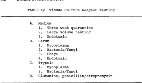
Medium (Benton et al., 1975} is generally produced in 200 1 lots without glutamine. Each lot of medium (Table IV) is held in quarantine at room temperature for 3 weeks prior to transfer to storage at 4C. During the course of filtration, 500-ml samples are taken for additional sterility testing. These random samples are held at room temperature for 7 days and then submitted for sterility testing at 37C and room temperature using five broth media and blood agar plates. These sterility tests are held for 21 days. Medium is usually released for use 28 days after its preparation.
Medium is not routinely submitted for endotoxin testing.
However, it has been our experience that medium found to contain endotoxin fails to support cell growth.
Â. Serum
Prior to purchase of a serum lot, two 500^nl samples are ob- tained from the manufacturer. These samples are tested (Table IV)
582 ROGER W. JOHNSON et al.
TABLE IV Tissue Culture Reagent Testing
A. Medium
1, Three week quarantine 2 , Large volume testing 3. Endotoxin
B. Serum
1. Mycoplasma 2. Bacteria/fungi 3. Phage
4. Endotoxin C. Trypsin
1. Mycoplasma 2. Bacteria/fungi
D. Glutamine, penicillin/streptomycin
for mycoplasma, bacteria, fungi, and bacteriophage. Several sam- ples have recently been tested for endotoxin. Lots of serum have been rejected due to contamination with one or more of each con- taminant mentioned with the exception of bacterial contamination
(Benton et al., 1975).
C. Trypsin
Trypsin is tested for mycoplasma and bacterial and fungal con- tamination when it is dispensed from the manufacturer's bottle in- to smaller aliquots and refrozen (Table IV).
D. Glutamine and Penicillin/Streptomycin
Glutamine and penicillin/streptomycin solutions are frozen as soon as they are filtered and random samples are submitted for bacteriological testing (Table IV). Glutamine is tested by in- oculation of duplicate sets of broths and blood agar plates.
Penicillin/streptomycin solution is filtered and the filters are placed in bottles of broths and on blood agar plates.
VIII. Contamination Monitoring During Scale-up, Production, and Final Product Testing
A. Scale-Up
Tissue culture media (Table VI used for scale up are incubated 72 hr at 37C prior to addition of serum, glutamine, and penicil- lin/streptomycin. Complete media are then held at room tempera- ture until used. As previously described, culture supernatants, at the time of transfer or at intermediate feedings are pooled and 100-ml amounts filtered. The filters are put on blood agar plates and incubated. Cell pools, obtained when transfers are made, are plated in blood agar and inoculated into trypcase soy broth. Portions of the complete cell culture medium are also in- cubated at 37C.
As the scale up cultures increase in number, the lot is di- vided into groups of cultures and sterility checks are made on these groups rather than individual cultures.
Cells used in the final scale-up step are submitted for myco- plasma testing. This allows for retrospective testing on the production lot.
B. Production Cultures
Prior to harvesting and refeeding of production level cul- tures, each bottle is examined before it is processed. Before the harvested culture fluids are clarified of cellular debris, a 500-ml sample of the pool of unclarified fluid is obtained for contamination testing. After centrifugation, 500-ml samples are obtained from each of several carboys containing the clarified material. These samples are filtered and the filters incubated on blood agar plates. Before the clarification step, the centri-
fuge hose lines and rotor are flushed with 20 1 of sterile water.
Five hundredritil amounts of the water are obtained both before and after flushing and are filtered and incubated to determine that the water, rotor and all the hose lines were sterile.
584 ROGER W. JOHNSON et al
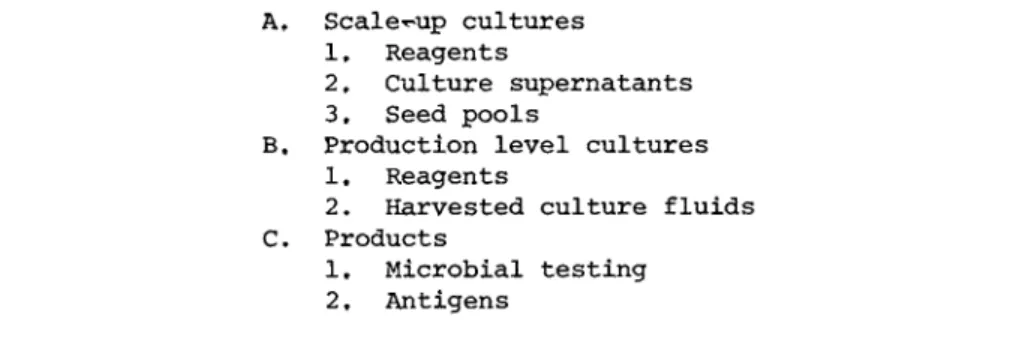
TABLE V Contamination Monitoring
During Scale^up, Production, and on the Final Product
A, Seale-up cultures 1, Reagents
2, Culture supernatants 3. Seed pools
B. Production level cultures 1. Reagents
2. Harvested culture fluids C. Products
1, Microbial testing 2, Antigens
The medium, in 15-1 carboys, used to feed the production cul- tures is incubated at 37C for 3 days before it is used. The incu- bation serves the dual purpose of enhancing growth of microbial agents to a visible level and warming the medium to 37C prior to use. Samples of the medium and the medium plus the individual constituents, i.e., glutamine, serum and penicillin/streptomycin are obtained in 500-ml amounts and incubated at 37C (Table V ) .
If the sterility results indicate freedom from microbial con- tamination, the harvested tissue culture fluids are then processed by continuous flow zonal centrifugation to concentrate and purify the virus. The final viral product is tested for freedom from bacterial and fungal contaminants, for antigen content, biochemi- cal and immunological characterization, and in vitro and, in some cases, in vivo bioassays.
The production level represents the highest level of monetary investment in the production process. Each harvest averages around 50 1 of material at a cost of several hundreds of dollars, exclusive of the cost of the glassware preparation.
The procedures outlined above include greater than 100 tests on each production lot. Although cumbersome, these procedures have precluded the use of contaminated reagents, prevented the
further processing of contaminated material and provided us with clues as to the probable sources of contamination,
ACKNOWLEDGMENTS
We express our appreciation to Drs, J, Gruber and H. J, Hearn, Jr. of the National Cancer Institute, for their continued support of this program, and to Dr. L. Herman, Environmental Services, National Institutes of Health, for his help and suggestions in en- viornmental contamination control and monitoring. We wish to ack- nowledge Dr. W. Nelson-Rees for the karyology of all our cells and Dr. C. Stulberg for the fluorescent antibody staining of them.
We are indebted to Mr. 0. R. Robinson, Jr. for his valuable sug- gestions on testing and to Mr. W. I. Jones and Mrs. R. Herring for reagent and product microbial contamination testing. We also wish to acknowledge the Frederick Cancer Research Center, Departments of Virus Production and Virus Purification staffs for their tech- nical assistance.
Research was sponsored by the National Cancer Institute under Public Health Service Contract NOl-CO-25423 MOD 27 with Litton Bionetics, Inc.
REFERENCES
Barile, M. F. (1973). In "Tissue Culture Methods and Applications"
(P. F. Kruse, Jr. and M. K. Patterson, Jr., eds.), pp. 729-735.
Academic Press, New York.
Barile, M. F. (1975). In this volume.
586 ROGER W. JOHNSON et ai
Benton, C. V., Johnson, R. W., Perry, Á., Jones, W. I. and Shibley, G. P. (1975). In this volume.
Boone, C. W. , Mantel, H., Caruso, T. D,, Jr., Kazam, E. and Stev-^
enson, R. E. 0-972). m Vitro 7, 174-189.
Chu, F. C., Johnson, J. B,, Orr, H. C., Probst, P. G. and Petric- ciani, J. C. C1973Î. m Vitro 9, 31-34.
Collins, M. J., Jr. and Parker, J. C 0-972), J, Nat, Cancer Inst, 49, 1139-1143.
Crawford, Y. E. and Kraybill, W. H., 0-967). Ann. N.Y. Acad. Sei.
143, 411-421.
Federoff, S., Evans, V. J., Hopps, H. E., Sanford, Ê. I. and Boone, C. W. 0-972). In Vitro 7, 161-167.
Giradi, A. J., Hamparian, V. V., Somerson, N, L, and Hayflick, L.
0-965). Proc. Soc. Exptl. Biol. Med. 120, 760-771.
Hayflick, L. (1965). Texas Reports. Biol. Med. 23, 285-303.
Jorgensen, J. H, and Smith, R. F. (1973). Appl. Microbiol. 26, 43-48.
Low, J. E. (1974). Appl. Microbiol. 27, 1046-1052.
Macpherson, I. (1966). J. Cell Sei. 1, 145-168.
Merrill, C. R., Friedman, T. B., Attalluh, A. F. M., Greier, M.
R., Krell, K. and Yarkin, R. (1972). m Vitro 8, 91-93.
Molander, C. W., Kniazeff, A. J., Boone, C. W., Paley, A. and Imagawa, D. T. (1972). In Vitro 7, 168-173.
Moody, E. E. M., Trousdale, M. D., Jargensen, J. H. and Shelokov, A. (1975). J. Inf. Dis. 131, 588-591.
Nelson-Rees, W. Á., Flandermeyer, R. A. and Hawthorne, P. K.
(1974). Science 184, 1093-1096.
O'Connell, R., Witten, R. G. and Faber, J. E. (1964). Appl. Micro- biol. 12, 337-342.
Petricciani, J. C , Chu, F. C. and Johnson, J. B. (1973) „ proc, Soc. Exptl. Biol, Med, 144, 789^792,
Stulberg, C. S., Coriell, L. Lf, Kniazeff, A. J. and Shannon, J.
E. 0-9701, m Vitro 5, 1-16,
Tumilowicz, J, J,, Alexander, A, D, and Stafford, K, 0-9741, In Vitro 10, 238^242,
Voxakis, A. C , Lomy, P. P, and Herman, L, G. C19741. Formula Management 12, 14-18,
Wildfeuer, Ç, Â., Schleifer, Ê, Ç, and Haferkamp, Ο, 0-9741. Appl.
Microbiol, 28, 867-871.