AUTOCLAVABLE LOW COST CELL CULTURE MEDIA
LEONARD KEAY
Department of Microbiology and Immunology Division of Biology and Medical Sciences Washington University School of Medicine
St. Louis, Missouri
I. INTRODUCTION
For the last twenty-five years animal cells have been grown in chemically-defined media supplemented with some type of serum.
These chemically-defined media consist of inorganic salts, vita- mins, amino acids and sometimes other organic compounds (Morgan et al., 1950; Eagle 1955a; Healy et al., 1954; Waymouth 1955; Ham 1963). Even in these media the term chemically-defined (that is knowing all of the components present and their precise amounts) may be questioned since the purity of many components is generally not completely established.
The supplementation of media with serum creates several prob- lems (Fedoroff et al., 1972). The most obvious is economic. The supplementation of medium with 10% serum accounts for 75-90% of the material costs (based on the cost of dry powder media).
513
514 LEONARD ΚΕÁ Y
However, more recently, serum has been implicated as a source of mycoplasmal and viral contamination of cell cultures (Molander et al,f 1972; Hopps, 1974}, The final reason is the variation between various samples of sera (Boone et al.f 1972} and the de- sirability of a totally chemically-defined medium.
The precise role(s) of serum is unknown. It may be a source of hormones, macromolecules required for protecting cell surfaces, free fatty acids, sterols, trace minerals or vitamins, and at- tempts to replace serum usually involve substitutions for these possible serum components (Higuchi, 1973; Taylor, 1974).
The problem of mycoplasma and virus contamination is the reason for seeking an autoclavable medium. Serum or other native protein cannot be autoclaved without coagulation and filtration will not remove mycoplasma or viruses. Therefore any completely autoclavable medium will have to also be serum-free medium.
Other desirable characteristics of any serum-free autoclavable medium are that it be totally chemically-defined, that it support the growth of a wide range of cells, that cells grow in it with- out an extensive period of adaptation, that high cell yields be obtained without repeated refeeding, and that the cells remain unchanged in karyotype, cell surface antigens, virus susceptibili- ty, etc., when grown in such a medium over an extended period of time.
Since the pioneer work of Eagle on the amino acid and vitamin requirements of HeLa and L-cells (Eagle 1955b and c) leading to the formulations of BME and MEM, few detailed nutritional studies have been carried out, although many formulations have been pub- lished.
Serum-free formulations ML 192/2 and MB 752/1 in which bovine serum albumin and bactopeptone were used as supplements to a salts/amino acids/vitamins medium were developed by Waymouth
(1956, 1959). The more complex chemically-defined medium NCTC109 supported the growth of adapted L-cells (NCTC 2071) in stationary systems but not in agitated systems. Cell disintegration rapidly
LOW COST CELL CULTURE MEDIA 515 occurred unless methylcellulose was added (Bryant et al.r 1961).
Macromolecules such, as methylcellulose, polyvinylpyrrolidone, protamine sulfate and serum albumin have been added to various serum-free media, although it has been shown that exhaustively dialyzed serum does not support cell growth, i.e. it is more than just "macromolecular."
The nutritional requirements for cell growth have been found to be dependent on the density of the cell inoculum and even in the case of the complex medium NCTC109, cell growth does not oc- cur in the absence of serum if the inoculum is too small (Bryant et al., 1961; Fioramonte et al., 1958; Eagle and Piez, 1962). A complex chemically defined medium supporting the growth of L-cells was also developed by Healy et al. (1955). At the other extreme, attempts were made to cultivate HeLa and L-cells in systems con- taining bacteriological media (Mayyasi and Schuurmans, 1956; Gins- berg et al., 1955), but the addition of serum was necessary and the growth and quality of the cells rather poor.
Other additives formulated into serum-free systems include un- saturated fatty acids, putrescine, insulin, thyroxine, pyruvate, ethanol, gluconolactone, and nucleotide precursors. Most serum- free media contain high levels of amino acids (both essential and nonessential), usually 3-10 fold higher than in MEM. Also, the vitamin levels are higher especially inositol and choline and oth- er vitamins such as vitamin B1 2' biotin and lipoic acid often in- cluded (Ham, 1965; Nagle et al., 1963; Higuchi, 1963; Tribble and Higuchi, 1963; Blaker et al., 1961; Lieberman and Ove, 1959;
Higuchi and Robinson, 1973; Lasfargues et al., 1973; Morrison and Jenkin, 1972; Birch and Pirt, 1970; Takaoka and Katsuka, 1971;
Holmes and Wolfe, 1961).
The role of insulin is uncertain, but it has been shown (Temin, 1967) that the growth of chick embryo fibroblasts is stim- ulated by insulin in serum-free systems and low molecular weight fractions have been isolated from serum which have this insulin- like activity but are not suppressed by antiinsulin sera (Pierson
516 LEONARD ΚΕÁ Y
and Temin, 1972]. Insulin has been shown to markedly stimulate the growth of HeLa cells in serum-free systems, as have vitamin B1 2 and biotin (Higuchi and Robinson, 1973; Blaker et al., 1971).
Bactopeptone has been used as part of several serum-free media (Waymouth, 1956, 1959), either alone or in conjunction with other additives such as lactalbumin hydrolysate, polyvinylpyrrolidone, and yeast extract (Lasfargues et al., 1973). Bactopeptone pos- sesses the advantages that it is both low cost and autoclavable, but it is not chemically-defined. In fact, probably less is known about the composition of bactopeptone than is known about serum. Recently the growth promoting activity of bactopeptone has been shown to be in the low molecular weight dialysate frac- tion (Taylor et al., 1972b) and some peptide fractions shown to promote cell growth (Taylor et al., 1974; Hsueh and Moskowitz, 1973a, 1973b) although no full characterization of the components have been made. Other peptones including soybean derivatives have been shown to support cell growth (Taylor et al., 1974; Hsueh and Moskowitz, 1973a, 1973b) although no full characterization of the components have been made. Other peptones including soybean derivatives have been shown to support cell growth (Taylor et al., 1972a; Healy and MacMorine, 1972).
Autoclavable media have been less extensively studied than serum-free media. Nagle developed a heat stable medium for the suspension culture of L-cells, HeLa and cat kidney cells (Nagle, 1968). The medium contained high levels of amino acids and after autoclaving, the medium was completed by the addition of sodium bicarbonate, methyl cellulose'and glutamine, each autoclaved sep- arately (the glutamine autoclaved as a dry powder). This medium was subsequently modified by increasing the level of choline 50-
fold (Nagle, 1969), and then by elimination of the glutamine by replacement with a series of amino acids (Nagle and Brown, 1971).
Yamane et al., 1968 found that autoclaved MEM would support the growth of baby hamster lung and L-cells provided that the autoclaving was carried out at pH 4.0^4.5 (maintained by a sue-
LOW COST CELL CULTURE MEDIA 517 cinate buffer] and that glutamijie and serum were added after autoclaving OTamane et al., 19681.
Pumper et al, 0.965] found that autoclaved bactopeptone or peptone dialysate when combined with autoclaved medium 199 or MEM
Cwithout glutamine] supported a 7 fold increase in rabbit heart myocardial cells in 7 days.
It is interesting to note that only a few of these serum-free or autoclavable media are commercially available. A formulation of Waymouths 752/1, bovine serum albumin and linoleic acid is available in liquid form (HWIO^) and autoclavable BME and MEM are available in dry powder form.
The approach taken in this laboratory is that the most useful medium would be based on a simple medium commercially available in dry powder form which with a minimum of additions or manipula- tions would be autoclavable and support the growth of many common- ly used cell types. The starting point was a simple medium (MEM) plus a supplement (bactopeptone), the latter being chosen because of low cost and autoclavability.
Initial studies were on the growth of commonly used cell lines in this serum-free system, followed by an investigation of the autoclavability of those serum-free systems shown to support cell growth when sterilized by filtration.
Finally some studies will be carried out on the problems of scale up of these systems for cell and virus production.
II. MATERIALS AND METHODS
Dry powdered media and serum were obtained from Microbiologi- cal Associates, Bethesda, Md., and KG Biologicals, Lenexa, Kansas.
Bactopeptone was obtained from Difco Laboratories. Inorganic salts (ACS Reagent grade] were obtained from Fisher Scientific Co., St. Louis, Mo, Darvan #2, a polyalkylbenzene sulfonate deter- gent, was obtained from R. T. Vanderbilt Co,, New York, Ν. Õ.,
518 LEONARD KEAY
and other additives and organic compounds were obtained from Sigma Chemical Co., St, Louis, Mo. Unless otherwise stated, all solutions were sterilized by membrane filtration.
Growth studies in monolayer or stationary suspension cultures were generally carried out in 75 cm2 plastic flasks containing 25 ml medium and incubated at 37° in a 5% C02/95% air atmosphere.
Agitated suspension cultures were carried out at 37° in glass spinners with teflon coated magnetic stirring bars. Cell counts were determined microscopically with a hemocytometer (after tryp- sinization in the case of monolayers) and cell viability deter- mined by the trypan blue dye exclusion method. Media and serum
substitutes were autoclaved at 10 psi for 10 minutes. Bactopep- tone was prepared as a 20% w/v solution and generally 5% v/v added to the medium to give a final concentration of 1% w/v. SSK7
(Lasfargues, 1973) contained bactopeptone 5%, PVP-360 3.5%, lac- talbumin hydrolysate 2.5%, yeastolate 0.5%, glucose 0.5%. Insu- lin was generally prepared as a 1 mg/ml solution in phosphate- buffered saline.
III. RESULTS AND DISCUSSION
A. The Growth of Established Cell Lines in Serum Free Media Initial experiments were carried out with L-cells (L-929 and a derivative L-60TM which grows more readily in suspension) and then extended to other established cell lines.
The results obtained with L-cells and BHK have been published (Keay, 1975) and will therefore only be summarized here.
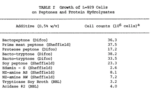
A number of peptones and protein hydrolysates were examined for their ability to support the growth of L-cells when they were added to supplement the simple MEM medium, The results (see Table I) show that a number of peptones support the growth of L-929 cells, whereas the more complete hydrolysates do not support cell growth. In fact the addition of hydrolysates reduces the growth
LOW COST CELL CULTURE MEDIA 519
Additive C0f5% w/v) Cell counts CIO6 cells)a
Bactopeptone CDifco) 36,3
Prime meat peptone CSheffield! 37.5
Proteose peptone CDifco) 17.2
Bacto-tryptone CDifco) 38.2
Bacto-tryptose CDifco) 33.5
Soy peptone CSheffieId) 23.3
Edamin - S CSheffield) 2.6
NZ-amine AS (Sheffield) 8.1
NZ-amine AW (Sheffield) 7.2
Trypticase Soy Broth (BBL) 3.2
Acidase #2 (BBL) 4.0
aInitial cell count was 1.15 x 1 06.
stimulating effect of the peptones. The addition of 0.5% lactal- bumin hydrolysate reduces the growth on bactopeptone by more than
50%. It was observed that as cell growth continued more of the cells detached and grew in suspension until the whole system con- sisted of bunches of floating cells. Although this may be a dis- advantage if a monolayer system is necessary, it is an advantage if a suspension culture is desired. It was found that the addi- tion of insulin or oleate had no effect on the growth of L-cells in MEM supplemented with 0.5% bactopeptone.
Using 0.5% bactopeptone as a supplement to Joklik-modified MEM, excellent growth was obtained L-60TM cells in spinner cul- ture.
The L-cells grew at once in this simple medium without any period of adaptation, and have been cultured in this serum-free system for several months at a time.
When attempts were made to culture BHK cells using bactopep- tone as a serum substitute two facts were quickly noted; the cells did not grow well on MEM or Joklik-modified MEM, but grew
TABLE I Growth, of L^929 Cells on Peptones and Protein Hydrolysates
520 LEONARD KEAY
1 06 cells/75 cm2 flask
Medium Expt Aa Expt Bb
MEM, 10% FCS 19.0
MEM, 1%BP 18.0
MEM, 1%BP, insulin0
-
12.2MEM-á 10%FCS 27.0
MEM-a insulin0 4.2
-
MEM-a 1%BP 24.0 23.0
MEM-a 1%BP, insulin0 19.0 17.5
aIn experiment A, monolayers were refed after 5 days, counted at 8 days.
bIn experiment B, monolayers were refed after 3 and 6 days, counted at 9 days.
°Insulin concentration 0.23 I.U./ml.
better on richer media such as MEM-alpha (without the ribosides and deoxyribosides. Stammers et al., 1971), F-12, or RPMI 1640;
and the cells grew as rather nodular clumps some of which detached from the surface and grew in suspension. The addition of a poly- alkylbenzene sulfonate detergent Darvan #2 (300 yg/ml) was suf- ficient to convert the system to a completely suspension culture even in stationary systems, although clumping still occured es- pecially in the MEM-alpha and RPMI 1640 systems. Monolayer sys- tems of BHK cells could be produced but were difficult to maintain because of subsequent cell detachment. It was found that growth of BHK cells on F-12 supplemented with 0.5% bactopeptone was unaf- fected by the addition of insulin.
The methods and media developed with the L and BHK cells were used in studies on the growth of five other cell lines (CHO, 3T3, BSC-1, HeLa and KB) (Keay, 1976).
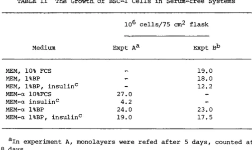
In the case of BSC-1 cells Can established African Green Mon- key Kidney Line) it was found that the cells would grow with bac-
TABLE II The Growth of BSC-1 Cells in Serum-free Systems
L O W COST CELL CULTURE MEDIA 521 TABLE III The Growth, of CHO Cells in Serum^free Mediae
Cells/ml
MEM,BP MEM-á, BP, MEM, BP insulin*3 MEM-a, BP insulin*3
Day 4 230,000 275,000 145,000 200,000 Day 5 185,000 365,000 180,000 340,000 Day 6 165,000 850,000 135,000 675,000 Day 7 120,000 930,000 100,000 810,000
aThe initial cell density was 50,000 cells/ml.
bThe insulin concentration was 0.11 I.U./ml.
topeptone (1%) as a serum substitute (see Table II). The cells remained an anchorage dependent line with very few detached cells observed. The confluent monolayers appeared to be healthier when MEM-alpha was used than when MEM was used, although the cell num- bers are not too different. At confluency the monolayers devel- oped the whorls observed in confluent monlayers grown in MEM + 10%
FCS. The addition of insulin appears to be unnecessary for cell growth. It was found that BSC-1 cells grown to confluency with bactopeptone as a serum substitute could be infected with SV40 and that the yield of virus produced was about the same as in sys- tems containing serum.
Chinese Hamster Ovary cells (CHO) were found to grow with bactopeptone (1%) as a serum substitute in MEM or MEM-á provided that insulin was added (see Table III). The optimal level of in- sulin has not been determined, but little difference was observed between 0.11 and 0.56 I.U./ml. It was observed that whereas with MEM-á + 10% FCS the CHO cells grew as a monolayer until close to confluency, when bactopeptone was used as a serum substitute the cells grew in stationary suspension in grape-like clusters. It was observed that the cell viability dropped rapidly as the cells
522 LEONARD ΚΕÁ Y
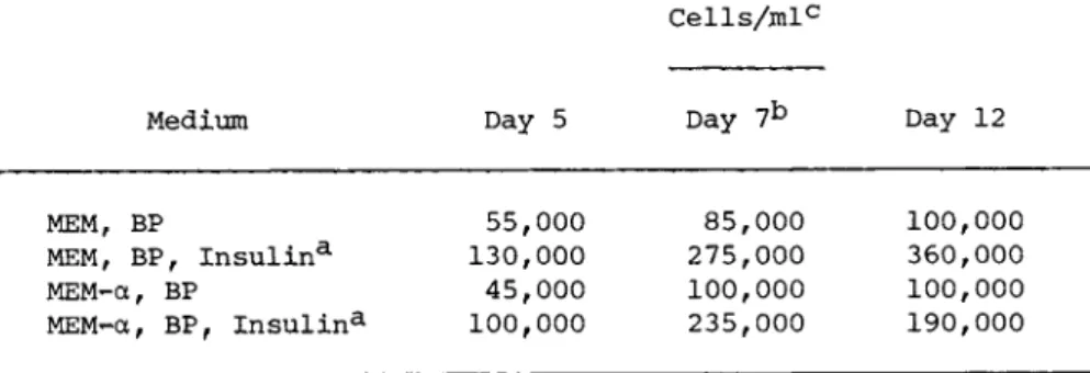
TABLE IV The Growth of Balb/c 3T3 Cells in Serum-free Systems
Cells/mlc
Medium Day 5 Day 7b Day 12
MEM, BP
MEM, BP, Insulina MEM-á, BP
MEM-a, BP, Insulin3-
55,000 130,000 45,000 100,000
85,000 275,000 100,000 235,000
100,000 360,000 100,000 190,000
aInsulin concentration was 0.11 I.U./ml.
bCells centrifuged down and refed with fresh medium.
cThe initial cell density was 25,000 cells/ml.
multiplied to about 1 06 cells/ml and immediate subculturing by centrifugation and resuspension in fresh medium was necessary.
The CHO cells were cultured in this manner in the system MEM-á + BP + insulin for 3 months after which they were recultured in MEM- á + 10% FCS whereupon they immediately resumed the monolayer growth and previous morphology.
Balb/c 3T3 cells are a mouse line which has only previously grown as a contact-inhibited monolayer. However, when the 3T3 cells were subcultured into MEM-alpha supplemented with 1% bacto- peptone and insulin it was observed that although most of the cells attached initially, they subsequently dissociated and grew in stationary suspension in grape-like clusters. The clumps grew quite large and were difficult to break up for cell counting which with the initial attachment made quantitative growth data less easy to obtain. This tendency to attach initially was only ob- served when new plastic flasks were used but not when used flasks were reused. Table IV shows the effect of insulin on the growth of 3T3 cells in the serum-free systems. Little difference was ob- served between growth in MEM and ÌÅÌ-á. 3T3 cells were grown in a spinner bottle in MEM-á, bactopeptone and insulin, polyvinylpyr-
LOW COST CELL CULTURE MEDIA 523 TABLE V The Effect of Medium on the Growth,
of HeLa, Cells in the Presence of Bactopeptone3- and Insulin*5
Cells/ml
Day 0 Day 2 Day 4
MEM 200,000 400,000 820,000
1st 200,000 405,000 930,000
Expt. MEM-alpha 200,000 700,000 1,100,000 200,000 500,000 1,150,000 Day 0 Day 3 Day 4 Day 5 Day 6
MEM 100,000 310,000 390,000 600,000 660,000 2nd 100,000 360,000 460,000 600,000 730,000 Expt. MEM-alpha 100,000 480,000 880,000 1,060,000 1,120,000
100,000 510,000 750,000 1,010,000 900,000
aThe bactopeptone supplement was 5% of a 20% solution.
bThe insulin level was 0.06 I.U./ml.
rolidone being added as a possible macromolecular protective agent. In a 100 ml spinner, the cell density rose from 1 χ 10^
to 4.8 x 1 05 in 3 days. However, sometimes the cell density pla- teaued at 3 x 1 05 cells/ml and the system does not appear to have any practical value.
HeLa cells have been grown in several serum-free systems and it was found that they grew readily in the system MEM-alpha plus bactopeptone (1%) provided that insulin is added. The cells grew as a stationary suspension. The results shown in Table V show that growth in MEM-á was considerably better than in MEM. This is probably because the MEM-á contains the vitamin B1 2 and biotin which have been shown to be necessary for the growth of HeLa cells. There was little cell growth unless insulin was added, but as little as 0.011 I.U./ml insulin had a pronounced effect on cell growth.
524 LEONARD KEAY
Medium Day 4 Day 7 Day 10
MEMa, BP 80,000 50, 000
-
MEMOC, BP, 0.01 I.U,/ 140,000 440, 000 746,000 ml insulin
MEMa, BP, 0.06 I.U./ 150,000 370, 000 755,000 ml insulin
MEMa, BP, 0.23 I.U./ 110,000 300, 000 620,000 ml insulin
MEM, BP, 0.23 I.U./ 150,000 270, 000 200,000 ml insulin
MEM, BP, 0.23 I.U./ 190,000 360, 000 592,000 ml insulin, B^2,
biotin
aInitial cell density was 70,000 cells/ml. Bactopeptone concen- tration was 1% w/v.
HeLa cells grew well in agitated suspension in the system MEM-á, bactopeptone, insulin and polyvinylpyrrolidone the density rising from 2 x 1 05 to 1.4 x 1 06 in 5 days.
Results similar to those obtained with HeLa cells were ob- tained with KB cells. Insulin was required for cell growth, and apparently due to its vitamin B1 2 a nd Biotin content, MEM-á was much more effective than MEM when bactopeptone was used as a serum
substitute (see Table VI). The cells grew as a stationary sus- pension in large grape-like clusters.
HeLa and KB cells were grown in spinner cultures in MEM-a, bactopeptone, PVP and insulin and used to prepare adenovirus.
The yield and quality of the virus produced was the same as that produced in Joklik-MEM supplemented with horse serum.
TABLE VI The Growth, of KB Cells in Serum-free Systems3-
LOW COST CELL CULTURE MEDIA 525
B. Growth, of Primary and Normal Cells in Serum-Free Media Much of the work on cell nutritional requirements has been criticized because it has all been carried out with HeLa or L- cells (Ham, 1974). Several of the serum-free media developed support the growth of a wide range of established cell lines, but not primary or normal cells (Higuchi and Robinson, 1973; Lasfar- gues et al., 1973).
Recently some progress has been made on extending our work to primary and secondary chick embryo fibroblasts. Chick embryo pri- mary cells or secondary cells (grown in MEM + 3 % FCS) (Keay and Schlesinger, 1974) were centrifugea and washed with PBS to remove residual fetal calf serum and trypsin, and resuspended in PBS for dilution into selected media. The results (Table VII) show that the complex serum substitute SSK7 (3% v/v) (Lasfargues, 1973) or the simpler bactopeptone, lactalbumin hydrolysate, polyvinylpyr- rolidone combination (3% v/v) (Keay, 1975) promote cell growth as well as does fetal calf serum provided that insulin is also added.
Insulin alone does not promote cell growth in this simple MEM sys- tem.
No attempts have yet been made to continuously culture the chick embryo cells in this serum-free system or to extend the method to other diploid or normal cells.
C. Autoclavable Low Cost Cell Culture Media
As described above, it is clear that many established cell lines will grow continuously on simple commercially available media supplemented with an autoclaved bactopeptone solution (or a more complex combination), insulin having to be added in some cases.
The question is whether an autoclavable medium can be devel- oped from this system. Previously Yamane CI968Î has grown cells on MEM autoclaved at low pHs provided glutamine is added after autoclaving. The autoclaving of glutamine and insulin represent
00 Η Ο Ο (Í . . . é é . . Ο CO CO CO <tf
co CN ^
ó> co I t · I · I ·
<sT CM LD
ï ó>. . . é . é . . . H ï ï rH CM VD Ο LH CO Φ
• é . . . é é co oo ^ m
ï CM vo
CO ^ O CN Ο M h ^ Φ
• H rH Ñ
g: g i
Ë C M
I t 1-3 t-3
•Η
W C
- H
s s" S S S S S* C S S
526
LOW COST CELL CULTURE MEDIA 527 specific problems. It was originally claimed that glutamine was a requirement for the groth of L cells and HeLa cells, but sub- sequently several glutamine^free media have been devised. Of course one problem is that when undefined additives such as serum or peptones are used, it is not clear whether glutamine or a glu- taminine-substitute is being added. It is interesting to note that the glutamine-free media contain pyruvate without which cell growth is reduced.
Initial experiments were carried out with L-60TM cells in stationary suspension in plastic flasks. The cells had been grow- ing in MEM supplemented with autoclaved bactopeptone solution
(final concentration of bactopeptone 1%). The medium used was AutoPowR MEM (Flow Laboratories) and autoclaving was carried out at 10 psi for 10 min with and without prior addition of solid bactopeptone and with and without prior neutralization with sodium bicarbonate. Glutamine was added to all media after autoclaving.
The results (see Table VIII) show clearly that AutopowR MEM and bactopeptone do not support the growth of L-60TM cells as well when autoclaved together as when autoclaved separately and then combined. Further experiments were carried out with regular MEM
(i.e., with glutamine and without succinate buffer). After neu- tralization with sodium bicarbonate, the medium was autoclaved at 10 psi for 10 min, cooled, the insoluble material solubilized by acidification with HCl, followed by immediate reneutralization with NaOH solution. Bactopeptone (20% w/v, autoclaved separately) was added to give a final concentration of 1% w/v, and glutamine
(292 mg/1) was also added to compensate for any loss of glutamine during autoclaving.
L-60TM cells have been grown in stationary suspension in this system for several months by subculturing several times weekly by centrifugation. Preliminary experiments show that BHK cells will grow in autoclaved F-12 medium supplemented with bactopeptone and Darvan and this is being investigated further.
rH
CO
ï
-Ρ co
U ù
>1 Ο rd
>1
Ù
>1
Q rd
Q fd
- H c
>
co rd
73 fd -P ö MH fd
Cn
•H 4-> fd fd
>
ïrH •
a û CM
auto
• H 73 ö
0>] Ο H lD h- rH CN CO CN Ol
Ο Γ- 00 Ο CN
^ CN CT» CO 00
i n (Í o c o h
^ ^ σ » o o c o
ï co m m CN CO CN CN CN
ß β rH rH
CT» cn β ß - rH rH CM Pu
cn CT. m
528
LOW COST CELL CULTURE MEDIA 529 How does the above system compare with the ideal medium? It meets essentially all of the requirements except one, namely that it is not chemically defined. The problem of chemical definition will probably be solved when the precise composition and roles of both serum and bactopeptone can be determined and both replaced by their purified growth promoting components.
It has not yet been established how long various types of cells will continue to proliferate in these autoclaved media or what changes may occur in the cells, or what problems may be en- countered on scale up. The economics of these systems cannot yet be compared with serum-containing systems since cell yields and growth rates have not yet been accurately determined. It is clear however that low cost formulations based upon commercially avail- able dry powder media supplemented with bactopeptone will support the growth of many cell lines and if the. conditions are carefully selected can be converted to totally autoclavable systems for cell and virus production.
ACKNOWLEDGMENTS
The dedicated technical assistance of Mr. Fred Anderson is gratefully acknowledged. This work was supported by the Human Cell Biology Program of the National Science Foundation (Grants
#GB 38657 and BMS 73-07013-A01).
REFERENCES
Birch, J. R. and Pirt, S. J. (1970) J. Cell Sc. 7, 661-670.
Blaker, G. J., Birch, J. R. and Pirt, S. J. (1971). J. Cell Sc. 9, 529-537.
Boone, C. W., Mantel, Ν., Caruso, T. D., Kazam, Å., and Stevenson, R. E. (1972). In Vitro 7, 174-189.
530 LEONARD KEAY
Bryant, J. C., Evans, V. J., Schilling, E. L., and Earle, W. R.
(1961). J. Natl. Cancer Inst. 26, 239-252.
Eagle, H. (1955a). Science 122, 43-46.
Eagle, H. (1955b), J. Expt. Med. 102, 37-48.
Eagle, H. (1955c). J. Expt. Med. 102, 595-600.
Eagle, H., and Piez, K. (1962); J. Expt. Med. 116, 29-43.
Fedoroff, S., Evans, V. J., Hopps, H. E., Sanford, K. K. and Boone, C. W. (1972). In Vitro 7, 161-167.
Fioramonte, M. C., Evans, V. J. and Earle, W. R. (1958). J. Natl.
Cancer Inst. 21, 579-583.
Ginsberg, H. S., Gold, E. and Jordan, W. S. (1955). Proc. Soc.
Expt, Biol. Med. 89, 66-71.
Ham, R. G. C1963). Expt, Cell Res, 29, 515-526.
Ham, R. G. C1968). Proc. Nat. Acad, Sei, 53, 288-293.
Ham, R. G. C1974). m Vitro 10, 119-129.
Healy, G, M. and MacMorine, Hf G, C19721, Progress in Immunobiol.
45, 202ô*208.
Healy, G. M., Fisher, D. C, and Parker, C. C1954) . Canad, J.
Biochem. Physiol, 32, 327-337.
Healy, G. M., Fisher, D. C. and Parker, C. (1955). Proc, Soc.
Expt, Biol. Med. 89, 71-77.
Higuchi, K. (1963). J. Infect. Dis. 112, 213-220.
Higuchi, K. (1970). J. Cell Physiol. 75, 65-72.
Higuchi, M. (1973). Adv. Appl. Microbiol. 16, 111-136.
Higuchi, K. and Robinson, R. C. (1973). Jn Vitro, 9, 114-121.
Holmes, R. and Wolfe, S. W. (1961). J. Biophys. Biochem. Cytol., 10, 389-401.
Hopps, H. E. (1974). Jn Vitro 10, 243-246.
Hsueh, H. W. and Moskowitz, M. (1973a). Expt. Cell Res. 77, 376- 382.
Hsueh, H. W. and Moskowitz, M. (1973b). Expt. Cell Res. 77, 383- 390.
Keay, L. (1975) Biotechnol. Bioeng. 17, 745-764.
Keay, L. (1976) Biotechnol. Bioeng. 18, 363-382.
LOW COST CELL CULTURE MEDIA 531
Keay, L. and Schlesinger, S. (1974). Biotechnol. Bioeng. 16, 1025-1044.
Lasfargues, E. Y., Continho, W. G., Lasfargues, J. C. and Moore, D. H. (1973). In Vitro 8, 494-500.
Lieberman, I. and Ove, P. (1959). J. Biol. Chem. 234, 2754-2758.
Mayyasi, S. A. and Schuurmans, D. M. (1956). Proc. Soc. Expt.
Biol. Med. 93, 207-210.
Molander, C. W., Kniazeff, A. J., Boone, C. W., Paley, A. and Imagawa, D. T. (1972). In Vitro 7, 168-173.
Morgan, J. F., Morton, H. J. and Parker, R. C. (1950). Proc. Soc.
Expt. Biol. Med. 73, 1-8.
Morrison, S. J. and Jenkin, Hf M. (1972). In Vitro 9, 94^100.
Nagle, S, C. (19681. ¢ññß. Microbiol, 16r 53^55, Nagle, S. C. C1969). Appl. Microbiol. 17, 318-319.
Nagle,. S, C, and Brown, B. L. (1971). J. Cell Physiol. 77, 259- 264.
Nagle, S. C., Tribble, H. R., Anderson, R. E. and Gary, N. D.
(1963). Proc. Soc, Expt. Biol. Med. 112, 340-344.
Pierson, R. W. and Temin, H. M. (1972). J. Cell Physiol. 79, 319- 330.
Pumper, R. W., Yamashiroya, H. M. and Molander, L. T. (1965).
Nature 207, 662-663.
Stammers, C. P., Elicieri, G. L. and Green, H. (1971). Nature New Biol. 230, 52-53.
Takaoka, T. and Katsuta, H. (1971). Expt. Cell Res. 67, 295-304.
Taylor, W. G. (1974). J. Natl. Cancer Inst. 53, 1449-1457.
Taylor, W. G., Dworkin, R. Á., Pumper, R. W. and Evans, V. J.
(1972a) Expt. Cell Res. 74, 275-279.
Taylor, W. G., Taylor, M. J., Lewis, N. T. and Pumper, R. W.
(1972b). Proc Soc. Expt. Biol. Med. 139, 96-99.
Taylor, W. G., Evans, V. J. and Pumper, R. W. (1974). In Vitro 9, 278-286.
Temin, H. M. (1967). J. Cell Physiol. 69, 377-384.
532 LEONARD KEAY
Tribble, H. R. and Higuchi, K. (1963). J. Infect. Dis. 112, 221- 225.
Waymouth, C. (1955). Tex Reports Biol. Med. 13, 522-536.
Waymouth, C. (1956). J. Natl. Cancer Inst. 17, 315-325.
Waymouth, C. (1959). J. Natl. Cancer Inst. 22, 1003-1017.
Yamane, I. , Matsuya, Y. and Jimbo, K. (1968). Proc. Soc. Expt.
Biol. Med. 127, 335-336.