IN A LARGE-SCALE VIRUS PRODUCTION LABORATORY
CHARLES V. BENTON ROGER AT. JOHNSON
ALBERT PERRY W, J. JONES GEORGE P. SHIBLEY
National Cancer Institute Frederick Cancer Research Center
Frederick, Maryland
I. INTRODUCTION
In the Viral Resources Laboratory at the Frederick Cancer Research Center, Frederick, Md. (FCRC), several oncogenic murine viruses are produced in large-scale tissue culture. These agents
include (1) Rauscher leukemia virus, produced in JLS-V9 mouse bone marrow cells, (2) Gross leukemia virus, progapated in NIH-3T3 mouse fibroblasts, and (3) mouse mammary tumor virus, grown in Mm5mt/c-L mouse mammary carcinoma cells. Approximately 600 liters per week of tissue culture fluids containing 1 07 - 10^0 virus par- ticles/ml, depending on the system, are produced. This effort in- volves, on a weekly basis, the use of CD 750 1 tissue culture media, C2) Γ75 1 fetal bovine sera, and C3) 2 to 15 1 each of peni-
559
cillin-streptomycin solutions, L-glutamine, phosphate buffered saline and trypsin solutions.
The frequent turnover of such large volumes of tissue culture reagents necessitates an efficient and relatively rapid method of reagent quality assurance testing. Ideally, any system for
achieving quality assurance will strike a balance of time, effort, cost, and value of the results obtained. Little is gained, for example, by enriching a bacteriological medium with expensive or unusual nutrients to detect a contaminating microbe that in most situations will not proliferate during routine tissue culture operations.
The present disscussion has been prepared primarily to sum- marize the results obtained at the FCRC with our current methods of reagent quality assurance. It should be kept in mind, however, that the methods employed are constantly being improved as more data is accumulated and analyzed.
II. SERUM
The cost and the nature of quality control in the processing of serum makes thorough quality assurance testing of each serum lot a necessity prior to its utilization in tissue culture.
A. Mycoplasma
With the use of routine antibiotic incorporation, which we find necessary in a large-scale system, mycoplasma screening re- ceives the major emphasis of our serum quality assurance program.
Data presented at a workshop on serum at the W. Alton Jones Cell Science Center (Fedoroff et al., 1971) indicated that serum con- taminated with bovine mycoplasma is a major source of mycoplasma contamination of tissue culture. The most common species found in bovine serum are Mycoplasma arginini and Acholeplasma laidla- wii CBarile, 1975). Barile C1973) and others (Rodwell, 1969;
Stanbridge et al,, 1969; Markov et al., 1969} have demonstrated that mycoplasma may alter the karyology and metabolism of cultured cells as well as affect the production of virus in certain cell systems.
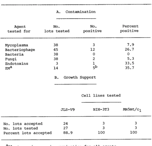
Our testing consists of the inoculation of a total of 25 ml of serum into several types of liquid and solid media: CI) BBL medium (Vera, 1974}, (2) Medium No. 243 (Hayflick, 1965), (3) M96 medium (Frey, 1973), and (4) U.S. Department of Agriculture medi- um. We have found that excluding any one of these media could cause the occasional lack of detection of certain species of strains at low concentrations. Samples are incubated aerobically and anaerobically at 37°C with both positive and negative con- trols. Broth culture material is used as a source of material for retesting and identification of corresponding positive agar plates. Observations are made at 7, 14, and 21 days post-inocu- lation. Results indicate a relatively low incidence of mycoplas- ma in the 38 commercial serum lots that we have screened (Table
I).
Currently, biochemical and physical methods of mycoplasma de- tection are being evaluated to determine the relative sensitivi- ties as compared to the biological culture approach. Schneider et al. (1973) have reported increased sensitivity using the de- tection of 16S and 23S mycoplasma ribosomal RNA (Markov et al., 1969; Harley et al., 1970; Levine et al., 1968) isolated by Poly- acrylamide gel electrophoresis (Grossbach and Weinstein, 1968).
Todaro et al. (1971) demonstrated that these entities could be physically separated based on their characteristic density of 1.22-1.24 g/cm3. Mycoplasma species are also noted for their characteristic cytoplasmic labeling in autoradiographs and for their ability to deplete arginine from tissue culture growth me- dium (Schneider et al., 1973; Barile, 1973).
A, Contamination
Agent No. No, Percent tested for lots tested positive positive
Mycoplasma 38 3 7.9
Bacteriophage 45 12 26.7
Bacteria 38 0 0
Fungi 38 2 5.3
Endotoxins 3 1 33.5
EMa 14 5b 35.7
B. Growth Support
Cell lines tested
JLS-V9 NIH-3T3 MmSmt/c-L
No. lots accepted 24 3 3 No. lots tested 27 3 3 Percent lots accepted 88.9 100 100
aElectron microscopic examination for all agents.
^Positives were: 1 bacteriophage, 1 C-type virus-like, 1 myco- plasma and 2 lots bacteria.
Â. Bacteriophage
The second group of biological contaminants for which serum is routinely tested before purchasing is bacteriophage. Bacterio- phage testing is relatively simple and indicates the existence or former presence of other contaminants, i.e., the homologous host bacterium or bacterial endotoxins. Also, bacteriophage Lambda has been demonstrated to alter the growth of human fibroblasts in tissue culture CMerril et al., 1971).
TABLE I Quality Assurance Testing of Commercial Serum Lots
Briefly, the test employs 50*100 ml of serum concentrated 50- fold by pelleting at 100,000 x g for 4 hr. This concentrate is mixed with 2 ml agar at 45°C and a suspension of Escherchia coli C, employed because of its wide range of bacteriophage suscepti- bility, is added and mixed. This inoculum is then added to a warm tryptone agar plate and the culture is incubated 24 hr at
37°C with positive and negative controls. Results (Table I) in- dicate that a high perce ntage of serum lots are contaminated with bacteriophage. Generally, the level of contamination is low, in the range of 1 plaque-forming unit (PFU) per ml; however, samples with as high as 2,000 PFU/ml have been encountered.
C. Animal Viruses
The commercial serum sources that we utilize routinely screen their product for several animal viruses. The viruses most often encountered are (1) bovine enteroviruses, (2) bovine viral diar- rhea virus, (3) bovine herpes viruses, (4) parainfluenza type 3, and (5) adenoviruses (Fedoroff et al., 1972; Molander et al., 1972; Boone et al., 1972). At the present time, no testing of serum for possible viral contamination is performed at FCRC.
However, as the current picture continues to develop concerning the possible origin and/or tropism of some oncogenic viruses, in- creasing concern has been generated over the possibility of the introduction of oncogenic viruses, viral components, or even anti- sera to these agents into our tissue culture systems. Since the logistics and, in some cases, the technology of screening for such agents is overwhelming, the problem, if it is real, might best be circumvented by the use of serum free or chemically de- fined media. This approach obviously would also reduce sources for other types of contamination, not to mention the probable economic advantage.
D. Bacteria and Fungi
Our bacteriological and fungal screening involves the routine use of 90 ml of the following media inoculated with 10 ml of serum: (1) yeast-mold broth, acidified to reduce bacterial growth,
(2) Sabouraud's dextrose broth, primarily for culturing fungi, (3) tryptose phosphate broth for streptococci and pneumococci, (4) tryptic soy broth for pathogenic and saphrophytic microbes, and
(5) brain heart infusion broth for staphylococci and other fas- tidious organisms. In addition, blood agar plates are inoculated.
All media are incubated both at 26° and 37°C and observed on days 7, 14, and 21 post-inoculation. Such a battery of media is re- quired to insure detection of the bulk of the contaminants rou- tinely encountered (Boone et al., 1972). Our screening has re- vealed a minimal amount of bacterial and fungal contamination
(Table I).
E. Endotoxins
Recently, testing of serum lots for endotoxins has been in- itiated by an independent source utilizing the method of Levine and Bang (1964), which relies on the clotting of Limulus amebocyte lysate in the presence of endotoxin. Although the metabolic ef- fect, if any, of endotoxins on the growth and virus production of tissue culture cells is unknown, the presence of these lipopoly- saccharide components of the outer bacterial cell wall does never- theless signal the former or concurrent presence of gram-negative bacteria. For this reason lots of serum that have detectable, picagram amounts of endotoxin are rejected. To date, endotoxin has been detected in one of three serum lots tested (Table I).
F. Electron Microscopy
Our final screening of serum for contamination, a procedure also recently established, involves the electron microscopic ex- amination of serum concentrated 100-fold by ultracentrifugation.
The observations are used as collaborative data to substantiate the results of biological testing, That is, unless the micro- graphs are overwhelmingly conclusive on their own merit, a serum lot will not be rejected unless another test concurs with the electron microscopic finding, Interestingly, bacterial entities were detected in two instances with the electron microscope that were not detected biologically - perhaps due to lack of viability
(Table I).
G. Growth Support
Before final acceptance, a serum lot must be tested for its growth-supporting abilities. Basically, the test involves seed- ing in duplicate 1 04 cells/cm2 into two sets of 75 cm2 flasks - one with control serum previously demonstrated to be adequate for growth support and one with test serum. All other medium con- stituents are the same in each group. Cells are fed every 72 hr and passed when confluent, usually after 5-7 days; this process is repeated for three passages. Observations are made daily and include (1) relative plating efficiency, (2) degree of spindling,
(3) refractility and granularity, (4) membrane integrity, (5) degree of floating cells or cellular debris, (6) percent con- fluency, and (7) cell counts at each passage. At the last pas- sage the tissue culture fluid is assayed for virus particles and viral polymerases. Table I demonstrates that most lots were ac- ceptable by these criteria.
III. MEDIUM
The tissue culture reagent that receives the next largest amount of quality assurance testing is the medium itself. At the FCRC this is a two-step process due to the fact that we prepare our media from commercial powders. The first step involves the initial testing of a representative reconstituted sample of the
powder lot, which, if purchased will usually be large enough to make 2,000-5,000 1 of medium, primarily for cellular growth sup- port and virus production and, secondarily, for contamination.
The basic procedures for testing media are similar to those out- lined for serum. Few commercial powders fail to pass these quali- ty assurance tests (Table III).
A commercial powder is reconstituted to make multiple 50-200 1 lots. During membrane filtration, 500 ml samples for sterility testing are taken at the beginning, in the middle and at the end of the aliquoting of each lot. The bulk of the lot is held at room temperature for 3 weeks on a quarantine status. A small portion of each lot is incubated at 37°C until that particular
lot is either rejected or utilized in the production unit. At the end of the first week of the quarantine period, the 500 ml samples are tested for sterility by inoculation into broths and agar plates and for ability to support cellular growth as previ- ously described. Bacterial and fungal contamination has repre- sented the only major problem in our processing of liquid medium lots (Table II). Once these lots pass all quality assurance tests they are incubated for 72 hr at 37°C just prior to use to allow for detection of any overt contamination heretofore missed.
IV. MISCELLANEOUS REAGENTS
The other tissue culture reagents utilized in our production laboratories are (1) L-glutamine, (2) penicillin-streptomycin so- lutions, (3) trypsin solutions, and (4) phosphate buffered saline.
Bacterial and fungal screening is performed on these reagents.
Even though mycoplasma have not been isolated from trypsin solu- tions, the source of the raw material for this enzyme makes it suspect for possible mycoplasma contamination (Barile, 1975); we therefore routinely screen these solutions for mycoplasma (Table III).
w u ù 1 ï
Ρ-é
• Ç ϋ
U Ö U Ï
Τ 3 Ö - Ñ CO
• Ñ ù β 0 0)
• Ç • Ñ - Ñ 0 ϋ rH TJ CO \ 0 •ñ ft ¹ 0 CO ö
•Η -Η (3
> ft ö
ϋ ϋ
¼ Ö
• Ñ co - Ñ ö
× - Ñ CO - Ñ ï
£ ΓÇ
Ï \ n en en - Ñ
ï
Ö Ö
U r-j - Ñ ft ö ϋ ϋ
ϋ
\ • Ñ
>
é h)
CO
• P T J cH - Ñ Ï Ö
• ö CO - ρ
CM
õ co
• Ñ
• Ç
&
• Ç
Ο ιΗ Ο
Cn
• Ç Ö - Ñ >
CO - Ç Ö - Ñ
• Ñ · Ç CO
a
- Ñ CO
ï
- Ñ Ï
- Ñ Ö CO Ö - Ñ - Ñ CO
_ ï
ft ιΗ ft \ 3 CO CO - Ñ
× Η - Ñ
ï ö
• 3 - Ñ
á. Ö õ õ
×
ï
- Ñ ï õ
ù
• Ç en
\ m
• Ç cd
•p Ö õ
m
i fφ
ιΗ ft Ï ü
S
CO E-«
c o
i
H
- ÑCO τι
rH 4 J Ï Ö
• Ö CO in rH \
00 m m
c o c o
567
TABLE III Quality Assurance Testing of Tissue Culture Reagents
Reagent No, lots tested9. No. positive Percent positive
L-glutamine P/Sb
19 22 10 13
1 1 0 2
5.3 4.5 0 Trypsin
PBSC 15.4
aTested for bacteria and fungi. Trypsin also tested for myco- plasma.
^Penicillin-streptomycin solutions.
cPhosphate buffered saline,
V. DISCUSSION
The final criterion on which any quality assurance program is based is the integrity of the product produced. At present, 7- 10% of all virus-containing tissue culture fluids that we produce are rejected due to contamination or, infrequently, poor quality.
The determination of the origin of these contaminants has, in virtually every instance, demonstrated that the reagents used were not the source. Moreover, it is estimated that if we were to eliminate our reagent quality assurance program, the rejection level of the viral fluids would approach 50% - an estimate that clearly justifies the continued monitoring of our reagents.
ACKNOWLEDGMENTS
Appreciation is extended to Drs. J. Gruber and H. J. Hearn, Jr. of the National Cancer Institute for their support of this program. The technical assistance of Mrs. Ruth Herring and the FCRC Virus Production staff is gratefully acknowledged.
Research, was sponsored by the National Cancer Institute under Public Health Service Contract N01-COTV25423 MOD 27 with Litton Bionetics, Inc.
REFERENCES
Barile, M. F. (1973Î, In "Contamination of Tissue Cultures" (J.
Foghf ed.) p. 136, Academic Press, New York, Barile, M. F. C1975Î. These proceedings.
Boone, C. W., Mantel, Ν., Caruso, T. D., Kazam, Å., and Stevenson, R. E. (1972), In Vitro 7, 174-189.
Fedoroff, S., Evans, V. J., Hopps, Ç. Å., Sanford, Ê. Ê., and Boone, C. W. (1972). In Vitro 7, 161-167.
Frey, M. L. (1973). In "Standardization Methods in Veterinary Microbiology" Nat. Acad. Sei., Washington, D. C..
Grossbach, U., and Weinstein, I. B. (1968). Anal. Biochem. 22, 311-320.
Harley, Å. Ç., Rees, K. R., and Cohen, Á. (1970). Biochim. Bio- phys. Acta 213, 171-182.
Hayflick, L. C1965). Texas Rpts. Biol. Med. 23, 285-303.
Levin, J., and Bang, F. B. (1964). Bull. Johns Hopkins Hosp. 115, 265-274.
Levine, Å. Ì., Thomas, L., McGregor, D., Hayflick, L., and Eagle, Ç. (1968). Proc. Nat. Acad. Sei. U.S.A. 60, 583-586.
Markov, G. G., Bradvarova, I., Mintcheva, Á., Petrov, P., Shish- kov, Ν., and Tsaneu, R. G. (1969). Exptl. Cell Res. 57, 374- 384.
Merril, C. P., Geier, M. R., and Petricciani, J. C. C1971).
Nature (London) 233, 398-400.
Molander, C. W., Kniazeff, A. J., Boone, C. W., Paley, Á., and Imagawa, D. T. 0-972). In Vitro 7, 168-173.
Rodwell, A. (1969), In "The Mycoplasmatales and the L Phase of Bacteria" (L. Hayflick, ed,1 p. 413. Appleton-Century-Crofts,
New York,
Schneider, E, L,, Epstein, C, J,, Epstein, W. L., Betlach, M,, and Halbasch, G. A. C19731. Exptl. Cell Res, 79, 343^349.
Stanbridge, E., Onen, Ì., Perkins, F. T., and Hayflick, L. (1969).
Exptl. Cell Res. 57, 397-410.
Todaro, G. J., Aaronson, S. A,, and Rands, E, C1971). Exptl. Cell Res. 65, 256-257.
Vera, H. D. (1974). Personal communication.