Cytoplasm of Plant Tissue Culture Cells
1PAUL G . MAHLBERG
Department of Biological Sciences, University of Pittsburgh, Pitts b u rgh, Pennsy ha η ία
Introduction
Streaming is usually evaluated in terms of the velocity of organelles of various sizes embedded in the cytoplasm. In the absence of microscopi- cally visible organelles or other particles, it is very difficult to determine motion in the groundplasm. Currently, emphasis on the streaming pro- cess is centered upon so-called lower plants such as myxomycètes (e.g., Physarum) and certain of the Characeae, exemplified by Nitella and
Chara (Kamiya, 1959, 1962) and, indeed, these organisms are excellent subjects in which to study streaming.
The streaming pattern may be correlated with the differentiation process of individual cells. This was suggested by Denham (1923) and Martens (1940) for Tradescantia stamen hairs where the streaming pattern appears to follow the wall ridges, and by Probine and Preston (1958) in Nitella where the helical pattern of streaming along the wall appears to be correlated with the orientation of the cellulose microfibrils of the wall. More effort, I believe, should be directed toward an analysis of streaming in cells of higher plants to determine the influence of stream- ing on differentiation. A possible point for departure can be the use of individual cells under continuous culture conditions where the cells are capable of division and possess the capacity to differentiate.
Various studies have been made of streaming in higher plant cells with emphasis on how different physical and chemical factors affect the rates of organelle movement. Several excellent reviews of these data have been published (Kamiya, 1959, 1962).
At present I wish to emphasize the variations in the streaming rate and pattern which can exist in one cell over a given interval of time.
Convenient materials for these investigations are the individual cells of callus origin which can be maintained under tissue culture conditions.
Such cells are parenchymatous in character and possess a large central
ι This work was supported in part by a grant (C-5714) from the National Institutes of Health, Department of Health, Education and Welfare.
43
vacuole. A peripheral layer of cytoplasm is in contact with the inner sur- face or the thin cell wall, and cytoplasmic strands traverse the vacuole, extending in different directions across the vacuole. Individual cells were chosen for analysis to avoid any intercellular influences upon the streaming process.
Very precise measurements of organelle speed can be performed by analyzing cine-film recordings of their movement. By this method it was possible to measure simultaneously the rates of movement for several organelles at different locations within the same cell. In this manner the streaming rates were analyzed in the peripheral cytoplasm, in trans- vacuolar strands of different diameters, in transvacuolar strands of diverse configurations, in cells of different genera, and for cells maintained in the culture chambers for varying durations of time.
Materials and Methods
The materials employed in this study consist of individual cells of Euphorbia marginata and genetically pure strains of Nicotiana tabacum grown as callus in tissue culture. These strains included a pigmented form, homozygous dominant for one of two factors controlling chloro- phyll synthesis, and an albino form where both factors controlling chlorophyll synthesis are homozygous recessive. The callus was derived from the hypocotyl of the germinated embryo and cultured on an agar- base medium containing mineral salts, sucrose, and specific growth regulators.
The culturing procedures have been described elsewhere for Euphorbia (Mahlberg, 1962) and for Nicotiana grown on a synthetic medium (Ven- keteswaran and Mahlberg, 1962). All cultures were maintained under continuous fluorescent lighting (250 ft-c) at 24-25°C.
T o secure individual cells a small piece of callus was introduced into the liquid form of the culture medium and shaken (170 rpm) for a period of 2-6 days to dissociate cells from the callus. Small drops of the liquid medium containing individual cells were micropipetted into aseptic chambers for viewing. A chamber consisted of a ring of sterile petroleum jelly separating a standard slide and a 7/8-in. coverslip. The depth and volume of the chambers were approximately 0.2 mm and 0.03 ml, respec- tively. The chambers were maintained and viewed at a temperature of 24-25°C.
The cells selected for observation were photographed on 16-mm cine film at 16 frames per second under phase contrast (40 χ , Ν. Α. 1.0) within 8 hr after mounting in the microchamber unless otherwise indicated. A strand selected for photographing occupied most of the film frame and
was in focus along its entire length. Measurements were performed with a film analyzing projector by projecting the film onto a 3 χ 4 ft piece of white paper. T h e position of individual organelles was followed on succes- sive frames and recorded at 0.5-sec intervals. The magnification was such that positional changes corresponding to 0.15-0.25 μ could be detected.
Measurements of transvacuolar strand diameter were approximate but adequately meaningful for comparative purposes. Filmed and measured in this way, the rate and direction of movement of several organelles in different portions of the cytoplasm could be determined simultaneously.
Observations
The materials selected for this investigation are those which I am currently employing in studies on cellular differentiation. Individual cells in suspension cultures vary in size and shape from spherical cells 40 μ in diameter to elongated cells 1500 μ in length and about 50 μ in diameter. The cells possess a large population of organelles showing striking uniformity in size (ca. 0.2-0.4 μ) and spherical in form. Initial electron microscope studies on these organelles in Euphorbia have re- vealed the fine structure characteristic of mitochondria. These studies will be expanded and extended to the tobacco strains in the future.
Larger organelles, at present assumed to be plastids, were distributed throughout the cytoplasm of the Euphorbia and green strain of tobacco;
however, in the latter they were few in number. The albino tobacco cells contained no large organelles which could be interpreted at this time as plastids.
The uniformity in size of the organelles was advantageous for the comparison of their speeds. Large organelles did not interfere with the streaming movements being plotted for organelles of small size. Since no data are presently available on the mass of the small organelles, it will be assumed for the present purposes of comparison that they are similar in mass. The transvacuolar strands are a very convenient and even ideal location within the protoplast to record the rate of streaming. Organelles can be easily and accurately followed in such strands.
The comparisons of speed for organelles were made in three, more or less spherical, albino tobacco cells with the largest dimensions of 120, 140, and 174 μ; a spherical green tobacco cell with a maximum diameter of
125 μ; and two Euphorbia cells 180 and 200 μ along the longest axis.
T H I N TRANSVACUOLAR STRANDS
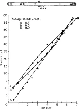
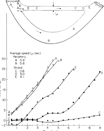
Organelles in a very thin transvacuolar strand approximately 0.5 μ in diameter were observed to move at a rather uniform speed as illustrated for a short strand in tobacco (Fig. 1) or a longer strand in Euphorbia
(Fig. 2). In both cells the rate of movement for each organelle was linear with little change in speed evident for an organelle over a maximum period of 7 sec. These observations represent the ideal condition. It should be emphasized that important differences in average speeds did exist between the organelles. In the tobacco cell (Fig. 1), the organelles designated by the letters Β and D were the first to appear on the strand separated from each other by distance of 4.4 μ. Each organelle moved at a different speed, one, B, moving at an average speed of 8.8 μ/sec or 1.4 μ/sec faster than the other, D. T h e organelle A appeared on the strand 13
0 C A DB
to.3
AVERAGE SPEED {μ/sec A 9.2 Β 8.8 C 8.2 D 7.4
FK;. 1. Transvacuolar strand, 0.3 μ in diameter, in an albino tobacco cell 120 μ in diameter. T h e four organelles moved with a uniform velocity along a transvacuolar strand approximately 30 μ in length. (Interval = i/2 sec.) T o p diagram—relative posi- tions of organelles at beginning and end of plotting.
sec after the appearance of D, moving linearly at an average speed of 9.2 μ/sec. T h e fourth organelle, C, appeared 2 sec later moving at the somewhat slower speed of 8.2 μ/sec.
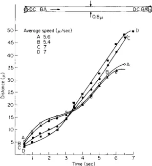
In thin strands of considerable length the movement of organelles follows a somewhat similar pattern as represented by Euphorbia (Fig. 2).
The organelle C moved at a nearly uniform speed for a distance of 56 μ.
A second organelle, A, accelerated over a distance of 52 μ, whereas the third organelle, B, 5 μ behind A, progressively decreased in velocity over a distance of 58 μ.
It should be noted that the speed of an organelle during the first
1/2-sec interval upon entering the strand can vary considerably (note time period at 1/2 sec in Fig. 1). T h e organelle C moved into the strand at a speed of 1.6 μ/sec, the other two organelles entered with speeds of 9.2 and 10.2 μ/sec. T h e organelle C which entered the strand at the slower speed was 1 and 6 sec, respectively, in advance of A and B. Other
£B AC —•» " Β ACS 10.5/4
Time (sec)
FIG. 2. Transvacuolar strand 0.5 μ in diameter in a Euphorbia cell 200 μ in length. Organelles entered the strand at different speeds and moved either linearly with time (C), accelerated (A), or decelerated (B) slowly along the 50-60 μ course during plotting. (Interval — i/2 s e c) T o p diagram—relative positions of organelles at begin- ning and end of plotting.
organelles followed similar patterns of movement when entering the strand.
The movement of an organelle in a thin strand may not be linear with time (Fig. 5). In a strand 0.8 μ in diameter the two organelles Β and A separated by a distance of 6.5 μ decelerated during the first 4 sec and subsequently accelerated during their movement along the strand. Both organelles followed a similar pattern which was quite irregular when compared with the previous graphs. Eight seconds after their appear- ance, two new organelles, C and D, appeared on the strand. Their velo-
cities were quite different as noted by the curves. These organelles pro- gressively accelerated for 3 sec whereupon their velocities became more or less constant along the strand. In contrast to the first pair of organelles, the pattern of movement for organelles C and D was not synchronized along the strand.
The rate of streaming along a thin strand can be subject to even greater variations than indicated in the previous figure when movements
feDC BA —» J DCBAQ-
L_^i ι • ι , ι , ι , ι , ι I 2 3 4 5 6 7
Time (sec)
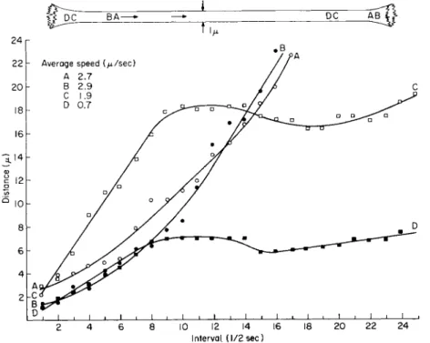
FIG. 3. Transvacuolar strand 0.8 μ in diameter in an albino cell 174 μ in diameter.
T h e four organelles followed two curve patterns. See text. (Interval = i/2 sec.) T o p diagram—relative positions of organelles at beginning and end of plotting.
in a strand are followed for extended periods of time. This is exemplified in Fig. 4 where the appearance of two sets of organelles, A, Β and C, D, in a strand 1 μ in diameter is separated by a 9-sec interval. T h e organelles appearing first, A and B, traversed the strand during a period of 8.5 sec, slowly accelerating during their movement. Their average speeds were 2.7 and 2.9 μ/sec, respectively. Note in the diagram that organelle Β accelerated to a greater extent than A, passing the latter along the strand.
In Fig. 4, the organelles C and D separated by a distance of 1.5 μ en- tered the strand 9 sec after the first set. T h e second set moved at different average speeds of 0.7 and 1.7 μ/sec, respectively. After these organelles had traveled for a period of 4 sec, they were separated by a distance of 10.5 μ. Both organelles then underwent rapid deceleration to zero velocity
for several seconds, temporarily reversed direction, and then resumed movement in their original direction. The organelles entered and moved along the strand with different initial velocities but note that their decel- eration and reversal in directions of movement were synchronized through
time. This pattern of movement for these organelles separated from each
Interval. (1/2 sec)
FIG. 4. Transvacuolar strand 1 μ in diameter in an albino cell 120 μ in diameter.
Organelles A and Β accelerate along the course, Β passing A. Organelles C and D which appear 9 sec after A and B, moved at different speeds but experience similar degrees of impedance along the strand although they are separated by 10.5 μ. T o p diagram—
relative positions of organelles at beginning and end of plotting.
other by 10.5 μ is suggestive that a unit of groundplasm at least 10.5 μ in length stopped, reversed direction, and then proceeded forward again.
STRAND OF LARGE DIAMETER
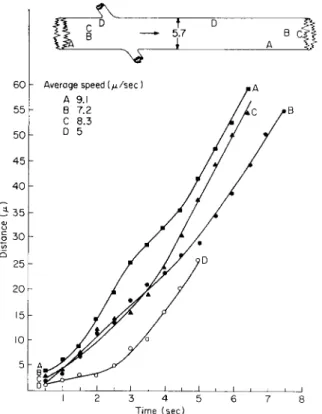
The streaming pattern of organelles was followed in a thick strand approximately 5.7 μ in diameter in an albino cell (Fig. 5). Initially I attempted to follow the movement of several organelles both along the surface and in the center of the strand to detect positional differences in speed for the organelles, as one would expect for laminar flow in a physical system. Lateral movement of the organelles, from the surface to
a more central position in the strand and vice versa, prevented me from gaining adequate data on this point.
Numerous organelles were moving in the thick strand, all moving in the direction indicated by the arrow. T h e recorded variations in velocity characterize the movement of the organelles. Those near the surface of the strand as well as those within moved readily in a lateral direction.
Time (sec)
FIG. 5. Transvacuolar strand 5.7 μ in diameter in an albino cell 1 7 4 μ in diameter.
Organelles were plotted simultaneously at different positions in transvacuolar strand.
(Interval — !/£ sec.) All organelles moved with different average speeds. T o p diagram—
relative positions of organelles at beginning and end of plotting.
Organelles A and D emphasized recorded ranges in velocities. Organelles Β and C remained adjacent to each other for 3.5 sec, whereupon C in- creased in velocity and moved ahead of B. There was little to suggest synchronization of organelle movement in strands of large diameter.
THIN-THICK TRANSVACUOLAR STRAND COMBINATION
Upon viewing the transvacuolar strand reticulum in the cell, one can observe combinations of strands which vary in their diameters. A typical
pattern is the form of the letter " Y " as in Fig. 6 consisting of a strand 4 μ in diameter and a divergent thin strand 0.5 μ in diameter.
Organelle movement was traced in the Y strand combination to deter- mine the velocity of organelles when moving from the strand of large diameter into the small strand. The rate of flow of a Newtonian sol in a Y tube should be greater in the thinner arm than in the base. Actually such a change in velocity did not occur. The speed of the organelles A and Β along the thick strand averaged 9.3 and 8.9 μ, respectively. The
55 50 45 40 35
-So
V, 25 b
20 15 10
Average speed {μ/sec) Thick strand
A 9.3 Β 8.9 Thin strand
C 7.8 D 6.8
FIG. 6.
diameters.
Time (sec)
Transvacuolar strand configuration involving strands 4 and 0.5 μ in Organelles either remain in thick strand or enter the thin strand. Average speeds of organelles entering the thin strand were somewhat less than those for the organelles remaining in the thick strand. (Interval = i/2 sec.) Diagram at right shows relative positions of organelles at beginning and end of plotting. Vacuole, v.
organelles C and D which moved from the thick strand into the thin strand exhibited no great acceleration. The velocity of these organelles, having average speeds of 6.8 and 7.8 μ, respectively, was actually less than that recorded for the organelles in the thick strand. Similarly, the aver- age speeds for two additional organelles which were plotted along the thin strand were lower than the speeds of the organelles along the thick strand.
PERIPHERAL CYTOPLASM-TRANSVACUOLAR STRAND COMBINATION
The simultaneous comparison of organelle movements was prepared for the peripheral cytoplasm and a transvacuolar strand 1 μ in diameter
(Fig. 7). Each organelle exhibited somewhat different average speeds and curve patterns. The average speed of two organelles (A = 5.9 μ/sec) and (B = 5.8 μ/sec) exceeded the speed of the organelles along the transvacuo- lar strand.
Average speed {μ/sec)
Time (sec)
FIG. 7. Combination of a transvacuolar strand 1 μ in diameter and the peripheral cytoplasm in an albino cell 120 μ in diameter. Certain organelles in strand were impeded or reversed their direction during their movement. (Interval — i/2 sec.) T o p diagram—relative positions of organelles at beginning and end of plotting; (-(-) and (—) indicate changes in direction of movement.
The movement of organelles along the strand was subject to inter- ference. Organelle C, the first to appear in the strand, moved at a slow speed averaging 0.6 μ/sec during a 9-sec period of time. Organelle E, appearing 3 sec after C, initially moved in the direction indicated as (—) for 1 sec, thereafter it reversed, to move in the (-f-) direction along the strand with a rather constant velocity. T h e path of organelle D which entered the strand 4 sec after C was impeded along its course for 6 sec after which it followed a linear time-distance relationship. T h e average
speed of D was 3 μ/sec, somewhat slower than the 4.1 μ/sec recorded for E.
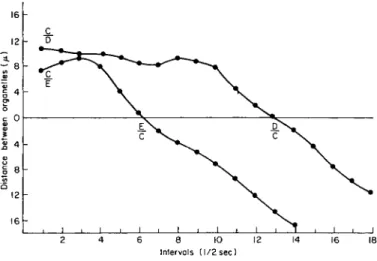
A most interesting phenomenon occurred when organelles D and Ε entered the strand (Fig. 8). Organelle Ε passed the impeded C after 3 sec and continued unimpeded along the strand. The distance between these organelles increased initially when Ε moved in the (—) direction (— 2 on the graph) and thereafter altered its course to move in the (-(-) direc-
16 h
Intervals (1/2 sec)
FIG. 8 . Passing of organelles within a transvacuolar strand 1 μ in diameter (as in Fig. 7 ) . Since organelles C, D, and Ε moved at different average speed, interceptions occurred along the transvacuolar strand. Symbols C/D and C / E indicate that the organelle C initially was ahead of either D or E. Positions were transposed D/C and E / C at the time interval when the organelles D and Ε passed C.
tion. Organelle D, at first moving slowly along the strand, increased in speed to converge upon and pass organelle C at interval 13, and there- after, continued to move unimpeded along the strand.
The average speeds for various organelles, both in broadly grouped categories of transvacuolar strands with diameters of two magnitudes and in the peripheral cytoplasm, are presented in Table I.
TWO-DIRECTIONAL MOVEMENTS ALONG ONE STRAND
Frequently movement can occur in two directions along one strand as in a strand 1.7 μ in diameter in an albino cell (Fig. 9a). T h e first or- ganelle, A, plotted along the strand at an average speed of 2.2 μ/sec, decelerated and at the time intervals 2 and 4 sec reversed the direction of movement for very short periods of time. The second organelle, B , moved along the strand linearly for a distance of 26 μ at an average speed
of 6.5 μ/sec. This organelle Β passed organelle A during its course along the strand.
Organelle C moving in the opposite direction progressed linearly for a distance of 14 μ during the first 2.5 sec of movement, whereupon it decelerated to zero speed and oscillated during the successive 3 sec while measurements were being made.
T A B L E I
AVERAGE AND RANGE IN SPEEDS FOR ORGANELLES IN DIFFERENT POSITIONS WITHIN THE CELLS, AND IN CELLS MAINTAINED IN MICROCHAMBERS FOR VARYING LENGTHS OF T I M E
Speed range
Position No. of in any one Average
or Diameter organelles interval speed
condition (μ) plotted (μ/sec) (μ/sec)
Transvacuolar 0.3-1.0 33 0-19 6.0
strand 1.1-6.0 29 0-19 5.8
Peripheral cytoplasm — 7 2.8-7.6 6.3
Constriction of
transvacuolar 1 4 1.4-10.8 7.9
strand 6 6 2.2-14.2 6.5
Age:
8 hr or less (from
above) — 79 0-19 6.5
6 days — 6 1.0-8.4 4.7
10 days — 4 3.0-9.0 6.7
Organelles C and A traveling in opposite directions were subjected to deceleration and reversal in direction at two positions during their course along the strand (Fig. 9b). These positions and times at which the reversal occurred could not be correlated.
These movements in opposite directions and at different velocities is indicative that the streaming cytoplasm possesses a structured character consisting of numerous streams or possibly fibrils along which the or- ganelles are transported.
REVERSAL IN DIRECTION OF MOVEMENT
Organelles traveling in the same strand can be observed to change their direction more dramatically than illustrated previously. This change may persist for periods of time during which the organelle can move a considerable distance (Fig. 10). Each organelle appeared to respond in- dependently of other organelles along the strand. T h e average speed of the organelles was plotted for each interval of time to illustrate the mag- nitudes of movement. Organelle Β moving at an average speed of 2.4 μ/sec traveled the length of the thick strand in about 10 sec. Organelle C,
after moving along the thick strand a distance of 11 μ, altered its direction to enter the thin strand. There was no increase in the velocity of this organelle upon entering the thin strand 1 μ in diameter.
The path of organelle A was subject to change. During the initial 2.5 sec it moved in the same direction as the other organelles, whereupon
J ι I 1 1 1 1 1 » 2 3 4 5 6
Time (sec)
FIG. 9. Transvacuolar strand 1.7 μ in diameter in an albino cell 120 μ in diameter.
Movement of organelles is evident in opposite directions along strand—(a) the time- distance curve and average speed of the organelles; (b) the magnitudes of speed for an organelle during an interval. (Interval = \/2 sec.) Arrows indicate direction of movement.
Note that organelles A and C, moving in opposite directions, underwent periodic reversals in direction (-{-) or (—) as they moved. T o p diagram—relative positions of organelles at beginning and end of plotting.
A reversed direction to travel a distance in excess of 8 μ along the thick strand. Again, this organelle reversed direction and after traveling a distance of 19 μ in the thick strand, it altered its direction again to enter the thin strand. The magnitude of interval speed per second for organelle A varied considerably attaining a maximum value of nearly 7 μ/sec in either direction and with average speeds of (-[-) 3.4 and (—) 2.5 μ/sec.
One interpretation of this strand complex is to suggest that several
or many cytoplasmic streams exist within the thick strand. T h e stream or streams transporting organelle Β move it along the large strand in the (-{-) direction, whereas other streams diverge from the thick into the thin strand (organelle C ) . T h e movement of organelle A may well represent shifting in position from one stream to another, these moving in different directions and at different velocities.
Time (sec)
FIG. 10. Transvacuolar strand configuration involving a 2 and a 1 μ in diameter strands in a chlorophyllous cell 125 μ in diameter. T h e three organelles followed dif- ferent patterns of movement: Β along the thicker strand; C entering into the thin strand from the thick strand; and A reversing directions at several points while following the general path indicated in diagram. T h e maximum speed of A in (-f) direction during a given interval was as great as that attained in the (—) direction during the plotting. (Interval = \/2 s e) T o p diagram—relative positions of organelles at beginning c
and end of plotting.
CONSTRICTION OF A TRANSVACUOLAR STRAND
T h e form of the transvacuolar strand can be exceedingly variable.
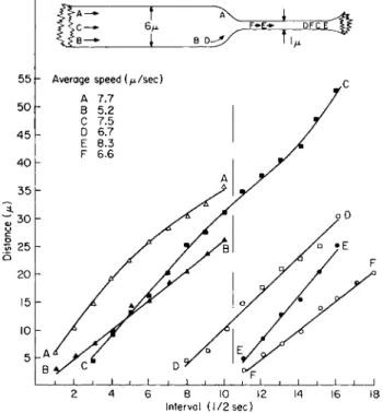
One form which was most instructive is represented in Fig. 11 in which a thick strand 6 μ in diameter suddenly constricted to a thin strand 1 μ in diameter. Organelle positions and period of movement relative to the constriction are represented on the abscissa; the dashed vertical line be- tween interval 10-11 represents the time interval when organelles C and D entered the strand constriction. T h e other organelles ( A and B) were
plotted either for the thick region of the strand (left of the dashed vertical line) or for organelles (E and F) in that portion of the strand which was small in diameter (right of dashed vertical line).
The rates of movement for organelles in the strand 6 μ in diameter were plotted simultaneously. T h e organelle Β moved linearly at an aver-
Β — [ Β I 1^
j I ι I ι I ι 1 L _ _J ι I • ι I L _J
2 4 6 8 10 12 14 16 18 Interval (1/2sec)
FIG. 1 1 . Constricted strand in a Euphorbia cell 1 8 0 μ in diameter. T h e vertical line at interval 1 0 - 1 1 represents interval when an organelle entered constriction (1 μ diameter) from the thick portion ( 6 μ diameter) along the strand. Some variation in average speeds are evident for different organelles. No significant change in speed is evident for organelles C and D entering the constriction from the thick portion of the transvacuolar strand. T o p diagram—relative position of organelles at beginning and end of plotting.
age speed of 5.2 μ/sec whereas organelle A progressively decelerated along the strand and was recorded to average a higher speed of 7.7 μ/sec.
These organelles could not be followed into the constriction.
The organelles F and E, plotted simultaneously along the constricted portion of the strand 1 μ in diameter, moved at a rather uniform rate averaging 6.6 and 8.3 μ/sec, respectively. Note that both organelles moved at different rates of speed in the thin strand.
The movement of the two organelles C and D was traced as they
moved both in the thick strand and in the thin strand. No appreciable in- crease in speed is evident for either organelle. T h e interval distances for organelle D plotted as a straight line with an average speed of 6.7 μ/sec.
Organelle C decelerated at the neck of the constriction and subsequently accelerated at a low rate of speed in the constricted strand, averaging 7.5 μ/sec along the entire strand. When the average speeds of these organ- elles, C and D, were examined relative to the region of large diameter (6 μ), it was found that they moved with an average speed of 7 and 7.7 μ/sec; their average speeds in the constricted strand were 6.6 and 7.2 μ/sec, respectively. Note that they decreased rather than increased in speed when entering the thin strand. T h e average speeds of all organelles plot- ted along the strand revealed only a slight increase in speed in the 1 μ sector compared to the portion 6 μ in diameter (Table I).
In a physical system the velocity of a pressure-induced laminar flow for a liquid will increase when passing from a tube of large diameter to one of small diameter. T h e change in velocity within a fluid can be expressed by the following equation:
Where J\ and V2 represent mean velocities, and âx and d2 represent diameters along the large and small diameter sections of the tube.
This law of classic fluid dynamics is not applicable to the flow of cytoplasm. T h e organelles when entering into the constricted portion of the strand 1 μ in diameter should increase in velocity nearly 30 times the recorded values for the organelles in the strand 6 μ in diameter.
The velocities for the organelles in this as well as other strands em- phasize that the cytoplasm possesses a structural character, possibly con- sisting of numerous fibrils or streams. T h e minor changes in velocity detected in the constricted strand emphasize that streaming of cytoplasm is not a pressure-induced flow.
EFFECT OF T I M E ON RATE OF STREAMING
The previous observations were made upon cells within 8 hr after preparation of the microchambers. Observations were continued on the individual cells daily for a period of 10 days and irregularly thereafter for 30 days to determine any changes in the pattern or rate of streaming.
Recordings on film were restricted to 6 and 10 days after chamber prep- aration. All the tobacco cells mentioned previously were streaming after 6 days.
The analyses of organelle movement along a thin strand, 0.5 μ in
diameter, for an albino cell maintained in the microchamber for the 6-day interval is indicated in Fig. 12. The three organelles, A, B, and C, illus- trated for the thin strand were plotted at varying velocities along the strand. The organelle A moved at an average speed, 6.4 μ/sec, consider- ably greater than the other organelles. Note that this organelle A is positioned between the two slow moving organelles.
The same strand, after 11 sec, increased to 1 μ in diameter (Fig. 13).
The change in diameter did not appreciably affect the velocity of the
^ B Ä C —» J B A C J ^ t 0.5/1
Time (sec)
FIG. 12. Transvacuolar strand 0.5 μ in diameter in an albino cell 174 μ in diameter after 6 days in microchamber. Average speeds of the organelles were somewhat variable.
(Interval — i/2 sec.) T o p diagram—relative positions of organelles at beginning and end of plotting.
organelles. The average speed recorded over a distance of approximately 35 μ for the three organelles A, B, and C was 4.2, 4.6, and 4.7 μ/sec, respectively, with only minor deviations from a linear time-distance relationship.
Active streaming was evident in the cells after 10 days enclosure in the microchambers (Fig. 14). The velocities for the organelles in the peripheral cytoplasm did not decrease appreciably after 10 days in the chamber from those speeds registered for organelles during the first 8 hr. Whereas the organelles A, B , and C moved at a rather uniform velocity, others were observed to move more irregularly along a course.
50 45 40 35 3- 3 0
ω υ
I 25 ω Ο
20 15 10
I 2 3 4 5 6 7 8 9
Time (sec)
FIG. 13. Cell as in Fig. 12. Same transvacuolar strand after 11 sec increased to 1 μ in diameter. Average speeds of the organelles corresponded closely with the slow moving organelle in Fig. 12. (Interval — i/2 sec.) T o p diagram—relative positions of organelles at beginning and end of plotting.
to measurably affect the streaming rate in other materials, were not known for the sealed microchambers.
Alterations in the form of the cytoplasm in the cultured cells became evident over prolonged periods of observation. Initially, the peripheral cytoplasm and that along a transvacuolar strand appeared optically homogeneous except for the organelles and the vacuolar membrane. After
10 days in culture, however, other cells were observed to develop an extensive system of phases or membranes which were arranged more or less parallel to each other and at irregular intervals were joined together into an anastomosing network. This system was very mobile and portions The average speeds for all the organelles plotted in the cells after 6 and 10 days in the microchambers are indicated in Table I.
Streaming continued in all the cells employed for these measurements until all cells plasmolyzed and appeared to die at the end of 18 days. T h e cultured cells appeared to be a very tolerant form of cell since the stream- ing rates of the above magnitude were evident in other cells 30 days after being mounted. After this period of time, the observations were discon- tinued. T h e oxygen and carbon dioxide gas tensions, both gases known
of membranes could be observed to undergo contortions during move- ment in the cell. Segments of the new membrane varying from 5-40 μ in length were observed to separate from one position, move for some dis- tance, and reattach to the membrane or phase system at another position within the cell.
Further observations indicated that each membranelike structure may be tubular in form. As viewed on film, a membrane periodically formed dilations which contained a volume of so-called groundplasm and or- ganelles. These dilations, too, were subject to change and would rapidly
I 1 1 1 I ι I ι I ι I ι I ι l •
0 1 2 3 4 5 6 7
Time (sec)
FIG. 1 4 . Peripheral cytoplasm in chlorophyllous cell 1 2 5 μ in diameter after 1 0 days in the microchamber. T h e period of time that the cell has been maintained in the culture chamber has not noticeably decreased the average speed of the organelles when their speeds are compared with organelles in many of the other graphs either for the transvacuolar strands or for the peripheral cytoplasm. (Interval = i/2 sec.)
constrict to assume the form of the tubular membrane structure. The organelles were not necessarily confined to the groundplasm within these tubelike structures, but often were observed between the parallel tubes.
At other locations in the cell, the organelles were observed to cross the membrane system at oblique angles.
Discussion
Heilbrunn (1952), Kamiya (1959), and Lundegârdh (1922) have demon- strated the presence of a cortical gel layer of cytoplasm in the internodal cell of Nitella and Chara as well as the plasmodium of myxomycètes. One interpretation of the streaming mechanism centers upon the physical
differences between the gel and sol phases and that forces generated at the interface between these two phases are responsible for the movement of the sol phase. The observations of Kamiya and Kuroda (1957a,b, 1958) upon isolated drops of Nitella emphasize the importance of a gel phase, it has been difficult to demonstrate a gel layer in the cells of higher plants and, indeed, the streaming activity in the cultured cells viewed on film indicate that no conspicuous gel layer was detectable to the inside of the plasma membrane (ectoplast), and none to the inside of the vacuolar mem- brane (tonoplast) in a transvacuolar strand.
It is not probable, in my interpretation, that the diverse movements of the organelles are to be explained by changes in cytoplasmic viscosity.
The rates of movement of organelles can be similar in transvacuolar strands of varying diameter. The cytoplasm in transvacuolar strands of small diameter does not appear to be more viscous than that in strands of large diameter when one evaluates the average speed of organelles along the linear part of the curves (compare Figs. 1, 2, 6, and 11). Two-directional movement and passing of organelles along a trans- vacuolar strand are most difficult phenomena to explain relative to changes in viscosity along the length or across the diameter of a trans- vacuolar strand.
The occurrence of a rhythm in the streaming cytoplasm of Physarum polycephalum has been demonstrated repeatedly in many excellent experi- ments by Kamiya (1962) and his co-workers. Indeed, Kamiya in analyzing plasmograms has indicated that a series of rhythms coexist within the protoplasmic system of Physarum. My initial efforts in analyzing the movement of organelles in cultured cells were directed in part toward determining whether a rhythm could be detected in the cytoplasm of the higher plant cells.
In cultured cells the pattern of cytoplasmic strands along the wall is constantly changing and the cytoplasm does not exhibit a rhythmic re- versal in direction of flow such as observed in P. polycephalum. Similarly the transvacuolar strands in toto are in motion, some disappearing from the strand reticulum, others appearing from the peripheral cytoplasm (Küster, 1956; Mahlberg, 1961; Mahlberg and Venketeswaran, 1962, 1963).
Superimposed upon these changes are the movements of the cytoplasm and the organelles within a strand. T o complicate the problem further, quantities of cytoplasm in cultured cells are observed to aggregate and move en mass and later disperse, the entire phenomenon appearing to lack periodicity (Mahlberg, 1961).
Comparison of organelle velocities revealed that no apparent rhythm existed in the cytoplasm which involved 1 /2-sec intervals of time or mul-
tiples of this unit of time. The time-distance relationships plotted for
many organelles in different positions within the cell demonstrated that an organelle during any one interval of time can move at different rates of speed varying from 0-19 μ/sec. Some organelles were observed to de- celerate to a speed of 0 μ/sec. Other organelles accelerated along their path even when confined to strands of small diameter. The speed of several organelles was evident as a sigmoid curve. Many organelles moved with a nearly constant velocity—perhaps surprisingly so—as represented by their linear time-distance relationships in several transvacuolar strands.
As noted for nearly all curves, variations in velocity for a single or- ganelle can occur for each 1/2 sec. This nonconstant speed for the or- ganelle is emphasized in Figs. 9 and 10 in particular. T h e changes in speed appear to be quite irregular, and my initial attempts to detect a synchronism for organelle movements have not been successful.
Organelles moving with a constant velocity would represent most accurately the streaming rate of the cytoplasm. However, each of several organelles can move with constant but different speeds along the same
strand, making it difficult to select the speed representing the true rate of streaming. Attempts to correlate movement of different organelles along the same strand over a period of 20 sec and more did not reveal a rhythmic or pulsative character for the cytoplasm. For example, two organelles can move at different speeds when crossing the same zone in thin strands, either at the same time or at different times. As noted in Fig. 4, however, there were instances along a strand when organelles (such as C, D) separated from each other by a considerable distance re- sponded with very similar patterns in movement. These nearly identical movements suggested that a filamentous mass of cytoplasm of consider- able length was undergoing changes in the rate of flow.
The most difficult patterns of movement to interpret are those where organelles move in opposite directions, where organelles pass each other, or where individual organelles reverse directions during their movement along the strand. These variations suggest that the cytoplasm within a strand is not moving at a similar velocity; rather that numerous indi- vidual streams (microstreams) of flow, or possibly that fibrils (Jarosch,
1958) constitute the cytoplasm of a strand. The cytoplasm, thereby, can be interpreted as a form of structured fluid as suggested by Allen (1962) for Amoeba. This is supported by the observations upon organelle velocity in constricted strands and those of varying diameters. The flow rate does not correspond to predictable changes in velocity, as indicated in Eq. (1), when the cytoplasm moves through strands of different diameters. The cytoplasm, therefore, does not appear to be a homogeneous or nonstruc- tured matrix, nor does it appear to possess an internal pressure which controls the movement of cytoplasm. It should be emphasized that the
interpretation of the structured character of cytoplasm as microstreams or fibrils must be sufficiently flexible to be definable at the molecular level as more data are accumulated on the energetics of streaming.
The microstreams are interpreted to be of various lengths and are interdigitated with each other; they represent the bulk flow of cytoplasm, are capable of sliding over one another, and move at varying rates in dif- ferent directions.
Some impression of the maximal diameters of microstreams can be gained from the following observed movements of organelles. Tentatively,
the diameter of a microstream, one or more moving an organelle, can be related with the varying velocities for organelles along a transvacuolar strand. In Fig. 4, the organelle Β passed A along a strand 1 μ in diameter.
Since the speed of both organelles was nearly linear, at least two micro- streams about 0.5 μ in diameter should be present in that strand. T h e three organelles (Fig. 7) in the thin strand, 1 μ in diameter, moved at different rates, two of the organelles crossing the path of the third. Their movement represented at least three different microstreams approximately 0.3-0.4 μ in diameter, since all organelles were moving at different rates of speed.
In a constricted transvacuolar strand (Fig. 11) the thick portion of the strand is interpreted to consist of numerous interdigitated microstreams moving at different velocities in both directions whereas the constricted portion contained fewer such interdigitated microstreams. Various micro- streams are interpreted to terminate or become folded in the enlarged portion of the strand. This interpretation is supported from observations which indicate that some organelles decelerate appreciably upon ap- proaching the zone of constriction and reverse directions. It was too dif- ficult to plot the course of these organelles for any distance since their path was quickly obscured by other organelles. Microstreams in the thick portion of the strand were interpreted to move individually into the con- stricted portion of the strand. Moving as interdigitated units the micro- streams would not increase appreciably in speed when they became com- ponents of the cytoplasm of the constriction.
The movement of organelles is associated primarily with the micro- streams, one or more carrying the organelles at different velocities or in different directions. The most generally accepted interpretation of or- ganelle and particle movement emphasizes the role of contractile or fibrous proteins which effect the movement of all cytoplasm and which carry the particles and organelles around the cell (Frey-Wyssling, 1955; Pfeiffer, 1953; Schmidt, 1941; Seifriz, 1944). However, the capacity of independent motility for metabolizing organelles must not be disregarded although
direct evidence for the mechanism of self-propulsion is presently un- available (Bünning, 1935; duBuy and Olson, 1940).
As interpreted here the sudden changes in direction or in velocity of an organelle reflect a shift in positions between different microstreams,
these moving at different rates or in different directions. T h e metabolizing organelles are being carried within or upon, but not affixed to, the micro- streams. Organelle transfer is effected by its capacity of self-motility where- by it moves from one microstream to another.
A streaming pattern observed in Acetabularia, described by Kamiya (1962) as multistriated streaming, is also apparent in higher plant cells.
T h e plasmic network, representing either phase differences or membrane- like structures, which became apparent in some of the cultured cells after
10 days, may be similar to the membranes observed by other investigators in different cells (Boresch, 1914; Honda et al., 1962; Küster, 1956; Scarth, 1942) or the striated tracks in Phy corny ces and Acetabularia (Kamiya, 1959). Possibly the fibrils identified by Jarosch (1958) in Chara may be identical to the membranous structures observed in the cultured cells.
The movement and contortions exhibited by segments of this mem- branelike system was associated with the streaming of the cytoplasm in general. During the initial phases of development numerous dilations were evident along the membranelike complex, but these became less conspicuous with time. The groundplasm confined within the compart- mentalized cytoplasm continued to stream as indicated by the movements of the organelles. T h e apparent segregation of the cytoplasm did not significantly alter the average speed of the organelles even after 10 days in the microchambers. Characterization of the membranelike structures in the cytoplasm will require further investigation.
Summary
The velocity of an organelle within streaming cytoplasm was recorded to vary from 0-19 μ/sec over a period of several seconds when plotted at 1 /2-sec intervals. In this initial study it was not possible to detect a pulsa- tive or rhythmic pattern for such variations in velocity.
Each of several organelles can move at different velocities along a strand, frequently passing each other as they move in the same direction.
Bidirectional streaming often was detected in strands of different diam- eters; in several instances individual organelles were observed and re- corded to alter their direction of movement along a strand.
Any two organelles can follow very similar, essentially synchronized, patterns of movement along a strand. Such patterns can be complex in
that they may include variations in velocities as well as changes in direc- tion of movement.
In a constricted strand the velocity of an organelle does not increase appreciably when the organelle enters the constriction, indicating that streaming cytoplasm is not under pressure.
The cytoplasm appears to possess structural characteristics, possibly consisting of microstreams of unknown dimensions. The numerous micro- streams interdigitate with each other and are interpreted to move at vary- ing velocities and in different directions.
Streaming may represent a dual problem involving motion at two dif- ferent levels: movement of the ground cytoplasm and movement of metab- olizing organelles. Movements of the organelles represent, primarily, transport by the microstreams.
Several organelles being carried by one or more microstreams moving at similar velocities would exhibit a synchronized pattern of movement.
It is presently interpreted thai metabolizing organelles may possess the capacity for self-motility. Their rapid changes in velocities and in direc- tions may reflect their capacity to utilize energy to shift their position from one microstream to another which is moving at a different speed or in a different direction.
ACKNOWLEDGMENT
The author is indebted to Dr. S. Venketeswaran and Dr. E. de Fulvio for assistance in preparation of portions of the film.
REFERENCES Allen, R. D. (1962). Sei. Am. 2 0 6 , 112-122.
Boresch, K. (1914). Z. Botan. 6 , 97-156.
Bünning, E. (1935). Z. Wiss. Mikroskopie 5 2 , 166-172.
Denham, H. J . (1923). ./. Textile Inst. Trans. 14, Τ 8 5 - Τ Π 3 . duBuy, H. G., and Olson, R. A. (1940). Am. J. Botan. 2 7 , 401-413.
Frey-Wyssling, A. (1955). Protoplasmatologia 2 , A2, 1-244.
Heilbrunn, L. V. (1952). "An Outline of General Physiology." Saunders, Philadelphia, Pennsylvania.
Honda, S. I., Hongladarom, T., and Wildman, S. G. (1962). In "Organelles in Living Plant Cells." Film. Educational Film Sales, University of California, Berkeley, California.
Jarosch, R. (1958). Protoplasma 5 0 , 93-108.
Kamiya, N. (1959). Protoplasmatologia 8 , 3a, 1-199.
Kamiya, N. (1962). In "Encyclopedia of Plant Physiology" (W. Ruhland, ed.), Vol.
XVII, pp. 979-1035. Springer, Berlin.
Kamiya, N., and Kuroda, K. (1957a). Proc. Japan Acad. 33, 149-152.
Kamiya, N., and Kuroda, K. (1957b). Proc. Japan Acad. 33, 201-205.
Kamiya, N., and Kuroda, K. (1958). Proc. Japan Acad. 34, 435-438.
Küster, Ε. (1956). "Die Pflanzenzelle." Fischer, Jena, Germany.
Lundegärdh, H. (1922). "Linsbauers Handbuch der Pflanzenanatomie," Vol. 1. pp. 1- 402. Gebrüder Borntraeger, Berlin.
Mahlberg, P. (1961). In "Streaming in Cultured Cells of Euphorbia marginata."
Film. W. R . Smith, Pittsburgh, Pennsylvania.
Mahlberg, P. (1962). Exptl. Cell. Res. 2 6 , 290-295.
Mahlberg, P., and Venketeswaran, S. (1962). In "Formation of Transvacuolar Strands in Cultured Cells of Euphorbia marginata." Film. W. R . Smith, Pittsburgh, Penn- sylvania.
Mahlberg, P., and Venketeswaran, S. (1963). Am. J. Botan. 5 0 , 507-513.
Martens, P. (1940). Cellule Ree. Cytol. Histol. 4 8 , 247-306.
Pfeiffer, W. (1953). Bet. Deut. Botan. Ges. 6 6 , 2-5.
Probine, M. C , and Preston, R . D . (1958). Nature 1 8 2 , 1657-1658.
Scarth, C. W. (1942). In "Structure of Protoplasm" (W. Seifriz, ed.), pp. 99-107. Iowa State Univer. Press, Ames, Iowa.
Schmidt, W. J . (1941). Ergeb. Physiol. Biol. Chem. Exptl. Pharmakol. 4 4 , 27-95.
Seifriz, W. (1944). In "Medical Physics" (Ο. Glasser, ed.), pp. 1127-1145. Year Book Publ., Chicago, Illinois.
Venketeswaran, S., and Mahlberg, P. (1962). Physiol. Plantarum 1 5 , 639-648.
DISCUSSION
DR. ALLEN: I must say I am quite struck by the similarity of your material to Foraminifera which I have studied and will report on later. One thing I have observed both in your moving pictures and in Foraminifera is that particles, if you analyze them over periods of time such as a few seconds, appear to be going at more or less uniform velocities; but, if you analyze them over shorter intervals, they move in jerks.
I got the definite impression, that in your film many or most of the particles move in a more or less jerky fashion. I think this shows in some of your graphs in which the points lie first above and then below the line.
DR. MAHLBERG: Yes, I agree that the motions are somewhat irregular. At first we thought there might be a periodicity in the motion, but we were not able to demon- strate this.
DR. NAKAI: I am very much interested to see that your film shows bidirectional streaming. As I will show in my film, neuronal pseudopodia also show bidirectional movement, however, different kinds of particles are involved and the character of the movement is different. I noticed that some of the fibers have a thickness of about 1 μ, and that the particles are hardly smaller. Therefore, I wonder whether there is enough room in a cytoplasmic strand 1 μ thick to generate separate streams, each carrying particles and sometimes going in opposite directions. Would you consider the hypothesis that the particles are actively metabolizing cell organelles which can interact with the immediate cytoplasmic surroundings and can generate a local streaming rather than simply following predetermined pathways?
DR. D E BRUYN: One could certainly imagine that a metabolizing organelle must take in molecules of very small size for synthesis, then must allow other molecules to diffuse out.
There are apparently in mitochondria little holes, or sieves, which may pass the large molecules unable to go through the membrane proper. If this were so, then you might get surface tension differences which then would shoot these organelles off under self-motility. This is one possibility.
DR. BISHOP: Are you identifying these particles? Are they metabolic particles or are they uncertain large granules? You didn't say much about them.
DR. MAHLBERG: T h e preliminary work on Euphorbia indicates that these are very likely mitochondria. Assuming all of the particles are as similar as they appear, we could call them metabolizing organelles. We can also see other dissimilar granules. I
didn't point any out, but there are small granules in these cultured cells that exhibit very, very little motion at all.
DR. REBHUN: We have been studying so-called independent movements in the cyto- plasm and their organization during mitosis. It is rather interesting that the class of particles which can participate in movement such as this, i.e., moving in one direction up to 30 μ and suddenly reversing or stopping, is extremely wide. It may be melanin granules in melanocytes and granules that have been identified as crystalline heme in certain eggs, and a variety of other particles in other cells. However, perhaps the most telling results in this direction were gotten by Andrews in Stentor and various other ciliates in which he observed carmine particles, India ink, and starch granules par- ticipate in these peculiar movements. Chambers saw these movements in the oil droplets "sliding" along astral rays; movements very similar to those of other naturally included particles. In such cases it would be, of course, impossible to ascribe this to metabolic processes within the particle.
DR. MAHLBERG: T h e cytoplasm is definitely moving. All that I am suggesting for self-motility of organelles is that they are able to transfer themselves from one stream to another. I am not sure how the inert granules do it. Perhaps they are in the boundary between.
On the other film, one can see the development of what appeared to be an entire membrane system within these cells. It was possible to see along one strand a series of membranes with movement going in opposite directions.
DR. D E BRUYN: It looks as if you might have something like elastic ropes moving sometimes in one direction and sometimes going back. They may not be really streams HI the hydrodynamic sense.
CHAIRMAN THIMANN: Perhaps something like Miss Kuroda's polygons.
DR. MAHLBERG: It could be something of that type. I am suggesting here there is an end, so to speak, to a stream.
DR. JAHN: I would like to emphasize the point made by Dr. Allen, that the phenomena you described are very similar to those in Foraminifera. Furthermore, in forams the "ropes" not only are elastic but they are on the surface so that insoluble dye granules can attach to the elastic rope and be pulled along, then become transferred to another "rope," and return.
DR. MAHLBERG: T h e organelle itself does not appear to be attached to the kind of membrane system I described; they are quite able to move around indiscriminately.
They can aggregate for a while and then disperse.