Role of the Cortical Gel Layer in Cytoplasmic Streaming
TO S H I O HA Y A S H I Biological Institute, College of General Education,
Tokyo University, Komaba, Tokyo, Japan
Although several types of cytoplasmic streaming are known in plant cells, rotational streaming will be dealt with here for the reason that it is simplest in dynamic organization.
The materials used in our experiments have been the internodal and rhizoid cells of the following Characeae: Chara Braunii, Chara corallina, and Nitella flexilis. These cells, some of which are truly enormous, meas- ure from a few millimeters to 10 cm in length and several tenths of a millimeter in diameter. They exhibit vigorous and typical rotational streaming and are suitable for many kinds of experimental approaches to the cytoplasmic streaming problem.
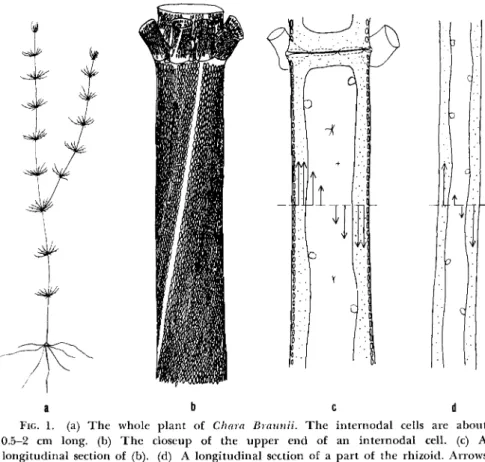
First, we should like to describe briefly the general organization of a typical characeous cell, C. Braunii. Figure 1 shows a series of line draw- ings, and Fig. la shows the general location of the leaf, internodal, and rhizoid (or root) cells of the whole plant. The second drawing (Fig. lb) shows a close-up of one end of an internodal cell in which the chloroplasts are arranged in spiral rows with a gentle slope. The streaming cytoplasm follows the path of these rows in a continuous belt around the cell. The cytoplasm is quiescent only at the indifferent or neutral zones marked by a lack of chloroplasts. Figure le, a longitudinal section, shows that the streaming endoplasm is bounded externally by a thin cortical layer in which most of the chloroplasts are found and internally by the tonoplast or vacuolar membrane. The cell wall and vacuole are nonliving parts of the cell. Between the two opposing streams there are two narrow zones, the so-called "indifferent zones," which are marked by a lack of chloro- plasts. The cytoplasm is quiescent only at these zones. Each of the two opposed streams has a uniform speed over the circumference covering about 165° (Kamiya, 1962). Only in the narrow zones close to the in- different lines the endoplasm streams with smaller rates.1 The last draw- ing (Fig. Id) is of a rhizoid cell, which is similar to internodal cells except
1 The cross section of a Nitella or Chara intcrnodal cell wall is circular. The contour of the tonoplast is also roughly circular because of local difference in endoplasmic thickness, but the endoplasm is thinner in the region of the indifferent zone.
19
for a lack of chloroplasts; the endoplasm streams as vigorously as that in the internodal cell.
The velocity of cytoplasmic streaming in adult internodal cells is very high as compared with that in other plant cells, and is kept almost con- stant under constant environmental conditions. It is usually 60 μ/sec at 20°C. But in young apical internodal cells, the cytoplasm streams much slower.
a b c d
FIG. 1. (a) T h e whole plant of Chara Braunii. T h e internodal cells are about 0.5-2 cm long, (b) T h e closeup of the upper end of an internodal cell, (c) A longitudinal section of (b). (d) A longitudinal section of a part of the rhizoid. Arrows indicate the direction and the relative rate of the streaming in the cell.
Streaming velocity increases rapidly as the temperature rises. T h e acceleration of the streaming was found to be produced mainly by the decrease in viscosity of the cytoplasm and not by the increase in the motive force behind the streaming (Hayashi, 1960). These results sub- stantially support the view of Lambers (1925) and are also in accord with the results obtained from myxomycete plasmodia (Kamiya, 1953).
There are two current theories about the site of motive force genera- tion of rotational streaming in plant cells; one is to attribute it to the
Cortical Gel Layer in Cytoplasmic Streaming 21 streaming cytoplasm itself, whereas the other takes the view that the motive force is generated at the boundary between the streaming endo- plasm and the cortical gel plasm (cf. Kamiya, 1959). I should like to describe some recent experiments which may help establish the locus of motive force generation.
Exfoliation of Chloroplasts by Means of Centrifugation A direct test for whether the cortical gel layer is of fundamental im- portance for the mechanism of the cytoplasmic streaming is to injure locally or totally destroy the cortical gel layer. In this case streaming should be impaired, modified, or prevented. On the other hand, if the whole mechanism of streaming should be located in the streaming endo- plasm itself, then the endoplasm might be able to move after destruction of the cortical gel layer. It was from this viewpoint that the following experiment was conducted.
One way of damaging the cortical layer is by centrifugation. For these experiments we have used a home-made electrically driven centrifuge microscope of very simple design with a rotor only 4 cm in diameter (Hayashi, 1957a). With this it is possible to subject our material to ac- celerations up to 4000 g within 4 sec, yet stop the rotor quickly when the desired degree of cortical injury was attained.
The effects of centrifugation are as follows. First the endoplasm is dis- placed centrifugally and the vacuole centripetally without apparent in- jury to the cortical layer. At higher accelerations, from 400 to 1000 g depending on the species and season, the chloroplasts are exfoliated or torn off the cortex, sometimes singly, but more often in longitudinal rows.
A substantial part of the cortical gel accompanies them.
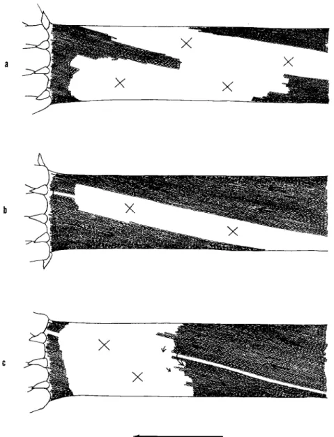
Figure 2 indicates three examples of cells which suffered exfoliation of chloroplasts to an extent suitable for observation. The white area, in the cell in this figure is the site where the chloroplasts have been torn off and have fallen to the centrifugal end of the cell.
In Fig. 3 the cytoplasm is transported continuously from the left- hand side of the cell along the peninsula-like area where the chloroplasts are embedded normally, but it stagnates and piles up at the white area.
Although the endoplasm can stream normally when in contact with the inner surface of chloroplast-containing, cortical, gel layer, the very same endoplasm immediately loses the ability to stream when it reaches the cortical layer from which the chloroplasts have been removed. The arrows in Fig. 3 show the direction and speed of the streaming at different loci which were obtained through cinematographic analysis.
We can infer that the cortical gel layer at the white area must have
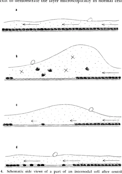
received rather severe injury, as is shown schematically in Fig. 4. Figure 4 shows the normal state of the cortical gel layer. Immediately after the centrifugal treatment there was no sign of any movement of the cytoplasm
a
FIG. 2. Cells immediately after centrifugation at 400 g for 3 min (in winter). No cytoplasmic streaming is observable at the white area marked with X where the chloroplasts have been exfoliated, whereas normal streaming continues at the area where the chloroplasts have remained attached at their original positions. Arrows in- dicate the direction of the cytoplasmic streaming.
Cortical (xel Layer in Cytoplasmic Streaming 23 except for the rotation of the exfoliated chloroplasts and the normal Brownian motion of small particles, though the streaming continued at the part where the chloroplasts remained attached at their original posi- tions (Fig. 4b). When the treatment of centrifugation is strong enough, the white area where the chloroplasts are exfoliated becomes too large for the cell to survive. This degree of cortical injury was achieved after about 3 min of centrifugation at an acceleration of 400 g in winter.
At any rate, the previously mentioned fact is a further confirmation of the findings of previous authors that, when the plasmagel layer of Nitella or Chara cells is injured locally by a needle or anesthetics, the
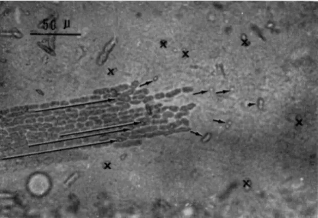
FIG. 3. Surface view of a part of the cortical gel layer of an internodal cell after centrifugation. Chloroplast layers are mostly exfoliated except at a peninsula-like area of the left. Arrows show the direction and speed of flow at different loci. T h e length of each arrow shows the distance covered by a particle in 5 sec.
endoplasmic flow no longer occurs at that spot but goes around it (Nichols, 1930; Linsbauer, 1929; Péterfi and Yamaha, 1931). These facts indicate the importance of the role played by the cortical gel layer in the mechanism of cytoplasmic streaming.
Recovery of Streaming
The cytoplasm at the white area where the chloroplasts have been exfoliated begins to move within 5 to 10 hr after the treatment, though the movement is feeble compared with that in other areas where chloro- plasts have not come off (Fig. 4c). Prolonged observations on these in- jured cells showed first that the initially feeble movement became pro- gressively stronger as time passed. Over a period of days the chloroplasts that had fallen to the centrifugal end of the cell returned to and settled down at the injured white area. Along with restitution of the chloroplasts
to the white area, the cytoplasmic streaming at that area is recovered (Fig.
4d). The direction of flow in this case was always the same as before.
Although the cortical gel layer is indicated schematically in Fig. 4, it is difficult to demonstrate the layer microscopically in normal cells. This
a
FIG. 4. Schematic side views of a part of an internodal cell after centrifugation showing the recovery of cytoplasmic streaming and restitution of the chloroplasts to the white area, (a) Before centrifugation; (b) 5-10 hr after centrifugation; (c) 3-5 days later; (d) about 2 weeks after the treatment.
is because the layer is so thin and attached so closely to the cell wall.
Moreover, in the case of the internodal cell, the chloroplasts directly inter- fere with observations of the cortical gel layer.
From the evidence presented so far, one might conclude that either
Cortical Gel Layer in Cytoplasmic Streaming 25 the cortical layer or the chloroplasts within it was required for streaming.
However, from the fact that the rhizoid cells, which are devoid of chloro- plasts, stream, we are inclined to believe that it is the cortical gel layer itself, and not the chloroplasts within it, which is essential to streaming.
Streaming in the Plasmolyzed Cell
Since the cortical layer is so difficult to see in internodal cells because of the chloroplasts, the ideal place to study the cortex is in rhizoid cells, which as we said, lack chloroplasts. Here it is possible to ascertain the presence or absence of this layer by plasmolysis (Hayashi, 1960). When the rhizoid cell of C. corallina is plasmolyzed by a 0.2 M solution of cal- cium chloride (Hayashi and Kamitsubo, 1959) the endoplasm streams normally along the inner side of the cortical layer which is microscopically visible and about 1 μ thick. On the other hand, at the site where no cor- tical gel as such is observable, the endoplasm does not stream.
In the protoplast plasmolyzed with calcium chloride it occasionally happens that the gel layer is found not at the periphery but along the tonoplast. In such a case the cytoplasmic movement is most active in the immediate proximity to the tonoplast and becomes weaker the greater the distance from the vacuole. In normal cells, however, the endoplasm streams, together with the tonoplast, with almost the same velocity throughout its thickness (Kamiya and Kuroda, 1956).
All the above facts show clearly that the boundary between organized cytoplasmic gel layer and the endoplasm plays an essential role in gen- erating the motive force of the cytoplasmic streaming in these cells.
Behavior of the Endoplasm under Moderate Centrif ligation Next we shall talk briefly about the behavior of the endoplasm when the internodal cell is centrifuged at low accelerations, at which chloro- plasts do not come off from the cortical gel layer. Instead, at these low accelerations the endoplasm accumulates at the centrifugal end of the cell. Most of the chloroplasts usually remain in position when the cen- trifugal acceleration is below 400 g. With the aid of a centrifuge micro- scope, we can observe directly the displacement of the endoplasm while the cell is being centrifuged (Fig. 5). When the centrifugal acceleration is decreased below 40 g, the sedimented endoplasm can go back toward the centripetal end in the form shown at the bottom of Fig. 5. T h e restitu- tional streaming toward the centripetal end of the cell always takes place only along one side of the cylindrical cell wall, the side along which the endoplasm flowed in that same direction before the cell was centrifuged.
This fact was established by Breckheimer-Beyrich (1954).
Kamiya and Kuroda (1958), who calculated the motive force respon- sible for rotational streaming from measurements of the balance-accelera- tion required to stop centripetal streaming, found that the thinner the endoplasmic layer, the greater the centrifugal acceleration required to stop streaming. This constitutes further support for the conclusion that the site of the motive force of rotational streaming is not distributed throughout the streaming endoplasm but localized at the boundary be-
2 00 X g
15 0 X g
10 OX g
30XS
FIG. 5. Schematic profiles of the endoplasm collected at the centrifugal end of the internodal cell under various accelerations. At 200 g, the endoplasm accumulates at the centrifugal end of the cell, where no cytoplasmic streaming toward the centripetal end is visible. Below 200 g endoplasm starts streaming against the centrifugal force in an extremely thin layer. When the centrifugal acceleration is decreased down to 40 g, the restitutional streaming of endoplasm becomes more conspicuous.
tween the outer edge of the endoplasm and the inner surface of the cortical gel layer.
L o c a l C h e m i c a l F i x a t i o n
At this point it would be pertinent to touch upon a recent experiment (Hayashi, 1960) in which the cell was fixed locally by bringing a micro- pipette filled with various fixatives in contact with the cell. Under such a situation, cytoplasmic streaming could not be observed for several hours at the spot where the cortical gel was fixed. T h e chloroplasts at that spot fell off together with the endoplasm into the vacuole, while the normal streaming kept going elsewhere. By plasmolyzing the cell, it was clearly ascertained that the cortical gel layer disappeared soon after the treat- ment but appeared again several hours later followed by the recovery of
Cortical Gel Layer in Cytoplasmic Streaming 27 the cytoplasmic streaming there. This may also be interpreted as evidence supporting the significance of the cortical gel layer in the rotational streaming typical of this plant.
Behavior in a Pathological Cell
T o conclude, I would like to add one more observation on an interest- ing abnormality of streaming in internodal cells of C. corallina infected by an unknown species of the oribatid mite (Hayashi, 1957b). No rota- tional streaming can take place in these cells, but the chloroplasts rotate independently around their own axes (Fig. 6b). This phenomenon may
a
sib cs> *gl <ss> Mj ^ ^><sl> §
FIG. 6. Schematic representation of longitudinal sections of a normal (a) and an abnormal cortical gel layer of the internodal cell infected by an oribatid mite (b).
imply that the cortical gel layer of the cell in question can no longer hold the chloroplasts firmly enough and such an abnormal gel layer cannot drive the endoplasm along that surface.
Summary
T o sum up, we can say that rotational streaming of cytoplasm found in the cells of the Characeae stops as soon as the cortical gel layer is destroyed by any means whatever, and that streaming is resumed as the cortical gel layer is reformed. T h e motive force for rotational streaming is an active shearing force generated at the boundary between the cortical gel and endoplasm. What comes into focus concerning the mechanism of cytoplasmic streaming in this case is, therefore, the key problem of how the active shearing force is produced at this boundary.
RE F E R E N C E S
Breckheimer-Beyrich, H . (1954). Ber. Deut. Botan. Ges. 67, 86.
Hayashi, T . (1957a). Botan. Mag. (Tokyo) 70, 168.
Hayashi, T . (1957b). / . Japan. Botan. 37, 82.
Hayashi, T . (1960). Sei. Papers Coll. Gen. Educ. Univ. Tokyo 10, 245.
Hayashi, T . , and Kamitsubo, E. (1959). Botan. Mag. (Tokyo) 72, 309.
Kamiya, N . (1953). Ann. Rep. Sei. Works Fac. Set. Osaka Univ. 1, 53.
KAMIYA, N. (1959). Protoplasmatologia 8, 3A, 1.
KAMIYA, N. (1962). In "HANDBUCH DER PFLANZENPHYSIOLOGIE" (W. RUHLAND, ED.), VOL.
X V I I , PART 2, P. 9 8 0 . SPRINGER, BERLIN.
KAMIYA, N., AND KURODA, K. (1956). Botan. Mag. (Tokyo) 6 9 , 5 4 4 . KAMIYA, N., AND KURODA, K. (1958). Protoplasma 5 0 , 144.
LAMBERS, M. H . R . (1925). Proc. Koninkl. Ned. Akad. Wetenschap. 2 8 , 340.
LINSBAUER, Κ. (1929). Protoplasma 5, 5 6 3 .
NICHOLS, S. P. (1930). Bull. Torrey Botan. Club 57, 153.
PÉTERFI, T . , AND YAMAHA, G. (1931). Protoplasma 12, 2 7 9 .
DISCUSSION
C H A I R M A N T H I M A N N : MAY I ASK, SIR, HOW IT IS THAT THE CHLOROPLASTS GET BACK INTO THE WHITE AREAS? THERE IS NO STREAMING TO CARRY THEM THERE. DO THEY GET BACK UNDER THEIR OWN MOTILITY?
DR. T O S H I O H A Y A S H I : THE CHLOROPLASTS COME BACK VERY SLOWLY. THE ENDOPLASM AT THE WHITE AREA DOES NOT STREAM, BUT IS SOMEHOW "PUSHED" INTO THE WHITE AREA.
DR. Z I M M E R M A N : DR. HAYASHI, IS THERE ANY RELATIONSHIP TO THE THICKNESS OF THE CORTICAL GEL LAYER AND THE RATE OF PROTOPLASMIC STREAMING?
DR. T O S H I O H A Y A S H I : I CANNOT GIVE YOU A DEFINITE ANSWER FOR YOUR QUESTION, SINCE THE CORTICAL GEL LAYER IS SO THIN THAT IT IS DIFFICULT TO DETERMINE ITS EXACT THICKNESS WITH AN OPTICAL MICROSCOPE. PROBABLY WHAT IS IMPORTANT FOR THE STREAMING IS AN ORGANIZED INNER SURFACE OF THE CORTICAL GEL LAYER AND NOT ITS THICKNESS.
DR. BURGERS: WOULD YOU REPEAT YOUR CONCLUSION? YOU SAID THE MOVEMENT IS LOCALIZED AT THE BOUNDARY BETWEEN THE CORTICAL GEL LAYER—AND WHAT FOLLOWED?
DR. T O S H I O H A Y A S H I : THE MOTIVE FORCE FOR ROTATIONAL STREAMING IS GENERATED AT THE BOUNDARY BETWEEN THE CORTICAL GEL AND ENDOPLASM.
DR. MARSLAND: I WOULD LIKE TO ASK HOW YOU DIFFERENTIATE BETWEEN THE CORTICAL GEL AND THE ENDOPLASM. HOW DO YOU FIND THE DIFFERENCE?
DR. T O S H I O H A Y A S H I : THE CORTICAL GEL IS STATIONARY, AND THE ENDOPLASM IS MOVING.
DR. JACOBS: WHEN YOU EXFOLIATE THE CHLOROPLASTS WITH CENTRIFUGAL ACCELERATION, DO SOME CHLOROPLASTS "DIFFUSE" IN AND THEN DIVIDE IN THE EXFOLIATED AREA OR ARE THEY ALL MOVED IN FROM OTHER AREAS?
DR. T O S H I O H A Y A S H I : THEY GROW, BUT I AM NOT QUITE CERTAIN ABOUT WHETHER THEY DIVIDE.
DR. MARSLAND: I THINK THESE OBSERVATIONS FIT VERY WELL WITH ONES I MADE MANY YEARS AGO ON Elodea, WHICH SHOWED THAT THE APPLICATION OF HIGH PRESSURE CAUSED SOLA- TION, PRESUMABLY OF THIS GEL LAYER. THE RATE OF MOVEMENT DROPPED OFF IN PROPORTION TO THE DEGREE OF SOLATION. THIS WOULD ALSO CERTAINLY INDICATE THE IMPORTANCE OF THE GEL STRUCTURE.
DR. LING: FIRST, IN ONE OF THE MOVIES YOU SHOWED, WHEN THE CHLOROPLASTS WERE MORE OR LESS RANDOMLY DISTRIBUTED, THE CYTOPLASM SEEMED TO ACCELERATE ALONG THE PATH WHERE THERE WAS A STRING OF THESE CHLOROPLASTS. IS THIS OBSERVATION CORRECT? SECOND, IF THIS IS THE CASE, IS IT POSSIBLE THAT THE CHLOROPLASTS MAY STILL ADD AN EFFECT ONTO THE CORTICAL CYTOPLASM IN SPITE OF THE FACT THAT IN RHIZOID CELLS LACKING CHLOROPLASTS STREAM- ING STILL TAKES PLACE.
DR. T O S H I O H A Y A S H I : CYTOPLASM SOMETIMES STREAMS FASTER ALONG THE PATH FORMED BY ALIGNED CHLOROPLASTS. THIS, I SUPPOSE, IS PROBABLY BECAUSE THESE CHLOROPLASTS ARE CONNECTED TO ONE ANOTHER BY CYTOPLASMIC FIBRILS. THE FIBRILS ARE PROBABLY LAID ON THE INNER SURFACE OF THE CHLOROPLAST ROWS IN THE NORMAL CELL AND ARE VERY LIKELY
Cortical Gel Layer in Cytoplasmic Streaming 29 responsible for driving the endoplasm. What is important for the mechanism of endo- plasmic streaming is not the chloroplast per se but these fibrils adhering to them.
CH A I R M A N TH I M A N N: We are going to hear this from Dr. Kuroda. I understand there is only rotation and not translation.
DR. AL L E N: Single chloroplasts can, however, be translated under some circum- stances.
CH A I R M A N TH I M A N N: That is important.
DR. GR I F F I N: In the rhizoid can you detect a difference between the cortical gel and the endoplasm?
DR. TO S H I O HA Y A S H I : NO, I cannot, with any certainty, in the normal rhizoid, but if it is plasmolyzed we can detect a thin cortical layer which has a refractive index higher than that of the endoplasm.
DR. AN D R E W G. SZ E N T -GY Ö R G Y I: What is the difference between the cortical gel and the endoplasm? Is it a matter of aggregation? Are they composed of different mate- rials? Is there an equilibrium between the endoplasm and cortical gel, or are they both structurally stable?
DR. TO S H I O HA Y A S H I: They are structurally stable and different in function, but probably there is a transformation of material from one to the other.
DR. IN O U É: I would like to put the same question in a little more specific form.
What happens to the cortical gel layer when you centrifuge the cell and the cortical gel is peeled off? I gather that is what you are saying does happen?
DR. TO S H I O HA Y A S H I: When chloroplasts are exfoliated, there still remains a damaged cortical layer. Once cortical gel layer is injured, however, it is no longer capable of producing the streaming. What is essential, I believe, is the organized inner surface of the cortical gel layer. Whether it contains chloroplasts or not is immaterial.
DR . JA R O S C H: We observe frequently that a piece of a chloroplast chain is detached from the cortical layer. At the moment this occurs, the piece of the chloroplast chain moves independently in the direction opposite to that of the endoplasmic streaming.
What is inferred from this fact is that cortical gel structure responsible for producing the active shifting force is attached to the surface of the detached chloroplasts.