Isolated from Plant Cells
1KlYOKO KURODA Department of Biology, Faculty of Science, Osaka University, Osaka, Japan
A number of studies (Donné, 1838; Dutrochet and Brongniart, 1838;
Velten, 1876; Stâlberg, 1927; Valkanov, 1934; Yotsuyanagi, 1953a,b;
Jarosch, 1956a,b, 1958, 1960; Kamiya and Kuroda, 1957b) have been made on the movement in fragments of cytoplasm isolated from cells of Characeae. Most previous workers have squeezed out the cell con- tents mechanically in order to obtain cytoplasmic drops in vitro. These cytoplasmic fragments naturally contain both streaming endoplasm and cortical gel plasm. In the light of our knowledge about the role played by the cortical gel plasm in the mechanism of cytoplasmic streaming, however, it would seem especially significant to study how the endoplasm behaves when it is isolated from the cell free from the cortical plasm.
E f f u s i o n o f E n d o p l a s m
Kamiya and Kuroda (1957a) have found that amputation of a portion of an internodal cell of Nitella is not fatal, provided the cell is somehow protected from collapsing after the cell is cut.
For this purpose, Kamiya and Kuroda (1957a) prepared a special glass chamber. As shown in Fig. 1, an internodal cell (c) is placed through a narrow groove in the wall of the chamber (A) so that a greater part of the cell remains inside and the smaller, lower end is left outside. The chamber is filled with a medium which is not only isotonic but also similar in its cationic constitution to the natural cell sap. It contains 0.08 Μ K N 03, 0.05 M NaCl, and 0.004 M C a ( N 03)2. The portion of the cell sticking out of the chamber is put in a cuvette (B) filled with the same solution. In order to prevent the cell from collapsing when it is cut and open, we applied weak negative hydro- static pressure of about 2 cm of mercury to the interior of the chamber.
Under these conditions the cell was cut in the solution with sharp scissors at the site indicated with the dash-dot line. Owing to the
ι This work has been supported partly by a grant for Fundamental Scientific Re- search from the Japanese Ministry of Education.
31
negative pressure in the chamber, the cell maintained its original form even when it was cut and left open.
After the cell is cut the endoplasm continues to stream normally along the cortical gel layer as far as the cell opening, where it comes
J IF
C _
—Γ- 3
FIG. 1. Operation setup for amputating one end of a Nitella cell. While apply- ing negative hydrostatic pressure in chamber (A), the cell (c) is cut in the cuvette (B) at the site indicated with the dash-dot line.
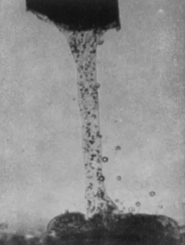
FIG. 2. Effusion of the endoplasm from the cell opening about 5 min after amputation of one end.
out gradually into the medium and falls down continuously onto the bottom of the cuvette without being dispersed into the medium (Fig. 2).
The effusion continues as long as 20-40 min without interruption; when the supply of endoplasm becomes scanty, a continuous strand of endo-
plasm is broken to form intermittent pendent drops, each of which is de- tached from the cell by gravity, and falls onto the bottom where it forms a sessile drop.
The cytoplasm isolated in this manner consists of endoplasm alone and suffers little injury, if any. These endoplasmic drops can survive usually for 10-50 hr in the medium (Kamiya and Kuroda, 1957a).
Movement in an Isolated Endoplasmic Drop
When the endoplasm comes out of the cell in the form of a con- tinuous strand, we notice that the entire strand falls down as a complete unit, there being no velocity gradient within it. What is noted here, however, is that the chloroplasts in the falling endoplasm rotate vigor- ously around their own axes, just as the few chloroplasts in the stream- ing endoplasm do in the intact cell.
When the endoplasm has settled on the bottom in the form of a sessile drop, both the nuclei and chloroplasts tend to sink toward the bottom, since they have a greater specific gravity than the cytoplasmic matrix. For this reason, the observation of their motion was best car- ried out with an inverted microscope with which the moving chloroplasts and nuclei can be seen most clearly in the same focal plane.
Endoplasm which streamed when in the intact cell no longer ex- hibits any mass streaming when isolated in vitro by this procedure.
This does not mean, however, that there is no motion inside the drop.
Besides Brownian motion, what is noticed here is vigorous rotation of chloroplasts and nuclei around their own axes (Kamiya and Kuroda, 1957b). The rate and direction of the rotation vary with each individual chloroplast and nucleus; the rate is usually within the range of 0.5-2 rps for the chloroplasts and 2 rpm for the nuclei.
An important question arises here as to why there is no mass streaming in the isolated endoplasm that was once streaming in the cell. Certainly injury caused by the operation of cutting the cell is not the cause of this, since chloroplasts and nuclei continue to rotate.
Through analysis of the intracellular velocity distribution of cyto- plasmic streaming in the internodal cell of Nitella, we came to the conclusion that the motive force of the streaming is an active shifting force generated at the boundary between the endoplasm and cortical gel layer (Kamiya and Kuroda, 1956). This is to say that the endoplasmic sol alone is not capable of streaming. Evidence presented by Dr.
Hayashi in this Symposium provides further support for this conclusion.
The fact that there is no velocity gradient within the falling endoplasm also may be understood from this viewpoint. Thus, it is reasonable to
believe that the absence of cortical gel is the reason that no mass stream- ing occurs in the isolated endoplasmic drop.
This same idea can also explain the fact that chloroplasts and nuclei continue to rotate in the isolated endoplasm, if we assume that the boundary between these organelles and the ground cytoplasm gen- erates the same active shifting force as the corticoendoplasmic boundary.
Thus we can say that when the solid phase is fixed (e.g., the cortical layer), cytoplasmic streaming takes place as observed in the intact cell, whereas when it is suspended (e.g., rotating organelles), the solid phase itself moves around as in the case of rotation of chloroplasts or nuclei.
When a chloroplast rotates slowly in the isolated endoplasm, it was observed, sometimes, that the endoplasm just around the chloroplast moves in the opposite direction. In order to clarify the relation of the movement of the chloroplast to the surrounding endoplasm, we tried to stop a chloroplast from rotating with a microneedle. As soon as rotation of the chloroplast was stopped, a local streaming of endoplasm was observed to take place along the surface of the chloroplast in a direction opposite to the original chloroplast rotation. If the microneedle was removed, the chloroplast started rotating again in the original direction, and there was no observable countercurrent of the matrix around the chloroplast.
These facts indicate that the rotation of chloroplasts observed in the isolated endoplasm is essentially the same phenomenon as the cytoplasmic streaming found in intact cells. Both are believed to be caused by the sliding force generated at the boundary between the plasmasol and plasmagel. The nature of this force is, however, still unknown.
Moving Cytoplasmic Fibrils
Recently, it was reported by Jarosch (1956a,b) that isolated proto- plasmic drops squeezed out of an internodal cell of Chara foetida con- tain many moving cytoplasmic fibrils. According to Jarosch these moving fibrils are so thin that only a well trained eye can recognize them in the dark field under high magnification. Both Kamiya and I confirmed many of his observations with Nitella flexilis. It was postulated by Jarosch (1956b, 1958, 1960) that these moving fibrils are oriented in an organized fashion on the inner surface of the cortical gel layer and that the protoplasmic streaming is brought about by the interaction between the endoplasm and those cytoplasmic fibrils. Moving fibrils, found in the cytoplasmic drops obtained from Chara or Nitella, may be considered as having detached themselves from the cortical gel plasm which is generally sloughed off when a drop of cytoplasm is
squeezed out of the cell mechanically. However, in order to account for the rotation of the nuclei in the cytoplasm streaming in the intact ceil, the presence of the fibrous elements is to be expected also in the endo- plasm. As a matter of fact, the moving fibrils could be found in the isolated drop of endoplasm without containing cortical gel plasm. They
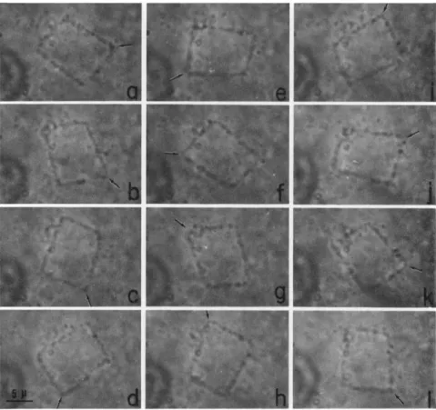
FIG. 3. Circular and polygonal loops of cytoplasmic fibrils in the isolated endo- plasm observed with an inverted microscope in bright field. Moving fibrils first appear in the form of ring (a). Later, they assume different shapes (b-f).
can be seen also in the bright field fairly well. Since the fibrils have a tendency to appear along the surface of the drop, they are observed clearly in one focal plane with the aid of an inverted microscope.
The isolated endoplasm contains only a small number of moving fibrils right after isolation; but the number increases gradually. Many undulating polygons of fibrils appeared in the same drop in a few hours.
This seems to indicate that the streaming endoplasm in the intact cell contains only a few visible moving fibrils, but contains a large number of elements capable of forming moving fibrils.
In the isolated endoplasm, moving fibrils first appear in the form of an extremely fine filament or loop which is in active motion. T h e
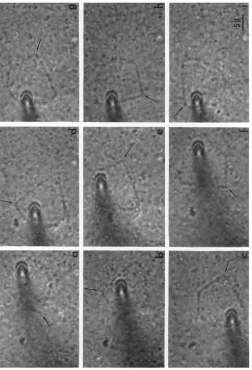
FIG. 4. T h e propagation of angles of a polygon as waves. Particles attaching to the fibril do not change their positions. Intervals of successive pictures: i/2 sec.
Arrows show a corner which propagates as a wave. (Observed with an inverted bright- field microscope.)
behavior of moving fibrils in the endoplasmic drops is quite similar to that in the squeezed out, cytoplasmic drop described by Jarosch (1956b).
Later these fibrils assume most fascinating shapes, such as semicircles, triangles, squares, pentagons, hexagons, and other polygons (Fig. 3).
It must be stressed that these thick fibrils display a remarkable un- dulating motion (Fig. 4); these undulations are observed only in thick fibrils which do not rotate vigorously as Jarosch pointed out. However,
the undulating motion propagates a wave in the direction usually op- posite to that of rotation. T h e polygons in which corners propagate as waves counterclockwise is about the same in number as those in which corners travel clockwise. By means of a cinematographic technique, we determined the rate of propagation of the undulating motion to be 9.8 μ/sec in an average of 15 polygons at 19-20°C and 13.1 μ/sec in an
T A B L E I
RA T E O F WA V E TR A N S M I S S I O N IN T H E LO O P S O F CY T O P L A S M I C FI B R I L S
Length of Rate of wave
Temp. Shape of fibril transmission
(°C) polygon (μ) (μ/sec)
19 Semicircle 37 9.3
19 Fan-shaped 37 10.4
19 Square 27 10.0
19 Pentagon 52 5.8
19 Pentagon 50 11.0
19 Pentagon 40 11.0
19 Pentagon 60 12.0
19 Pentagon 62 13.0
19 Hexagon 51 10.5
19 Hexagon 41 10.7
19.5 Pentagon 45 10.8
20 Fan-shaped 50 8.0
20 Triangle 41 5.3
20 Pentagon 43 9.0
20 Pentagon 57 10.8
24 Triangle 25 11.5
24 Square 37 15.5
24 Pentagon 48 9.6
24 Pentagon 40 13.2
24 Pentagon 72 13.0
24 Pentagon 73 14.6
25 Semicircle 39 12.9
25 Pentagon 49 14.7
average of 8 polygons at 24-25 °C. The rate of propagation increases with temperature, but it is independent of the number of angles, the shape and the size of the polygons; the angular velocity of wave propa- gation is greater the smaller the polygons (Table I).
Since moving fibrils in the isolated endoplasm could be observed in bright field, several micromanipulations could be made in order to in- vestigate the mechanical properties of fibrils. We first tried to disturb the polygons mechanically when they were in an early stage of forma- tion. When one of these polygons was dragged by a microneedle, it was split into a number of fine fibrils, each of which rotated very furiously without any sign of undulation. The fact to be pointed out in this
FIG. 5. A polygonal cytoplasmic fibril dragged back and forth by a microneedle. During this procedure the polygon scarcely changed its form and continued wave transmission just as before the dragging. Arrows show a corner which propagated as a wave. Intervals between a-b, b-c, d-e, e-f, g-h, h-i: 2 seconds; c-d: 5 seconds; f-g: 24 seconds. (Observed with an inverted microscope in bright field.)
case is that the newly formed fibrils always rotate in the direction op- posite to that of the transmission of the corners in the original polygon.
Next, the same operations were performed on polygons a few hours later when they had become thicker. Unlike the newly formed polygons, polygons at this later stage were easily dragged back and forth by the microneedle. During this procedure the polygons scarcely changed their form, and the corners continued to travel along the fibrils just as if they were free from dragging (Fig. 5).
The facts mentioned previously indicate that the polygon is com- posed of many finer fibrils, each of which is capable of moving rapidly.
During the formation of a polygon, these fine fibrils apparently form thicker fibrils by accretion until eventually they become strong enough to continue wave transmission even after some mechanical disturbance.
It was shown that the isolated endoplasm contains fibrous elements which could form moving fibrils visible at the microscopic level. In spite of this, no mass streaming takes place in the isolated endoplasm.
This may be because the moving fibrils are scattered around and have no organized orientation and coordination. If the moving fibrils are gradually arranged in an orderly fashion during the process of incuba- tion, mass streaming could begin to occur also in the isolated endoplasm.
A few experiments were performed under aseptic conditions with the purpose of prolonging the survival time of the isolated drop of the endoplasm. T h e incubation medium consisted of a $/Α concentrated solu- tion mentioned previously to which was added glucose, biotin, vitamin Bx, indoleacetic acid, the microelements of Heller, and a i/2 concentrated Knop's solution. After 1 day's incubation, some of the incubated endo- plasm began to show local cytoplasmic streaming. With the present technique the isolated endoplasm could survive about 3 days outside the cell. Naturally, experiments along this line await further improve- ment in the technique, but the results obtained so far are rather en- couraging. Studies on a fragment of cytoplasm in vitro and the analysis of its behavior have been conducted by many workers in the past.
Systematic work in this field will without doubt help us gain an in- sight into the mechanism of intracellular motions as well as many other important physiological phenomena.
RE F E R E N C E S
Donné, A. (1838). Ann. Sei. Nat., Bot. [2]10, 346.
Dutrochet, M., and Brongniart, A. (1838). Ann. Sei. Nat., Bot. [2]10, 349.
Jarosch, R. (1956a). Oesterr. Akad. Wiss. Math.-Naturw. Kl., Anz. No. 6, 58.
Jarosch, R. (1956b). Phyton (Buenos Aires) 6, 87.
Jarosch, R. (1958). Protoplasma 50, 93.
Jarosch, R. (1960). Phyton (Buenos Aires) 15, 43.
Kamiya, Ν., and Kuroda, Κ. (1956). Botan. Mag. (Tokyo) 69, 544.
Kamiya, N., and Kuroda, K. (1957a). Proc. Japan Acad. 33, 149.
Kamiya, N., and Kuroda, K. (1957b). Proc. Japan Acad. 33, 201.
Stâlberg, Ν. (1927). Botan. Notiser. p. 305.
Valkanov, A. (1934). Protoplasma 20, 20.
Velten, W. (1876). Oesterr. Botan. Z. 26, 77.
Yotsuyanagi, Y. (1953a). Cytologia 18, 146.
Yotsuyanagi, Y. (1953b). Cytologia 18, 202.
DISCUSSION
CHAIRMAN TH I M A N N: It is a fascinating picture. I am sure there are a lot of comments.
DR. RE B H U N: Your pictures seem to indicate two kinds of phenomena concerned with motion of the fibrils in the cytoplasm. When these fibrils move, they appear to have a countercurrent stream moving in the opposite direction. When they are aggregated into polygons, I have looked to see if there was a countercurrent stream and it appeared to me there was not. That is, it appeared when these fibrils formed polygons, whatever the process deforming the shape of the polygons was entirely within the fibril and not coupled to motion of the surrounding cytoplasm. Does that agree with your observations?
DR. KURODA: I think so.
DR. COUILLARD: Your polygons appear remarkably coplanar all the time. Have you considered the possibility that you might be observing an optical section of a three- dimensional body such as a polyhedron?
DR. KURODA: The polygons we observed are not an optical section of a three- dimensional body. The reason why they lie in the same plane is because they tend to appear in the peripheral layer of the drop. The polygons which are found in the inner space of the drop can take all possible orientations. In this case we observe only a fraction of them in one plane of focus.
DR. MAHLBERG: DO the polygons ever stop moving; and, if so, do they reverse direction?
DR. KURODA: They continue moving without interruption for hours. Sometimes the motion of the polygons lasts more than 24 hr. The direction of wave trans- mission never reverses.
DR. MAHLBERG: It appeared some of them slowed down during the filming. I wondered if the organelles continued moving independently, that is, in a wave action.
DR. KURODA: After the polygons have been formed, wave propagation generally con- tinues at nearly a constant speed for a few hours.
DR. MAHLBERG: Does the wave propagation ever stop?
DR. KURODA: I have never seen polygons that stopped completely. Only in a degenerating drop, cytoplasmic fibrils accrete in a needlelike structure which is stationary.
DR. GOLDACRE: DO you ever see short lengths of the polygon material moving in a straight line?
DR. KURODA: There are no such short pieces of the polygon material observable.
DR. GOLDACRE: Can you not snap the loop by pulling on two needles?
DR. KURODA: I tried to pull the loop using two microneedles. If it is stretched,, the polygon is split into finer fibrils which are scarcely visible. I suppose they are motile, but they are almost beyond the resolution of the optical microscope and it is hard to observe their motion.
DR. BO U C K: First, do you ever see fibrils in an intact cell or are they only in these isolated drops? Second, what are the particles?
DR . KU R O D A: The polygons have been also found in a centrifuged cell of Nitella
by Dr. Kamitsubo (unpublished) quite recently. Naturally this is not an intact cell but it is still living. My answer to the second question is that these granules are not identified cytologically or electron microscopically. According to Dr. Jarosch's classifica- tion of the cytoplasmic inclusions, they are probably prespherosomes.
DR. JA R O S C H: As to the first question of Dr. Bouck, I have seen moving fibrillar rings showing undulatory movement in the intact cell also. In a few cases it was possible to observe them with dark-field illumination lying in the stationary proto- plasm along the indifferent line. But this seems to be an exception. In most cases, there are no visible fibrillar structures in the intact cell.
DR. DEBR U Y N: One ought to get a motion something like that observed in your polygons if you could take a flagellum and tie the tail to the other end. Is any- thing known of the fine structure of these polygons?
DR. KURODA: NO. Nothing is known electron microscopically of the fine structure of these polygons. Dr. Terada studied both endoplasmic drops and internodal cells of Nitella, with an electron microscope, but he has not been successful in finding fibrous structures or any structure which seems to have a bearing on the polygons.
DR. IN O U É: I would like to point out the possibility that the polygonal move- ment could be explained by a twisting motion as seen in the axostyle of certain protozoa. If this is so, I would make two predictions which one could verify with the electron microscope; one, the cross section of the so-called fibrils ought to be asymmetrical instead of circular, and two, one should see not a regular sine wave but a three-dimensional saw-tooth wave. Judging from your photographs, I think there is a good likelihood that you are observing the propagation of such a twisting motion.
DR. AL L E N: One idea that comes to mind is the possible relationship between the movement of these fibrils and streaming of the intact cell. These fibrils might be oriented along the cortex in such a manner that some kind of waves passing along the fibrils (such as the straightening or stiffening waves) could drive the endoplasm.
DR . GO L D A C R E: I wonder about the membrane that appears to be around the isolated drops of cytoplasm. You say this is permeable to the kind of substances that will not enter the living cell, such as certain vital dyes.
DR . KU R O D A: There is a definite membrane at the surface of the isolated endo- plasmic drop. This membrane, I suppose, is more like a tonoplast than the proto- plasmic surface membrane. It has a weak tension amounting to 0.001-0.006 dyne/cm.
It is semipermeable to solutes. This drop can be vitally stained with some dyes, such as erythrosine, chrysoidine, and eosine, but details of permeability characteristics of this membrane are still unknown. When the drop degenerates, there is often a
"ghost" left.