Dynamic Organization: Morphophysiological Study of Ameboid Movement, IV
TOHRU H . A B E Laboratory of Biology, Hosei University, Tokyo, Japan
Since the first observations of ameboid movement and of protoplasmic streaming, an extensive literature dealing with either or both of these subjects has grown, offering us many different unestablished hypotheses on which variously detailed theories have been based. T h e methods of modern biology have tended to oversimplify the rather formidable com- plexities of the phenomena observed which take place in living organ- isms. Analytical studies of these phenomena have had to follow physical or chemical lines. In highly differentiated organisms, the application of physical and chemical methods is often well justified; but the ameba, lacking obvious structural differentiation, is often treated in analytical studies as if it contained no differentiation whatever. Bodies of amebae, however, are actually differentiated, but their structural organization, being dynamic rather than static, has often been overlooked. Earlier research workers who failed to recognize this dynamic organization tended to oversimplify the phenomena of ameboid movement in order to attempt to explain it in terms of seemingly analogous phenomena of physics and chemistry. This trend still prevails in biology, and serves, on the one hand, to advance the science and, on the other, to intro- duce almost insoluble problems, especially in the study of ameboid movement.
It is of greatest importance to question the reason why most, or perhaps all, hypotheses and theories of ameboid movement and proto- plasmic streaming are controversial and conflicting. This situation is apparently due to the fact that, first, there are many different types of ameboid movement and protoplasmic streaming, seemingly quite different from one another, which have in most cases been lumped together and discussed as a single phenomenon, second, cytoplasmic structure and its changes during cell deformation are not discernible, and third, there is a lack of suitable techniques to determine whether the mechanism of streaming is located within the stream itself or in some other part of the cell and whether the distal extension of pseudopods is active or passive.
221
Because of these difficulties, we have had either to explain the mech- anism in purely symbolic terms, to resort to the unanalytical process of
"labeling" phenomena as "Chemotaxis," "thigmotaxis," etc., or to find seemingly analogous phenomena in physics, chemistry, or physiology, such as contractility, extensibility, viscosity, attachment, spreading, ex- pansion, and viscoelasticity. It is often difficult to justify the use of these terms rigorously in biology.
There is a further fact which needs to be considered. There seems to be a tradition in science to accept experimental results at their face value as valid. Sometimes, however, it appears that we are not sufficiently critical in our interpretations. In such irritable cells as amebae, changes in body contour and protoplasmic streaming in response to a stimulus are rapid and distinct, but well regulated according to the dynamic organiza- tion of the body (Abé, 1961, 1962). If based on these considerations, critical reviews of the literature (De Bruyn, 1947; Allen, 1961a) should force us continually to re-examine old and new theories in order to determine what can be accepted as valid.
We should also bear in mind that the variation in types of cell de- formation and patterns of cytoplasmic streaming is so enormous that there may well be hidden cause-and-effect relationships between events occurring in different parts of an ameba.
Another problem confronting us is physiological, or at least related to our sensory physiology. One tends to become trapped in a pattern of thought which revolves about some hypothesis or theory, sometimes to an extent involving important aspects of phenomena observed under the microscope. T h e detection and study of motion falls also in this category.
It is difficult or, more probably, utterly impossible to follow accurately, and with the same reliable exactness, by the methods hitherto used, two or more events occurring simultaneously in different parts of a body.
This is important because measurements of particle velocities in different regions of the cytoplasm and at different instants are without significance unless combined with the knowledge of events in the posterior and other regions, particularly in the anterior frontal end (Abé, 1963).
Mechanism of ameboid movement and organization of amebae have been discussed in general ways by Pantin (1923), Mast (1926), Goldacre (1956, 1961), and Allen (1961a, b). I have also investigated these prob- lems to some extent using striata and verrucosa groups of amebae, which showed the existence of what is called "dynamic organization" (Abé, 1961,
1962, 1963). At least the first three of the four authors just cited presented the hypothesis that the probable site from which the motive force is generated to induce distal extension of pseudopods can be found in the posterior portion of an ameba body. Contrary to them, Allen presented
a new theory in which he suggested that the site is located in the distal end of extending pseudopods. In this respect, my conclusions (Abé, 1961, 1962, 1963) agree with Allen's suggestion, though they differ from it in some points. In this paper I should like to mention principally the ejecting process of the unusually large and solid diatom shell, which naturally tells us something about the dynamic organization and mech- anism of ameboid movement in Amoeba proteus.
Materials and Methods
The material used in this investigation was a large ameba of proteus type, 200-300 μ in body length, which had been collected from a suburban region of Tokyo and kept in culture for more than 10 years in our laboratory. Usually they are polypodial, but can be induced to take a monopodial form; in this shape the problem of analyzing and describing the inner structures of the cell are considerably simplified.
Photographic exposures of 1 sec duration were taken at appropriate intervals (10-30 sec) during the ejection of a digested diatom. Care was taken to include all portions of an ameba's body in every photograph except Fig. 4, so that one could study, correlatively, all the events occur- ring in different portions of the body. The longitudinal median portion of the body has a considerable thickness. Thus, granules clinging to the dorsal and ventral walls and their inner structures are out of focus in this thickened portion, although the author used a lens of low magnifying power ( χ 20, Olympus) to take the photographs. However, this is not true of the much thinner peripheral portions of the body.
The structural differentiations presented in the figures are limited to those which can be seen within the plane of focus; this can be confirmed in many other photographs, some of which will be published in my forthcoming works. T h e events which occurred during ejection of this food residue were so complicated that their analysis required several years, during which time so many supporting observations were made that it seemed best to document the observations with the actual photo- graphs, which may then be allowed to tell their own story.
There are only a few reports in the literature of the ejection of food residues by amebae (e.g., Grosse-Allermann, 1909; Neresheimer, 1905), and none of these have dealt with large particles. I have often observed the ingestion of large diatoms, but rarely their ejection. One such event had been recorded photographically and was analyzed in a manner which may shed some light on the relationships between cell deformation and cytoplasmic streaming. The slow-shutter streak-photograph method im- mediately shows where cytoplasmic streaming or, more probably, move- ment of granules has occurred; cell deformation is also shown, less clearly
in single photographs, as blurred regions of body contour, but, more clearly, by comparing the cell outlines in successively taken photographs.
The method must be used intelligently, for different lengths of exposure give quite different pictures. As discussed in my previous paper (Abé,
1963), all particles suspended in the flowing endoplasm move irregularly and at irregular points. An excessively long exposure invariably results in a figure full of granular streaks, whereas too rapid exposure results in a figure with entirely motionless particles. It is also essential to use a phase-contrast or other optical system so that the particles to be registered on film will usually be brighter than their background.
Observations
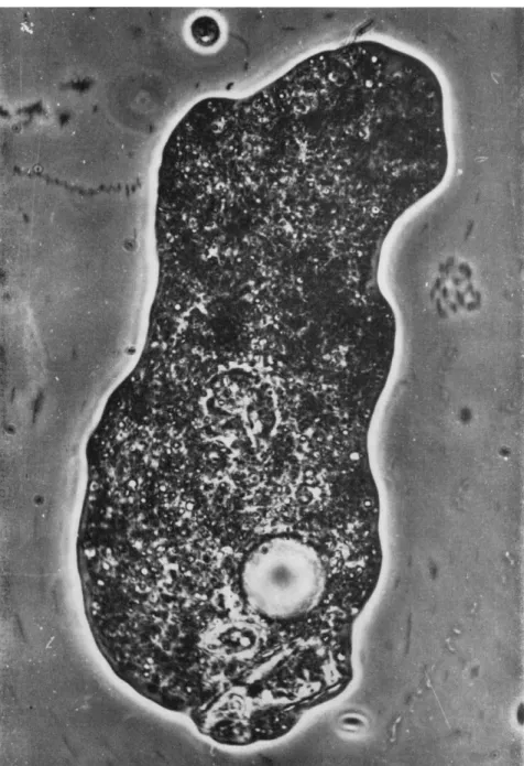
The sequence of photographs beginning with Fig. 1 shows the entire ejection process of a large and nearly emptied diatom shell lodged in the caudal end of a proteus type of amebae. This cell body had been floating motionless in an attached state owing to mechanical disturbance of the water, and had come to rest finally upon the substratum, to which it adhered. This indicates that the ameba is not flattened in any way, at any instant, from above the body. In the anterior end there is some slow cytoplasmic streaming in the forward direction, as evidenced by the slightly rod-shaped blurred images of particles in the subfrontal and anteriolateral regions. In contrast, no movement was detected in the posterior half of the body. In this stage, the pointed posterior end of the diatom was seen still covered with a thin layer of substance, pre- sumably the plasmalemma.
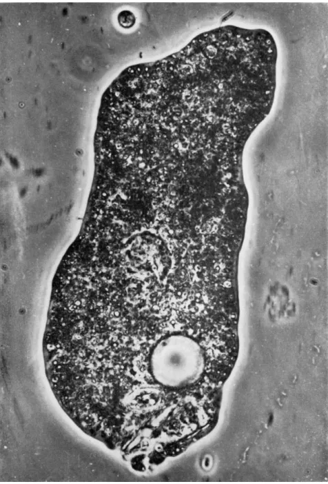
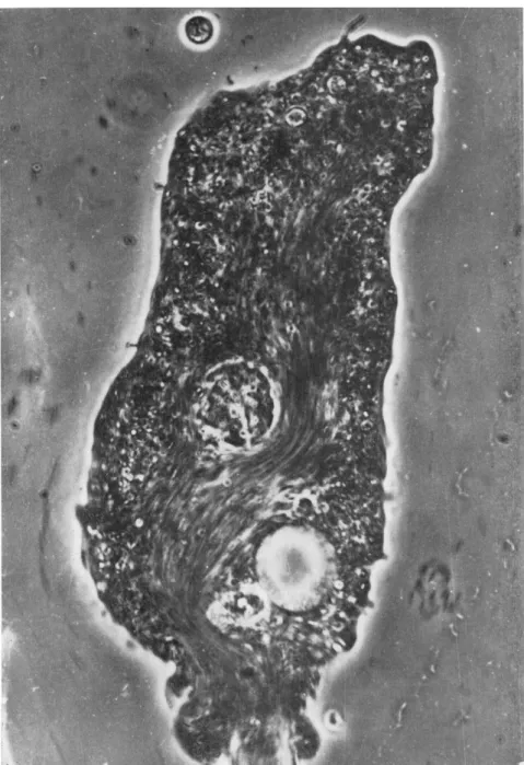
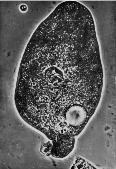
In Fig. 2 a new surface protuberance is clearly visible (compare Fig. 1) around the posterior end of the diatom, the distal end of which is exposed. This suggests fusion of the plasmalemma and the mem- branous wall of the food vacuole and their subsequent rupture to form a pore. In its vicinity weak, yet distinct, granular motion can be ob- served (compare Fig. 5); in spite of this, of the five photographs repro- duced here, Fig. 2 shows the least trace of granular movement. At the next moment (Fig. 3), the posterior bulge is rapidly extending, ejecting the diatom from the ameba. At the same time a vigorous cytoplasmic streaming is seen along the axial portion in the direction of the ejected diatom. The stream itself meanders, steering clear of the stationary nucleus and contractile vacuole. The streaks of various lengths through- out the body give a fairly accurate impression of the speeds and direc- tions of particles in motion. Judging from these streaks, motion is most rapid within the narrow channel near the contractile vacuole, and be- comes progressively weaker toward the old anterior end (top of the pho- tograph). Stationary particles are seen in various regions of the body.
They are crowded more densely or thickly anteriorly than posteriorly and toward peripheral than toward the median regions. In Fig. 3, vigorous cytoplasmic streaming can be seen along the posterior major portion covering about 9 6 % of the body length and 20 to 6 0 % of the body width. Slow, but distinct, particle movement can be seen even in the so-called ectoplasmic region, apparently contributing to the axial endoplasmic flow. This fact demonstrates the heterogeneity and spongy texture of the so-called ectoplasm (Abé, 1961, 1962, 1963; Goldacre, 1956).
More specifically, the so-called ectoplasmic gel layer is so constituted, at least in Fig. 3, that it is penetrated by canaliculi of fluid material in which small particles are suspended and can move.
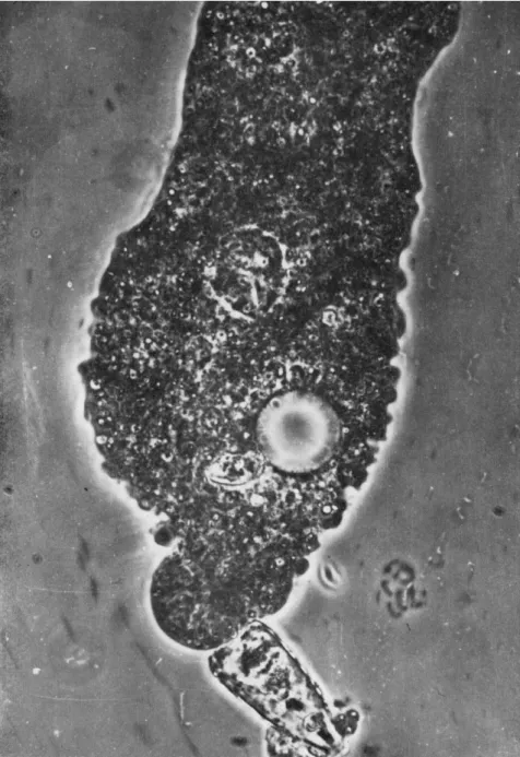
By the time Fig. 4 was recorded, the ejection process was nearly com- plete. T h e caudal bulge is about to cease its growth in length and in transverse dimensions judging from its sharply definable outer contour and very slow motions of particles lying within it.
Another striking feature of the period represented by Figs. 3 and 4 is the formation and growth in number and size of tiny knoblike surface projections along either side of the posterior half of the body.
Figure 5 was reconstructed by tracing Figs. 1 to 4 on the same sheet of paper, taking the positions of the nucleus and contractile vacuole as points of reference. From this reconstruction several changes can be seen which were not obvious from the photographs themselves:
1. T h e strongest deformation of the body contour occurred in the interval between Figs. 2 and 3 in the original posterior portion just an- terior to the bulge: in all probability this reflects a compensation for formation of the bulge.
2. T h e linearly arranged projections from the lateral edges of the body are not caused by contraction or by wrinkly shrinkage of the body.
3. In various parts of the body there were extensions and retractions.
It is particularly noteworthy that the anterior-most portion of the body (top right of figure) continues to advance in spite of the fact that the large particles A and Β move posteriorly. This would suggest that fluid matrix in the subfrontal region moved backward in accordance with the vigor- ous backward flow of the endoplasm in spite of the extension of the very surface of the pseudopod.
4. T h e stream lines shown in the posterior portion (Fig. 5) were taken partly from Fig. 2 (left side) and partly from Fig. 3 (right side).
The former represents the flow preliminary to expulsion of the diatom shell, and the latter is that accompanied by expulsion. The flows repre- sented on the right and left sides of the diagram are correlated clearly with the deformations occurring on the same side. Measurements showed that the direction of the stream on the right-hand side turned at an
FIG. 1. Diatom lodged in caudal end of proteus type of amebae. In anterior region some slow cytoplasmic streaming in forward direction is seen (rod-shaped blurred images). Posterior end of diatom covered with thin layer of substance.
FIG. 2 . Posterior end of diatom has new surface protuberance. Weak granular motion is visible in its vicinity.
FIG. 3. Posterior bulge ejecting diatom from ameba. Vigorous cytoplasmic stream- ing in direction of diatom.
FIG. 4. Ejection of diatom almost completed. Caudal bulge has sharp outer contour.
respectively, the body contours of Figs. 1-4.
230
FIG. 6. Ameba is now longer and wider (especially in posterior region) and ap- parently endoplasmic flow has resumed. A narrow hyaline zone is visible around the body.
angle of approximately 340 degrees from its original anteromedial direction toward the posterior end. This acute change of direction and accompanying phenomena are very unusual and cannot be explained by the assumption of active movement of the fluid matrix or of the granules.
5. The ameba seemed to exert some degree of coordination in eject- ing the diatom. The exact manner in which the caudal bulge and bi- lateral retractions of the body surface were brought forth is suited for rotation of the diatom to minimize its frictional resistance. Rotation was enhanced in part by the initial axial flow (Fig. 2) which pushed the distal end of the diatom in the posterior direction. The retraction of the body surface on the right side then completed the rotation process and partly performed the ejection.
6. The anterior displacement of the arrested smaller food residue (FV) seems paradoxical unless one takes into consideration the fact that the retracted part of the body wall, which happened to push the flared end of the diatom inward, undoubtedly forced, in turn, the food residue (FV) through a gelated region which otherwise would have served to keep it stationary.
7. First there was a slight clockwise, then a much more pronounced counterclockwise, rotation of the nucleus. T h e former appears to be caused by the vigorous but unusual rearward flow of the endoplasm in contact with the nuclear surface; the latter might have been caused by the normal forward flow of endoplasm in the interval between Figs. 4 and 6, which can be seen, in part, in Fig. 6.
8. The distance between the nucleus and contractile vacuole in- creased in the interval between Figs. 4 and 6 (longer than the intervals between the other pictures). This movement is apparently not due to contraction of the body, but to the reopening of endoplasmic flow which results in elongation of the body, particularly in its anterior half, and by dint of which the nucleus is carried forward.
By the time Fig. 6 was recorded, the organism had become even flatter than before, and therefore was longer and wider (especially in its posterior region); yet its original organization remains, as suggested by the reopened, forward, endoplasmic flow in the anterior portions of the body. A narrow hyaline zone is formed almost all around the body.
This may in all probability be due not to inward retractions of periph- eral granules but to extension or formation of hyaline structures.
Discussion and Conclusions
The foregoing appears to be the first documented example of the ejection of a large food residue from an ameba. The details of the proc- ess are such that my original conclusions about the importance of dy-
namic organization in amebae (Abé, 1961, 1962, 1963) appear to be gen- erally applicable, regardless of differences in taxonomic situations of amebae and in size, shape, or types of deformation and locomotion of the cell body.
Perhaps the most noteworthy feature of the ejection process is that the so-called strongest "contraction" of the body occurred not in the region of the body where the rearward endoplasmic streaming originated
(i.e., the original front end), but rather in the temporarily formed sub- frontal (i.e., old posterior) region of the flow, close to the bulge. Thus, the site of body contraction remained the same even at or after the time when the direction of vigorous streaming reversed temporarily, and the ordinary forward flow reappears invariably in the old anterior end of the body. This seems difficult to account for on the basis of the tail contraction theory (Goldacre, 1956, 1961; Mast, 1926; Pantin, 1923) and perhaps others (Allen, 1961a).
The morphological peculiarities generally accepted as occurring in the portions of the body in which the ordinary endoplasmic stream originates cannot be seen in the old anterior end in which the temporarily reversed vigorous stream starts in this case. The distinct diminution of the body dimensions or the contraction of the body could be seen in the subfrontal portions of the reversed endoplasmic flow (e.g., the old posterior most portions of the body surrounding the unusually formed caudal bulge).
In other words, continued solation and carrying away of fluid matrix, which under normal conditions induce so-called "contraction," could not be seen in the old anterior region. Furthermore, the vigorous backward stream did not prevent the continued (although slight) advance of the original front end, nor did it cause continued locomotion of the body in reversed direction. These observations seem particularly difficult to ex- plain in terms of ectoplasmic contraction or of the simplified sol-gel trans- formation hypothesis. At the same time, they suggest some degree of inde- pendence of the pseudopodial extension from the seemingly distinct endoplasmic streaming within. It is to be recalled in this connection that in Fig. 3 the stationary granules are seen along the entire breadth of the original front end, suggesting the existence there of gelled struc- tures; however, not a single arrested particle can be seen along the new frontal surface and within the extending bulge. It will be recalled that in striata amebae, there is a fenestral opening in the posterior end of the body.
There has been a traditional tendency to treat the endoplasmic stream as an entity. But Fig. 3 and subsequent ones, it seems to me, may sug- gest some differences in physiological properties between the temporarily induced rearward flow and the normal forward flow of endoplasm, be-
cause of the fact that the latter is invariably associated with solation in its streamhead regions, whereas the former is not, but instead seems to be supplied with newly evolved sol in its downstream portion. The author's previous writings concerning, e.g., the anteroposterior differences in physiological activities and some other properties of the normally flowing endoplasm of striata ameba (Abé, 1961, 1962) may serve to some degree to explain these peculiarities. It may be permissible for me to suggest that endoplasm lying in the posterior end of the body is newly evolved from gel structures by their solation, and has the least capability for gelation (Abé, 1961, 1962) and the strongest capability for solation acting on gel structures regardless of what it encounters. This may ex- plain why vigorous gelation does not occur along the surface of the bulge into which the newly evolved sol might have been poured, and, at the same time, why the nucleus was least influenced by the rearward stream but was strongly carried forward, showing distinct rotation in accord- ance with the normal forward flow (Abé, 1961, 1962). According to the views I have already expressed elsewhere (Abé, 1961, 1962), gelation oc- curs wherever active extension of a pseudopodial tip is carried on. Based on these, one may be allowed to conclude that the formation of the caudal bulge in this case is apparently passive and never active.
The position or line along which the knoblike surface projections are formed corresponds to that of the so-called "lateral fin" in striata amebae. T h e same kind of structure is seen fairly frequently along the laterals of locomoting, large amebae and also partly or entirely around small species of Amoebidae. T h e flattening of the body seen in Fig. 6 is not due to passive flattening of the body because no pressure was applied to the body from above. It is due to growth not only in length of the body but also in breadth of the bilateral projections forming a conjoined broad membranous pseudopod, by which the ameba can ad- here firmly to and also extend along the substratum. Adherence of the ameba body at the time of ejection of an unusually large food residue is of frequent occurrence and may be, it seems to me, reasonable. Based on the frequent splitting and reunion of the peripheral membranous pseudopods seen quite often in the smaller amebae, the body-contour change occurring in the interval between Figs. 4 and 6 corresponds ex- actly to that described for tissue culture cells under the heading of
"spreading" or "expansion" (Abercrombie, 1961; Ambrose, 1961; Taylor, 1961; Weiss, 1961). However, the use of these terms should not mislead us into believing that the process has more than passing similarity to the spreading or expansion of a liquid drop.
Under normal conditions of locomotion, the posterior end of the ameba body is the ordinary site of the most vigorous solating activity
(Abé, 1961, 1962, 1963). In the present case, it is certain that the diatom surrounded by its vacuolar wall lies in this part of the body filled with freshly evolved sol. At the time of ejection of the diatom, it is apparent that there is formed, through the gelled body wall, a large pore, the diameter of which is a little smaller or larger than that of the diameter of the diatom. Taking into consideration the fact that this region nor- mally has the least gelating activity, there cannot occur rapid gelation all over the pore so as to cover it completely, just as is the case with the discharging pore of the contractile vacuole in striata ameba (Abé, 1961, 1962). Then the membranous wall of the food vacuole might nat- urally cover the pore and subsequently be pushed out so as to cover at least a major portion of the surface of the caudal bulge, if the fluid endo- plasm moves passively under the influence of pressure from within the body.
The explanation for these phenomena is not based on any of the old or current hypotheses, but solely on this author's interpretation of the dynamic organization of the ameba's body. This entails organized gel structures with a considerably higher order of physiological and morphological complexity than has hitherto been recognized. T h e ob- servations and discussions given in this paper demonstrate that this or- ganization persisted throughout the period in which the polarity of streaming temporarily reversed.
Though the tail contraction hypothesis is not supported, in my opin- ion, by the facts presented here, it is, of course, possible that the vacu- olar wall surrounding the diatom might serve to eject the diatom by dint either of local contractility or turgidity of the body as a whole. But this assumption is insufficient to explain the continued distal extension of the pseudopodial tip. However, it may be a matter of question whether or not the ejection process proper is related to the normal mechanism of ameboid movement. In the case of small food residues, no sign of disturbance in ameboid movement can be detected. T h e disturbance was shown in such an exaggerated degree in the present case presumably because of the considerably larger size and rigid structure of the food residue, both bringing forth unusually strengthened and causally induced activities in different portions of the body. Not rarely, a much larger diatom is captured and completely digested by Amoeba proteus before its ejection. In such cases, locomotion usually stops just as in this case. Therefore, the processes described in this paper are by no means abnormal or unusual.
By analyzing the ejection process, the author has been led to con- clude, in agreement with Allen (1961a, b), that the site of generating force or mechanism to bring forth the distal extension of pseudopods
and protoplasmic streaming must be sought for in the anterior-most portion of a body or of a psevidopod.
ACKNOWLEDGMENT
The author wishes to express his sincere thanks to Assistant Professor K. Ishii of Hosei University for the extreme care he exercised in taking the photographs in conformity to the author's ideas and suggestions.
REFERENCES Abé, T. H. (1961). Cytologia (Tokyo) 2 6 , 378.
Abé, T. H. (1962). Cytologia (Tokyo) 2 7 , 111.
Abé, T. H. (1963). / . Protozool. 1 0 , 94.
Abercrombie, M. (1961). Exptl. Cell Res. Suppl. 8 , 188.
Allen, R . D . (1961a). In "The Cell" ( J . Brächet and A . E . Mirsky, eds.), Vol. II, pp. 135- 216. Academic Press, New York.
Allen, R . D . (1961b). Exptl. Cell Res. Suppl. 8 , 17.
Ambrose, E. J . (1961). Exptl. Cell Res. Suppl. 8 , 154.
De Bruyn, P. P. H. (1947). Quart. Rev. Biol. 2 2 , 1.
Goldacre, R . (1956). Proc. Intern. Congr. Cybernetics, 1st Namur, 1956, p. 715. Gau- thier-Villars, Paris.
Goldacre, R . (1961). Exptl. Cell Res. Suppl. 8 , 1.
Grosse-Allermann, W . (1909). Arch. Protistenk. 1 7 , 203.
Mast, S. M. (1926). / . Morphol. Physiol. 5 1 , 347.
Neresheimer, E. (1905). Arch. Protistenk. 6 , 147.
Pantin, C. F. A . (1923). / . Marine Biol. Assoc. U.K. 1 3 , 24.
Taylor, A . C. (1961). Exptl. Cell Res. Suppl. 8 , 154.
Weiss, P. (1961). Exptl. Cell Res. Suppl. 8 , 260.
DISCUSSION
DR. WOLPERT: I would just like to ask whether the diatom which was egested had a vacuole around it.
DR. ABÉ: Apparently not.
DR. INOUÉ: In your photograph (Fig. 3), it appeared that there was streaming in the cytoplasm of the ameba near the top of the picture after the diatom was ejected.
DR. ABÉ: Streaming began there, not in the posterior.
DR. INOLÉ: Then, the first streaming always appears near the surface in the di- rection in which the ameba moves. Is that correct?
DR. A B É : Yes.