Ameboid Movement
R . J . G O L D A CE R
Chester Beatty Research Institute, London, England
A. Introduction
Ameboid movement is perhaps one of the most striking visible mani- festations of life in the cell, and attempts to explain this movement have been made for about 130 years (see review by De Bruyn, 1947). In this century, most investigators have agreed that locomotion is a result of the contraction, at one end, of a tube of plasmagel, which encloses plasmasol.
This is expressed in the theories of Pantin (1923) and Mast (1926). It was not possible to say much about the mechanism of the contraction until the isolation of muscle proteins and their reaction with adenosine triphosphate (ATP) (Szent-Györgyi, 1947) suggested that something similar to this reaction was going on in the contracting part of the ameba (Goldacre and Lorch, 1950). Considerable support for the involvement of A T P came from the experiments of Loewy (1952), Ts'o and colleagues (1956), Hoffmann-Berling (1960), Simard-Duquesne and Couillard (1962), and others. T h e theories of Pantin (1923) and Mast (1926) have been elaborated by Goldacre (1952a, b, 1953, 1954, 1958a, b, 1961) to account for the behavior of amebae in a variety of activities and for the control of locomotion by a feedback process.
In the last few years there has been a considerable increase in interest in the mechanism of ameboid movement, and several new theories have been proposed (Allen, 1961a,b,c; Ambrose, 1961; Bell, 1962; Bingley and Thompson, 1962; Kavanau, 1963). I propose to discuss some of these in the light of experimental evidence presented here.
In this paper I shall consider the location of the motor mechanism of ameboid movement, its nature, and how it regulates itself.
Unless otherwise stated, the species of ameba used in the new experi- ments reported is Amoeba proteus.
B. Is Ameboid Movement a Result of a Pull from the Front or a Push from the Back?
The "fountain-zone" theory of Allen (1961a,b,c) requires a pull from the front as opposed to the theories of Pantin (1923), Mast (1926), and
237
Goldacre and Lorch (1950), which postulate an active contraction at the back. T h e following points are opposed to a pull from the front.
1. In a polypodial ameba, a persisting pull from the front of each pseudopod advancing in different directions would pull the cell into pieces. Cytoplasmic movements are strong enough to do this, as, for example, in cell division, and in the tearing of captured prey into pieces by pseudopods (Mast and Root, 1916; Beers, 1924). On the other hand, the existence of more than one rear contracting region (such as
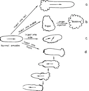
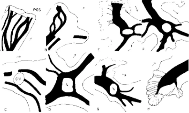
FIG. 1. Effect of microinjection of A T P ( 2 % ) into various parts of the ameba. In each, a vigorous contraction of plasmagel occurred at the site of injection immediately, resulting in the wrinkling of the cell membrane over it. These secondary "tails" soon fused with the original tail material. Note reversal of streaming after injection into front end, which then became a tail region.
a tail region and a retracting pseudopod) merely leads to the ultimate fusion of the two contracting regions through the contraction of the part of the cell between them (see, for example, Fig. lc and d). As cell fragmentation does not occur in ordinary locomotion, the advancing tips must be moving passively.
2. A pull from the front requires the "pumping-out" of the tail, resulting in the wrinkling of the membrane in it; if the membrane at the tail region is broken, the external medium should be sucked into the cell. This does not happen—cytoplasm always escapes (de Bary, 1864;
Goldacre, 1961).
3. A glass spring microbalance, made in the shape of a two-pronged fork on the de Fonbrune microforge, showed, by the squeezing together of the prongs when it was pushed into the tail region of amebae, that there was an active contraction there (Goldacre, 1961). There was no effect on the prongs when it was placed in the "fountain zone," in- dicating an absence both of a contraction [which should occur according to Allen's (1961a,b,c) theory], and of an expansion.
4. While the shape of the ameba is continuously changing, and the cytoplasm circulates and occupies each different part of the cell in turn, the tail is the only permanent surface feature. The leading pseudo- pod in a polypodial ameba may retract from time to time, but the tail remains as a permanent contracting region of the cell (Goldacre, 1961).
If the motive force were located in the anterior end of the cell, it would be extinguished when the leading pseudopod retracted. Analysis of cine film of the polypodial Amoeba discoïdes showed that this happened about every 3 min.
Now follow a number of experiments which indicate that the tail is a special, unique region of the cell, which is compatible with its being a motor region and incompatible with the location of the motor in the front.
5. The results of the microinjection of A T P into the ameba were briefly described by Goldacre and Lorch (1950) and more extensively by Goldacre (1952c). Figure 1 summarizes the results of microinjection into several hundred amebae of small amounts of 2 % A T P solution (neutral- ized with NaOH) into various sites of the cell. In each case, a vigorous contraction began immediately, and the membrane near the site of in- jection wrinkled as if from the contraction of the plasmagel underlying it. This wrinkling was similar in appearance to a large "tail" or uroid, and suggests that in the normal ameba's tail there is a continuous release of A T P by some enzyme reaction.
The effect of the injected A T P lasted about 1 min, and the solution could be injected repeatedly to produce the same effect.
It is noteworthy that when two wrinkled tail regions were simulta- neously present in the cell (Fig. lc and d), they soon fused into one, owing to the eventual contraction of the region between them, for each produced a propagated contraction, the contracted material liquefying to flow away along the central channel of the cell.
Control injections were done with other substances, none of which caused a contraction or wrinkling of the membrane. These were:
Chalkley's medium, Ringer's solution, MgCl2 equimolar with the A T P injected, distilled water, 0 . 1 % neutral red, and 2 % lissamine green.
That the contraction observed with 2 % A T P was not produced by
a mere mechanical stimulation of the cell was also shown by injection of a series of progressively more dilute solutions of ATP, which produced progressively less effect. Below 0.4% ATP, there was hardly any response.
It is to be noted that the actual effective concentration was much lower than these figures, owing to diffusion beyond the point of injection.
An interesting effect owing to the reversal of the direction of stream- ing by the injection of A T P into the advancing tip of the ameba (Fig.
Id) was the appearance of the nucleus and contractile vacuole in the new advancing tip of the ameba (which had previously been the tail end). Since the nucleus normally maintains a fairly central position in the cell, and is not carried along in the stream like the small granules, this suggests that the nucleus is held back or "filtered out" by a net- work of plasmagel strands in front of it which are absent behind it, so that when the stream is artificially reversed, the nucleus can enter the tail. Further evidence for this plasmagel network is given below (Figs.
5 to 7).
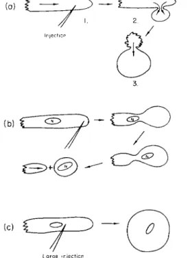
6. T h e microinjection of heparin (1:1000 solution, in amounts of about 3 times the volume of nucleus) produced results which were the opposite of those made by A T P (Goldacre, 1952c). Heparin caused a spherical pseudopod to be blown out at the site of injection (Fig. 2). This was in contrast to the cylindrical one formed normally when the walls were stiffened by the plasmagel tube. Often the whole of the cell would blow out into the pseudopod, as if that were the weakest part, giving way under the pressure within the cell (Fig. 2a). Sometimes the spherical pseudopod was pinched off. Often the nucleus moved into the spherical pseudopod as if all plasmagel network resistance to its passage along with the plasmasol stream were dissolved.
A large quantity of heparin liquefied the cell completely. T h e heparin influenced a volume of cytoplasm many tens of times greater than the volume injected. It appears that heparin both liquefies existing gel and prevents the formation of fresh gel.
T h e local absence of plasmagel caused by heparin in the advancing end of the cell indicates that no active contractile process can go on there and that events there are passive.
7. When an ameba is placed in neutral red solution for a few seconds, the plasmagel tube is stained red. If the ameba is then replaced in its natural medium, the dye is progressively released into the tail as the tail overtakes each portion of the red plasmagel tube in turn, and the plasmasol which streams forward from the tail is colorless (Goldacre,
1952a), so that the tail contains all the dye by the time the ameba has streamed through its own length. Nothing happens at the anterior end. It is interesting in this connection that stretched and unstretched
muscle have a different adsorptive power for dyes. Margaria (1932, 1934) found an apparent change of pH of 1.4 units in muscle stretched in the presence of phenol red. This was reversible. T h e release of neutral red thus suggests an active process in the ameba's tail, i.e., a contraction producing a decrease in dye adsorption.
It has been suggested by Bingley and Thompson (1962) that the neutral red accumulates in the tail because of an electrical potential gradient which they reported in the cell. This cannot be so, because when
L a r g e i n j e c t i o n
FIG. 2. Microinjection of heparin (1:1000 solution). Note liquefaction of plas- magel at site of injection resulting in the blowing out of a spherical rather than a cylindrical pseudopod.
oppositely charged dyes (such as eosin and the common anionic in- dicators) were injected into the cell, they spread evenly throughout the cytoplasm (Clark, 1943) and did not accumulate at the anterior end of the cell. This experiment also disproves Kavanau's (1963) suggestion that neutral red goes to the tail because water is driven in the opposite direction to the stream of particles which, he says, are jet-propelled.
Kavanau's mechanism should apply to all dyes, and not exclude the anionic ones; moreover, it would merely cause circulation, not accumula- tion of the dye solution.
8. When amebae are exposed to about one-quarter saturated, aqueous solutions of volatile fat-soluble substances, including anesthetics (such as chloroform, ether, and benzene), the plasma membrane becomes raised away from the granular cytoplasm owing to an increase in thickness of the hyaline layer, except at the tail, where the membrane and plasmagel remain attached (Goldacre, 1952b). This again shows that the tail is a special region.
9. If A T P is being used up continuously in the ameba's tail, one might expect to find phosphate ions from its breakdown there. A rather long-
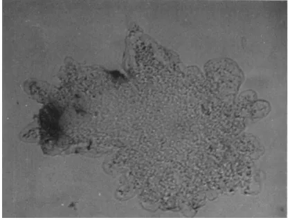
FIG. 3. Ameba showing, as blackening, the intense blue color produced in the tail when a reagent producing a blue color with phosphate ions is dropped over it when streaming actively (see text).
shot experiment was carried out in the hope of testing this. T h e medium was sucked away from amebae streaming actively in a drop on a slide and replaced by a drop of a solution which turns an intense blue with phosphate ions (ammonium molybdate, 0.1%, and stannous chloride, 0.1%, in Ν sulfuric acid). This solution, though toxic, permitted ame- boid movement to continue for 2 to 3 min, but the tails began to turn blue within a few seconds, so that the experiment was over before the amebae had stopped streaming. It was also found that prodding the ameba with a blunt needle [which would normally produce a local contraction (Goldacre, 1952b)] produced a blue color at the place touched (Fig. 3).
Control amebae previously fixed in osmic acid gave no blue color, as
would be expected, for phosphate ions would soon diffuse away in the fixative. These results are consistent with the production and breakdown of A T P at the tail and site of stimulation, respectively. There might be other interpretations of the blue color, however, owing to the extreme sensitivity of the reagent and to the fact that the amebae were not alive (though fixed by the reagent) at the end of the experiment.
These nine experiments indicate that the active process, which drives the ameba along, is located in the rear of the cell and cannot be in the front.
C. The Nature of the Ameba's Tail
The experiments in Section Β suggest that the ameba is driven along by the continuous production, in the tail region, of ATP, which would cause contraction and then liquefaction of the plasmagel in the tail. T h e resulting plasmasol would flow up the central liquid channel of the ameba to gel once more on the walls of the advancing pseudopods.
The question arises, how would the continuous production of A T P in the tail come about? T h e continuous production of anything at any fixed part of the ameba seems at first sight impossible, since the cyto- plasm circulates every 2 min and occupies each part of the ameba in turn, so that, for example, any enzyme which might be producing the A T P in the tail would be moving continually out of place; moreover, it is possible to cut off the tail (or the front end) of the ameba without interfering, except momentarily, with ameboid movement (Radir, 1931);
a new tail (or front end, respectively) forms again to replace the old one immediately.
A clue was given by the fact previously mentioned that anesthetics raise the membrane except at the tail. Inspection of the ameba under high-power magnification showed that in the normal ameba, also, the membrane was in contact with the granular plasmagel in the tail, but elsewhere was insulated from it by a hyaline layer which, however, was much thinner than in anesthetized amebae (Goldacre, 1952b, 1961). This is in contrast to Mast's (1926) much-reproduced diagram which shows hyaline layer over the whole surface of the ameba including the tail.
It seemed likely that this membrane-plasmagel contact in the tail was the cause of the enzyme reaction there, and this was tested by causing the membrane to contact the plasmagel by five different meth- ods:
1. Prodding with a blunt needle. Whenever the membrane was pressed right across the hyaline layer, a response to touch occurred (Gold- acre, 1952b).
2. Passing an electric current (Fig. 4). Membrane and cytoplasm moved
in opposite directions, and cytoplasm pressed against the membrane on the anodal side of the cell. These then contracted and became the tail (Goldacre, 1958a), causing the well-known streaming toward the cathode (Mast, 1931).
3. Sucking sharply at one end of a capillary tube in which an ameba has been squeezed. Hyaline fluid could be sucked from one end of the cell to the other (like water through a sponge), raising the membrane at one end of the cell and pressing it against the plasmagel at the other.
The latter end became the tail. T h e end which was the tail could be reversed at will by this method (Goldacre, 1958a).
F i g . 4. Effect of passing a direct current across the cell. Cell membrane and granular cytoplasm move in opposite directions to press together on the anodal side (-(-) of the cell. This contact causes a contracting tail region there, and the ameba then streams actively toward the cathode (—).
4. Pressing on the cover slip over an ameba, to flatten it to several times its original area. This caused the membrane to contact the plas- magel over most of the surface of the ameba, which contracted all at once when the cover slip was raised, pulling the ameba into a spherical shape. A similar effect was produced by sucking an ameba repeatedly in and out of a narrow tube so as to squeeze its sides. On release it rounded up. This is a well-known technique for producing spheres for volume measurement.
5. Replacing the external medium with oil. A striking phenomenon occurred. T h e amebae were washed in distilled water, the water was sucked away as much as possible, and about half a minute allowed for the evaporation of the last trace, shown when gentle blowing caused the surface of the ameba (as seen in the low-power microscope) to wrinkle.
T h e ameba was immediately covered with oil, whereupon pulsations began. Under high power, it was seen that the granular cytoplasm slowly (1-3 μ/sec) moved outward until it touched the cell membrane; immedi- ately it pulled away rapidly (10-20 μ/sec) by about 20 μ. This plasmagel
+
4- 2
expansion-contraction cycle of absorption of hyaline fluid followed by contraction and syneresis to produce insulating hyaline fluid between membrane and plasmagel again occurred about every 10 sec. A somewhat similar phenomenon has been reported by Kavanau (1963).
These five experiments indicate that it is the membrane-plasmagel contact which causes contraction in the tail. This explains why cutting off the tail has no effect—new membrane becomes pressed against the plasmagel at once.
One supposes that there is an enzyme on the membrane which has a nondiffusible substrate in the plasmagel, from which it is insulated over the surface of the ameba except in the tail [and also retracting pseudopods (Goldacre, 1961)]. Contact in the tail would produce A T P continuously, and this would cause continuous contraction in the tail.
How this is controlled by feedback is discussed in Section E,l.
The presence of an occasional clear blister or occasional trapped food vacuole in the tail does not invalidate the argument above, since each contact between plasmagel and membrane causes contraction over a considerable area beyond it (as does an injection of ATP), and any pockets of passive material in between contacts would merely be pulled along until they were eventually disposed of.
D. Experimental Evidence for Plasmagel Network The heterogeneous structure and consistency of the cytoplasm is of interest in relation to the maintenance of the position of the larger organelles, such as the nucleus and contractile vacuole. Information about this was obtained as follows:
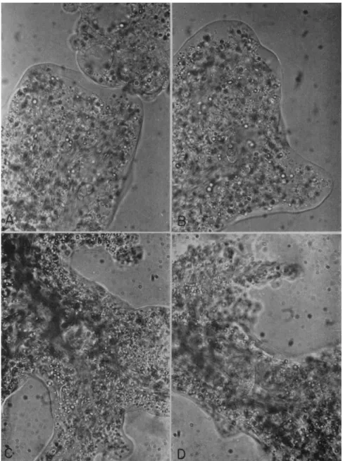
Actively streaming uncompressed specimens of Amoeba proteus were photographed with a high-power objective ( χ 9 5 , N.A. 1.3) with an ex- posure of 3 sec. This gave an optical section in which the particles in the moving cytoplasm were streaked in the direction of their movement, whereas those in the stationary cytoplasm gave sharp images (Figs. 5 and 6). The apparent plasmagel network structure is correlated with the observation that the finest cytoplasmic granules flow smoothly forward, larger ones start and stop and may move sideways before proceeding forward (as if moving around an invisible obstruction), and the largest particles, such as nucleus and large food vacuoles, are held back com- pletely (Goldacre, 1952c; Marsland, 1950; and, in a striata ameba, Abé, 1961). Similar observations have been reported for the movement of these cytoplasmic particles in a centrifugal force field (Allen, 1960;
Harvey and Marsland, 1932).
Figure 7 shows diagrammatically the distribution of plasmagel and
FIG. 5. Photos showing existence of interlocking plasmasol-plasmagel network in Amoeba proteus. High-power optical section with long exposure (3 sec) causing streaking of moving particles along direction of movement. Note how nucleus is held back in C and D by the narrower channels in front of them. For corresponding drawings showing position of plasmasol-stream network in black and plasmagel network in white, see Fig. 6A, B, F, G.
plasmasol in the ameba (drawn in the monopodial form for simplicity), based on evidence from many photos of the type shown in Fig. 5, from direct microscopical observation, and from time-lapse microcinematog- raphy. The hyaline layer and most of the finer plasmasol channels are omitted for clarity.
The diagram shows at once why the nucleus is not carried forward with the other particles surrounding it in the axial stream, but main-
FIG. 6. Drawings showing position of plasmasol-stream network (black) and plasmagel network (white); taken from many photos similar to those in Fig. 5, at various parts of the ameba. Note how nucleus is held back in D, E, F, and G, and the contractile vacuole in C, by the narrower channels in front.
Fig. 7. Composite picture showing distribution of plasmagel (white) and plasma- sol stream (black), based on many photographs similar to those in Fig. 5, on cine films, and on direct observation. T h e smallest plasmasol streams are omitted for clarity.
This distribution of plasmagel explains why the nucleus and contractile vacuole maintain a central and posterior position, respectively.
tains a position in the cell a little behind the center. The plasmasol stream behind the nucleus is wide enough to allow its passage, but the plasmasol streams in front of it are too narrow (Figs. 5 and 6D, E, F, G).
The nucleus is thus "sieved out." Thus, when A T P was injected into the front end of the ameba so as to cause reversal of flow (Section B,5), it was possible for the nucleus to move up to the anterior end (the former tail) because there was a wide channel with no plasmagel network to obstruct it in this region (Fig. 7). Other means of liquefying the resisting plasmagel network in the anterior half of the cell also allowed the
nucleus to move to the front, for example, microinjection of heparin (Section B,6) and heating to just below the point (about 40°C) when the plasmagel completely liquefied and the cell rounded up; this was preceded by very fast streaming, and the nucleus appeared in the front end of the cell. T h e contractile vacuole appears to be trapped in the thick, soft plasmagel in the tail formed by the pulling inward of the walls by the tail contraction, so that there is no free wide channel in front of it. Large food vacuoles sometimes became trapped in the tail region, also, being pulled around for long periods. They sometimes appeared on cine films, especially in amebae slightly flattened by the cover slip for interference microscopy. T h e nucleus is rarely trapped in this position because it seems to have the power to liquefy slowly the plasmagel network in front of it, and slowly ploughs a channel through it, as Figs. 5C, and D show. T h e power of the nucleus to liquefy plasma- gel very near it is suggested also by the following observation: Small pieces were cut with a micromanipulator from amebae; nucleated fragments of volume several times that of the nucleus soon became quite spherical and still, as if entirely composed of plasmasol, whereas anucleate fragments, of the same size and at the same time, had irregular shapes, as if containing some plasmagel. Larger nucleated fragments, which had some cytoplasm further away from the nucleus, behaved quite differently and streamed actively.
The tail has a relatively high proportion of "ectoplasm" (Mast and Prosser, 1932; Marsland, 1956) which is now seen to be plasmagel; but this is penetrated by numerous very fine channels leading to the mem- brane (Fig. 7). These are probably the residues of the much wider channels at the anterior end of the cell, which have been overtaken by the tail and squeezed thinner by the tail contraction. Through these channels can be seen, in some amebae, trains of cytoplasmic granules moving in single file toward the axial streamlike red cells in fine capil- laries. It is possible that finer ones exist which are difficult to detect.
Only the broader ones could be photographed by the technique used in Fig. 5, owing to blurring by the advance of the tail region during the intentionally long exposure given. However, by detaching amebae from their grip on the glass so that they fell to the bottom of a hanging drop, and streamed without advancing, this difficulty could be partly overcome.
When an injection of A T P was made just below the cell membrane, sometimes a large, clear bubble or blister was formed under the mem- brane; this always then contracted vigorously, driving the fluid inward and away from the site of the injection (Goldacre, 1952c). This shows that the production of A T P by membrane-plasmagel contact in the tail region (Section C) would tend to drive the contracted and ATP-
liquefied plasmagel inward into the posterior axial stream, and the fine channels in the tail plasmagel would provide for this. This observa- tion is thus consistent with the continuous production of A T P in the tail, at the membrane-plasmagel interface.
E. Self-Regulating Mechanism
Now follows one of the most interesting consequences of this system—
its power to regulate itself. No other theories of ameboid movement deal with self-regulation in the cell.
1. SPEED REGULATION
It is noticeable that in fast amebae the hyaline cap is thick, about 20 μ thick in A. proteus, whereas in sluggish amebae, such as those in an old culture, it is only 1-2 μ thick or even less. T h e hyaline layer is only visible at the anterior end of sluggish amebae, whereas in fast-moving amebae it extends down the sides back almost to the tail. This hyaline layer volume is a function of the speed of the ameba and appears to be the physical basis of a speed governor based on the feedback principle (Goldacre, 1954, 1958a, 1961). Briefly, the more vigorous the tail contrac- tion, the greater the amount of hyaline fluid squeezed out from the tail's spongelike plasmagel. This flows forward like a tide (Allen and Roslan- sky, 1958) and raises the membrane off the surface of the plasmagel, to reduce the plasmagel-membrane contact and hence the rate of A T P production in the tail, resulting in a reduced contraction there. This is a negative feedback process in which an increase in tail contraction increases the hyaline fluid which reduces the membrane-plasmagel con- tact which reduces the tail contraction. This tends to keep the speed of streaming constant; the speed is, in fact, constant in monopodial amebae (Pantin, 1923; Pitts, 1933; Hahnert, 1932; Goldacre, 1958a, 1961).
T h e speed decreases as the number of pseudopods increases (Pitts, 1933) and an explanation for this, in terms of the above feedback mechanism, has been given by Goldacre (1961).
Negative feedback regulators can be induced to generate oscillations under suitable conditions of time lag, etc. (Wiener, 1948), and a spec- tacular demonstration of the existence of the feedback speed regulator in the ameba was provided by the cytoplasmic oscillations that occurred when the ameba was flattened with a cell compressor to a critical thick- ness (Goldacre, 1954, 1958a, 1960, 1961). These had a frequency oî about 1/sec—a rate about 50 times as fast as pseudopod formation and retraction.
All living things are goal-seeking organizations, and their apparent purposiveness is a result of built-in negative feedback governors of vari-
ous kinds (Ashby, 1960; Potter and Auerbach, 1959). T h e first mechano- chemical feedback regulator in the cell was described by Goldacre (1954, 1958a), and the first biochemical self-regulating device (feedback inhibi- tion) was described by Umbarger (1956) and extended by Magasanik (1961). The mechanism described previously for regulating speed in amebae seems also to be involved in regulating where they go, as in- dicated below.
2. TROPHIC MECHANISMS IN THE AMEBA
An ameba is a goal-seeking organism and, for example, discovers where food is and stays there; it also avoids noxious substances. Although amebae congregate densely and remain almost stationary near a local supply of food organisms, when unfed they move rapidly in an explor- ing manner and do not appear to be influenced in their direction of movement by individual food organisms unless almost touching them.
Observations and cine films revealed that pseudopods of an exploring and unfed ameba frequently retracted from a food organism when a further 10% protrusion would have engulfed the organism, whose suitability as food was demonstrated shortly afterward when it was en- gulfed by another pseudopod.
T h e explanation appears to be as follows. Unfed amebae move with only one or a few pseudopods in a direction unrelated to that in which food lies and at relatively high speed until by chance they contact food organisms. These induce pseudopods in greatly increased numbers over the surface of the amebae, causing a reduction in speed, according to the inverse relation between speed and number of pseudopods observed empirically by Pitts (1933) and explained in terms of a feedback mecha- nism by Goldacre (1961). As a result, the amebae would congregate where their speed is reduced, i.e., in places of food, like molecules of liquid distilling from a warm to a cold spot.
Thus it seems that the feedback mechanism regulating speed of loco- motion in terms of number of pseudopods would also serve to lock the ameba on to its goal, i.e., food. What happens when amebae get within very close range of food is described by Jeon and Bell (1962) and Bingley et al. (1962), who found that certain tissue extracts provoked pseudopod formation. It is interesting that one of the active substances was heparin, which was also found by Goldacre (1952c) to cause a pseudopod to be pushed out at the site where microinjected (in strong contrast to ATP, which caused the ameba to move away from the site of injection). These opposing effects are illustrated in Figs. 1 and 2.
It is a curious fact that whereas amebae will avoid a drop of toxic substance in a wide dish by veering away from it, they are unable to
avoid it if they are placed in a narrow capillary tube in which they can- not turn around and are orientated so as to move toward the toxic sub- stance at the far end. Being unable to reverse, they are moved forward by the inexorable contraction of their tail region until they meet the poison and are killed (Goldacre, unpublished).
A clue to the mechanism of negative C h e m o t a x i s was provided by the observation that many toxic substances induce the m o n o p o d i a l form in A. proteus in concentrations well below the rapidly lethal concentration;
for e x a m p l e , neutral red, which was extensively used in this work because of its visibility, induced the monopodial form at about 0.002% (Gold- acre and Lorch, 1950) and killed rapidly at about 0.05%, and a wide variety of fat-solvent type anesthetics made the cell monopodial below the concentration at which the cells were immobilized (Goldacre, 1952b).
In the induced monopodial state, the contraction of the tail and the advance of the leading pseudopod were unaffected. In a naturally poly- podial ameba, this must mean that the reagent inhibited selectively the formation of lateral pseudopods, of which there are usually several on each side of the main axis. Analysis of cine films of normal ameboid movement in Amoeba discoïdes revealed that about every 3 min the leading pseudopod retracted and one of the lateral pseudopods became the anterior end of the cell, so that the cell moved off at about 50 degrees to its previous direction. In this way the cell followed a wavy, exploring path. Photos of an ameba rendered monopodial by neutral red and of a normal polypodial ameba in water are given by Goldacre and Lorch (1950).
It seems clear from the observations above that negative Chemotaxis would operate as follows: the ameba would move about in the concentration gradient until one side of the ameba arrived at a con- centration capable of causing the monopodial form; pseudopod forma- tion on that side would be inhibited and motion would only be possible in other directions.
F. Summary
1. Reasons are given for locating the motor mechanism of ameboid movement in the rear end or "tail" of Amoeba proteus rather than in
the front.
2. T h e tail is a distinct physiological, as well as morphological, region of the cell, and behaved like a region of the cytoplasm into which A T P had just been injected with a micropipette.
3. When cell membrane was brought into contact with the plasmagel across the intervening hyaline layer by each of five different methods, contraction occurred in the region of contact.
4. A theory of ameboid movement was elaborated which explains how a continuous supply of A T P could be provided at the site of cyto- plasmic contraction in the tail by membrane-plasmagel contact, and how the movements are sustained and regulated by a negative feedback proc- ess involving the variable volume of the hyaline layer. This theory was extended to explain the details of the processes whereby amebae con- gregate where food is and avoid harmful substances.
5. Reasons for the central and posterior positions of nucleus and contractile vacuole were given in terms of the distribution of a plasmagel network extending throughout the cell, which was demonstrated by photographs taken by time exposures.
6. Microinjection of heparin prevented formation of plasmagel and caused local solation of existing plasmagel. This enabled the nucleus to enter the anterior end of the cell, which is normally forbidden to it by the plasmagel net.
ACKNOWLEDGMENTS
This investigation has been supported by grants to the Chester Beatty Research Institute (Institute of Cancer Research: Royal Cancer Hospital) from the Medical Research Council, the British Empire Cancer Campaign, the Anna Fuller Fund, and the National Cancer Institute of the National Institutes of Health, U.S. Public Health Service.
REFERENCES Abé, T . H. (1961). Cytologia 2 6 , 378.
Allen, R. D. (1960). / . Biophys. Biochem. Cytol. 8 , 379.
Allen, R. D. (1961a). In "Biological Structure and Function" (T. W. Goodwin and O. Lindberg, eds.), Vol. 2, p. 549. Academic Press, New York.
Allen, R. D. (1961b). Exptl. Cell Res. Suppl. 8 , 17.
Allen, R. D. (1961c). In "The Cell" (J. Brächet and A. E. Mirsky, eds.), Vol. 2, p. 135.
Academic Press, New York.
Allen, R. D., and Roslansky, J . D. (1958). / . Biophys. Biochem. Cytol. 4, 517.
Ambrose, E. J . (1961). Exptl. Cell Res. Suppl. 8 , 54.
Ashby, W. R. (1960). "Design for a Brain," 2nd ed., p. 55. Chapman and Hall, London;
see also ibid. Electron. Eng. 2 0 , 379 (1948).
Beers, C. D. (1924). Brit. J. Exptl. Pathol. 1, 335.
Bell, L. G. E. (1962). / . Theoret. Biol. 3 , 132.
Bingley, M. S., and Thompson, C. M. (1962). / . Theoret. Biol. 2 , 16.
Bingley, M. S., Bell, G. E., and Jeon, K. W. (1962). Exptl. Cell Res. 2 8 , 208.
Clark, A. M. (1943). Australian J. Exptl. Biol. Med. Set. 2 1 , 215.
de Bary, A. (1864). "Die Mycetozoen," W. Engelman, Leipzig, Germany.
De Bruyn, P. P. H. (1947). Quart. Rev. Biol. 2 2 , 1.
Goldacre, R. J . (1952a). Intern. Rev. Cytol. 1, 135.
Goldacre, R. J . (1952b). Symp. Soc. Exptl. Biol. 6 , 128.
Goldacre, R. J . (1952c). Ph.D. Thesis, London University, pp. 109-115.
Goldacre, R. J . (1953). Nature 1 7 2 , 593.
Goldacre, R. J . (1954). Excerpta Med. 8 , 408.
Goldacre, R. J . (1958a). Proc. Intern. Congr. Cybernetics, 1st, Namur, 1956, pp. 715 and 726. Gauthier Villars, Paris.
Goldacre, R. J . (1958b). In "Surface Phenomena in Chemistry and Biology" (J. F.
Danielli, K. G. A. Pankhurst, and A. C. Riddiford, eds.), p. 278. Pergamon, New York.
Goldacre, R. J . (1960). Cybernetica 2, 117.
Goldacre, R. J . (1961). Exptl. Cell Res. Suppl. 8, 1.
Goldacre, R. J . , and Lorch, I. J . (1950). Nature 1 6 6 , 497.
Hahnert, W. F. (1932). Physiol. Zool. 5 , 491.
Harvey, Ε. N., and Marsland, D. A. (1932). / . Cellular Comp. Physiol. 2, 75.
Hoffmann-Berling, H. (1960). In "Comparative Biochemistry" (M. Florkin and H. S.
Mason, eds.), Vol. 2, p. 341. Academic Press, New York; see also ibid. Biochem.
Biophys. Acta 1 4 , 188 (1954).
Jeon, K. W., and Bell, L. G. E. (1962). Exptl. Cell Res. 27, 350.
Kavanau, J . L. (1963). / . Theoret. Biol. 4 , 124.
Loewy, A. G. (1952). / . Cellular Comp. Physiol. 4 0 , 127.
Magasanik, B. (1961). In "Biological Approaches to Cancer Chemotherapy" (R. J . C.
Harris, ed.), p. 35. Academic Press, New York.
Margaria, R. (1932). Boll. Soc. Ital. Biol. Sper. 7, 557.
Margaria, R. (1934). / . Physiol. (London) 82, 496.
Marsland, D. A. (1950). J. Cellular Comp. Physiol. 3 6 , 205.
Marsland, D. A. (1956). Pubbl. Staz. Zool. Napoli 28, 182.
Mast, S. O. (1926). J. Morphol. 4 1 , 347.
Mast, S. O. (1931). Z. Vergleich. Physiol. 15, 309.
Mast, S. O., and Prosser, C. L. (1932). / . Cellular Comp. Physiol. 1, 333.
Mast, S. O., and Root, F. M. (1916). / . Exptl. Zool. 21, 33.
Pantin, C. F. A. (1923). J. Marine Biol. Assoc. U. K. 13, 24.
Pitts, R. F. (1933). Biol. Bull. 6 4 , 418.
Potter, V. R., and Auerbach, V. H. (1959). Lab. Invest. 8, 495.
Radir, P. (1931). Protoplasma 12, 42.
Simard-Duquesne, N., and Couillard, P. (1962). Exptl. Cell Res. 28, 85, 92.
Szent-Györgyi, A. (1947). "Muscular Contraction." Academic Press, New York.
Ts'o, P. O. P., Bonner, J . , Eggman, L., and Vinograd, J . (1956). / . Gen. Physiol. 39, 325.
Umbarger, H. E. (1956). Science 123, 848.
Wiener, N. (1948). "Cybernetics," Wiley, New York.
DISCUSSION
DR. BOVEE: I hesitate to cast myself in the role of a partial peacemaker in the wonderful argument that seems to have been engendered here between these two theories. I think it is generally agreed there is some sort of fibrous network formed at the anterior end. Whether it actually begins contraction there seems unresolved.
In my short paper presented in 1952, I suggested that in a pressure system the development of the gel at the anterior end might be in the form of fibrous network which, because of the pressure of the flowing endoplasm, might enter into a con- traction that would be, in the older terminology, isometric and show no appreciable shortening at first. But as this ectoplasm reached the rear end, under different con- ditions favoring isotonic contraction there, a visible shortening would result.
Presumably this shortening might be related to the membrane contact Dr. Goldacre has proposed. This would produce pressure from the rear and permit both theories to have at least some relationship to one another.
DR. MARSLAND: I would prefer to think in terms of a tube-wall contraction rather than tail contraction; very likely contraction can occur in any part of the tube wall.
Is it not more likely that the locus of action of the ATPase enzyme is the con- tractile protein itself, as in the case of muscle? It might then be the A T P that is being carried on the membrane rather than the enzyme.
DR. GOLDACRE: T h a t is a possibility, if enough A T P were carried on the membrane.
DR. GRIFFIN: Your film of the twitching movements demonstrates very clearly the oscillation in the cytoplasm, but I could see no movement of the membrane and no change in thickness of the hyaline layer. I wondered if you could see such a change in the films you have made?
DR. GOLDACRE: I looked for that, but the amplitude of the oscillation is only a few microns, and if you work out what it would be in the membrane itself, it comes to very much less than that.
DR. GRIFFIN: DO you think that a change in hyaline-layer thickness might be occurring above or below the plane of focus?
DR. GOLDACRE: NO. I think it would be too small to see.
DR. GRIFFIN: Your postulated control mechanism invokes a movement of the membrane and the making and breaking of cytoplasm-membrane contacts. I had somehow had the impression that your observations supported this hypothesis.
DR. GOLDACRE: There is a diagram to show the feedback mechanism of a speed governor controlling the constant speed.
DR. GRIFFIN: Clearly, an oscillation occurs, but there is in your film no indication that the membrane is involved. Also, I know of no other clear evidence that mem- brane contact does induce cytoplasmic contraction.
DR. GOLDACRE: There are the 5 experiments reported in my communication. In a rapidly moving ameba, the hyaline layer is thick, perhaps 20 μ; in sluggish amebae, such as in old cultures, it may be only a micron thick. This suggests that the volume of the hyaline fluid is related to the speed of the ameba. I think I have shown how the variable volume of the hyaline layer could be the physical basis of a speed governor.
DR. GRIFFIN: It is an interesting hypothesis. I just don't see any evidence for it.
That some sort of feedback occurs during ameboid movement, I have no doubt.
DR. GOLDACRE: I think the hyaline fluid flows in the way Allen and Roslansky showed with their earlier interference microscopy with flattened ameba. T h a t study was done under optical conditions which were better than those used for later work with roughly cylindrical amebae. There they postulate the syneretic fluid flows forward as a tide from the contracting tail.
DR. KITCHING: I should like to ask Dr. Goldacre how this self-regulating mecha- nism of propulsion is modified in response to external conditions; in other words, what is the basis of behavior in the ameba?
DR. GOLDACRE: I think you can use this feedback mechanism to show two things:
First it explains Pitts' observation on polypodial amebae. They move more slowly than monopodial specimens because the total contraction rate in the cell is constant and shared over all the retracting pseudopods, leaving less for the tail. It also explains why they congregate near food. Food induces many pseudopods by some unknown mechanism which slows them down and, therefore, they congregate where food is. It is like molecules going from a hot place to a cold place. They distill over on to the cold place because their movement is slower there.
DR. KITCHING: T h a t does not really explain the question. It is why the pseu- dopods grow where there is food.
DR. GOLDACRE: I am not attempting to explain that, but am using that fact to
explain why they congregate. Recently Bell and Jeon at Kings College have shown that heparin and extracts of hydra and various ciliates, when placed in a capillary tube near an ameba, will cause pseudopods to form. I have shown that injected heparin does the same thing. A T P and heparin have the opposite effect on locomo- tion, for A T P causes the cell to move away from and heparin toward the site of injection.
DR. KITCHING: Schaeffer in 1917 demonstrated responses toward food material. But the problem is: How does the food material affect the moving ameba?
DR. GOLDACRE: Heparin will do it, and heparin-like extracts of hydra. A most beautiful film Bell and Jeon have made at Kings College shows amebae "playing football" with small round pieces of hydra tissue put into a petri dish. T h e rolling ball, pushed forward by any ameba nearby, activates other amebae to move toward it whenever it comes within range of them.
DR. INOUÉ: Could you tell us what forces you found with your elegant micro- spring balance in different portions of the ameba?
DR. GOLDACRE: T h e force in the tail or the contracting pseudopod—it was of the order of a hundredth of a milligram. Elsewhere there was no pulling whatever.
DR. INOUÉ: NO pulling or expanding?
DR. GOLDACRE: NO.
DR. INOUÉ: Only in the tail?
DR. GOLDACRE: Only in the tail and contracting pseudopods.
DR. MARSLAND: I would like to know whether the mucoprotein is considered to be equivalent to what Mast called plasmalemma, or is it possible that we have a mucoprotein layer, a protein layer (plasmalemma), and then the true cytoplasmic membrane?
DR. GOLDACRE: I think they are two quite separate things, and the electron mi- croscope suggests the mucoprotein is the fringe which is seen on the outside of the Danielli double-layered plasma membrane, elements about 1000 A long and 100 A in diameter.
DR. WOLPERT: Really, it is much more protein than polysaccharide. T h e mem- brane analysis is about 32% lipid, 12% polysaccharide, and about 2 4 % protein; so far we cannot account for 100% of the mass in terms of these substances.