Cytoplasmic Streaming and Locomotion in Marine Foraminifera
1ROBERT D . ALLEN
Department of Biology, Princeton University, Princeton, Nexv Jersey and Marine Biological Laboratory, Woods Hole, Massachusetts
The Foraminifera (or "forams") are a group of highly diversified protists with complex life cycles. They emit long, filamentous pseudo- podia ("filopodia"), which usually branch and fuse to form complex networks ("reticulopodia"). Bidirectional cytoplasmic streaming in all parts of this network is one of its most striking characteristics.
There are a number of cogent reasons for studying cytoplasmic streaming in forams. T h e first is the intrinsic fascination of these organ- isms: in no other animal cells can such rapid, widespread, and complex cytoplasmic streaming be seen.
Second, it is a matter of considerable theoretical importance to know what taxonomic relationship the Foraminifera (and the Radiolaria, which show similar streaming) bear to the other ameboid organisms.
T h e gross morphologies and patterns of movement differ, but does this mean that the basic underlying mechanisms are different? Could one mechanism have evolved from the other, or both from a common mecha- nism? The taxonomy of the sarcodines will remain somewhat obscure until we are in a position to answer these questions.
Third, cellular and developmental biologists are becoming increas- ingly aware of the important role that autonomous tissue cell move- ments play in embryonic development in organisms as different as slime molds (Shaffer, 1963), sea urchins (Gustafson, 1963), and amphibians (Holtfreter, 1946). The problem of understanding tissue cell movements runs parallel, in many respects, to those in the sarcodines. Pseudopodia are found in both kinds of cells with nearly every conceivable dimensions and shape, from cylindrical "lobopodia" with rounded or pointed tips to flat sheets (referred to as "hyaloplasmic veils" by tissue culture work- ers) on the one hand, and filopodia or reticulopodia, on the other.
Neurons, in particular, exhibit a movement pattern which is surpris- ingly similar to that in forams, except that it is perhaps 100-200 times
ι Supported by a Research Grant (RG-8691) from the Institute of General Medical Sciences, National Institutes of Health, U. S. Public Health Service.
407
slower (Godina, 1955; Nakai, 1956, 1963). This similarity in movement pattern suggests a similar mechanism. The relative ease with which forams can be grown in the laboratory and handled in experimental studies might lead one to expect that they would be the material of choice as a "model" for neuron motility studies which are basic to an understanding of brain development and perhaps to some important aspects of brain function.
Earlier Descriptive Accounts and Theories of Foraminiferan Movement
There have been many fascinating descriptions of streaming and locomotion in living forams, some written in very poetic terms. For example, in one oft-quoted passage from Leidy's (1879) monograph, the motions of the reticulopodial network were compared to the motions a spider's web might exhibit if it were made from streaming cytoplasm under control of the spider. If the fact were added that streaming in every part of the network is bidirectional, then the description would be remarkably complete. Among the other authors who have described streaming and locomotion in forams are Dujardin (1835), Schultze (1854), Doflein (1916), Sandon (1934, 1944), Le Calvez (1938), Jepps (1942), and Jahn and Rinaldi (1959).
Unfortunately, all of the former studies on this subject have been hampered by one or more of the following considerations:
1. All have dealt only with fully developed reticulopodial networks.
In some respects, observations on developing, degenerating, or regenerat- ing networks are more revealing.
2. Until the recent study of Wohlfarth-Bottermann (1961), there was virtually no information available on reticulopodial ultrastructure.
Even now, much remains to be learned.
3. Up to the present time there have been no published quantitative studies of the dynamic aspects of streaming or pseudopodial movement.
4. Nearly every investigator has approached the problem of stream- ing and locomotion with certain mistaken or at least preconceived notions about either the nature of flow itself, the rheological properties of the cytoplasm, pseudopodial ultrastructure, or the molecular basis of contractility. Furthermore, there has been a tendency to describe observa- tions in terms of a particular model or theory, instead of allowing the observations first to stand alone, then be examined in the light of one theory or another.
The theories themselves have evolved about as one might expect.
Virtually all the fluids with which we are familiar flow only when pressure is applied to them. It is, therefore, quite natural that biologists
Streaming and Locomotion in Foraminifera 409 with mechanismic leanings would assume, often tacitly, that cytoplasmic streaming must be caused by pressure. However, that this generalization need not be true should have been obvious to anyone who has stirred raw egg-white with a fork. This viscoelastic fluid, and others like it can be displaced by tensile forces, whereas purely viscous fluids require pressure to make them move. It seems now to have been proven in the case of the rotatory cytoplasmic streaming in Characeae that pressure is not the motive force; instead a shearing force is generated at a "gel-sol interface" (Kamiya, 1959; see also papers of Hayashi and Kuroda in this Symposium).
In the case of Foraminifera, it was first pointed out by Sandon (1934) that the velocity of streaming is independent of pseudopodial diameter, and that there was no sign of any sol-gel differentiation similar to that in the ameba. Doubtless, it must have occurred to earlier workers that if pressure were the cause of flow, the pseudopod would have to consist of two tubes, one each to accommodate the inward and outward streams. A single-channel pseudopod could not accommodate bidirectional stream-
ing; yet streaming in both directions does occur in all parts of the network, as Jahn and Rinaldi (1959) have stressed, and these and other workers have asserted that particles in opposing streams collide and reverse. In view of the ultrastructure which has been shown for these pseudopodia, these "collisions" may have been misinterpretations of spontaneous reversals of particles belonging to "streams" which terminate and reverse direction at various points along the network.
The objections to pressure as the cause of cytoplasmic flow in retic- ulopodia became very convincing after publication of the observations of Jahn and Rinaldi (1959). They stressed the presence of bidirectional streaming in all parts of the network, even in parts of it which had been excised from the body. Furthermore, they observed flow in a loop pattern in the very smallest pseudopodial branches. On these bases, they rejected the pressure hypothesis in favor of an "active-shearing" model which assumed a particular ultrastructure for reticulopodia and an
"active-shearing mechanism," the exact nature of which was left open.
It was assumed that pseudopodial branches contained one or more folded filaments, the arms of which were contiguous over most or all of their length; a shearing force was assumed to be developed between the two arms, displacing them in opposite directions. As well as making a definite break with the then popular theory of ameboid movement, the active-shearing model offered the possibility of explaining foram stream- ing along the lines in which muscle contraction (Hanson and Huxley, 1955) and Nitella streaming (Kamiya and Kuroda, 1956) had been ex- plained.
The active-shearing theory seemed plausible in the absence of de- tailed evidence to the contrary from studies either of the ultrastructure or of the dynamics of reticulopodia. However, the electron-microscopic study of Allogromia laticollaris by Wohlfarth-Bottermann (1961) has left little doubt that the specific ultrastructure demanded by the active- shearing theory is absent. In fact, both light- and electron-microscopic studies now point to the pseudopodia consisting largely of a loose bundle of fibrils2 of different sizes separated by empty spaces. If this information is correct, it would be difficult to imagine how an active shearing mechanism could operate over the distances represented by these empty spaces.
Wohlfarth-Bottermann also pointed to the existence of filaments within the fibrils. It seems more likely that contraction might occur at the level of the fibril because of interactions at the filament level. So far, none of the ultrastructural data shed any light whatever on the mechanism of streaming or of contraction if it is involved.
Materials and Methods for the Present Study
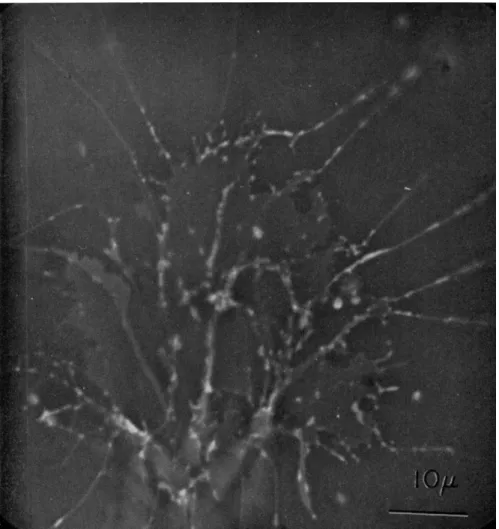
The organism adopted for the present study is a new species of Allogromia (Figs. 1 and 2) discovered and cultivated by Dr. John Lee.
It is currently designated Allogromia sp. strain N F (Lee, 1963). The organisms are cultivated on 1% agar in sea water on petri plates sprinkled with milligram quantities of bakers' yeast. Within a few days, the Allo- gromia grow to large numbers.
Single organisms are obtained for study by adding a few milliliters of sea water to a plate and brushing the surface of the agar lightly. The sea water containing dislodged specimens is then decanted into a small dish, from which single organisms can be selected before they become attached again.
Movements are recorded on Plus-X film at either 8 or 16 frames/sec with an Arriflex camera driven by a synchronous motor. Of a variety of optical systems which have been tried, Reichert Anoptral appears to yield the best contrast for both the hyaloplasm and inclusions. Inter- ference contrast and polarized light have also been used, but the results of these studies are not ready for publication.
- T h e terms "fibril" and "filament" have been used in many different situations in biology and require some redefinition in each situation. Fibrils in this case are membrane-bounded protoplasmic units, barely visible under best optical conditions, several of which make up a pseudopod. These "fibrils" are displaced in toto and are not tubes through which cytoplasm flows. Each fibril, in turn, has within it sub- microscopic filaments.
Streaming and Locomotion in Foraminifera 411 A Vanguard Motion Analyzer3 and a Perceptoscope4 projector were used for plotting the motions of particles.
Results
OBSERVATIONS OF CYTOPLASMIC STREAMING IN Allogromia SP. STRAIN NF.
Extension and Retraction of Pseudopodia
Advancing pseudopodia almost always extend straight ahead into the medium from the cell body until they make contact with, and ad- here to, the substratum. Once attached, pseudopodia seldom advance any great distance in a straight line; instead they tend to bend at inter- vals at angles of from 5 to 30 degrees usually in the same direction (Figs. 1 and 2).
Attached pseudopodia with definite bends in them tend to straighten out as the most proximal attachment points are lost. Apparently tension between a more distant attachment point and the cell body is responsible for this straightening. If a retracting pseudopod becomes detached, the pseudopod shows some elastic return, indicating that it was under ten- sion, and then begins to wave about, showing that it is considerably less rigid than when originally extended; sometimes pseudopodia bend at angles, wave back and forth, or form a loose coil, which may flail about briefly before disappearing into the cell body. Smaller branch pseudo- podia shrivel and disappear within seconds on becoming suddenly detached.
Pseudopodia which retract slowly show what will henceforth be referred to as the "fibril-droplet transition": although originally most of the body of the pseudopodia has been more or less cylindrical in shape with only a few accumulations of cytoplasm at attachment points and the tip (Fig. 2), on slow retraction, cytoplasm originally in the form of the fibrillar pseudopodial mass becomes converted into the substance contained in "droplets" which may appear at certain places in the net- work, or may be transported along the network and into the cell body.
T h e conversion of this droplet material to fibrils during pseudo- podial extension will be discussed later.
The most obvious difference between extending and retracting pseudopodia is their rigidity; the former are quite stiff and unyielding, whereas the latter are flaccid and bend easily when stressed by weak water currents.
3 T h e Vanguard Motion Analyzer is available from the Vanguard Instrument Co., 184 Casper St., Valley Stream, New York.
4 Perceptual Development Laboratories, 6767 Southwest Avenue, St. Louis, Missouri.
Proliferation of Pseudopodia by Branching and Splitting
Newly formed pseudopodia almost invariably form branches diverg- ing at acute angles (usually 15-30 degrees) from the direction of the main tip. T h e most common method of branch formation is as follows:
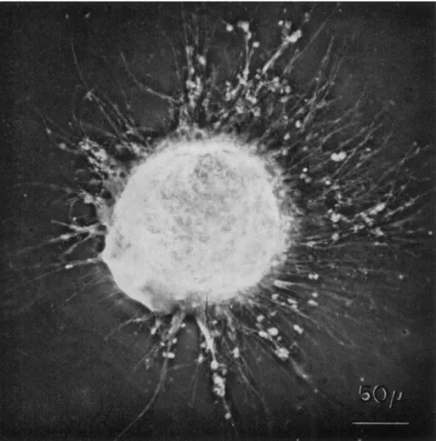
FIG. 1. A specimen of Allogromia sp. strain N F . (obtained from Dr. John Lee) feeding on dried bakers' yeast and bacteria. Scale, 50 μ.
after the original "trunk" pseudopod has made one or more slight bends, tension develops in the pseudopod as a whole between the body and the most proximal attachment point. When the pseudopod pulls away from this attachment point, a slender thread of cytoplasm remains (Fig. 2);
immediately it begins to grow in length and diameter and serves as a branch pseudopod. We can infer from this either that the branch pseudopod (or a loop of cytoplasm that formed it—see discussion to
Streaming and Locomotion in Foraminifera 413
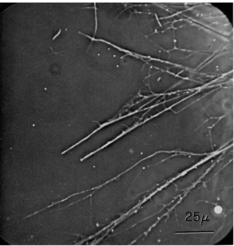
FIG. 2. A portion of the reticulopodial network of a specimen of Allogromia sp.
strain NF. larger than that shown in Fig. 1. Note the difference in the sizes of various pseudopods, droplets present at tips, junctions, and attachment points; note also small sheetlike areas apparently attached to the substratum. Scale, 25 μ.
diverge at wide angles and develop tension. At the point of splitting, there is usually a thin web of apparently fluid cytoplasm (Fig. 2). Ob- servations on living organisms suggest that the moving fibrils are em- bedded in some kind of tacky matrix which is apparently not preserved by fixation for electron microscopy.
follow) pre-existed at the attachment point, or that a loop was formed by pulling a straight fibril away from an attachment point. As we shall see later, the former possibility seems more likely.
Branched pseudopodia characteristically split when their branches
The splitting process is reversible; tips which have moved apart cause splitting of the trunk pseudopod behind them. If they approach one another, reducing the tension which causes splitting, then the branches fuse again.
Fusion of Bremckes to Form the Reticulopodial Network
Blanches from different trunk pseudopodia may meet at any angle whatever and still fuse. The composite pseudopod then shows particles moving at the velocities characteristic of the two branches that fused;
these velocities may be either quite similar or quite different.
A fused branch may show splitting into the original two streams as the result of tension developed within the network. In newly developed networks splitting typically progresses proximally, but in older networks, in which some of the branches point toward the cell body after making several turns in the same direction, splitting may also proceed distally.
The Fibril-Droplet Transition
Filopodia and reticulopodia of forams may appear almost perfectly cylindrical at low magnification, but when examined with high-resolution optics they can be seen to have thickened regions and various other irregularities, particularly at the pseudopodial tips, at attachment points where dropletlike masses are usually present, and at the junction of branches where weblike structures can be seen (Figs. 1 and 2).
For descriptive purposes, I have used the terms "droplet" and "fibril"
to denote only the shape of the portions of the cytoplasm that we see in the microscope; their possible functional significance will be discussed later. However, the formation and disappearance of these droplets ap- pears to be an important aspect of cytoplasmic streaming and locomotion in forams. For example, when a pseudopod is diminishing in mass, it does so not so much by decreasing its length as by converting its fibril- lar pseudopodia to the form of these droplets which are then transported along the network to the cell body. Presumably the material in these droplets is reused to form new pseudopodia, for one sees such droplets being transported toward sites of pseudopodial extension. In extending pseudopodia, accumulations tend to occur mostly at attachment points and near or at the advancing tip.
There are two experimental situations which demonstrate the fibril droplet transition in an exaggerated form; in one of these its reversibil- ity is particularly well seen.
1. As was pointed out by Sandon (1934), injurious stimuli sometimes cause a pseudopod to break up into a series of short droplets and rods.
By chance, we were able to record a case of pseudopodial contact between
Streaming and Locomotion in Foraminifera
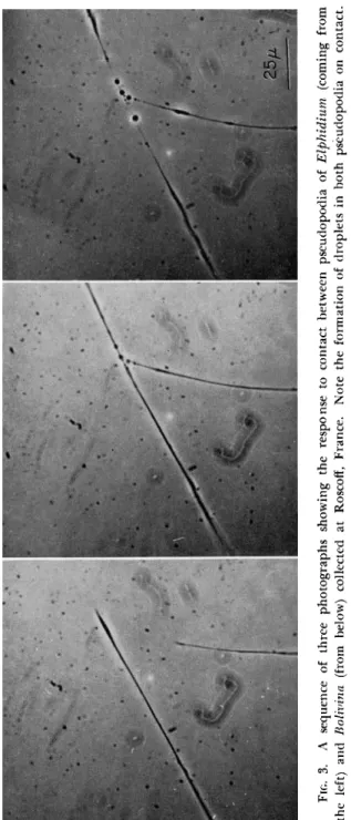
FIG. 3. A sequence of three photographs showing the response to contact between pseudopodia of Elphidium (coming from the left) and Bolivina (from below) collected at Roscoff, France. Note the formation of droplets in both pseudopodia on contact. Scale, 25 μ.
specimens of the genera Elphidium and Bolivina at Roscoff, France. In Fig. 3 it can be seen that, although both organisms responded to this contact, in one, the pseudopod rapidly disintegrated into large droplets.
Subsequently, one of these droplets was carried away intact, and the material of the others was somehow "spun" into fibrils again.
FIG. 4. An excised portion of the reticulopodia network of Allogromia sp. strain NF. about 20 min after excision. Note the beginning of droplet formation. Scale, 50 μ.
2. Experimentally excised portions of the reticulopodial network continue to show virtually normal streaming, as Jahn and Rinaldi (1959) showed, but within minutes begin to show the first effects of a gradual degeneration process, the end result of which is the appearance of drop- lets and thickened regions throughout the network (Fig. 4). During the degeneration process, the fibrillar parts of the network become thinner and show less and less vigorous cytoplasmic streaming. T h e final stages of this streaming process involves unidirectional streaming of a few par- ticles toward a droplet, into which they disappear. If an intact portion
Streaming and Locomotion in Foraminifera 417 of the network makes a chance contact and fuses with the excised portion, there is an immediate response. T h e material within the droplets is
"rescued" by being rapidly reincorporated into the fibrillar organization of the network, and bidirectional streaming is again established through- out the whole fused network.
A Preliminary Analysis of Particle Motion Velocities in Reticulopodia Although the initial impression one receives of the streaming in the reticulopodial network of a foram is one of extreme complexity, there
13- 12- I I- 10- 9- c 8- ο υ ω y «Λ ι -
1
6-Ο
^ 5- 4-·
3- 2- I -
0 2 4 6 8 10 12 14 16 18 20 22 24 26 28 30 32 34 Microns
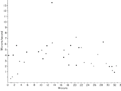
FIG. 5. A record of the velocities exhibited by various particles during a 5-sec interval of time along a section of a trunk pseudopodium between two attachment points 33 μ apart. T h e one closer to the body is the origin. Note the two distally moving particles which are traveling at a velocity several times that of the slowest particles.
are certain features of this streaming which appear to be universal and, therefore, probably important.
1. The tips of branch pseudopodia or unbranched filopodia may extend, remain stationary, or retract, but in any event bidirectional streaming continues.
2. Branch pseudopodia from the same trunk pseudopod quite often exhibit streaming at different velocities.
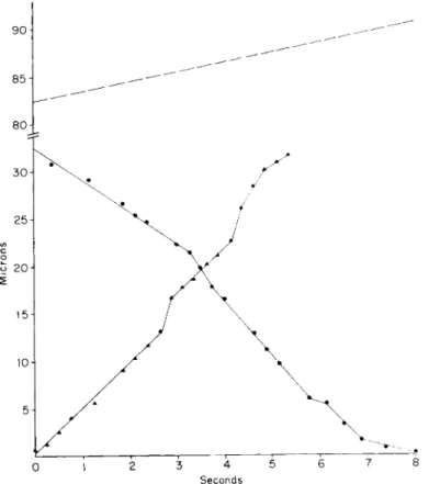
3. Large, trunk pseudopodia may show at one time particles travel- ing at different velocities. T h e most rapid particles may be moving at several times the velocity of the slowest ones (Fig. 5).
4. Larger pseudopodial branches (over 1 μ in diameter) usually show particles moving at several velocities in both directions (Fig. 6).
7 0
0 1 2 3 4 5 6 7 8 9 10 II 12 13 14 15 16 17 18 Seconds
F i g . 6. A plot of the distance traversed in time by the tip of a pseudopod 3 μ in diameter ( ) , with various distally ( ) and proximally ( ) moving particles. T h e attachment points and the times at which they yielded to tension transmitted along the pseudopod are shown as broken lines parallel to the χ axis.
(Figure 8 shows the morphology of the pseudopod during the period in which these measurements were made.)
5. In larger branches, a single particle may be found to change its velocity abruptly in the vicinity of points of attachment to the sub- stratum (Figs. 6 and 7). Frequently these attachment points can be dis- cerned only after the pseudopod pulls away from them, leaving new branch pseudopodia (Fig. 8).
6. In the exceedingly fine pseudopodial branches of young organ- isms (Fig. 9), the velocity at which the tip advances is not nearly so uniform as in larger pseudopodia (cf. Figs. 7 and 10). T h e velocity of
Streaming and Locomotion in Foraminifera 419 particles is much more variable and can be followed accurately except
at the very tip where we have never been certain that we have tracked the same particle passing over the tip and returning. Characteristically there is a bulge or a droplet at or near the tip into which the particles disappear. Some appear to come out again after only a little delay, but
90
85
80
30
25
(/) c ο
ο 20
15
10
5
0 1 2 3 4 5 6 7 8
Seconds
FIG. 7. A record of the motions of two particles which could be traced for a period of 8 and 5 sec, respectively, in relation to the advancement of the tip. This analysis was made on the same pseudopod as that in Fig. 6. (Figure 8 shows the morphology of the pseudopod during the period in which the measurements were made.)
others remain in the droplet for some time. Particle e in Fig. 10 illus- trates this problem: it may or may not be identical with particle g.
If so, then it returned from the tip much more slowly than it approached it. We thought we could see this quite often in casual observations, but when faced with the problem of recording and analyzing such an event, we could not be certain of having kept the same particle in view while passing over the tip region.
7. It was first noted in the analyses, and subsequently confirmed in casual observations, that a number of peculiar events take place at attachment points. First, there is characteristically a thickening there which does not remain constant in size or shape (Figs. 2 and 8). Second, particles enter the attachment-point region and characteristically speed up and slow down; they may then either (1) pass on (usually at a differ-
Seconds
FIG. 8. Tracings from the motion picture records analyzed in Figs. 6 and 7.
Arrows show positions of known attachment points revealed by the presence of a branch pseudopod as the main pseudopod was pulled away.
ent velocity as in Figs. 6, 7, and 10), (2) disappear in the thickened cytoplasm at the attachment point, or (3) reverse their direction.
Each organism is somewhat different from the last; therefore, it can- not be stated that these analyses will apply to all organisms similarly analyzed in the future. However, the features pointed out above have been seen enough times to instill some confidence that they are wide- spread, and, therefore, of importance. Data of this type are the raw material of which intelligent models to explain this and other kinds of motility must be made.
Streaming and Locomotion in For amini) er a 421 Cytoplasmic Streaming in, and Movement of, the Cell Body
The cell body of Allogromia sp. strain NF. is so transparent that cytoplasmic streaming can be seen at the entire periphery by changing the level of focus of the microscope. The pattern of streaming within the body is very complex. Streaming seems to be most active in the
FIG. 9. A portion of the network of a young organism (Allogromia sp. strain NF.) showing pseudopodia close to the resolution limit of the microscope emanating from a thin sheet of hyaline cytoplasm traversed at points by barely visible fibrils. Particles streamed along these fibrils (not elsewhere on the hyaline sheets) and out into the pseudopodia. Some remained stationary while others moved. (The analysis shown in Fig. 10 was made of one of the pseudopodia in the group shown, but at a later time when the morphology had changed somewhat.) Scale, 10 μ.
cortical region. Often the cytoplasm contains many clear vacuoles, around which thin streams of particles stream in a tortuous path, as if the whole cortical region were a mass of reeled-in pseudopodial fibrils.
The velocity of streaming in the cell body is not significantly different from that in the pseudopodia.
/
/
Seconds
FIG. 10. An analysis of the advance exhibited by the tip of a reticulopod such as shown in Fig. 9, and the motions exhibited by particles in or on the pseudopo- dium. Note the irregular tip velocity (cf. Figs. 7 and 8) and the changes in the ve- locities of particles advancing to the same region of the pseudopod (particles d and f) while particles c and e went through the same region unimpeded. It is not certain whether or not particles e and g are identical because e disappeared momentarily into a droplet at the tip of the pseudopod.
The cell body exhibits independent "ameboid" movements which are apparently not related to events in the reticulopodial network. In fact, some cells have been observed to move slowly in this manner with- out any pseudopodia. T h e cell becomes deformed, but not continuously as is the case with amebae. Instead, the cell surface moves in "lurches,"
more or less as it would be expected to if some kind of strain were applied to the cell surface until a yield point was reached.
Streaming and Locomotion in Foraminifera 423 Discussion
RETICULOPODIAL ULTRASTRUCTURE
There are at least seven separate lines of evidence that bear on reticulopodial ultrastructure.
1. The obvious mechanical rigidity of extending pseudopodia is particularly remarkable in the light of their fineness. This must indicate the existence of structural anisotropy, perhaps fibrillar or filamentous structures.
2. T h e optical anistropy expected of structurally anisotropic material is present. Schmidt (1937) reported positive, axial birefringence in Mili- ola filopodia. Using Dr. Shinya Inoué's rectified, oil-immersion system, I was able to see the birefringence of the network and noticed that cer- tain sections of it increased gradually in birefringence over a few seconds, but then suddenly became isotropic again. This could indicate the development of temporary strain in elastic structures which was relieved by flow.
3. It is even possible to see with the light microscope some of the fibrillar structure in trunk pseudopodia at places where they spread out. As Jahn and Rinaldi (1959) reported, the particles in a particular region follow tracks at the same velocity; in addition, under good optical conditions one can see the fibrils themselves.
4. The dynamic behavior of reticulopodia, especially their branch- ing, splitting, and fusion, all point to a fibrillar organization of the network.
5. The recent electron-microscopic study of Wohlfarth-Bottermann (1961) has confirmed the suspicions of earlier authors that the reticulo- podium does, in fact, consist of a loose bundle of fibrils of different sizes surrounded largely by empty space (empty, at least, after fixation and exposure to the electron beam). Each fibril is a protoplasmic unit containing such structures as mitochondria and surrounded by a unit membrane. The electron microscope also has revealed a filamentous ultrastructure for the fibrils.
6. The behavior of the pseudopodia in fusion, splitting, and branch- ing suggests that there may be a tacky substance or "glue" or some sort which keeps the membrane-bounded fibrils, of which the pseudopod is composed, together. If this is true, this material does not survive the preparatory procedures for electron microscopy.
7. The fiber-droplet transition is a feature which tells us little, so far, about reticulopodial ultrastructure other than that the fibrils are unstable under certain local physiological or environmental conditions.
We need a great deal of further information about the structure of the droplets when forming and when disappearing.
Although there remain many fundamental uncertainties regarding the details of reticulopodial ultrastructure, a concept is beginning to emerge which offers an explanation for several previously incompre- hensible aspects of foram movement and behavior. Jahn and Rinaldi's (1959) concept of the smallest branches being made up of one or more folded fiber loops would seem to be probably correct except that each fiber (and not the whole filopod) is surrounded by a membrane. T h e only reservation concerning this concept springs from the fact that the existence of a true loop at the filopodial tips remains to be proven, either morphologically or by a clear demonstration of a single particle following an uninterrupted U-shaped path at the tip.
One of the most puzzling observations which this new concept explains is the flow of membrane in different directions in different parts of the pseudopodial surface. As long as the pseudopod was con- sidered as a single protoplasmic unit, the alternative explanations of this involved either high-velocity gradients within the membrane, or the absence of a membrane—an interpretation which Jahn and Rinaldi (1959) correctly pointed out was "physiological heresy."
The manner in which branches form at attachment points suggests that not all the folded fibrils terminate at the tips of branches. T h e termination of some of these loops at points along the trunk pseudopods and branches would explain the gradual (and apparently stepwise) diminution of diameter toward the periphery and the instantaneous ap- pearance of branches when a trunk or branch pseudopod is pulled away from an attachment point.
T H E FIBRIL-DROPLET TRANSITION
Although the appearance of droplets due to stimulation or injury had been noted in passing by earlier workers (esp. Sandon, 1934), the normal occurrence of a similar phenomenon throughout the network during establishment and destruction of the network has not received the attention it deserves. T h e observations presented here would suggest that it is a phenomenon of central importance. Unfortunately, we do not know in which of several ways it should be interpreted.
It must be borne in mind that what we have described as "droplet formation" is merely deformation of a body which could be either an active (i.e., contractile) or passive process. If passive, it could represent either deformation due to "solation," that is, to decreased internal resis- tance to tension at the surface of the fibrillar unit (i.e., surface tension and/or elastic forces); alternatively the fibrillar unit might be "reeled
Streaming and Locomotion in Foraminifera 425 in" intact at attachment points and other places and stored in droplets to be later "paid out" again.
In interpreting the fibril droplet transition, it should be emphasized that in the extreme case the whole filopod breaks up into droplets, usually connected by a fine fibril (Fig. 3). This situation is quite reminis- cent of the breakup of Amoeba proteus pseudopodia into droplets on sudden application of very high hydrostatic pressures (Marsland and Brown, 1936). In the latter case, the breakup appears to be due to sur- face tension acting on a cylindrical mass of fluid that has suddenly lost its gel structure. However, we cannot exclude the possibility that the foram fibrils contract actively into the little droplets which we see. In normal reticulopodial activity, only some of the fibrils in any part of the network transform into droplets. Under these circumstances, it is difficult to see why solated fibrils should respond to surface tension when the normal tackiness of fibril units in a pseudopodium would suggest that adhesive forces should tend to keep them elongated.
It is probably premature to draw any serious analogies between the fibril droplet transition and simple, abstract models of complex rheo- logical events in ameba such as the Landau (1959) scheme for "sol-gel reactions." It should be pointed out, however, that the application of this model would place the following restrictions on events in the organ- ism: (1) Since the model assumes that contracted gel must solate before it can contract again, droplet formation would have to occur only in contracted material; (2) solated material in the droplet would have to be formed into fibrils again without the aid of contractile forces; and (3) contraction could play no role in the droplet—>fibril direction of the
transition.
A second analogy that could be made is to actomyosin sols and gels on the addition of adenosine triphosphate (ATP). Actomyosin sols show a viscosity drop on the addition of A T P but cannot perform mechanical work or develop tension because they lack elastic structure, being in the sol state. Actomyosin gels, on the other hand, show contraction and syneresis on the addition of ATP, but do not solate between contraction cycles. These analogies, then, are also not very helpful.
A third possible analogy might be to the behavior of intact muscle, in which the primary contractile event is an elasticity change resulting in shortening and stiffening of elastic components, but which also in- volves an increase in the viscous resistance against which the muscle must work (Gasser and Hill, 1924; Pryor, 1952). T h e elasticity change of
intact muscle is imitated by muscle models, and by actomyosin gels and fibers, but the contractility of these model systems does not necessarily involve the same changes in viscous resistance.
While not wishing to propose a formal model to explain the con- sistency changes associated with cell movement here, I would like to point out that it may be unnecessary to assume, as it is assumed in the Landau model (and others) that a change of viscosity is a necessary and integral part in the recovery from contractile processes. This concept originally grew out of the Mast (1926) theory of ameboid movement, which rests on rheological interpretations which have been questioned (Allen, 1960, 1961a). It may be more generally true that contractile events involve primarily only the elastic components of cytoplasmic structure; what has been termed solation may involve changes in the viscous properties, stiffness, and yield point of the material, and may be either secondary to or unrelated to the contractile event.
T H E MECHANISM OF PSEUDOPODIAL EXTENSION AND CYTOPLASMIC STREAMING
The readily observed fact that the reticulopodia exert tension and perform work would suggest strongly that they are contractile. The absence of close association between fibrils in a pseudopod seems to rule out an active-shearing force between fibrils as an explanation of
contractility at the pseudopod level. Therefore, it must be the fibrils themselves that are contractile, probably through events occurring at the level of the filaments, observed by Wohlfarth-Bottermann (1961), or below.
Leaving aside for the present all problems of how contractility occurs at the submicroscopic level because of lack of evidence, let us see how pseudopodial extension and cytoplasmic streaming could be ex- plained in terms of fibrillar contractility.
The growth of a rigid filopodium indicates that some kind of gel structure must be built up in it. This could be done either by addition and stiffening of less rigid material (not necessarily sol!) on either the proximal or the distal side. T h e first possibility can be rejected as it would result in only forward cytoplasmic streaming in the advancing pseudopod. T h e addition of gelated material at the tip in the same manner as in the ameba would account for bidirectional streaming unless the gelled part of the fibril were attached to the substratum.
The model by which, in principle, a folded contractile fibril can displace its two ends in opposite directions by a propagated contraction anchored at the bent portion has been described in detail elsewhere (Allen, 1961a,b).5 In the case of pseudopod extension in forams, it is
5 A further discussion of the mechanics of the "contraction anchored at a bend principle" is to be found in the Free Discussion following Parts III and IV of this Symposium.
Streaming and Locomotion in Foraminifera 427 only necessary to assume that a contraction is anchored at the bend of the folded fibril (in this case at the tip of a pseudopod) so that the material passing away from the tip is contracted, and that passing toward the tip is relaxed and about to contract. If we assume that the stiffness of the "springs" in contracted fibrils increases as in muscle, then we have a satisfactory explanation for the existence of a stiffened gel rod in newly formed pseudopodia.6 A drop in the stiffness of this rod on relaxation could account for the relative flaccidity of pseudopodia in retraction.
The "contraction anchored at a bend principle" described previously and elsewhere also serves as a convenient explanation of bidirectional streaming throughout the network. The force of contraction is applied as tension to one side of the loop and compression to the other side, forcing the two sides to move in opposite directions. This model has the same advantage as the active-shearing hypothesis in that it invokes directed forces (in this case tension and compression) rather than the nondirectional force of pressure which would be clearly inapplicable here. The contraction model has a distinct advantage over the active- shearing model in that it assumes no ultrastructure which cannot be demonstrated, and it invokes no new and mysterious processes or forces.
This is not, however, to deny the possible existence of such forces at the filament level or below; at present we have no information about what occurs at that level.
An apparent disadvantage of the contraction hypotheses is that one would expect the resistance to movement to increase with the length of the pseudopod if all the force were applied at the tips of branches. In- stead of slower motion, long pseudopodia seem to show the most rapid motion (cf. Fig. 5). T o account for this it seems probable that long pseudopodia (and short ones as well) must have "booster engines" at intervals along them to keep the fibrils moving. A series of folded fila- ments extending different lengths along the pseudopodium would, in effect, deploy just such a series of booster engines (in the form of the bent regions of folded fibrils). When such a long pseudopod is pulled laterally by another pseudopod, it leaves behind tiny branch pseudopodia
6 Neither the present author, nor Jahn and Rinaldi (1959) were able to confirm reports in the early literature concerning the existence of a "stereoplasmic rod" in the reticulopodia of the marine foraminifera examined. It seems likely that earlier authors (e.g., Doflein, 1916 and others) were misled by diffraction artifacts and strong convictions about the necessity for a rigid rod to provide the observed stiffness. At present all that need be assumed is that at least one of the fibrils is rigid; since it is not discernible in phase contrast, it very likely does not differ significantly from other fibrils in refractive index.
at intervals of from 10-30 μ apart, showing the previous location of these boosters.
It is quite clear that there occur along the network regions in which particles move in both directions at a wide variety of velocities, some several times as rapid as the slowest movement. There are also some particles which are stationary. T h e slower particles can be explained as belonging to folded filament loop units extending for different distance out into the network; some doubtless extend out to the tips, while others terminate at various attachment points. T h e fastest particles (some over 13 μ/sec) are difficult to account for unless we assume some kind of "pick-a-back" mechanism in which one folded fibril unit func- tions while itself in motion riding on another one, so that the maximum streaming velocity observed is the sum of the velocities gained from the two (or perhaps more) units combined. Such a situation would not be hard to imagine in a loose bundle of fibrils and could easily give rise to the variety of velocities often observed.
Using the convenient contraction at a bend principle we could al- most construct a tentative model of reticulopodial dynamics, except for the fact that we know so little about the fibril droplet transition. In general, these droplets must represent "reservoirs" of "fibril-forming materials"—or, perhaps, folded fibrils themselves which have been reeled in for storage. In the latter case it would be easy to postulate a way in which they could be paid out to form the rigid structures present in pseudopodia. However, if the fibrillar material solates completely in forming droplets, it would be difficult to imagine how this could be spun out again by tension unless there were enough elasticity left to orient filaments into a form that could later contract. Similar problems are encountered in the organization of the mitotic spindle (Inoué, 1959;
see also paper in this Symposium).
T h e maintenance of bidirectional streaming into and out of ex- panding and retracting parts of the network would seem to require some activity on the part of the cell body, at least to the extent of reeling in contracted arms of folded filaments, which as we have suggested may be
"pushed" by the compressive force of the contraction at a bend mecha- nism toward the cell body. T h e presence of a complicated pattern of cytoplasmic streaming within the cell body would seem to indicate the possibility that the cell body participates to this extent. However, it must be recalled that streaming can continue for one to several hours in the absence of the cell body, but sooner or later fails.
As stated previously, the author's tentative model based on the con- traction at a bend principle is incomplete in many respects. However, it has the advantage of offering a similar model to that previously
Streaming and Locomotion in Foraminifera 429 suggested for the Chaos-Amoeba proteus group of amebae. We still lack sufficient data on the mechanical properties of the cytoplasm in either organism to construct a detailed mechanical model to illustrate exactly how the principle would work. T h e mechanical conditions are somewhat different in the cases of amebae and forams; in amebae one would expect the lateral components of the contractile forces to balance out because of the radial symmetry of the pseudopod. In the case of forams we have no reason to expect that such a symmetry exists; there- fore it seems likely that pseudopodia should be found to bend when the contractile force exerted at the tip exceeds the stiffness of the pseudopod.
This may explain the pronounced tendency of foram pseudopodia to bend at intervals, typically at attachment points. A close match between the lateral component of the force of contraction and the stiffness might easily result in the activities which have been reported for unattached pseudopodia, such as bending, waving, flailing about, and in extreme cases coiling.
Summary
The cytoplasmic streaming, locomotion, and general behavior of marine Foraminifera with reticulopodial networks has been discussed in general and with particular reference to observations carried out on Allogromia sp. strain NF.
The principal processes in the establishment of the network are pseudopodial extension, attachment, branching, splitting, fusion, and retraction.
Cytoplasmic streaming is bidirectional in all parts of the intact net- work as asserted by earlier workers, but unidirectional streaming may occur in excised portions. Data are presented to show how the velocity of particles varies along different parts of the network. Changes in velocity are particularly obvious at or near attachment points.
An important aspect of reticulopodial dynamics is the transition be- tween fibrillar material of the pseudopod and droplets which appear at attachment and branching points, at or near the tip, and at various other points along the network. T h e possible nature of this transition is discussed.
Two experimental situations were observed in which fibrils are readily converted into droplets. One case involved an intergeneric pseu- dopodial contact; the other occurred in excised portions of a network.
In one case the recovery of material in droplets formed in an excised portion of network was effected when a portion of the original network fused with it and thus "rescued" it from further degeneration.
The possible mechanisms of pseudopodial extension and cytoplasmic
streaming are discussed in the light of the present work and of a recent electron-microscopic study indicating that the pseudopodial structure is basically a loose bundle of fibrils of different sizes contain- ing filamentous material. On the basis of this and other information, the active-shearing model is rejected in favor of an incomplete, but more attractive model based on fibrillar contractility.
Pseudopodial extension and bidirectional cytoplasmic streaming can both be explained in terms of the contraction anchored at a bend principle applied to a folded contractile fibril. Long pseudopodia consist of many folded fibrils, many of which terminate along the pseudopodia.
These may serve as booster engines and help overcome the added fric- tion involved in streaming in long pseudopodia. Multiple velocities ob- served in trunk pseudopodia may require a "pick-a-back" deployment of folded fibrillar units, in which some function while themselves riding on top of others.
The model which is proposed rests on the same mechanical principle previously proposed as a partial explanation of ameboid movement in the Chaos-Amoeba proteus group.
ACKNOWLEDGMENTS
We wish to acknowledge the assistance of Mr. W. Reid Pitts, J r . , Mrs. Eleanor Benson Carver, and Mrs. Prudence Jones Hall in analyzing the motion picture films.
REFERENCES Allen, R. D. (1960). / . Biophys. Biochem. Cytol. 8 , 379.
Allen, R. D. (1961a). In "The Cell" (J. Brächet and A. E. Mirsky, eds.), Vol. II, pp.
135-216. Academic Press, New7 York.
Allen, R. D. (1961b). Exptl. Cell Res. Suppl. 8 , 17.
Doflein, F. (1916). Zool. Jahrb. Abt. Anat. Ontog. Tiere 3 9 , 335.
Dujardin, F. (1835). Ann. Sei. Nat. Zool. 1 4 , 343.
Gasser, H. S., and Hill, Α. V. (1924). Proc. Roy. Soc. B 9 6 , 308.
Godina, G. (1955). Atti Accad. Naz. Lincei Rend. Sei. Fis. Mat. Nat. 1 8 , 104.
Gustafson, T . (1963). This Symposium.
Hanson, J . , and Huxley, H. E. (1955). Symp. Soc. Exptl. Biol. 9 , 228.
Holtfreter, J . (1946). / . Morphol. 7 9 , 27.
Inoué, S. (1959). In "Biophysical Science" (J. L. Oncley et al., eds.), pp. 402-408. Wiley, New York.
Jahn, T . L., and Rinaldi, R. A. (1959). Biol. Bull. 1 1 7 , 100.
Jepps, M. W. (1942). / . Marine Biol. Assoc. U.K. 2 5 , 607.
Kamiya, N. (1959). Protoplasmatologia 8 , 1.
Kamiya, N., and Kuroda, K. (1956). Bot. Mag. (Tokyo) 6 9 , 546.
Landau, J . V. (1959). Ann. N. Y. Acad. Set. 7 8 , 487.
Le Calvez, J . (1938). Arch. Zool. Exptl. Gen. 8 0 , 163.
Lee, J . J . , and Pierce, S. (1963). / . Protozoal. 1 0 , 404.
Leidy, J . (1879). U.S. Geol. Surv. Rept. 1 2 , 324 pp.
Marsland, D. Α., and Brown, D. E. S. (1936). J. Cellular Comp. Physiol. 8 , 167.
Mast, S. O. (1926). / . Morphol. 4 1 , 347.
Streaming and Locomotion in Foraminifera 431
Nakai, J . (1956). Am. J. Anat. 9 9 , 81.
Nakai, J . (1963). This Symposium.
Pryor, M . G. M . (1952). In "Deformation and Flow in Biological Systems" (A. Frey Wyssling, ed.). Amsterdam.
Sandon, H. (1934). Nature 133, 761.
Sandon, H. (1944). Nature 154, 830.
Schmidt, W. J . (1937). Protoplasma 27, 587.
Schultze, M . (1854). "Uber den Organismus der Polythalamien." Leipzig, Germany.
Shaffer, Β. (1963). This Symposium.
Wohlfarth-Bottermann, Κ. E. (1961). Protoplasma 54, 1.
DISCUSSION
DR. JAHN: I would like to commend Dr. Allen on these very excellent motion pictures. This is the second time I have seen them, and I have been unable to detect any differences between the two species of Allogromia. So far as I know, anything that can be said about one can be said about the other. I would confirm the state- ment of mine that Dr. Allen quoted to the effect that particles go out to the tip, turn around, and come back.
I would also like to underscore the point that when the cell body is being moved by the pseudopodia, there is two-way movement in all of the pseudopodia. In a large pseudopodium, perhaps 80% of the granules may be going in one direction;
but if the microscope is focussed carefully, the other 20% of the granules will be found going in the opposite direction. There is always two-way streaming.
One other point I would like to make is that I am not disturbed by the fact the electron microscopists have not yet discovered the "legs of the millipede," because they have also not discovered whatever causes active sliding in Chara, Nitella, in the stamen hair cells of Tradescantia, and elsewhere where it has been described.
Possibly a single mechanism will turn up sooner or later that will explain all of these phenomena. It is also possible Dr. Allen has a good idea on this subject.
DR. ALLEN: Since you brought up the active-shearing idea, I should perhaps point out that the fibers which make up a pseudopod, and which are moving in opposite directions, consist apparently of cytoplasmic elements, each of which is surrounded by a membrane. It is rather difficult to imagine how your "millipede legs" could be acting between these membranes, which are separated (at least in fixed material) by several millimicrons.
CHAIRMAN MARSLAND: It is apparent from the film that hydrostatic pressure could not explain protoplasmic streaming in Foraminifera.
DR. L E E : I have been looking at these Foraminifera for some time, and am in- terested in their mechanism of movement. Polyoral organisms are even more in- teresting than these monoral ones; there is always a dominant "head" or "chief"
region which advances in one direction and all the other "heads" follow behind. It looks to me like a pulling action in which one chief region becomes stronger than the others.
In these polyoral individuals, occasionally the head end will bud off; about a half hour before this head end is budded off, a new chief takes over and the old head end then begins to trail behind until it is budded off.
The feeding process is also very interesting. These form "pools," as I like to call them, at the end of pseudopodia which are effective bacterial traps. One sequence you saw in the film was really very beautiful. T h e trap spreads over the bacterial
surfaces and all the bacteria are somehow caused to adhere and get carried back toward the body.
CHAIRMAN MARSLAND: According to this hairpin loop contraction idea that you have proposed, wouldn't it necessitate some backward movement in the contracting part of the loop as well as the forward movement?
DR. ALLEN: T h e mechanism I proposed requires relative movement of the two arms of the loop in opposite directions. Movement relative to the substratum de- pends on what part is attached. If, for example, the contracted arm were attached, then the bend would be expected to advance. T h e contracted arm would be "built forward" by new material becoming incorporated and stiffened at the bend.
CHAIRMAN MARSLAND: Yes; but wouldn't there have to be just an approximation of the particles as a result of the contraction which would be toward the point of attachment?
DR. ALLEN: They should come closer together in approaching the bend, or at any other place where contraction is postulated. Of course, approximation of particles only suggests contraction; physical evidence is required to demonstrate a contraction.