expression during lung cancer cell differentiation
Atousa Arbabian1,2, Jean-Philippe Brouland3,Agota Ap ati4,5, Katalin Paszty6, Luca Heged}us4, Agnes Enyedi 4,5, Christine Chomienne1,2and Bela Papp1,2,7,8
1 Institut National de la Sante et de la Recherche M edicale, UMR-S 940, Paris, France
2 Institut Universitaire d’Hematologie, Universite Paris Diderot, PRES Sorbonne Paris-Cite, France 3 Service d’Anatomie et Cytologie Pathologiques, H^opital Lariboisiere, Paris, France
4 Institute of Molecular Pharmacology, Research Centre for Natural Sciences, Hungarian Academy of Sciences, Budapest, Hungary 5 Department of Molecular Cell Biology, National Blood Service, Budapest, Hungary
6 Molecular Biophysics Research Group, Hungarian Academy of Sciences and Semmelweis University, Budapest, Hungary 7 Institut National de la Sante et de la Recherche M edicale, UMR U978, Bobigny, France
8 Universite Paris-13, PRES Sorbonne Paris-Cite, Bobigny, France
Keywords
calcium; differentiation; endoplasmic reticulum; lung cancer; SERCA Correspondence
B. Papp, UMR U978 Inserm, Universite Paris-13, UFR SMBH, 74 rue Marcel Cachin, 93017 Bobigny, France
Fax: +33 1 48 38 73 69 Tel: +33 6860 88309 E-mail: belapapp2@yahoo.fr
(Received 4 September 2012, revised 23 October 2012, accepted 7 November 2012) doi:10.1111/febs.12064
Cellular calcium signaling plays important roles in several signal transduc- tion pathways that control proliferation, differentiation and apoptosis. In epithelial cells calcium signaling is initiated mainly by calcium release from endoplasmic-reticulum-associated intracellular calcium pools. Because cal- cium is accumulated in the endoplasmic reticulum by sarco/endoplasmic reticulum calcium ATPases (SERCA), these enzymes play a critical role in the control of calcium-dependent cell activation, growth and survival. We investigated the modulation of SERCA expression and function in human lung adenocarcinoma cells. In addition to the ubiquitous SERCA2 enzyme, the SERCA3 isoform was also expressed at variable levels. SERCA3 expres- sion was selectively enhanced during cell differentiation in lung cancer cells, and marked SERCA3 expression was found in fully differentiated normal bronchial epithelium. As studied by using a recombinant fluorescent cal- cium probe, induction of the expression of SERCA3, a lower calcium affin- ity pump, was associated with decreased intracellular calcium storage, whereas the amplitude of capacitative calcium influx remained unchanged.
Our observations indicate that the calcium homeostasis of the endoplasmic reticulum in lung adenocarcinoma cells presents a functional defect due to decreased SERCA3 expression that is corrected during pharmacologically induced differentiation. The data presented in this work show, for the first time, that endoplasmic reticulum calcium storage is anomalous in lung can- cer cells, and suggest that SERCA3 may serve as a useful new phenotypic marker for the study of lung epithelial differentiation.
Introduction
Non-small-cell lung cancer is currently the most fre- quent fatal malignancy worldwide with more than a million deaths occurring annually [1,2]. Although much is known about risk factors such as tobacco
smoking, or regarding molecular and genetic events that occur during lung tumorigenesis, the overall sur- vival rate of lung cancer could not be significantly improved during the last 30 years [2]. This indicates
Abbreviations
CAG promoter, cytomegalovirus early enhancer/chickenbactin promoter; ER, endoplasmic reticulum; GFP, green fluorescent protein;
SB, Sleeping Beauty; SERCA, sarco/endoplasmic reticulum calcium ATPase.
that currently available knowledge of the molecular biological behavior of lung carcinoma cells is insuffi- cient for the development of efficient therapy. In par- ticular, knowledge about calcium signaling in lung cancer cells is scarce, although calcium signaling is an essential component of several regulatory networks involved in the control of cell growth, differentiation and apoptosis.
Calcium-dependent cell activation is initiated by the release of calcium ions stored in the endoplasmic reticu- lum (ER) into the cytosol through inositol-1,4,5- trisphosphate receptor calcium channels [3]. In addition, calcium in the ER lumen is required for the function of various ER located chaperones such as calreticulin or calnexin involved in the conformational maturation and post-translational modification of newly synthesized proteins that transit through the organelle [4–6]. Abnor- mal ER calcium homeostasis can lead to cellular stress responses, growth arrest and apoptosis [4,7–9].
Sarco/endoplasmic reticulum calcium ATPase (SERCA) enzymes are essential for the dynamic equi- librium of ER calcium homeostasis. Located in the ER membrane, these enzymes sequester calcium ions from the cytosol into the ER lumen by ATP-dependent active ion transport, and thus generate a steep calcium concentration gradient between the cytosol (approxi- mately 50–100 nM) and the ER lumen (approximately 0.1 mM) [10]. SERCA proteins are encoded by three genes (ATP2A1, 2 and 3) that can give rise to several protein isoforms by alternative splicing [11–15].
SERCA expression is tissue dependent and develop- mentally regulated. SERCA1 is expressed in skeletal muscle, and the SERCA2a isoform is mainly expressed in cardiomyocytes. Whereas SERCA2b is a ubiqui- tously expressed isoform thought to be involved in
‘housekeeping’ functions, in several cell types such as cells of hematopoietic origin, gastric and colonic epi- thelial cells, b-cells of islets of Langerhans or vascular endothelium, SERCA2b has been shown to be co-expressed with SERCA3 [15–26]. Because SER- CA2b and SERCA3 enzymes display distinct calcium affinities [27–32], their co-expression is involved in the fine-tuning of the parameters of ER calcium sequestra- tion according to the specific calcium storage and release requirements of the ER of a given cell type.
By controlling intra-ER calcium levels, SERCA enzymes are involved in the control of cell growth [33–
36]. Small molecular SERCA inhibitors are tumor promoters [37,38], and SERCA mutations have been identified in cancer [39–43]. In this context we have previously reported that the expression of SERCA enzymes is anomalous in breast, colon as well as gas- tric carcinoma due to the loss of expression of
SERCA3 [18,44,45]. It has also been shown that SERCA3 expression is induced during histone-deacety- lase inhibitor-induced differentiation of gastric and colon carcinoma cells [18], as well as following the inhibition of the APC/b-catenin/TCF4 axis [44], and that normal colonic and gastric epithelium expresses SERCA3 abundantly [44]. In addition, the pharmaco- logical modulation of SERCA activity has also been shown to enhance cell differentiation in colon carci- noma [44] and acute promyelocytic leukemia [46] cells.
Taken together, these observations indicate that the functional maturation of ER calcium homeostasis that occurs during normal cell differentiation is blocked due to deficient SERCA3 expression in malignant cells, and that this phenomenon is involved in the mainte- nance of the neoplastic phenotype [16,47].
In order to establish whether defects of ER calcium homeostasis can be found in pulmonary neoplasia, in this work we investigated SERCA expression in nor- mal lungin situ and in lung adenocarcinoma cell lines undergoing pharmacologically induced cell differentia- tion in vitro, and investigated functional calcium stor- age capacity by confocal microscope calcium fluorimetry using a green fluorescent protein (GFP) based calcium probe in differentiated and control cells.
Our results indicate that SERCA3 is abundantly expressed in normal fully differentiated bronchial epi- thelium, whereas expression is highly heterogeneous in carcinoma tissue. We also show that SERCA3 expres- sion is induced during the pharmacologically induced differentiation of lung adenocarcinoma cell lines, and that differentiation induction leads to decreased ER calcium storage.
These observations show that, similarly to other types of neoplasia, lung cancer cell calcium homeosta- sis can also be remodeled during drug-induced cell dif- ferentiation. This indicates that defects of SERCA3 expression in cancer may be a widespread phenome- non, and show for the first time that ER calcium homeostatic defects are involved in shaping the lung cancer phenotype.
Results
SERCA expression in lung tumor cell lines and primary tumor tissue
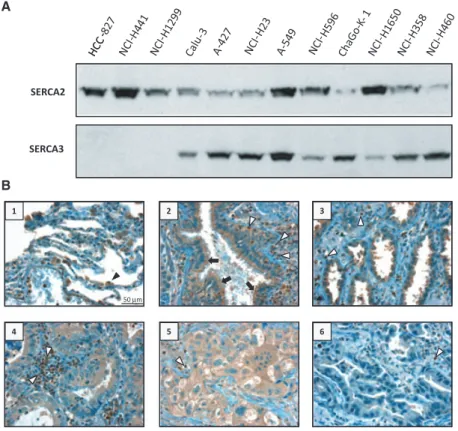
Western immunoblot analysis performed on equal amounts of proteins obtained from total cell lysates from various non-small-cell lung carcinoma cell lines indicates that the SERCA expression pattern of these cells is heterogeneous. As shown in Fig. 1A, whereas all cell lines express the ubiquitous SERCA2 isoenzyme,
SERCA3 expression displays a marked heterogeneity:
in cell lines such as HCC-827 or NCI-H441, SERCA3 protein expression is barely detectable or absent, but in other cell lines such as Calu-3, A-427, NCI-H23, A549, Cha-Go K-1, NCI-H1650, NCI-H358 or NCI-H460, SERCA3 protein is highly expressed and expression lev- els vary depending on cell line.
When investigated in situ by immunohistochemistry using a SERCA3-specific monoclonal antibody, SERCA3 expression could be detected in normal cili- ated bronchial epithelium and, to a variable degree, also in primary human lung adenocarcinoma tissue. As shown in Fig. 1B, whereas bronchial epithelial cells were markedly labeled (++, photograph 2, black arrow) in a highly reproducible manner for SERCA3, staining in lung adenocarcinomas was heterogeneous and varied between weak (+/ , photograph 6), moderate (+, pho- tographs 4 and 5) and marked (++, photograph 3).
SERCA3 labeling in normal lung parenchyma (photo- graph 1) was readily detectable in alveolar macrophages (black arrowhead), and normal infiltrating lymphocytes displayed strong SERCA3 labeling (+++, white arrow- heads) in normal (photograph 2) as well as tumor tissue (photographs 3–6).
These observations taken together show that SERCA3 expression is part of the normal differentiation program of lung epithelium, and that SERCA3 expres-
sion may be decreased or lost in lung adenocarcinoma.
In addition, these observations indicate that heteroge- neous SERCA3 expression in lung cancer cell linesin vitro reflects a pathophysiologically relevant phenomenon, because this is also observed in tumor tissuein situ.
Induction of SERCA expression by short chain fatty acids
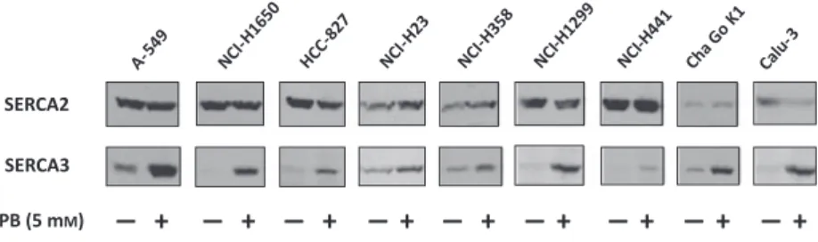
When various non-small-cell lung carcinoma cell lines were subjected to treatments with short chain fatty acids and analogs, a selective induction of SERCA3 expres- sion could be observed. As shown in Fig. 2, treatment with 5 mM phenylbutyrate for 5 days resulted in a marked and selective upregulation of the expression of the SERCA3 isoenzyme, whereas the expression of SERCA2 was not modified significantly or was decreased. SERCA3 induction could also be obtained byn-butyrate orn-valerate as well (not shown). As stud- ied in the A549 lung adenocarcinoma cell line, induction of SERCA3 expression was concentration dependent in the low millimolar range and reached a plateau above 2 mM n-butyrate or 4-phenylbutyrate concentrations (Fig. 3A,B). SERCA3 expression reached a plateau of four- to six-fold without significant toxicity compared with untreated cells over a period of 4–7 days in cells treated with 3 mM butyrate or phenylbutyrate,
SERCA2
SERCA3
1 2 3
50µm
4 5 6
A
B
Fig. 1.SERCA expression in lung adenocarcinoma cell lines and lung tumor tissue. (A) Expression of SERCA2 and SERCA3 proteins in untreated lung carcinoma cell lines, detected with the IID8 (SERCA2) and the PLIM430 (SERCA3) monoclonal antibodies by western blotting. (B) Immunohistochemical detection of SERCA3 protein in normal lung parenchyma (photograph 1), bronchial epithelium (photograph 2) and grade 1 (photographs 3 and 4), grade 2
(photograph 6) and grade 3 (photograph 5) lung adenocarcinomas. Black arrows, normal bronchial epithelial cells; white arrowheads, normal lymphocytes; black arrowhead, alveolar macrophage (original magnification 409).
respectively (Fig. S1A,B). Induction of SERCA3 expres- sion could be obtained by other short chain fatty acid analogs as well. As shown in Fig. S2, when A549 cells were treated with short chain fatty acids of increasing chain length (from acetate to caproate), maximal induc- tion could be obtained by butyrate and valerate. Among the tested branched chain fatty acid analogs, valproate was also active. Aryl-substituted analogs such as 3-phe- nylpropionate, 4-phenylbutyrate or 5-phenylvalerate also induced SERCA3 expression, with maximal activ- ity observed in the case of phenylbutyrate.
In parallel with SERCA2 and SERCA3, the expres- sion of gelsolin and p21CIP1/WAF1, two general markers of lung cancer cell differentiation [48,49], has been investigated by western blotting in several lung adeno- carcinoma cell lines treated with various short chain fatty acids for 5 days. As shown in Fig. 4, the selective induction of SERCA3 expression of A549, NCI-H358, NCI-H1650, Cha-Go K1 and HCC-827 cells with butyrate, valerate or phenylbutyrate was accompanied, in all investigated configurations, by increased gelsolin and p21CIP1/WAF1 expression. This indicates that the treatments induce phenotypic differentiation of the cells, and that the induction of SERCA3 expression is part of this process in all investigated model systems.
Induction of SERCA3 expression during the spontaneous differentiation of Calu-3 cells in post-confluent culture
Calu-3 lung adenocarcinoma cells have been shown to spontaneously undergo phenotypic differentiation towards a ciliated bronchial epithelial phenotype in post-confluent cultures [50]. The cells form a tight, polarized monolayer that displays trans-epithelial elec- trical resistance, barrier function and vectorial solute transport, as reflected by the formation of domes when grown on plastic, and express apical microvilli and tight junctional complexes [51]. Post-confluent Calu-3 cells are widely used in pharmacological research as an in vitromodel for bronchial epithelium [52]. As shown in Fig. 5, the expression of SERCA3 is markedly increased in Calu-3 cells after confluency, whereas SERCA2 expression is not modified significantly. Inter- estingly, short chain fatty acid treatment of confluent Calu-3 led to a further increase of SERCA3 expression.
Phenylbutyrate has already been shown to enhance CFTR protein expression in post-confluent, differenti- ated Calu-3 cells [52]. These data taken together indicate that the induction of SERCA3 expression takes place during the spontaneous differentiation of
SERCA3 SERCA2
PB (5 mM) – + – + – + – + – + – + – + – + – +
Fig. 2.Selective induction of SERCA3 protein expression in various lung adenocarcinoma cell lines. The cells were treated with 5 mM
phenylbutyrate (PB) for 5 days, and identical amounts of total cell protein lysates were analyzed using the IID8 (SERCA2-specific) and PLIM430 (SERCA3-specific) antibodies by western blotting. SERCA protein expression in phenylbutyrate treated cells (+) is compared with untreated controls ( ).
days 0 5 5 5 5 5 5 0 0 1 2 3 4 5 (mM)
Butyrate
days 0 5 5 5 5 5 5 Phenylbutyrate
0 0 1 2 3 4 5 (mM)
P = 0.152 P = 0.026 P = 0.020 P = 0.023 P = 0.022
P = 0.018 P = 0.016
P = 0.091
A B
Fig. 3.Dose–response relationship of SERCA3 induction. A549 cells were treated with various concentrations of butyrate (A) and phenylbutyrate (B), and SERCA expression was measured by semi-quantitative western blotting. An approximately six-fold induction of SERCA3 expression (black columns) is obtained in the low millimolar
concentration range for both molecules, whereas the expression of SERCA2 (empty columns) decreases slightly (butyrate) or is not modified significantly (phenylbutyrate;n=3).
post-confluent Calu-3 cells, a process that can be fur- ther enhanced by phenylbutyrate.
Endoplasmic reticulum calcium storage in short chain fatty acid differentiated cells
In order to investigate ER calcium storage capacity in lung carcinoma cells, we established 11 clonal cell lines (BWV543 cells) derived from A549 lung adenocarci- noma that stably express the GCaMP2 recombinant fluorescent calcium probe in the cytosol and performed confocal microscope measurements of fluorescence on cells treated with 5 mMphenylbutyrate for 5 days and on untreated control cells. Treatment of all clones led to increased SERCA3 expression similarly to the
parental A549 cell line. ER calcium storage capacity was tested by confocal microscopy on two randomly selected clones (BWV543 clones 4 and 2.4). As shown in Fig. 6, when clone 4 cells incubated in the absence of extracellular calcium were treated with thapsigargin, a significant calcium release from the ER to the cyto- sol could be observed in control cells, whereas thapsi- gargin-induced calcium release in cells in which SERCA3 expression was induced by phenylbutyrate was significantly smaller. After the completion of thapsigargin-induced calcium release measured in the absence of extracellular calcium, 2 mM Ca2+ was added to the extracellular medium in order to observe capacitative calcium influx. Despite the marked decrease of thapsigargin-induced calcium release in phenylbutyrate treated cells, capacitative calcium influx was of essentially identical amplitude in untreated and phenylbutyrate treated cells. Identical results were obtained on BWV543 clone 2.4 cells as well.
Discussion
Because calcium uptake into the ER is performed exclusively by SERCA enzymes, SERCA function con- stitutes a key nodal point in cellular calcium homeo- stasis and signaling. SERCA activity is required for calcium accumulation in a resting cell for intra-ER cal- cium-dependent chaperone functions, as well as for calcium accumulation in the ER for inositol-1,4, 5-trisphosphate-induced calcium release during cell activation. Moreover, because SERCA enzymes take up calcium from the cytosol even during an inositol-1, 4,5-trisphosphate-induced calcium release event, they can shape the amplitude and the duration of calcium peaks, or modulate the spatiotemporal characteristics of calcium oscillations in the cytosol. SERCA activity can therefore critically modulate the state of activation of a cell, and can influence the type and the amplitude of the response given by a cell to calcium-dependent stimuli. This has important consequences for tumor biology: direct pharmacological SERCA inhibitors such as thapsigargin or 2,5-di-tert-butyl-1,4-benzohy- droquinone are known tumor promoters, and SERCA2+/ knockout is known to lead to squamous tumorigenesis with a long incubation time. In addition, sequencing of the SERCA2 and SERCA3 genes in var- ious tumors including lung cancer and corresponding normal tissue suggests that inactivating mutations occur in these genes in a proportion of tumors [40,41], and it has thus been suggested that SERCA3 haploin- sufficiency may predispose to cancer development or may promote tumor progression [41,42]. In this con- text the induction of SERCA3 expression, as observed
A549
NCI-H358
NCI-H1650
HCC-827
Treatment – + – + – + – +
phenylbutyrate
butyrate
butyrate butyrate
valerate NCI-H1650
Cha-Go K1 butyrate
NCI-H358 valerate
Fig. 4.Expression of lung cancer cell differentiation markers by short chain fatty acids. Several lung adenocarcinoma cell lines (A549, NCI-H358, NCI-H1650, Cha-Go K1 and HCC-827) were treated with various short chain fatty acids (phenylbutyrate, 3 mM; valerate or butyrate, 5 mM) for 5 days, and SERCA expression as well as the expression of gelsolin (82 kDa) and p21CIP1/WAF1 (21 kDa) was observed by western blotting. Similarly to SERCA3, the expression of gelsolin and p21CIP1/WAF1 is increased in short chain fatty acid treated cells in all investigated drug/cell line configurations.
in our work, may therefore exert anti-oncogenic effects in lung epithelial cells.
The observations presented in this work show, for the first time, that the expression of SERCA-type calcium pumps in lung cancer cells is dynamic, as various cell- differentiation-inducing stimuli led to the selective induction of the expression of the SERCA3 isoenzyme in all investigated cell lines. In addition, strong SERCA3 expression was found in fully differentiated bronchial epithelium. These data, taken together, indicate that SERCA3 expression is part of the differentiation pro- gram of normal bronchial epithelium and that this phe- nomenon can be recapitulated to various degrees during the differentiation of all investigated cell lines.
SERCA3 induction in A549 cells, a widely studied lung adenocarcinoma line, was associated with a decreased calcium release signal upon SERCA inhibi- tion. The calcium affinity of SERCA3 is inferior (approximately 1.2lM) to that of the co-expressed SERCA2b isoform (approximately 0.2lM). The decreased calcium release signal observed in phenyl- butyrate treated A549 cells cannot be attributed exclusively to SERCA3 induction due to the intercon-
nectedness of calcium homeostatic mechanisms that operate simultaneously in a cell. Decreased calcium release upon SERCA inhibition, however, is compatible with the notion of the formation of a leakier or lower- affinity, SERCA3-associated intracellular calcium pool, similar to that observed previously in butyrate treated KATO-III gastric carcinoma cells [18].
The normal lung contains several distinct and spe- cialized epithelial cell types. In addition, lung carci- noma as a group consists of several histological tumor types that can display different degrees of histological differentiation. The ontogenic relationship between normal and tumoral cell types in the lung is not estab- lished in sufficient detail [1], and the molecular signal- ing mechanisms involved in the induction and the maintenance of tumoral phenotypes of increasing malignancy are not sufficiently known in the lung.
Consequently, lung cancer is currently not amenable to successful pharmacological intervention. The observa- tion that lung cancer cell calcium ER homeostasis is remodeled during differentiation is important for can- cer biology because this indicates that lung adenocarci- noma cells may display calcium homeostatic defects
SERCA3 SERCA2
butyrate valerate phenylbutyrate
– – – – +
30 100 100 100 100
– – – + –
– – + – –
SERCA2 SERCA3
Expression (fold increase)
P = 0.0001 P = 0.01 P = 0.0075
confluency (%) A
C D
B
Fig. 5.Induction of SERCA3 expression during the spontaneous differentiation of post-confluent Calu-3 cells–superinduction by short chain fatty acids. Calu-3 lung adenocarcinoma cells were grown in pre-confluent conditions and in post-confluency, in the absence or the presence of various short chain fatty acids (5 mM) for 5 days, and SERCA expression was determined by western blotting. (A), (B) Photomicrographs of pre- and post-confluent cultures. Formation of ‘domes’ in post-confluent cultures (arrowheads) is indicative of trans-epithelial solute transport, a feature of a tight, differentiated epithelial monolayer. (C), (D) Cells at day 1 of post-confluency were allowed to undergo differentiation for 5 days in the absence or the presence of 5 mM butyrate, valerate or phenylbutyrate. SERCA2 and SERCA3 expression was then detected by western blotting and quantified. The selective induction of SERCA3 expression by post-confluent growth is further enhanced by short chain fatty acids. White bars, pre-confluent untreated cells; grey bars, post-confluent untreated cells; black bars, post- confluent, phenylbutyrate treated cells (n=3).
similar to those observed in colon, gastric and breast carcinoma, as well as acute promyelocytic leukemia. In those systems the cross-talk of ER calcium homeostasis with other mechanisms of control of cell differentiation permits pharmacologically induced cell differentiation to be induced and potentiated and certain forms of resistance to differentiation-induction therapy to be overcomein vitro[44,46]. In addition, SERCA3 expres- sion has also been shown to be a useful new immuno- histochemical marker for the study of the state of differentiation and degree of malignancy of colon and breast tumors [44,45]. Data presented in this paper indicate, for the first time, that ER calcium biology is connected to cell differentiation also in pulmonary epi- thelium, and shows that ER calcium homeostasis pre- sents defects in lung cancer cells, which can be overcome by pharmacologically induced cell differenti- ation. Although the precise correlation of the loss of SERCA3 expression with lung adenocarcinoma histo- logical type, grade, molecular subtypes or clinical parameters requires further work, the demonstration of calcium homeostatic anomalies in lung cancer may help identify new calcium-dependent targets for the therapy
of the disease. In addition, our observations suggest that SERCA3 may prove useful for the immunohisto- chemical analysis of lung tumors and for the study of bronchic epithelial differentiation.
Materials and methods
Chemicals
n-Butyric and n-valeric, 4-phenylbutyric acid, as well as sodium acetate, sodium caproate, sodium valproate, isobu- tyric, 4-methylvaleric as well as phenylacetic, 3-phenylprop- ionic and 5-phenylvaleric acids were from Sigma-Aldrich (Saint-Quentin Fallavier, France). Free acids were dissolved to 300 mM final concentration in 300 mM sterile sodium bicarbonate solution, filtered through 0.2 lm membranes and stored at 25°C. Sodium salts were dissolved at 300 mMconcentration in sterile water and filtered through 0.2lm membranes.
Cell lines
The Calu-3, NCI-H23, NCI-H358, NCI-H441, NCI-H460, NCI-H596, NCI-H1299 and NCI-H1650 as well as the HCC-827 cell lines were obtained from ATCC (ATCC- LGC Standards Sarl, Molsheim, France). A549 and A427 cells were from DSMZ (Braunschweig, Germany), and Cha-Go K1 was purchased from ECACC (Porton Down, UK). The various cell lines were cultured according to the instructions of the cell line depository of origin. RPMI- based media contained Ultraglutamine-I (Lonza, Verviers, Belgium) in addition to 2 mMglutamine.
Treatments
Exponentially growing cells were trypsinized and plated into 60 cm² cell culture grade Petri dishes in new medium.
When cultures reached 40–60% confluency as examined by light microscopy, medium was renewed and drugs were added from concentrated stock solutions. Following treat- ments as indicated in the figures, cells were washed with ice-cold 150 mMNaCl and precipitated with 5% trichloro- acetic acid. The trichloroacetic acid pellet was quantified and dissolved in modified La€emmli-type sample buffer exactly as described earlier [17], and 50lg total cellular protein was deposited for SDS/polyacrylamide gel electro- phoresis per well [17]. Following electrophoresis and transfer to nitrocellulose membranes, equal loading of samples was controlled prior immunoblotting by Ponceau red staining of total protein deposited on the membranes. Ponceau red stained blots were scanned and equal protein loading was controlled by densitometry using the SCIONIMAGE software (Scion Corporation, Frederick, MD, USA) as described earlier in detail [17].
Time (minutes) RelaƟve fluorescence (F/F0)
1.0 1.5 2.0 2.5
3.0 thapsigargin Ca2+
SERCA2
SERCA3 control
treated
6 5 4 3 2 1 0
Fig. 6.Calcium release from SERCA-dependent intracellular calcium pools in A549 cells. A549 cells carrying the GCaMP2 fluorescent calcium probe (BWV543-4 cells) were treated or not with 5 mM phenylbutyrate for 5 days, and cytosolic calcium fluorescence was recorded by confocal fluorescence microscopy after complete inhibition of SERCA activity with 10lMthapsigargin in the absence of extracellular calcium. After completion of the thapsigargin-induced calcium release signal, extracellular medium was replenished with 2 mM Ca2+ and capacitative calcium influx was observed. Phenylbutyrate treatment leads to a significant decrease of calcium release from intracellular pools upon complete SERCA inhibition, whereas capacitative calcium influx remains similar. A total number of 36 treated and 48 untreated individual cells were recorded in four parallel experiments. Right panel:
similarly to parental A549 cells, SERCA3 expression was induced by phenylbutyrate in A549 cells carrying the GCaMP2-carrying calcium probe.
Western blotting
The SERCA3-specific PLIM430 mouse IgG1 monoclonal antibody was used for western immunblotting as hybrid- oma supernatant as described earlier in detail [17,18,44].
Purified mouse anti-gelsolin antibody (Cat # 610412; BD Transduction Laboratories, San Jose, CA, USA) (dissolved in NaCl/Tris milk at 0.05lgmL 1final concentration) and the mouse monoclonal anti-p21 antibody SC-71811 (Santa Cruz Biotechnologies, CA, USA) (dissolved in NaCl/Tris milk at 0.2lgmL 1final concentration) were used on heat treated samples (100°C for 12 min) run in 8% polyacryl- amide gels. Antibodies were revealed with an anti-mouse Ig horseradish peroxidase conjugate (Jackson Immuno- Research, Newmarket, Suffolk, UK) and the enhanced chemiluminescence system of Amersham (Courtaboeuf, France) as described earlier [17,18,44]. Non-saturated luminograms were scanned and densitometric analysis was done with theSCIONIMAGEsoftware [11–15].
Immunohistochemistry
Formalin fixed paraffin embedded lung cancer tissue micro- array slides were purchased from BioChain Institute, Hayward, CA, USA (T8235724, T8235732, Z7020066, Z7020067; CliniSciences, Montrouge, France), and from US Biomax, Rockville, MD, USA (BC04015, BC041114, BC041115, BC04119b, LC20810; Euromedex, Souffelwe- yersheim, France). Staining for SERCA3 was performed using the clone 2H3 monoclonal mouse anti-SERCA3 anti- body (IgG2a kappa) from Abnova (Tebu-bio, Le Perray en Yvelines, France) raised against a GST-tagged peptide sequence encompassing the 501–621 amino acid fragment of SERCA3, at 3lgmL 1final concentration diluted in Dako REAL antibody diluent (Dako France SAS, Trappes, France) as described in [45]. Briefly, after inhibition of endogenous peroxidase and antigen retrieval of deparaffi- nized sections by atris-hydroxymethyl-aminomethane-based reagent (CC1 solution; Ventana Medical Systems, Illkirch, France) at 95–100°C for 12 min, slides were incubated for 30 min at 37°C with the antibody, and staining was revealed using the Ventana I-View Biotin-Ig-streptavidin- biotin-horseradish peroxidase system with blocking of endogenous biotin activity and copper enhancement, accord- ing to the instructions of the manufacturer. As negative con- trol, isotype matched irrelevant antibody was used at the same concentration, and this gave no staining. As internal positive control, lymphocytes and vascular endothelial cells present in the samples and which express high levels of SERCA3 [17,24] were used. Slides were counterstained with hematoxylin and bluing agent (Ventana). SERCA3 immuno- staining of the samples was evaluated semi-quantitatively using a four-grade scale (0 to 3+). Photomicrographs were taken with a Zeiss Axio Scope.A1 microscope equipped with a Zeiss N-Achroplan 409/0.65 objective and an AxioCam
ICc1 camera, using the AXIOVISION 4.8.2 software. Tissue microarray slides were also processed using the ImmPRESS biotin-free polymerized enzyme staining system (Vector Lab- oratories, Clinisciences SAS, Nanterre, France) according to the instructions of the manufacturer, and SERCA3 staining with this system gave identical results.
Confocal fluorescent microscope calcium measurements
In order to generate the SB-CAG-GCaMP2 fluorescent cal- cium probe, a DNA fragment encoding GCaMP2 was first generated by PCR using the pN1-GCaMP2 plasmid, a gen- erous gift of Junichi Nakai, RIKEN Brain Science Insti- tute, Saitama, Japan [53]. Primer sequences were as follows: forward, 5′-CTACCGGTCTCGCCACCAATG-3′; reverse, 5′-AGATCTCCGCTCACTTCGCTGTC-3′. The forward primer adds an AgeI restriction site, the reverse primer adds aBglII restriction site. The complete GCaMP2 DNA fragment was cloned viaAgeI(5′)/BglII(3′) into an SB-CAG-AmaxaGFP vector from which Amaxa GFP was excised with the same enzyme pair. The GCaMP2 fragment is framed in the resulting vector by two left inverted repeat –direct repeat regions, which are the recognition motifs of Sleeping Beauty(SB) transposase [54–57], and possesses a cytomegalovirus early enhancer/chickenbactin (CAG) [58]
promoter. The efficiency of such a symmetrical transposon was proved by T. Orban (personal communication). This construct was used for stable transfection using the SB transposon delivery system as described earlier in detail [54 –57,59]. For the SB transposase, we used an enhanced ver- sion of the enzyme having an approximately 100-fold higher activity than the originally reconstructed transposase [59,60]. For transfection of exponentially growing A549 cells, the FuGENE® 6 (Roche Applied Science, Rotkreuz, Switzerland, http://www.roche-applied-science.com) reagent was used according to the instructions of the manufacturer.
Cells were cotransfected with transposon and transposase plasmids in a 10:1 ratio to avoid overproduction inhibition of the transposase [56,57]. Following transfections, cells were cultured for 3 days and then resuspended by trypsini- zation and sorted for GFP fluorescence using the Aria High Speed Cell Sorter of Becton-Dickinson (San Jose, CA, USA, http://www.bdbiosciences.com). The sorted cell population (containing approximately 60% GFP positive cells) was further cultured in bulk for 4 days, trypsinized and single cell cloned by limiting dilution in 96-well plates.
Individual GCaMP2 expressing clones were identified by GFP fluorescence using an inverted fluorescence microscope and expanded.
SB-CAG-GCaMP2-carrying A549 clones were grown to 20–30% confluency in eight-chamber Lab-Tek glass bottom chamber slides and treated with 5 mM phenylbutyrate for 5 days. At the end of treatment, the cells were washed with
calcium-free Hanks’ solution. Cells were then observed with an Olympus IX-81/FV500 laser scanning confocal micro- scope using an Olympus PLAPO 609 (1.4) oil immersion objective. For GCaMP2 imaging cells were excited with the 488 nm laser line and emission was collected between 505 and 535 nm. Under these conditions emitted fluorescence is proportional to the cytosolic free calcium concentration [53].
Calcium signal measurements were carried out in Hanks’
balanced salt solution supplemented with 20 mMHepes (pH 7.4) and 0.9 mM MgCl2. This medium was supplemented with 100lMCaCl2and 100lMEGTA for the measurement of thapsigargin-induced calcium release from the ER. Under these conditions free calcium concentration in the medium is approximately 4lM, and no calcium influx is observed.
Capacitative calcium influx was thereafter induced by the addition of 2 mM free CaCl2 to the medium. Time lapse sequences of cellular fluorescence were recorded and images were analyzed with the FLUOVIEW TIEMPO (v4.3; Olympus, http://www.olympusmicro.com) time course software. In order to compensate for variability of fluorophore expres- sion levels in individual cells, fluorescence values are expressed asF/F0. A total of 36 untreated and 48 treated cells were recorded individually in four parallel experiments.
Statistical analysis
Data are presented as the meanSEM and correspond to at least three independent experiments. Statistical analysis was done using Student’s pairedttest.
Acknowledgements
We express special thanks to Professor Neville Craw- ford (Department of Biochemistry, Hunterian Insti- tute, London, UK) for giving us the PLIM430 hybridoma, and to Dr Tamas Orban (Membrane Research Group, Hungarian Academy of Sciences, Hungary) for his contribution regarding the use of the transposon system used in this work. The excellent technical help of Mr Patrice Castagnet (Service d’Anatomie et Cytologie Pathologiques, H^opital Lari- boisiere, Paris, France) is gratefully acknowledged.
This work was supported by INSERM, the Associa- tion pour la Recherche sur le Cancer, France, and by the Hungarian Academy of Sciences grants CK80283 and K101064 (EA). This work is dedicated to the memory of Casper and Rose.
References
1 Travis WD (2004) World Health Organization, International Agency for Research on Cancer,
International Association for the Study of Lung Cancer, International Academy of Pathology: Pathology and
Genetics of Tumours of the Lung, Pleura, Thymus, and Heart. IARC Press, Lyon.
2 Youlden DR, Cramb SM & Baade PD (2008) The international epidemiology of lung cancer: geographical distribution and secular trends.J Thorac Oncol3, 819–831.
3 Berridge MJ (2009) Inositol trisphosphate and calcium signalling mechanisms.Biochim Biophys Acta1793, 933–940.
4 Berridge MJ (2002) The endoplasmic reticulum: a multifunctional signaling organelle.Cell Calcium32, 235–249.
5 Coe H & Michalak M (2009) Calcium binding chaperones of the endoplasmic reticulum.Gen Physiol Biophys28, F96–F103.
6 Malhotra JD & Kaufman RJ (2007) The endoplasmic reticulum and the unfolded protein response.Semin Cell Dev Biol18, 716–731.
7 Burdakov D, Petersen OH & Verkhratsky A (2005) Intraluminal calcium as a primary regulator of endoplasmic reticulum function.Cell Calcium38, 303–310.
8 Banhegyi G, Baumeister P, Benedetti A, Dong D, Fu Y, Lee AS, Li J, Mao C, Margittai E, Ni Met al.
(2007) Endoplasmic reticulum stress.Ann N Y Acad Sci 1113, 58–71.
9 Lai E, Teodoro T & Volchuk A (2007) Endoplasmic reticulum stress: signaling the unfolded protein response.Physiology22, 193–201.
10 Wuytack F, Raeymaekers L & Missiaen L (2002) Molecular physiology of the SERCA and SPCA pumps.Cell Calcium32, 279–305.
11 Vangheluwe P, Sepulveda MR, Missiaen L, Raeymaekers L, Wuytack F & Vanoevelen J (2009) Intracellular Ca2+- and Mn2+-transport ATPases.
Chem Rev109, 4733–4759.
12 Vandecaetsbeek I, Vangheluwe P, Raeymaekers L, Wuytack F & Vanoevelen J (2011) The Ca2+pumps of the endoplasmic reticulum and Golgi apparatus.Cold Spring Harb Perspect Biol3, a004184. doi:10.1101/
cshperspect.a004184.
13 Bobe R, Bredoux R, Corvazier E, Lacabaratz-Porret C, Martin V, Kovacs T & Enouf J (2005) How many Ca2+-ATPase isoforms are expressed in a cell type? A growing family of membrane proteins illustrated by studies in platelets.Platelets16, 133–150.
14 Dally S, Corvazier E, Bredoux R, Bobe R & Enouf J (2010) Multiple and diverse coexpression, location, and regulation of additional SERCA2 and SERCA3 isoforms in nonfailing and failing human heart.J Mol Cell Cardiol48, 633–644.
15 Hovnanian A (2007) SERCA pumps and human diseases.Subcell Biochem45, 337–363.
16 Arbabian A, Brouland JP, Gelebart P, Kovacs T, Bobe R, Enouf J & Papp B (2011) Endoplasmic
reticulum calcium pumps and cancer.BioFactors37, 139–149.
17 Dellis O, Arbabian A, Brouland JP, Kovacs T, Rowe M, Chomienne C, Joab I & Papp B (2009) Modulation of B-cell endoplasmic reticulum calcium homeostasis by Epstein–Barr virus latent membrane protein-1.Mol Cancer8, 59.
18 Gelebart P, Kovacs T, Brouland JP, van Gorp R, Grossmann J, Rivard N, Panis Y, Martin V, Bredoux R, Enouf Jet al.(2002) Expression of endomembrane calcium pumps in colon and gastric cancer cells.
Induction of SERCA3 expression during differentiation.
J Biol Chem277, 26310–26320.
19 Launay S, Bobe R, Lacabaratz-Porret C, Bredoux R, Kovacs T, Enouf J & Papp B (1997) Modulation of endoplasmic reticulum calcium pump expression during T lymphocyte activation.J Biol Chem272, 10746–10750.
20 Launay S, Giannı M, Kovacs T, Bredoux R, Bruel A, Gelebart P, Zassadowski F, Chomienne C, Enouf J &
Papp B (1999) Lineage-specific modulation of calcium pump expression during myeloid differentiation.Blood 93, 4395–4405.
21 Papp B, Enyedi A, Kovacs T, Sarkadi B, Wuytack F, Thastrup O, Gardos G, Bredoux R, Levy-Toledano S
& Enouf J (1991) Demonstration of two forms of calcium pumps by thapsigargin inhibition and radioimmunoblotting in platelet membrane vesicles.
J Biol Chem266, 14593–14596.
22 Papp B, Enyedi A, Paszty K, Kovacs T, Sarkadi B, Gardos G, Magnier C, Wuytack F & Enouf J (1992) Simultaneous presence of two distinct endoplasmic- reticulum-type calcium-pump isoforms in human cells.
Characterization by radio-immunoblotting and inhibition by 2,5-di-(t-butyl)-1,4-benzohydroquinone.
Biochem J288, 297–302.
23 Wuytack F, Papp B, Verboomen H, Raeymaekers L, Dode L, Bobe R, Enouf J, Bokkala S, Authi KS &
Casteels R (1994) A sarco/endoplasmic reticulum Ca2+-ATPase 3-type Ca2+pump is expressed in platelets, in lymphoid cells, and in mast cells.J Biol Chem269, 1410–1416.
24 Hadri L, Ozog A, Soncin F & Lompre AM (2002) Basal transcription of the mouse sarco(endo)plasmic reticulum Ca2+-ATPase type 3 gene in endothelial cells is controlled by Ets-1 and Sp1.J Biol Chem277, 36471–36478.
25 Diederichs F (2008) Ion homeostasis and the functional roles of SERCA reactions in stimulus-secretion coupling of the pancreatic beta-cell: a mathematical simulation.Biophys Chem134, 119–143.
26 Varadi A, Molnar E, Ostenson CG & Ashcroft SJ (1996) Isoforms of endoplasmic reticulum Ca2+- ATPase are differentially expressed in normal and diabetic islets of Langerhans.Biochem J319, 521–527.
27 Bobe R, Bredoux R, Corvazier E, Andersen JP, Clausen JD, Dode L, Kovacs T & Enouf J (2004) Identification, expression, function and localization of a novel (sixth) isoform of the human sarco/endoplasmic reticulum Ca2+ATPase 3 gene.J Biol Chem279, 24297–24306.
28 Dode L, Vilsen B, Van Baelen K, Wuytack F, Clausen JD & Andersen JP (2002) Dissection of the functional differences between sarco(endo)plasmic reticulum Ca2+-ATPase (SERCA) 1 and 3 isoforms by steady- state and transient kinetic analyses.J Biol Chem277, 45579–45591.
29 Poch E, Leach S, Snape S, Cacic T, MacLennan DH &
Lytton J (1998) Functional characterization of alternatively spliced human SERCA3 transcripts.Am J Physiol275, C1449–1458.
30 Wuytack F, Dode L, Baba-Aissa F & Raeymaekers L (1995) The SERCA3-type of organellar Ca2+pumps.
Biosci Rep15, 299–306.
31 Lytton J, Westlin M, Burk SE, Shull GE &
MacLennan DH (1992) Functional comparisons between isoforms of the sarcoplasmic or endoplasmic reticulum family of calcium pumps.J Biol Chem267, 14483–14489.
32 Chandrasekera PC, Kargacin ME, Deans JP & Lytton J (2009) Determination of apparent calcium affinity for endogenously expressed human sarco(endo)plasmic reticulum calcium-ATPase isoform SERCA3.Am J Physiol Cell Physiol296, C1105–1114.
33 Lipskaia L, Hulot JS & Lompre AM (2009) Role of sarco/endoplasmic reticulum calcium content and calcium ATPase activity in the control of cell growth and proliferation.Pflugers Arch457, 673–685.
34 Apati A, Janossy J, Brozik A, Bauer PI & Magocsi M (2003) Calcium induces cell survival and proliferation through the activation of the MAPK pathway in a human hormone-dependent leukemia cell line, TF-1.
J Biol Chem278, 9235–9243.
35 Brini M & Carafoli E (2009) Calcium pumps in health and disease.Physiol Rev89, 1341–1378.
36 Gill DL, Waldron RT, Rys-Sikora KE, Ufret-Vincenty CA, Graber MN, Favre CJ & Alfonso A (1996) Calcium pools, calcium entry, and cell growth.Biosci Rep16, 139–157.
37 Sakai A & Teshima R (2001) 2,5-Di-tert-butyl-1,4- hydroquinone enhances cell transformation
accompanied by an increase in intracellular free calcium ion concentration.Cancer Lett168, 183–190.
38 Hakii H, Fujiki H, Suganuma M, Nakayasu M, Tahira T, Sugimura T, Scheuer PJ & Christensen SB (1986) Thapsigargin, a histamine secretagogue, is a non-12-O- tetradecanoylphorbol-13-acetate (TPA) type tumor promoter in two-stage mouse skin carcinogenesis.
J Cancer Res Clin Oncol111, 177–181.
39 Prasad V, Okunade GW, Miller ML & Shull GE (2004) Phenotypes of SERCA and PMCA knockout mice.
Biochem Biophys Res Commun322, 1192–1203.
40 Korosec B, Glavac D, Rott T & Ravnik-Glavac M (2006) Alterations in the ATP2A2 gene in correlation with colon and lung cancer.Cancer Genet Cytogenet 171, 105–111.
41 Korosec B, Glavac D, Volavsek M & Ravnik-Glavac M (2008) Alterations in genes encoding sarcoplasmic- endoplasmic reticulum Ca2+pumps in association with head and neck squamous cell carcinoma.Cancer Genet Cytogenet181, 112–118.
42 Korosec B, Glavac D, Volavsek M & Ravnik-Glavac M (2009) ATP2A3 gene is involved in cancer susceptibility.Cancer Genet Cytogenet188, 88–94.
43 Liu LH, Boivin GP, Prasad V, Periasamy M & Shull GE (2001)Squamous cell tumors in mice heterozygous for a null allele of Atp2a2, encoding the sarco(endo) plasmic reticulum Ca2+-ATPase isoform 2 Ca2+pump.
J Biol Chem276, 26737–26740.
44 Brouland JP, Gelebart P, Kovacs T, Enouf J, Grossmann J & Papp B (2005) The loss of sarco/
endoplasmic reticulum calcium transport ATPase 3 expression is an early event during the multistep process of colon carcinogenesis.Am J Pathol167, 233–242.
45 Papp B & Brouland JP (2011) Altered endoplasmic reticulum calcium pump expression during breast tumorigenesis.Breast Cancer (Auckl)5, 163–174.
46 Launay S, Giannı M, Diomede L, Machesky LM, Enouf J & Papp B (2003) Enhancement of ATRA- induced cell differentiation by inhibition of calcium accumulation into the endoplasmic reticulum: cross-talk between RAR alpha and calcium-dependent signaling.
Blood101, 3220–3228.
47 Papp B, Brouland JP, Gelebart P, Kovacs T &
Chomienne C (2004) Endoplasmic reticulum calcium transport ATPase expression during differentiation of colon cancer and leukaemia cells.Biochem Biophys Res Commun322, 1223–1236.
48 Chang TH & Szabo E (2000) Induction of
differentiation and apoptosis by ligands of peroxisome proliferator-activated receptor gamma in non-small cell lung cancer.Cancer Res60, 1129–1138.
49 Chang TH & Szabo E (2002) Enhanced growth inhibition by combination differentiation therapy with ligands of peroxisome proliferator-activated receptor- gamma and inhibitors of histone deacetylase in adenocarcinoma of the lung.Clin Cancer Res8, 1206–1212.
50 Foster KA, Avery ML, Yazdanian M & Audus KL (2000) Characterization of the Calu-3 cell line as a tool to screen pulmonary drug delivery.Int J Pharm208, 1–11.
51 Mathia NR, Timoszyk J, Stetsko PI, Megill JR, Smith RL & Wall DA (2002) Permeability characteristics of calu-3 human bronchial epithelial cells:in vitro–in vivo correlation to predict lung absorption in rats.J Drug Target10, 31–40.
52 Loffing J, Moyer BD, Reynolds D & Stanton BA (1999) PBA increases CFTR expression but at high doses inhibits Cl secretion in Calu-3 airway epithelial cells.Am J Physiol277, L700–708.
53 Nakai J, Ohkura M & Imoto K (2001) A high signal- to-noise Ca2+probe composed of a single green fluorescent protein.Nat Biotechnol19, 137–141.
54 Ivics Z, Hackett PB, Plasterk RH & Izsvak Z (1997) Molecular reconstruction of Sleeping Beauty, a Tc1-like transposon from fish, and its transposition in human cells.Cell91, 501–510.
55 Ivics Z & Izsvak Z (2006) Transposons for gene therapy!Curr Gene Ther6, 593–607.
56 Izsvak Z & Ivics Z (2004) Sleeping beauty
transposition: biology and applications for molecular therapy.Mol Ther9, 147–156.
57 Izsvak Z, Ivics Z & Plasterk RH (2002) Sleeping Beauty, a wide host-range transposon vector for genetic transformation in vertebrates.J Mol Biol302, 93–102.
58 Alexopoulou AN, Couchman JR & Whiteford JR (2008) The CMV early enhancer/chicken beta actin (CAG) promoter can be used to drive transgene expression during the differentiation of murine embryonic stem cells into vascular progenitors.
BMC Cell Biol9, 2.
59 Mates L, Chuah MK, Belay E, Jerchow B, Manoj N, Acosta-Sanchez A, Grzela DP, Schmitt A, Becker K, Matrai Jet al.(2009) Molecular evolution of a novel hyperactive Sleeping Beauty transposase enables robust stable gene transfer in vertebrates.Nat Genet41, 753–761.
60 Orban TI, Apati A, Nemeth A, Varga N, Krizsik V, Schamberger A, Szebenyi K, Erdei Z, Varady G, Karaszi Eet al.(2009) Applying a ‘double-feature’
promoter to identify cardiomyocytes differentiated from human embryonic stem cells following transposon- based gene delivery.Stem Cells27, 1077–1087.
Supporting information
Additional supporting information may be found in the online version of this article at the publisher’s web site:
Fig. S1. Time course of SERCA2 and SERCA3 expression in short chain fatty acid treated A549 cells.
Fig. S2. Induction of SERCA3 expression in A549 cells by various short chain fatty analogs.