INTRODUCTION
The endoplasmic reticulum (ER) is a complex membrane structure in the cytoplasm of eukaryotic cells. It has important roles in protein and lipid biosynthesis, regulation of intracellular Ca2+ balance and most biotransformation reactions also take place in the ER. In eukaryotic cells, approximately one third of the cellular proteins, i.e. the secretory and membrane proteins are synthesized on the ER membrane-bound ribosomes, and attain their final conformation inside the lumen. This rapid folding mechanism assures the achievement of proteins’
biological activities. For proper protein folding, a dynamic balance between the ER protein load and the folding capacity must be maintained; a sophisticated quality control system guarantees that the final, folded proteins are devoid of errors.
However, ER homeostasis can be disrupted by pathological and physiological insults such as high protein demand, environmental toxins, viral infection or mutant protein expression resulting in an accumulation of unfolded or misfolded proteins in the lumen, the condition called ER stress.
Unfolded proteins are dangerous as they may aggregate and become toxic (1).
ER stress activates unfolded protein response (UPR), a complex signalling network, that is assigned to repair the
condition of unfolded/misfolded protein accumulation by increasing ER-resident chaperones, inhibiting protein translation, accelerating the endoplasmic reticulum-associated protein degradation (ERAD) and also autophagy components to promote clearance of unwanted proteins (2). If this procedure fails to restore the homeostasis in the lumen, cellular dysfunction and finally, apoptotic cell death occur, as can be observed in various diseases such as diabetes, inflammation, ischemia or neurodegenerative disorders including Alzheimer’s disease, Parkinson’s disease or autoimmune diseases (3, 4). According to the current model, UPR acts as a double switch between life and death during ER stress: directly regulates both apoptotic and anti-apoptotic pathways (5). Furthermore, recent results indicate that activation of UPR is also possible through lipid-dependent regulation; a mechanism independent of misfolded/unfolded proteins (6).
The devices with pulsed electromagnetic fields (PEMF) generate short burst of non-ionising, extremely low frequency electromagnetic field (0.5 – 100 Hz) without producing heat.
PEMF device was first applied on bone fracture where it could accelerate the healing process (7). Not much later this method has been approved by the Food and Drug Administration (FDA) in the USA. This non-invasive treatment is reportedly effective for treating nonunions, delayed unions (8, 9), pseudoartrosis E. KECZAN1, G. KERI2, G. BANHEGYI1, I. STILLER1,2
EFFECT OF PULSED ELECTROMAGNETIC FIELDS ON ENDOPLASMIC RETICULUM STRESS
1Department of Medical Chemistry, Molecular Biology and Pathobiochemistry, Semmelweis University, Budapest, Hungary;
2Pathobiochemistry Research Group of Hungarian Academy of Sciences, Budapest, Hungary
The maintenance of protein homeostasis in the endoplasmic reticulum (ER) is crucial in cell life. Disruption of proteostasis results in ER stress that activates the unfolded protein response (UPR); a signalling network assigned to manage the accumulated misfolded or unfolded proteins. Prolonged or unresolved ER stress leads to apoptotic cell death that can be the basis of many serious diseases. Our aim was to study the effect of pulsed electromagnetic fields (PEMF), an alternative, non-invasive therapeutic method on ER stressed cell lines. First, the effect of PEMF treatment on the expression of ER stress markers was tested in three different cell lines. PEMF had no remarkable effect on ER stress protein levels in human embryonic kidney (HEK293T) and human liver carcinoma (HepG2) cell lines. However, the expression of BiP, Grp94 and CHOP were increased in HeLa cells upon PEMF exposure. Therefore, HepG2 cell line was selected for further experiments. Cells were stressed by tunicamycin and exposed to PEMF. Grp94, PDI, CHOP and PARP expression as markers of stress were monitored by Western blot and cell viability was also investigated.
Tunicamycin treatment, as expected, increased the expression of Grp94, PDI, CHOP and inactivated PARP. Analysis of protein expression showed that PEMF was able to decrease the elevated level of ER chaperons Grp94, PDI and the apoptosis marker CHOP. The truncated, inactive form of PARP was also decreased. Accordingly, cell viability was also improved by PEMF exposure. These results indicate that PEMF is able to moderate ER stress induced by tunicamycin in HepG2 cells. However, our results clearly draw attention to that different cell lines may vary in the response to PEMF treatment.
K e y w o r d s : pulsed electromagnetic field, endoplasmic reticulum, stress, tunicamycin, cell viability
(10), diabetes mellitus induced complications (11-15), delayed wound healing (16), pain (17) or neurodegenerative disorders (18). A number of in vitro studies have revealed that electromagnetic fields affect cellular processes such as proliferation or cell death and models explaining its molecular background already exist. However, in most cases the whole mechanism of action is still unclear and sometimes results are controversial. One theory says that PEMF increases Ca2+binding to calmodulin, which induce nitric oxide synthases (NOS) (e.g.
eNOS) resulting in the production of nitric oxide (NO).
Increased NO level influence blood and lymph flow and cGMP production (19). These alterations can further activate and inactivate signal pathways resulting the effectiveness of PEMF mentioned above. Other researchers suggest its effect on cytosolic concentration of Ca2+ (20), protein phosphorylation (21) or conformational changes of macromolecules (22). It was also demonstrated that electromagnetic fields are able to change the amount of the lipid second messengers, diacylglycerol (DAG), phosphatidic acid (PA) (23) and of lipid peroxidation products (24).
We hypothesize that PEMF is a modulator of ER stress.
Studies have already been published discussing the impact of electromagnetic fields on the mechanism of cell death (20, 25- 29). However, the results were equivocal and the exact mode of action has not been clarified.
Our aim was to study the effect of PEMF on ER stress and consequent apoptosis provoked by a classic ER stressor, tunicamycin. This compound is a prototypic UPR inducer, which prevents the N-glycosylation of newly synthesized glycoproteins in the ER (30, 31).
MATERIALS AND METHODS Cell culture and maintenance
HepG2 (human liver carcinoma), HEK 293T (human embryonic kidney) and HeLa (human cervical cancer) cells were cultured in DMEM medium (Gibco), supplemented with 10%
heat inactivated fetal bovine serum (Gibco) and 1%
antibiotics/antimycotics (Gibco) at 37°C in a 5% CO2incubator.
Pulsed electromagnetic fields treatment
The PEMF device, PulsePad (Oxford Medical Instruments ltd.), is a commercially available system that works with a frequency of 8 Hz, maximum flux density of 70.5 mT and meant flux density of 0.56 mT. During the PEMF exposure of equal amount of cells the device was placed into the CO2 incubator underneath the cell plates.
Endoplasmic reticulum stress induction and pulsed electromagnetic fields treatment
To induce ER stress equal amount of HepG2 cells were treated with different concentration (0.1, 1, 10 µM) of tunicamycin (Sigma) for 24 hours. During the induction the cells were either PEMF stimulated for 24 hours or non-stimulated as detailed above.
Sodium dodecyl sulfate-PAGE and Western blot analysis After treatment cells were harvested and lysed with 20 mM Tris, 135 mM NaCl, 10% glycerol, and 1% NP40 at pH 6.8.
Lysates were centrifuged at 12,000 × g for 15 minutes at 4°C and the protein content was measured with the BCA Protein Assay Kit (Pierce). Equal amounts of protein was mixed with
sodium dodecyl sulfate (SDS) (Sigma) sample buffer, denatured by boiling and separated on SDS-polyacrylamide gels. Proteins were then electroblotted to 0.45 µm PVDF membrane and blocked with 5% non-fat milk in TBS-Tween buffer for 1 hour at room temperature. Immunoblotting was implemented with 1% non-fat milk in TBS-Tween buffer for overnight at 4°C or for 1 hour at room temperature. The following antibodies were used: Grp78 (Santa Cruz, sc-1050, 1:1000), Grp94 (Santa Cruz, sc-11402, 1:10,000), PDI (Santa Cruz, sc-20132, 1:10,000), CHOP (Cell Signaling #2895, 1:1000), PARP (Cell Signaling #9542, 1:5,000), GapDH (Santa Cruz, sc-32233, 1:10,000), and Hrp-conjugated secondary antibodies (Cell Signaling, 1:1000).
Cell viability assays
Cell viability was detected using Cell Titer-Blue Assay (Promega). Equal amont of cells were propagated and treated on 96-well plates. After 24 hours of tunicamycin treatment with or without PEMF, cells were incubated with an indicator redox dye, resazurin for 2 hours at 37°C. The viable, metabolically active cells are able to reduce resazurin into a fluorescent end product, resorufin and then the absorbance can be measured at 620 nm and is expressed in arbitrary unit, being proportional to cell viability. At least three parallel measurements were carried out.
Statistical analysis
The densitometry analysis of western blot data were carried out with ImageJ softwer. The relative band densities were normalised to appropriate GAPDH bands used as reference protein. Results after PEMF treatment of ER stressed cells were compared to their untreated pairs with Student’s t-test and are expressed as means ± S.D.
RESULTS
Comparison of the effect of pulsed electromagnetic fields treatment on different cell lines
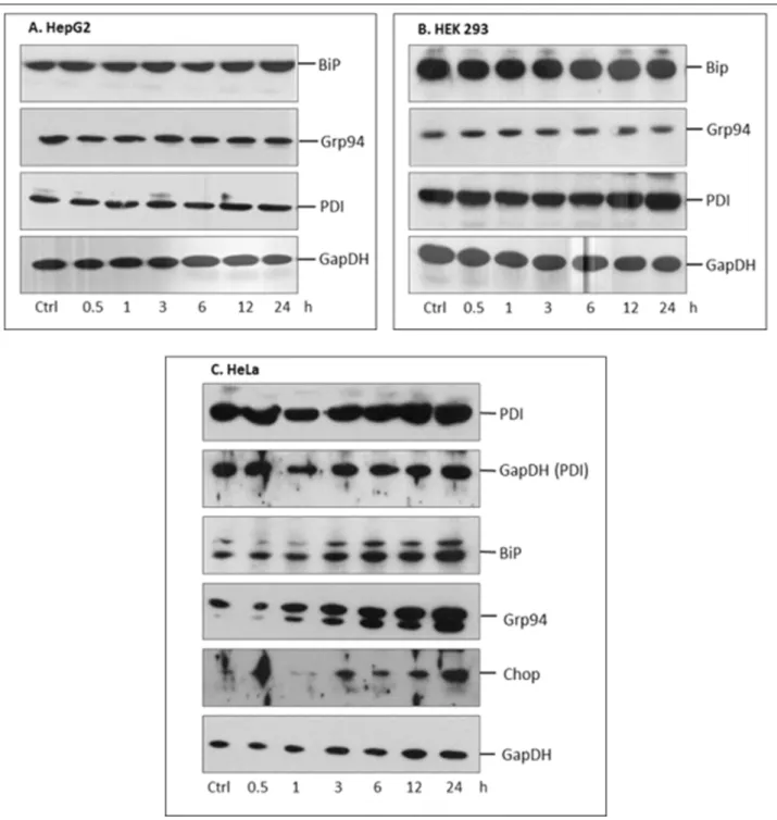
In ancillary experiments we tested how the PEMF exposure influences the expression of stress markers of the ER. Three selected cell lines - HEK, HepG2, HeLa - were treated with PEMF for different time period (0.5, 1, 3, 6, 12, 24 hours).
Expression of ER stress markers, (BiP, Grp94 and PDI) were detected by Western blot. No remarkable differences were demonstrated in HepG2 and HEK cells, while in HeLa cells an alteration in expression of BiP, Grp94 and also an apoptosis marker CHOP was found (Fig. 1).
Pulsed electromagnetic fields is able to attenuate endoplasmic reticulum stress in tunicamycin treated HepG2 cells
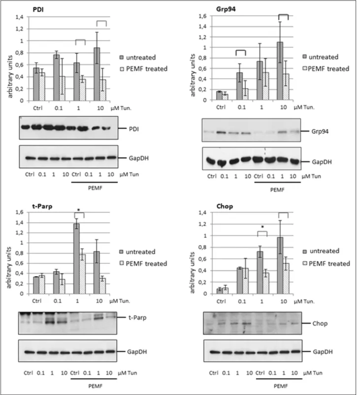
Since HepG2 cell line did not show any variation in expression of selected ER chaperons upon PEMF exposure, we chose this line for further experiments. For studying PEMF influence on cells under ER stress, equal amount of HepG2 cells were stressed with different concentration of tunicamycin for 24 hours, with or without simultaneous PEMF treatment. PDI, Grp94, CHOP expression and the inactivation of PARP were analysed. As shown on Fig. 2., PEMF exposure was able to reduce the tunicamycin-induced expression level increase of ER stress markers. In case of PDI and Grp94 markers, statistical analysis of three independent experiments indicated differences (P < 0.1) compared to untreated, stressed cells. Despite of the 770
band intensity variances between each independent experiment, statistical differences were not detected. The expression of BIP was also effected (data not shown).
Apoptosis markers CHOP and the cleaved, inactive form of PARP (t-PARP as truncated PARP) were also influenced by PEMF treatment: the diagram demonstrates a significant decrease (P < 0.05) in their inducibility compared to the non- PEMF cells. The figure also demonstrates that PEMF did not remarkably modulate the examined protein expression of unstressed cells.
Pulsed electromagnetic fields is able to increase cell viability in tunicamycin treated HepG2 cells
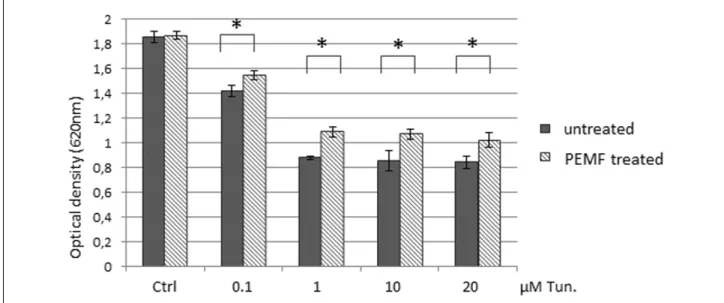
CHOP is regarded as a proapoptotic transcription factor of UPR (32) while cleavage of PARP by caspase-3 has been published to be required for apoptosis. To investigate further the effect of diminished CHOP and t-PARP, we monitored cell viability of stressed cells with and without PEMF exposure. Fig. 3 presents that PEMF was able to significantly elevate (P < 0.05) the viability of tunicamycin-treated cells at each treated concentration.
Fig. 1. The effect of pulsed electromagnetic fields (PEMF) treatment on endoplasmic reticulum (ER) varies in different cell lines.
HepG2 (A), HEK293T (B) and HeLa (C) cells were PEMF exposured for different time periods and expression of ER stress markers was tested by Western blot. (A – B): PEMF treatment did not effect the expression of ER chaperons and foldases of HepG2 and HEK293T cells. (C): In HeLa cells PEMF treatment increased the expression of BiP, Grp94 and CHOP proteins. A representative experiment out of two is shown.
DISCUSSION
The aim of this study was to investigate the effect of PEMF on ER stress and cell viability in tunicamycin-treated cell line.
Our main findings are that PEMF exposure (8 Hz and meant flux density of 0.56 mT) is able to reduce the elevated activity of ER
stress markers induced by tunicamycin, in HepG2 cell line. The cell viability was also significantly increased upon PEMF treatment.
ER stress-induced UPR is a highly controlled molecular pathway that mediates programmed cell death and regulates apoptosis, therefore able to protect cells from premature death.
772
Fig. 2. Pulsed electromagnetic fields (PEMF) exposure decreased the expression of endoplasmic reticulum (ER) stress markers, foldase and apoptosis markers in tunicamycin treated HepG2 cells. Cells were stressed with different concentration of ER stressor, tunicamycin (0.1, 1, 10 mM). Analysis of protein expression showed that PEMF was able to decrease the elevated level of ER chaperon and foldase Grp94, PDI and the apoptosis markers CHOP and cleaved PARP (t-PARP) induced by tunicamycin. Square brackets indicate differences by P < 0.1. *Asterisks over the brackets indicate statistically significant difference P < 0.05. Displaying no bracket indicates a P value ≥0.1.
This explains the significant on-going efforts to understand the aspects of this multilayer process. Since excessive ER stress/UPR can lead to several serious human diseases via apoptosis, finding appropriate, non-invasive tools to reduce ER stress is of high interest (33, 34).
PEMF is an alternative, non-invasive method that is reported to successfully treat several health problems including neurological disorders (Alzheimer-, Parkinson diseases, dementia) or ischemia, where apoptosis/anti-apoptosis and/or altered ER homeostasis plays important role in the pathogenesis.
First we tested three different cell lines on how their ER reacts on different lengths of PEMF exposure. In HepG2 and HEK cells no remarkable changes of chaperons BiP and Grp94, and PDI were observed. In contrast, HeLa cells showed increased expression of BiP and Grp94. The level of CHOP protein was also elevated. The transcription factor CCAAT- enhancer-binding protein homologous protein (CHOP) is a molecule involved in the regulation of ER stress induced apoptosis (32).
Our results on HeLa cells are in agreement with the observations by Chen et al. (25). They published that in HeLa cells, picosecond pulsed electric fields induced not only mitochondrial mediated apoptosis (27) but the increased expression of ER chaperons (Grp78, Grp94) and CHOP (25).
Nevertheless, our result clearly suggests that different cell lines vary in the response at PEMF treatment just as it has already been proposed (20, 35). Morotomi-Yano et al. (20) also showed that HeLa cells underwent cell death after picosecond pulsed electric fields but the preferential mode of cell death was necrosis, whereas Jurkat cells underwent apoptosis. The essential role of extracellular Ca2+was also demonstrated by a study on the effect of static magnetic field on apoptotic cell death in different cell lines (35). It was found that in some type of cell lines after apoptosis induction, static magnetic fields were able to increase cell survival via inducing Ca2+influx from the extracellular medium; however other lines showed no antiapoptotic effect on magnetic fields (35). They concluded that magnetic fields are protective in those cell lines where Ca2+
plays an antiapoptotic effect. It should be mentioned that the comparison of results from different studies are often difficult
because devices of PEMF treatment usually work on different frequencies and magnetic fields. The PEMF’s multiple cellular responses were suggested to vary depending on the intensity of the applied electric fields (27).
Since HepG2 cell line did not show alteration in ER chaperon expression upon PEMF exposure, it was selected for the investigation of PEMF on ER stress. After PEMF exposure of tunicamycin treated cells, reduced expression of ER stress markers Grp94, PDI, CHOP and diminished level of cleaved PARP were presented. The overexpression of CHOP indicates ER stress and promotes apoptosis of the cell (36). Poly (ADP- ribose) polymerase (PARP) is inactivated trough cleavage by active caspase-3 during apoptosis. Therefore we tested whether that reduction of stress markers’ amount upon PEMF is able to influence the viability of stressed cells. Our results show that PEMF exposure was able to significantly decrease the number of death cells compared to the unexposed control. Similar effect of PEMF has already been published by other groups; however an ER stress inducer, affecting the mechanism of luminal protein folding was not used before (26, 28, 29). Nevertheless, tunicamycin is a well-known inhibitor of N-glycosylation in the ER, it was also published that tunicamycin is able to decrease palmitoylation of the endothelial isoform of NOS thus stimulate NO synthesis and also increased intracellular calcium levels in a concentration-dependent manner in bovine aortic endothelial cells (37).
Kaszuba-Zwoinska et al. studied 50 Hz PEMF treatment on Mono Mac6 monocytic cells apoptotised by different agents (28). The puromycin-PEMF treated cells were less susceptible to apoptosis induction than cultures not stimulated with PEMF.
Puromycin was used as an inhibitor of telomerase activity;
however, it is also an ER Ca2+leak channel opener (38) and an ER stressor (39). The 50 Hz exposure also inhibited puromycin induced cell death in human neuroblastoma and rat neuroendocrine cells (26) and also in U937 cells (29).
In another experimental model on cultured hippocampal cells (40) electromagnetic fields did not affect the cell mortality.
However, when exposure happened during oxidative stress (NO) significant increase in cell death was observed compared to the unexposed stressed cells.
Fig. 3. Pulsed electromagnetic fields (PEMF) exposure restores cell viability of tunicamycin induced cells. HEK293T cells were treated by different (0.1, 1, 10, 20 mM) concentration of tunicamycin with/without PEMF exposure. Cell viability was detected using Cell Titer- Blue Assay Kit. Results are presented as mean values of optical density ± S.D. and were compared using Student’s t-test. Asterisks indicate statistically significant difference between PEMF treated and untreated, endoplasmic reticulum (ER) stressed cells: *P < 0.05.
The reduction of ER stress and apoptosis can be a mechanism how PEMF is able to influence the health problems mentioned above. Silencing of CHOP expression in rabbit is able to protect from AD-like disease (41). Other publications indicated that CHOP is a key molecule not only in apoptosis but also in inflammatory responses (32). Whether the induced ER stress is modulated through protein phosphorylation, altered Ca2+ fluxes or conformational changes of the ER membrane phospholipids, is still a question and requires further studies. Our results also indicate that PEMFs effect varies in different cell lines; the liver carcinoma cell line, HepG2, or the human embryonic kidney, HEK 293T, did not show any affect in ER stress markers upon PEMF, however the expression of ER markers were increased in the human cervical cancer cells HeLa. Investigations on primary cell cultures and animals are needed to clarify the effect of PEMF in the outcome of ER stress.
Acknowledgements: This work was supported by INKUBATOR grant of the Department of Medical Chemistry, Molecular Biology and Pathobiochemistry, Semmelweis University, Budapest, Hungary to I.S. and by OTKA 112696 grant.
Conflict of interests: None declared.
REFERENCES
1. Braakman I, Bulleid NJ. Protein folding and modification in the mammalian endoplasmic reticulum. Annu Rev Biochem 2011; 80: 71-99.
2. Mandl J, Meszaros T, Banhegyi G, Csala M. Minireview:
endoplasmic reticulum stress: control in protein, lipid, and signal homeostasis. Mol Endocrinol 2013; 27: 384-393.
3. Stiller I, Lizak B, Banhegyi G. Physiological functions of presenilins; beyond gamma-secretase. Curr Pharm Biotechnol 2014; 15: 1019-1025.
4. Eguchi K. Apoptosis in autoimmune diseases. Intern Med 2001; 40: 275-284.
5. Orsolya K, Lizak B, Stiller I, Banhegyi G. A Systems Biological perspective of cellular stress-directed programmed cell death. Comput Mol Biosci 2014; 4: 28-34.
6. Volmer R, Ron D. Lipid-dependent regulation of the unfolded protein response. Curr Opin Cell Biol 2015; 33:
67-73.
7. Bassett CA, Pawluk RJ, Pilla AA. Acceleration of fracture repair by electromagnetic fields. A surgically noninvasive method. Ann NY Acad Sci 1974; 238: 242-262.
8. Aaron RK, Ciombor DM, Simon BJ. Treatment of nonunions with electric and electromagnetic fields. Clin Orthop Relat Res 2004; (419): 21-29.
9. Sharrard WJ. A double-blind trial of pulsed electromagnetic fields for delayed union of tibial fractures. J Bone Joint Surg Br 1990; 72: 347-55.
10. Cebrian JL, Gallego P, Frances A, et al. Comparative study of the use of electromagnetic fields in patients with pseudoarthrosis of tibia treated by intramedullary nailing. Int Orthop 2010; 34: 437-440.
11. Jing D, Cai J, Shen G, et al. The preventive effects of pulsed electromagnetic fields on diabetic bone loss in streptozotocin-treated rats. Osteoporos Int 2011; 22: 1885- 1895.
12. Pan Y, Dong Y, Hou W, et al. Effects of PEMF on microcirculation and angiogenesis in a model of acute hindlimb ischemia in diabetic rats. Bioelectromagnetics 2013; 34: 180-188.
13. Weintraub MI, Herrmann DN, Smith AG, Backonja MM, Cole SP. Pulsed electromagnetic fields to reduce diabetic neuropathic pain and stimulate neuronal repair: a randomized controlled trial. Arch Phys Med Rehabil 2009;
90: 1102-1109.
14. Canedo-Dorantes L, Garcia-Cantu R, Barrera R, Mendez- Ramirez I, Navarro VH, Serrano G. Healing of chronic arterial and venous leg ulcers with systemic electromagnetic fields. Arch Med Res 2002; 33: 281-289.
15. Roland D, Ferder M, Kothuru R, Faierman T, Strauch B.
Effects of pulsed magnetic energy on a microsurgically transferred vessel. Plast Reconstr Surg 2000; 105: 1371- 1374.
16. Goudarzi I, Hajizadeh S, Salmani ME, Abrari K. Pulsed electromagnetic fields accelerate wound healing in the skin of diabetic rats. Bioelectromagnetics 2010; 31: 318-323.
17. Heden P, Pilla AA. Effects of pulsed electromagnetic fields on postoperative pain: a double-blind randomized pilot study in breast augmentation patients. Aesthetic Plast Surg 2008;
32: 660-666.
18. Varani K, Vincenzi F, Targa M, et al. Effect of pulsed electromagnetic field exposure on adenosine receptors in rat brain. Bioelectromagnetics 2012; 33: 279-287.
19. Strauch B, Herman C, Dabb R, Ignarro LJ, Pilla AA.
Evidence-based use of pulsed electromagnetic field therapy in clinical plastic surgery. Aesthet Surg J 2009; 29: 135-143.
20. Morotomi-Yano K, Akiyama H, Yano K. Different involvement of extracellular calcium in two modes of cell death induced by nanosecond pulsed electric fields. Arch Biochem Biophys 2014; 555-556: 47-54.
21. Wetzel BJ, Nindl G, Vesper DN, Swez JA, Jasti AC, Johnson MT. Electromagnetic field effects: changes in protein phosphorylation in the Jurkat E6.1 cell line. Biomed Sci Instrum 2001; 37: 203-208.
22. Delport PH, Cheng N, Mullier C, Sansen M, De Loecker W.
The effects of pulsed electromagnetic-fields on membrane- transport, protein and Atp synthesis in rat skin. Biochem Soc Trans 1984; 12: 437-438.
23. Clejan S, Ide C, Walker C, Wolf E, Corb M, Beckman B.
Electromagnetic field induced changes in lipid second messengers. J Lipid Mediat Cell Signal 1996; 13: 301-324.
24. Badzhinian SA, Meliksetian AM, Kazarian PA, Malakian MG, Egizarian DE, Grigorian DS. Electromagnetic fields of millimeter range: effect on the structure and functional properties of erythrocyte membranes. Radiats Biol Radioecol 2002; 42: 551-555.
25. Chen WJ, Xiong ZA, Zhang M, et al. Picosecond pulsed electric fields induce apoptosis in HeLa cells via the endoplasmic reticulum stress and caspase-dependent signaling pathways. Int J Oncol 2013; 42: 963-970.
26. Grassi C, D’Ascenzo M, Torsello A, et al. Effects of 50 Hz electromagnetic fields on voltage-gated Ca2+channels and their role in modulation of neuroendocrine cell proliferation and death. Cell Calcium 2004; 35: 307-315.
27. Hua YY, Wang XS, Zhang Y, Yao CG, Zhang XM, Xiong ZA. Intense picosecond pulsed electric fields induce apoptosis through a mitochondrial-mediated pathway in HeLa cells. Mol Med Rep 2012; 5: 981-987.
28. Kaszuba-Zwoinska J, Chorobik P, Juszczak K, Zaraska W, Thor PJ. Pulsed electromagnetic field affects intrinsic and endoplasmatic reticulum apoptosis induction pathways in MonoMac6 cell line culture. J Physiol Pharmacol 2012; 63:
537-545.
29. Kaszuba-Zwoinska J, Wojcik K, Bereta M, et al. Pulsating electromagnetic field stimulation prevents cell death of puromycin treated U937 cell line. J Physiol Pharmacol 2010; 61: 201-205.
774
30. Esko JD, Bertozzi CR. Chemical tools for inhibiting glycosylation. In: Essentials of Glycobiology, A. Varki, R.D. Cummings, J.D. Esko, et al., (eds.). Cold Spring Harbor (NY), Cold Spring Harbor Laboratory Press, 2009.
31. Park SH, Hong H, Han YM, et al. Nonsteroidal anti- inflammatory drugs (NSAID) sparing effects of glucosamine hydrochloride through N-glycosylation inhibition; strategy to rescue stomach from NSAID damage. J Physiol Pharmacol 2013; 64: 157-165.
32. Oyadomari S, Mori M. Roles of CHOP/GADD153 in endoplasmic reticulum stress. Cell Death Differ 2004; 11:
381-389.
33. Padilla J, Jenkins NT. Induction of endoplasmic reticulum stress impairs insulin-stimulated vasomotor relaxation in rat aortic rings: role of endothelin-1. J Physiol Pharmacol 2013;
64: 557-564.
34. Malhi H, Kaufman RJ. Endoplasmic reticulum stress in liver disease. J Hepatol 2011; 54: 795-809.
35. Fanelli C, Coppola S, Barone R, et al. Magnetic fields increase cell survival by inhibiting apoptosis via modulation of Ca2+influx. FASEB J 1999; 13: 95-102.
36. Li YM, Guo Y, Tang J, Jiang J, Chen Z. New insights into the roles of CHOP-induced apoptosis in ER stress. Acta Biochim Biophys Sinica (Shanghai) 2014; 46: 629-640. Erratum 2015; 47: 146-147.
37. Buckley BJ, Whorton AR. Tunicamycin increases intracellular calcium levels in bovine aortic endothelial cells.
Am J Physiol 1997; 273: C1298-C1305.
38. Hammadi, M., Oulidi A, Gackiere F, et al. Modulation of ER stress and apoptosis by endoplasmic reticulum calcium leak via translocon during unfolded protein response:
involvement of GRP78. FASEB J 2013; 27: 1600-1609.
39. Oguma T, Ono T, Kajiwara T, et al. CD4(+)CD8(+) thymocytes are induced to cell death by a small dose of puromycin via ER stress. Cell Immunol 2009; 260: 21-27.
40. Boland A, Delapierre D, Mossay D, Dresse A, Seutin V.
Effect of intermittent and continuous exposure to electromagnetic fields on cultured hippocampal cells.
Bioelectromagnetics 2002; 23: 97-105.
41. Prasanthi JR, Larson T, Schommer J, Ghribi O. Silencing GADD153/CHOP gene expression protects against Alzheimer’s disease-like pathology induced by 27- hydroxycholesterol in rabbit hippocampus. PLoS One 2011;
6: e26420. doi: 10.1371/journal.pone.0026420 R e c e i v e d : February 29, 2016
A c c e p t e d : September 30, 2016
Author’s address: Dr. Ibolya Stiller, Department of Medical Chemistry, Molecular Biology and Pathobiochemistry, Semmelweis University, H-1428 Budapest, POB 2, Hungary.
E-mail: stiller.ibolya@med.semmelweis-univ.hu.