Studying protective compounds against lipotoxicity on insulinoma cell line
PhD thesis outline
Simon-Szabó Laura
Semmelweis University Pharmacological Sciences Doctoral School
Supervisors: prof. Kéri György, DSc., kutatóprofesszor Dr. Csala Miklós, DSc., egyetemi docens Official reviewers: Dr. Törőcsik Beáta, Ph.D., adjunktus
Dr. Venekei István, Ph.D., egyetemi docens
Head of final examination committee:
prof. Török Tamás, DSc., egyetemi tanár Members of final examination committee:
Dr. Klebovich Imre, DSc., egyetemi tanár Dr. Bajtay Zsuzsa, DSc., egyetemi docens
Budapest
2016
Introduction
Type 2 diabetes is a widespread disease occurring predominantly in welfare society. The main provoking factors are excess food intake, lack of physical activity, and the consequent obesity. Nowadays overweight is becoming more and more prevalent even in childhood. Type 2 diabetes is a complex metabolic disorder, with permanently elevated high blood sugar levels (hyperglycaemia).
It is essential to maintain the normal blood glucose concentration within a range of 3.5 and 5.5 mM, to ensure the proper functioning of the body. Any alteration from the normal range could lead to pathological conditions. Blood glucose level is under strict control with two hormones playing fundamental role: insulin and glucagon.
Both are produced by the islets of Langerhans in the pancreas;
glucagon is made by α-cells and insulin is made by β-cells.
Glucagon causes elevation, while insulin results in a decrease of blood glucose concentration. Reduction of muscle-fat mass ratio leads to weaker insulin response, a phenomenon called insulin resistance. In the early stage of insulin resistance pancreatic β-cells produce excessive amount of insulin for compensation. If the counterbalancing hormone levels are not enough the blood glucose concentration starts rising. The surplus nutriment supply burdens the
intermedier metabolism of cells, triggering stress signal transduction pathways. This leads to first relative and afterwards absolute insulin deficiency. Insulin resistance is responsible for overproduction of glucose in liver and less uptake by muscle tissue. Moreover it boosts fatty acid release from adipose tissue, increasing the free fatty acid content of blood resulting in lipotoxicity in most tissues.
Lipotoxicity is a complex cellular stress condition including the endoplasmic reticulum (ER) stress. Extreme environmental disorders can disturb the delicate protein maturation processes in the lumen of ER which provokes the unfolded protein response (UPR).
The UPR is triggered by the accumulation unfolded or misfolded proteins in the lumen of the organelle and consists of three major pathways. The aim of all three is to repair the homeostasis of ER by decreasing the load and increasing the capacity of protein folding machinery and enhancing the misfolded protein degradation pathway. If these efforts fail to restore equilibrium the cells can die in apoptosis.
Recent discoveries suggest the role of c-jun N-terminal kinase (JNK) also known as stress activated protein kinase (SAPK) in development of insulin resistance. JNK is a stress kinase, which can be activated by one of the branches of UPR, the inositol-requiring
(IRS1), thus inhibiting the tyrosine phosphorylation of this signal transducer by the insulin receptor. Therefore signalling pathway of insulin will be damaged, binding of insulin to its receptor cannot be able to provoke the proper increase in glucose uptake and inhibition in glucose production.
Several compounds have been released to prevent the development of type 2 diabetes in the stage of insulin resistance. One of them is metformin from the group of biguanide molecules.
Metformin decreases glucose production in liver, increases glucose uptake in muscle, thus prevents hyperglycaemia. Decreases appetite and helps reducing weight, inhibits gluconeogenesis and mitochondrial cell death. One of its possible targets is the first complex of mithochondrial respiratory chain. However it is crucial to develop new drug molecules with fewer side effects, such as stomach problems or lactic acidosis in patients with kidney, lung or heart diseases. The latter, elevation of lactic acid in blood, could be a life threatening condition.
Signal transduction therapy has become widely accepted by now. It means selective inhibition of one of the stress activated signalling pathways. Such therapeutic approach for example the hindrance of IRS-1 serine phosphorylation directly or indirectly through the inhibition of JNK.
Drugs with these effects would help the restoration of off- balanced conditions, but will not change the fact that the primary prevention and treatment of obesity related metabolic disorders are founded in healthy life style, diet and exercise.
Aims
Due to the increasing prevalence of type 2 diabetes, research for new medication has become highly important. Signal transduction therapy has become widely accepted by now. Major points of our interest were lipotoxicity and lipoapoptosis. We studied the protective effect of the known antidiabetic drug, metformin on possible inhibition of ER stress on a suitable cell culture model. We used RINm5F rat insulinoma cell line, and provoked lipotoxicity by addition of albumin conjugated palmitate.
We were particularly interested in JNK activation and the concomitant IRS-1 phosphorylation at Ser307, as one of the most significant factor in development of insulin resistance and β-cell damage. Besides analysing effects of metformin, we aimed to find novel compounds inhibiting the aforementioned phenomenon on β- cell-lipotoxicity model system. For primary screening of kinase inhibitor candidates we used HEK293 human kidney fibroblasts and provoked IRS-1 serine phosphorylation by JNK activation upon anisomycin treatment.
Specific aims:
• Investigation of possible protective role of metformin against lipotoxicity and lipoapoptosis on pancreatic β-cell line.
• Reveal the effect of metformin on UPR activation during lipotoxicity in β-cells.
• Identification of novel compounds effectively inhibiting IRS-1 serine phosphorylation to develop possible antidiabetic agents,
Methods
Viability of RINm5F rat insulinoma cell line was assessed by trypane blue exclusion method following albumin conjugated palmitate treatment. Live and dead (stained) cells were counted using automatic cell counter.
Apoptotic and necrotic cells were examined by Annexin-V- Fluos Staining Kit and florescence microscopy. Annexin labelled, green fluorescence cells were considered apoptotic while propidium- iodide labelled, red fluorescence cells were counted as necrotic.
Double labelling (cells showing both red and green fluorescence) was also considered as necrosis. In each experimental condition, a minimum of 1000 cells was counted. The necrosis and apoptosis indexes were calculated as (necrotic or apoptotic cells)/(total cell number)×100.
Signs of UPR activation upon anysomicin or palmitate provoked ER stress in HEK293 or RINm5F cells were examined by Poliacrilamide Gelelectroforesis (PAGE) and Western blot analysis.
Splicing of XBP-1 was studied during lipotoxicity on insulinoma cells. cDNA was produced by reverse transcription of DNA-free RNA samples using SuperScript III First-Strand Synthesis System for RT-PCR Kit. Spliced and unspliced XBP-1 sequences
(421 or 447 bp, respectively) were amplified by PCR using SY121041268-007 XBP-1 sense (rat) and ST00450236-001 XBP-1 antisense (mouse, rat) primers and iProof High-Fidelity DNA Polymerase Kit. PCR products were purified by PEG precipitation and their concentration was measured with Nanodrop 1000 Spectrophotomete. PstI restriction endonuclease treatment was carried out, the amplified sequence of unspliced XBP-1 was cut in two fragments (153 and 294 bp) while the spliced variant remained uncut. Equal amounts of digested PCR products were resolved by electrophoresis in 2% agarose gel and visualized by EtBR staining.
Western blot results were quantified by densitometry using ImageQuant 5.2 software. Data are presented as mean values ± S.D.
and were compared using ANOVA with Tukey’s multiple comparison post hoc test. Differences with a P value below 0.05 were considered to be statistically significant.
Results
Palmitate-induced Apoptosis in RINm5F Cells
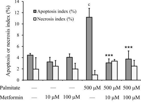
To study the protective role of metformin against lipotoxicity on RINm5F cell line we used palmitate treatment. Based on previous publications using the same cell type we applied albumin conjugated palmitate in 500 µM concentration.
Cell death was examined by differentiating apoptosis and necrosis using Annexin-V-Fluos Staining Kit. and fluorescence microscopy (Figure 1.). 500 µM palmitate for 6 h nearly tripled the apoptotic index. Metformin treatment alone did not affect cell death, however significantly decreased palmitate induced apoptosis to the level of untreated cells in 10 µM and 100 µM concentrations. In the same time neither palmitate nor metformin affected necrotic cell death (Figure 1).
Figure 1.: Apoptotic and necrotic index on RINm5F cell line
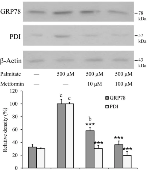
The effect of metformin at palmitate-induced ER stress.
The attendant of the UPR is the activation of the ER chaperons therefore it's a very common marker of the ER stress. In our experiments we measured two main ER-chaperons by Western-blot analysis (Figure 2.). GRP78 alias Bip and PDI quantity also were increased in the palmitate induced samples compare to the control level which represents that the lipotoxicity was acted ER stress in cells. The induction of the two chaperons were decreased by the simultaneous metformin treatment. Applying 100 µM metformin GRP78 and PDI were remained at the control level (Figure 2.).
Figure 2.: Induction of the ER chaperons: GRP78 and
Selection of possible inhibitors of JNK activation and IRS-1 Ser307 phosphorylation
JNK inhibitors were selected from the Vichem EVL library and primary tested on a well-documented and widely used model system, activation of JNK by anisomycin on HEK293 kidney fibroblasts. We choose the published BI-78D3 molecule as positive control for JNK inhibition, and first examined its effectivity and concentration dependence on our model.
The selected reference molecules and the newly synthetized compounds first were tested on anysomycin treated HEK293 cells by detecting their effect on JNK, c-Jun and IRS-1 phosphorylation in a 10 µM concentration. As we expected strong correlation was observed between the level of JNK and c-Jun phosphorylation. On the other hand rate of JNK and IRS-1 phosphorylation (density of P- JNK and P-IRS-1 Ser307) did not show tight correlation possibly due to the presence of other serine kinases phosphorylating IRS-1.
From primary screening of 57 compounds we selected 12 for further experiments. Our selection was based on criteria of JNK phosphorylation level under 65% and IRS Ser307 phosphorylation level under 15%. Compounds meeting these conditions all had 2,6- disubstituted 7-oxo-pyrido-[2,3-d]pyrimidin core structure.
Examination of selected inhibitors on lipotoxicity model
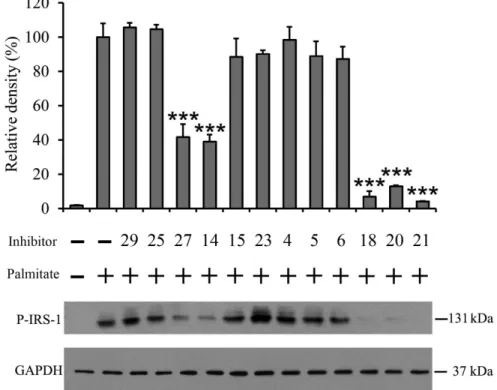
Inhibitory agents proved to be effective on anisomycin treated HEK293 cell line were further investigated on biologically more relevant lipotoxicity model. RINm5F rat insulinoma cells were stressed with albumin conjugated palmitate (500 µM) along with twelve different inhibitors (10 µM). After 8 hours of incubation JNK and IRS-1 Ser307 phosphorylation were detected by Western blotting (Figure 3.) Addition of two compounds (number 27 and 18) resulted in strong inhibition of JNK phosphorylation (54% and 59%
respectively). These two molecules also caused significant but slightly different level of inhibition (58% and 93% respectively) in IRS-1 Ser307 phosphorylation. Interesting to note that three additional agent also effectively decreased IRS-1 Ser307 phosphorylation (number 14, 20 and 721 causing 61%, 87% and 96%
inhibition), although they didn’t impinge on JNK phosphorylation (Figure 3.).
Figure 3.: Inhibition of lipotoxicity induced IRS-1 Ser307 phosphorylation in RINm5F cell line
Conclusions
In this study we focused on the research of lipotoxicity and lipoapoptosis in the development of insulin resistance, and looked for possible inhibitors of IRS-1 phosphorylation, a central player in the above disorders.
We analysed the effect of the known antidiabetic metformin on cell viability, rate of apoptosis and ER stress by using RINm5F rat insulinoma cell line treated with albumin conjugated palmitate.
We had special interest examining the activation of the central stress kinase JNK and consecutive IRS-1 Ser307 phosphorylation.
Our most important findings:
• Metformin prevented palmitate induced apoptotic cell death, which was also confirmed by significant decrease of caspase-3 activation, while did not affected necrosis.
• Addition of metformin strongly alleviated the events of lipotoxicity induced UPR, such as eIF2α phosphorylation by PERK, IRE-1 dependent splicing of XBP-1 mRNA and initial phosphorylation of JNK, IRS-1 and c-Jun through IRE-1. In
upregulation of ER chaperon PDI and GRP78 and last but not least the induction of proapoptotic CHOP.
RINm5F rat insulinoma cell line treated with albumin conjugated palmitate. Served as a model system for screening compounds inhibiting IRS-1 Ser307 phosphorylation and can be basis of new drug development for insulin resistance and β-cell death. We tested reference molecules and their newly synthetized derivatives from Vichem EVL library-altogether 57 potential JNK inhibitors- on anisomycin treated HEK293 cell line by detecting their effect on JNK activation and IRS-1 Ser307 phosphorylation.
• Five of the twelve selected molecules showed significant inhibition on IRS-1 Ser307 phosphorylation and four of these also reduced JNK activation. These compounds can be promising starting points for new drug development, thus it is reasonable to further investigate their effectiveness on in vivo experiments as well.
Publications
Publications related to doctoral thesis:
Simon-Szabo L, Kokas M, Mandl J, Keri G, Csala M. (2014) Metformin attenuates palmitate-induced endoplasmic reticulum stress, serine phosphorylation of IRS-1 and apoptosis in rat insulinoma cells. PLoS One, 9(6):e97868.
IF: 3,234
Simon-Szabo L, Kokas M, Greff Z, Boros S, Banhegyi P, Zsakai L, Szantai-Kis C, Vantus T, Mandl J, Banhegyi G, Valyi-Nagy I, Orfi L, Ullrich A, Csala M, Keri G. (2016) Novel compounds reducing IRS-1 serine phosphorylation for treatment of diabetes. Bioorg Med Chem Lett, 26(2):424-428.
IF: 2,420
Zambo V, Simon-Szabo L, Szelenyi P, Kereszturi E, Banhegyi G, Csala M. (2013) Lipotoxicity in the liver. World J Hepatol, 5(10):550-557.
Publications not related to doctoral thesis:
Varga A, Gyulavari P, Greff Z, Futosi K, Nemeth T, Simon-Szabo L, Kerekes K, Szantai-Kis C, Brauswetter D, Kokas M, Borbely G, Erdei A, Mocsai A, Keri G, Vantus T. (2015) Targeting vascular endothelial growth factor receptor 2 and protein kinase D1 related pathways by a multiple kinase inhibitor in angiogenesis and inflammation related processes in vitro. PLoS One, 10(4):e0124234.
IF: 3,234