Multiple system-level feedback loops control life-and- death decisions in endoplasmic reticulum stress
Orsolya Kapuy1 , Margita Marton1, Gabor Banhegyi1,2and P. K. Vinod3
1 Department of Medical Chemistry, Molecular Biology and Pathobiochemistry, Semmelweis University, Budapest, Hungary 2 Pathobiochemistry Research Group of the Hungarian Academy of Sciences and Semmelweis University, Budapest, Hungary 3 Centre for Computational Natural Sciences and Bioinformatics, International Institute of Information Technology, Hyderabad, India
Correspondence
O. Kapuy, Department of Medical Chemistry, Molecular Biology and
Pathobiochemistry, Semmelweis University, 1094 Budapest, T}uzolto utca 37-47, Hungary Tel: +36 1 266 26 15
E-mail: kapuy.orsolya@med.semmelweis- univ.hu
(Received 2 October 2019, accepted 14 November 2019)
doi:10.1002/1873-3468.13689 Edited by Alfonso Valencia
Scientific results have revealed that autophagy is able to promote cell survival in response to endoplasmic reticulum (ER) stress, while drastic events result in apoptotic cell death. Here, we analyse the important crosstalk of life-and-death decisions from a systems biological perspective by studying the regulatory modules of the unfolded protein response (UPR). While a double-negative loop between autophagy and apoptosis inducers is crucial for the switch-like charac- teristic of the stress response mechanism, a positive feedback loop between ER stress sensors is also essential. Corresponding to experimental data, here, we show the dynamical significance of Gadd34-CHOP connections inside the PERK branch of the UPR. The multiple system-level feedback loops seem to be crucial for managing a robust life-and-death decision depending on the level and durability of cellular stress.
Keywords:autophagy; apoptosis; endoplasmic reticulum stress; systems biology; feedback loops
Cellular protein homeostasis (called proteostasis) is essential in the dynamic changes required for the cell to respond various stimuli (such as nutrient availabil- ity, inflammatory mediators). Proteostasis is controlled by a complex regulatory system that involves intracel- lular and extracellular protein synthesis, folding, degradation, aggregation and disaggregation[1,2]. This process is mainly driven by the endoplasmic reticulum (ER) with the perception and control of these different changes for the support of the correct functioning of proteins [3]. The translation, formation and mainte- nance of the native state of proteins take places in the ER [4,5]. In addition, several biosynthetic, metabolic and signal transduction pathways are regulated by the ER [6]. For this complex operation, a high luminal Ca2+ environment and a special redox homeostasis are essential in the ER[7,8].
The disruption of the balance in the ER may gener- ate the turning on of the ER stress response
mechanism [3,6,9,10]. The unfolded protein response (UPR) of the ER is the principal signalling pathway that helps to deal with the imbalances in protein fold- ing and drives suitable protein quality control [11,12].
UPR immediately turns on when incorrectly folded or damaged proteins get accumulated in the cell. UPR has three well-defined ER-resident transmembrane sig- nal transducers, called IRE1 (inositol requiring 1 kinase), PERK (PKR-like ER kinase) and ATF6 (acti- vating transcription factor 6) [13]. Both IRE1 and ATF6 promote transcription of UPR target genes cru- cial in folding and quality control upon ER stress, while PERK-controlled pathway leads to the general inhibition of protein translation via eiF2aphosphory- lation[13,14].
It is already well known that harmful ER stress immediately accelerates autophagy-dependent cellular
‘self-cannibalism’ [15,16]. Autophagy carries out the degradation of unnecessary or damaged proteins and
Abbreviations
APOA, active form of apoptosis inducer; AUTA, active form of autophagy inducer; ER, endoplasmic reticulum; ERSS, ER stress sensor.
organelles, through the delivery into autophagosomes followed by lysosomal digestion. These degraded com- ponents can be reused later, which makes autophagy a cellular survival mechanism by ‘self-eating’ of damaged or not properly folded proteins during UPR-controlled ER stress [17–19]. However, excessive level of ER stress can occur apoptotic [9,15,20]or necroptotic cell death[21]. Cells can be ablated by apoptosis through a well-defined manner during which cells lose their connections with the surrounding cells, become shrun- ken with the condensation of nucleus and are finally engulfed by surrounding cells or macrophages. Apop- tosis can be triggered by several cellular signals coming from either outside or inside from the cell[22]. Differ- ent kind of cellular stress events can activate the intrinsic or so-called mitochondrial apoptotic pathway, which generates cytochrome c flux out of the mito- chondria. Besides, apoptosis can also be activated through death receptors, which initiates the activation of the extrinsic pathway[23].
Although both autophagy and apoptosis are regu- lated by complicated networks of signal transducers, there is growing evidence that a link between these two mechanisms also exists. This connection realized at various levels is developing a so-called crosstalk with even more comprehensive regulatory networks between autophagy and apoptosis [24,25]. While autophagy is essential in cell survival, apoptosis is definitively a programmed cell death mechanism; their connection looks substantial in a well-balanced cellu- lar response upon various stress signals (such as nutri- ent deprivation, ER stress) [25]. Our recent results based on molecular biological tools and system bio- logical methods have shown that autophagy always precedes apoptotic cell death even upon severe ER stress [26]. This test was confirmed by various reagents inducing ER stress (such as DTT, thapsi- gargin and tunicamycin) [26]. These ER stressors are able to generate a clear threshold for the apoptosis induction upon ER stress. With the use of either autophagy activator or inhibitor, the importance of autophagy-dependent survival was also explored previ- ously. In addition, transient high level of ER stress treatments was also performed, to further confirm the irreversible dynamical behaviour of apoptosis induc- tion. In order for the better understanding of the regulatory systems, a stochastic model was built illustrating the life-or-death decision-maker process induced by ER stress[26].
In the current work, we study the characteristic fea- tures of the autophagy–apoptosis regulatory network and their activation profiles upon various levels of ER stress by paying special attention to the UPR. A
mathematical model of a minimal network is devel- oped, which claims that not only the crosstalk between the branches of UPR (i.e. between PERK and IRE1), but crosstalk inside the UPR branches (e.g. between Gadd34 and CHOP, the targets of PERK) might be also essential during ER stress. Using the recent exper- imental findings, our analysis demonstrates that the system-level feedback loops are crucial to achieve all the desired characteristics upon ER stress.
Materials and methods
Building up a mathematical model
Life-and-death decision induced by endoplasmic reticulum (ER) stress is directly regulated by the three pathways of unfolded protein response (UPR for short)[11]. In order to understand the dynamical characteristic of the control net- work and therefore explain the life-and-death decision, our goal is to build up the simplest mathematical model by finding the key crosstalks of the regulatory network. The key assumptions and the limitations of the proposed mod- els are the followings:
1 A biological regulatory network can be translated into a set of ordinary differential equation (ODE) to describe how the concentration/activity of each con- trol element in the network changes with the time.
A deterministic model can give a precise explanation about the dynamical characteristic of the regulatory network of a cellular decision-making process (the detailed description of the models and codes can be found in Appendix S1).
2 Since the UPR is so complex containing so many redundant pathways and regulatory cascades, we did not build in all the molecules of the network separately, rather here we focus only those key ele- ments, which have important role in determining the dynamical features of the response mechanism. An
‘element’ in our model can be easily more than one molecule; for example, autophagy/apoptosis inducer means all those molecules, which induce autophagy/
apoptosis (the detailed description of the elements can be also found in Appendix S1).
3 Although UPR has three well-defined regulatory pathways, for simplicity here we postulate two ‘ER stress sensors’ only, called them ERSS1 and ERSS2, respectively. For the dynamical analysis of the con- trol network, a third pathway is not required. We claim that each sensor can induce both autophagy and apoptosis, but they have various strengths on the two stress response mechanisms (i.e. one of them is stronger on autophagy; meanwhile, the other one is stronger on apoptosis; biologically, it means that
sensor molecules can induce different downstream pathways).
4 In our hypothesis, ERSS1 and ERSS2, autophagy and apoptosis inducers refer to the active forms of those complexes, which are essential to the cellular stress-dependent turning on of ER stress response mechanism, autophagy and apoptosis, respectively (Fig.1, Table S1).
5 BIP/Grp78 is not included, and we assume that ER stressors directly induce ERSS1 and ERSS2. To understand the dynamical behaviour of the system, we do not take into account each molecule of UPR;
rather, we focus only on the effect of the ER stress sensors on its downstream targets, that is autophagy and apoptosis inducer.
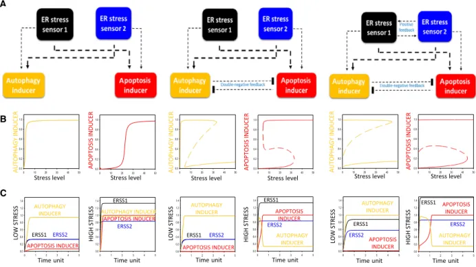
In our first scenario, there is neither feedback connection between ERSS1 and ERSS2, nor between autophagy and apoptosis inducers (see Fig.1A, panel left). The dynamical characteristic of this control network can be appropriately
illustrated by signal–response curve, where the activity of both autophagy and apoptosis inducers is plotted in the function of cellular stress level (Fig.1B, panel left). Under physiological conditions (i.e. stress=0), neither autophagy nor apoptosis are detected; however, both mechanisms show a sigmoid increase depending on the level of ER stress. Corresponding to the experimental data [26], it is easily manageable that autophagy-dependent survival has a quick activation already at low level of ER stress; mean- while, apoptosis remains inactive. In addition, similar to our results[26] autophagy precedes apoptotic cell death in time upon excessive level of ER stress (see the time course simulations on Fig.1C, panel left). However, both mecha- nisms seem to be active upon excessive level of cellular stress, which does not match to reality, as autophagy- dependent survival process has to be turned off when the cell initiates apoptosis[26].
Therefore, our simple wiring diagram is extended with a crosstalk between autophagy and apoptosis; namely, according to already published data[27] a double-negative
A
B
C
0 1 2 3 4 5
0.0 0.2 0.4 0.6 0.8 1.0 1.2 1.4
0 1 2 3 4 5
0.0 0.2 0.4 0.6 0.8 1.0 1.2 1.4
0 1 2 3 4 5
0.0 0.2 0.4 0.6 0.8 1.0 1.2 1.4
0 1 2 3 4 5
0.0 0.2 0.4 0.6 0.8 1.0 1.2 1.4
0 1 2 3 4 5
0.0 0.2 0.4 0.6 0.8 1.0 1.2 1.4
0 1 2 3 4 5
0.0 0.2 0.4 0.6 0.8 1.0 1.2 1.4
0 10 20 30 40 50
0.0 0.2 0.4 0.6 0.8 1.0
0 10 20 30 40 50
0.0 0.2 0.4 0.6 0.8 1.0
ERSS1 ERSS2
AUTOPHAGY INDUCER
APOPTOSIS INDUCER
LOW STRESS HIGH STRESS
ERSS1
ERSS2 AUTOPHAGY
INDUCER APOPTOSIS
INDUCER
Time unit Time unit
APOPTOSIS INDUCER
AUTOPHAGY INDUCER
Stress level Stress level
0 10 20 30 40 50
0.0 0.2 0.4 0.6 0.8 1.0
0 10 20 30 40 50
0.0 0.2 0.4 0.6 0.8 1.0
0 10 20 30 40 50
0.0 0.2 0.4 0.6 0.8 1.0
0 10 20 30 40 50
0.0 0.2 0.4 0.6 0.8 1.0
ERSS1
ERSS2 AUTOPHAGY
INDUCER APOPTOSIS INDUCER
LOW STRESS HIGH STRESS
SSERTSWOL HIGH STRESS
APOPTOSIS INDUCER AUTOPHAGY
INDUCER
ERSS1 ERSS2 ERSS2
AUTOPHAGY INDUCER APOPTOSIS INDUCER
ERSS1
ERSS1 ERSS2 AUTOPHAGY INDUCER
APOPTOSIS INDUCER
Time unit Time unit
Time unit Time unit
Stress level Stress level
Stress level Stress level
APOPTOSIS INDUCER
AUTOPHAGY INDUCER
APOPTOSIS INDUCER
RECUDNIYGAHPOTUA
Fig. 1.System-level feedback loops guarantee the robustness of the control network upon ER stress. Three simple models are presented:
(panel left) no connection between neither ER stress sensor 1 and 2 nor autophagy and apoptosis inducers; (panel middle) there is a double- negative feedback loop between autophagy and apoptosis inducers; (panel right) there is a double-negative feedback loop between autophagy and apoptosis inducers and there is a positive feedback loop between ER stress sensors 1 and 2. (A) The regulatory elements and their connections of life-and-death decision when the autophagy inducers, the apoptosis inducers and the ER stress sensors are grouped together in isolated yellow, red, black and blue boxes, respectively. Dashed lines show how the molecules can influence each other, thicker line assumes stronger effect. Blocked end lines denote inhibition. (B) The signal–response curves of (left) autophagy and (right) apoptosis inducers are shown with respect to increasing stress level. Solid lines denote stable state, while dashed lines denote the unstable state. (C) The temporal dynamics of ER stress sensor 1 (black) and 2 (blue), autophagy (yellow) and apoptosis (red) inducers upon low (left, stress = 5) and high (right, stress = 50) level of ER stress.
feedback loop is built in between autophagy and apoptosis inducers (Fig.1A, middle panel). In this case, the double- negative feedback loop generates an amplifying connection between the two mechanisms. Both activation and inactiva- tion of autophagy and apoptosis can generate a discontinu- ous switch, where the cellular response changes abruptly with a well-defined threshold of ER stress level (Fig. 1B, middle panel). An amplifying loop is essential for the pre- cise separation of two completely different states. In one state, when the stress level is tolerable, autophagy is active and blocks cell death (Fig.1B,C, middle panel); meanwhile, in the other state apoptosis turns on and autophagy turns off upon intolerable ER stress (Fig. 1B,C, middle panel).
However, in this simple model the double-negative feed- back loop is not enough to bring irreversibility into the control network. Since apoptosis induction is definitively a one-way process [28]; therefore, our model is required fur- ther extension.
To generate a one-way switch for apoptosis activation (autophagy inactivation) upon intolerable ER stress, a posi- tive feedback loop is built in between the two ER stress sensors (Fig.1A, panel right). The multiple feedback loops of the control network ensure the one-directional switch- like characteristic of the stress response mechanism, mean- ing that apoptosis inducer increases (autophagy inducer decreases) abruptly and irreversible as the magnitude of ER stress crosses a critical value (Fig.1B,C, panel right).
Results and Discussion
A positive feedback loop is present between PERK targets upon ER stress
Our mathematical analysis suggests that a positive feedback loop between two ER stress sensors might generate an essential irreversible stress response mech- anism, but we are also interested in the biological rele- vance of this regulatory connection.
This positive feedback loop was recently suggested between PERK and IRE1 upon ER stress[29], suppos- ing that a crosstalk can be observed between the two branches of UPR, but we cannot rule out the presence of other positive feedback loops in the control net- work. To further explore the importance of these posi- tive feedback loops in the ER stress response mechanism, we also investigated whether this connec- tion is present or not inside one of the branches of UPR, focusing here on PERK-controlled signalling pathway of UPR. The key downstream targets of PERK are Gadd34 and CHOP, respectively [30,31].
Many data have already proved that CHOP has an essential role in apoptosis induction [32], while novel results have also suggested that CHOP promotes the activation of various autophagy genes (such as p62,
Atg3, Atg12) upon early ER stress[33]. Gadd34 is able to enhance autophagy via mTOR downregulation[34];
however, excessive level of Gadd34 results in apoptotic cell death [35]. Marciniak et al. [36] have shown an impaired Gadd34 activation in tunicamycin-treated CHOP-/-cells; meanwhile, Gadd34 downregulation has a negative effect on CHOP activation upon cellular stress[35]. These results clearly suggest a direct and/or indirect positive feedback loop between Gadd34 and CHOP.
According to these above-mentioned experimental data, we suggest that positive feedback loops are pre- sent inside the UPR branches, that is between Gadd34 and CHOP (Figs2and3, Table S1). According to our simple model, we assume that Gadd34 is stronger on autophagy induction, while CHOP has more drastic effect to enhance apoptotic cell death. Our goal is to investigate the role of this connection in a robust life- and-death decision of the control network.
The fine-tuning of autophagy-dependent survival is managed by the precise dynamical control of its enhancers
To explain the dynamical characteristic of the control network, first we confirmed the effect of either Gadd34 or CHOP up- and downregulation upon enduring and excessive level of ER stress by using computer simula- tions (Figs2and3).
In the absence of CHOP, the threshold level of apoptosis induction moves to right; therefore, apopto- sis cannot turn on, and meanwhile, autophagy remains active even at high level of ER stress (Fig. 2B, panel left). According to the experimental data, the viability of CHOP depleted cells drastically increases [37], because they cannot induce apoptosis. Although Gad- d34 has some positive effect on apoptosis inducer, its effect is much stronger on autophagy, and together with the double-negative feedback loop between autop- hagy and apoptosis inducers, they keep autophagy active even upon intolerable level of ER stress (Fig. 2C, panel left). Besides, Igase et al. have shown that CHOP overexpression in vascular smooth muscle cells (VSMC) significantly reduced cell viability and induced apoptosis[38]. Since the signal–response curve of apoptosis inducer moves to left when CHOP is overproduced in the cells, the threshold of apoptosis induction gets to a lower level of ER stress (Fig. 2B, panel right). Therefore, apoptosis turns on even at low level of ER stress; meanwhile, autophagy becomes inactive (Fig.2B,C, panel right).
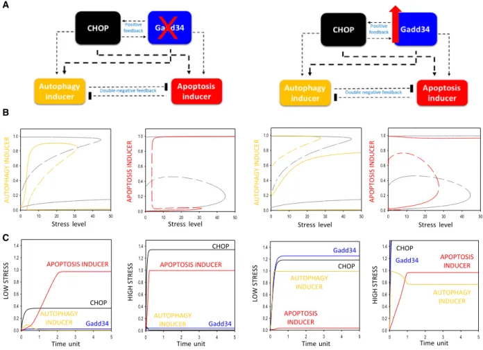
It is much more interesting that although Gadd34 is essential for autophagy-dependent survival upon ER
stress, both Gadd34 depletion [39] and over-produc- tion[35]result in early apoptotic cell death. To explain the dynamical characteristic of the control network, computer simulations were carried out (Fig. 3). In the absence of Gadd34, both the activation threshold for apoptosis inducer and inactivation threshold for autophagy inducer move to left, resulting in an apop- totic induction/autophagy inactivation upon lower level of ER stress (Fig. 3B, panel left). Without Gad- d34, autophagy inducer is not strong enough and CHOP can easily hyper-activate the cell death process even at low level of ER stress (Fig. 3B,C, panel left).
In addition, Gadd34 overexpression also moves the activation threshold of apoptosis inducer/inactivation threshold of autophagy inducer to a lower ER stress level supposing a drastic decrease in cell viability
(Fig. 3B, panel right). Although high Gadd34 level in the cell has a hyper-positive effect on autophagy induction due to the positive feedback between CHOP and Gadd34, Gadd34 accelerates the cell death mecha- nism via CHOP, as well. Besides, Gadd34 also has a direct positive effect on apoptosis inducer. Therefore, autophagy does not have any chance to win against apoptosis, and the cells enter the self-killing pathway already at lower level of ER stress (Fig. 3B,C, panel right).
Our dynamical analysis confirms that the proper balance of both ER stress sensors (i.e. CHOP and Gadd34) is essential to determine the cellular life-and- death decision upon ER stress. Since the proper acti- vation of autophagy-dependent cellular survival seems to be controlled by Gadd34 at excessive level of ER
0 10 20 30 40 50
0.0 0.2 0.4 0.6 0.8 1.0
0 10 20 30 40 50
0.0 0.2 0.4 0.6 0.8 1.0
0 1 2 3 4 5
0.0 0.2 0.4 0.6 0.8 1.0 1.2 1.4
0 1 2 3 4 5
0.0 0.2 0.4 0.6 0.8 1.0 1.2 1.4
0 1 2 3 4 5
0.0 0.2 0.4 0.6 0.8 1.0 1.2 1.4
0 1 2 3 4 5
0.0 0.2 0.4 0.6 0.8 1.0 1.2 1.4
HIGH STRESS
AUTOPHAGY INDUCER
SSERTSWOL
APOPTOSIS
INDUCER APOPTOSIS
INDUCER
AUTOPHAGY INDUCER
HIGH STRESS
LOW STRESS
AUTOPHAGY INDUCER AUTOPHAGY
INDUCER
A
B
C
0 10 20 30 40 50
0.0 0.2 0.4 0.6 0.8 1.0
0 10 20 30 40 50
0.0 0.2 0.4 0.6 0.8 1.0
Stress level Stress level
APOPTOSIS INDUCER
RECUDNIYGAHPOTUA AUTOPHAGY INDUCER
APOPTOSIS INDUCER
Stress level Stress level
Time unit Time unit Time unit Time unit
APOPTOSIS INDUCER CHOP
Gadd34
CHOP Gadd34
CHOP
Gadd34
CHOP
Gadd34
APOPTOSIS INDUCER
Fig. 2.Both upregulation and downregulation of CHOP level have a crucial effect on ER stress response mechanism. Two experimental protocols are simulated: (panel left) CHOP depletion by setting CHOP-T = 0.2 and (panel right) CHOP overexpression by setting CHOP- T = 4. (A) The regulatory elements and their connections of life-and-death decision when the autophagy inducers, the apoptosis inducers, CHOP and Gadd34 are grouped together in isolated yellow, red, black and blue boxes, respectively. Dashed lines show how the molecules can influence each other. Blocked end lines denote inhibition. (B) The signal–response curves of (left) autophagy and (right) apoptosis inducers are shown with respect to increasing stress level. Solid lines denote stable state, while dashed lines denote the unstable state.
Grey lines show the original signal–response curves. (C) The temporal dynamics of CHOP (black) and Gadd34 (blue), autophagy (yellow), and apoptosis (red) inducers upon low (left, stress = 5) and high (right, stress = 50) level of ER stress.
stress, these results also suggest that Gadd34 level might play the key role in switching between life and death.
A simple coherent feedforward loop between the regulators of autophagy and apoptosis inducers does not guarantee the proper dynamical features of the control network
Our dynamical analysis suggests that the positive feedback loop between Gadd34 and CHOP seems to be crucial in accelerating apoptotic cell death even when Gadd34 is overexpressed in the cell upon ER stress. To further investigate the importance of this positive feedback loop and explain its dynamical
characteristic in the control network, we assumed that Gadd34 is not able to activate CHOP (Fig. 4A, B). In this case, the positive feedback loop gets reduced to a simple coherent feedforward loop in the control network. Namely, CHOP induces the autop- hagy inducer both directly and indirectly via Gadd34 (Fig. 4A).
In the absence of the positive feedback loop between ER stress sensors, the activation threshold of signal–
response curve of apoptosis inducer moves to higher ER stress values (Fig. 4B, panel left). Besides, the washing out of ER stressor results in a theoretical turning off of cell death mechanism (Fig. 4B, panel left). This is very similar to the earlier findings that at least one positive feedback loop definitively is essential, B
C A
X
0 1 2 3 4 5
0.0 0.2 0.4 0.6 0.8 1.0 1.2 1.4
0 1 2 3 4 5
0.0 0.2 0.4 0.6 0.8 1.0 1.2 1.4
0 1 2 3 4 5
0.0 0.2 0.4 0.6 0.8 1.0 1.2 1.4
0 1 2 3 4 5
0.0 0.2 0.4 0.6 0.8 1.0 1.2 1.4
0 10 20 30 40 50
0.0 0.2 0.4 0.6 0.8 1.0
0 10 20 30 40 50
0.0 0.2 0.4 0.6 0.8 1.0
0 10 20 30 40 50
0.0 0.2 0.4 0.6 0.8 1.0
0 10 20 30 40 50
0.0 0.2 0.4 0.6 0.8 1.0
HIGH STRESS
AUTOPHAGY INDUCER
SSERTS WOL
APOPTOSIS INDUCER
APOPTOSIS INDUCER
AUTOPHAGY INDUCER
HIGH STRESS
LOW STRESS
AUTOPHAGY INDUCER AUTOPHAGY
INDUCER
Stress level Stress level
APOPTOSIS INDUCER
RECUDNI YGAHPOTUA APOPTOSIS INDUCER
Stress level Stress level
Time unit Time unit Time unit Time unit
APOPTOSIS INDUCER
CHOP Gadd34
CHOP
Gadd34
CHOP
Gadd34 CHOP
Gadd34
AUTOPHAGY INDUCER
APOPTOSIS INDUCER
Fig. 3.Both up- and downregulation of Gadd34 level has a crucial effect on ER stress response mechanism. Two experimental protocols are simulated: (panel left) Gadd34 depletion by setting Gadd34-T = 0.05 and (panel right) Gadd34 overexpression by setting Gadd34-T = 2.
(A) The regulatory elements and their connections of life-and-death decision when the autophagy inducers, the apoptosis inducers, CHOP and Gadd34 are grouped together in isolated yellow, red, black and blue boxes, respectively. Dashed lines show how the molecules can influence each other. Blocked end lines denote inhibition. (B) The signal–response curves of (left) autophagy and (right) apoptosis inducers are shown with respect to increasing stress level. Solid lines denote stable state, while dashed lines denote the unstable state. Grey lines show the original signal–response curves. (C) The temporal dynamics of CHOP (black) and Gadd34 (blue), autophagy (yellow) and apoptosis (red) inducers upon low (left, stress = 5) and high (right, stress = 50) level of ER stress.
since it has a key role in ensuring the irreversibility of apoptotic cell death[40].
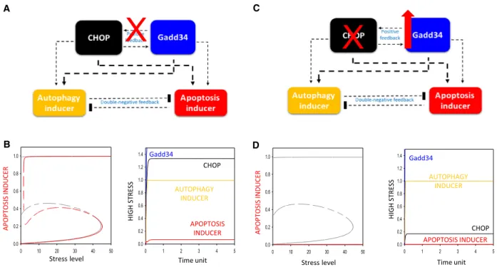
In our theoretical analysis, if Gadd34 does not have any positive effect on CHOP and only CHOP is able to promote Gadd34, the most interesting scenario is when Gadd34 gets overexpressed upon intolerable ER stress (Fig. 4B, panel right). In this case, a completely novel phenotype is observed. Since Gadd34 has both direct and indirect positive effects on autophagy-de- pendent survival, but it enhances apoptosis only directly (it cannot promote apoptotic cell death indi- rectly via CHOP due to the absence of Gadd34 ->
CHOP connection); therefore, Gadd34 overexpression results in a hyper-activation of autophagy, instead of apoptotic cell death, as it is observed in reality[35].
Here, we show that one of the important roles of Gadd34- to CHOP-positive feedback loop is to guar- antee a proper dynamical balance of autophagy–
apoptosis crosstalk. Namely, this connection has an essential role in ensuring that both Gadd34
upregulation and downregulation cause apoptotic cell death upon high level of ER stress.
Gadd34-dependent CHOP activation is essential to avoid fatal hyper-activation of autophagy upon ER stress
The question immediately arises what is the biological importance of that both Gadd34 up- and downregula- tion blocks the hyper-activation of autophagy? Why is the hyper-activation of autophagy so dangerous for the cell during high level of ER stress? Although autophagy-dependent self-cannibalism seems to be pos- itive for the cellular system to promote survival[15], it is also well known that sustained autophagy might be harmful and cause an uncontrolled cell death. Since apoptotic cell death is a much faster and more accu- rate way of removing damaged cells from the multicel- lular organism, the biological system assures itself that it dies by apoptosis rather than a not so exact
0 1 2 3 4 5
0.0 0.2 0.4 0.6 0.8 1.0 1.2 1.4
0 10 20 30 40 50
0.0 0.2 0.4 0.6 0.8 1.0
0 10 20 30 40 50
0.0 0.2 0.4 0.6 0.8 1.0
A
X
X
C
B D
0 1 2 3 4 5
0.0 0.2 0.4 0.6 0.8 1.0 1.2 1.4
HIGH STRESS
AUTOPHAGY INDUCER
Stress level
RECUDNI SISOTPOPA
Time unit CHOP Gadd34
Stress level
APOPTOSIS INDUCER
APOPTOSIS INDUCER
APOPTOSIS INDUCER Gadd34
CHOP AUTOPHAGY
INDUCER
HIGH STRESS
Time unit
Fig. 4.The presence of positive feedback loop between Gadd34 and CHOP determines the dynamical characteristic of the control network upon ER stress. Two different mutant phenotypes are simulated: (A) Gadd34 cannot enhance CHOP activation (kaers1" = 0) and (B) Gadd34 is overexpressed (Gadd34-T = 2); meanwhile, CHOP (CHOP-T = 0.2) is depleted. The regulatory elements and their connections of life-and- death decision when the autophagy inducers, the apoptosis inducers, CHOP and Gadd34 are grouped together in isolated yellow, red, black and blue boxes, respectively. Dashed lines show how the molecules can influence each other. Blocked end lines denote inhibition. (C) The signal-curve of apoptosis inducer with respect to increasing stress level in the presence (grey) and absence (red) of Gadd34 -> CHOP connection (left) and the time course simulation of its combination with Gadd34 hyper-activation (Gadd34-T = 2) upon high (stress = 50) level of ER stress (right) are shown. (D) The signal–response curves of apoptosis inducer with respect to increasing stress level (left) and time course simulation upon high (stress = 50) level of ER stress (right) are depicted. On signal–response curve solid lines denote stable state, while dashed lines denote the unstable state; on time course simulation, the temporal dynamics of CHOP (black) and Gadd34 (blue), autophagy (yellow) and apoptosis (red) inducers are plotted.
autophagy. This might be a very important issue for the cellular system, namely why the hyper-activation of autophagy is blocked so precisely upon ER stress.
Who plays the key switch in this process? Corre- sponding to already published experimental data, our analysis suggests that Gadd34 has to be one of the key molecules, which not only switches on autophagy when the cell promotes the survival process upon early ER stress, but it also blocks the hyper-activation of autophagy by enhancing apoptotic cell death with respond to excessive level of ER stress (Fig.4).
It is already well known that besides Gadd34 has a positive effect on apoptosis induction indirectly via CHOP, Gadd34 also has direct positive effect on induction of self-killing mechanism [41]. We investi- gated how important are these two pathways in terms of apoptosis induction when excessive level of ER is combined with Gadd34 overexpression. Therefore, to
further explore this question a mutant phenotype was predicted by our computer simulations (Fig.4C,D).
Firstly, Gadd34 level was increased (Gadd34-T=2) in the cell (i.e. simulating Gadd34 hyper-activation), and then, the total level of CHOP was depleted (CHOP-T =0.2) upon ER stress (Fig. 4D). According to the signal–response curves, autophagy inducer remains active, and meanwhile, the activation threshold for apoptosis gets completely diminished, suggesting that cells cannot enter apoptotic cell death even at high level of ER stress (Fig. 4D, panel left). We are able to confirm that autophagy gets hyper-activated; mean- while, apoptosis is not observed, suggesting that the presence of Gadd34- to CHOP-positive feedback loop is essential for the proper dynamical characteristic of the control network. With this predicted mutant pheno- type, we could also prove that the Gadd34-dependent apoptosis induction indirectly via CHOP is much more
Fig. 5.Schematic figure of the stress response mechanism and the various outcomes.
robust than the direct effect on the apoptosis inducer.
Although the direct positive effect is still present in this phenotype, it is not strong enough to win against the accelerated Gadd34-dependent autophagy induction.
We claim that Gadd34 is crucial to control the autophagy-dependent survival, but in the absence of CHOP its hyper-activation might generate a sustained autophagy response in the cell resulting in later a self- cannibalism-induced cell death. However, the dynami- cal feature of this mutant phenotype has to be proven experimentally in the near future.
Conclusions
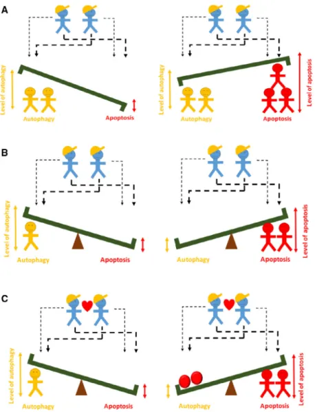
To fight for the maintenance of cellular homeostasis upon various extracellular and intracellular stress events is one of the crucial duties of the cells building up a multicellular organism. Here, we investigate the key regulatory motifs of the control network to show that multiple feedback loops guarantee the irreversible switch-like characteristic of life-and-death decision upon an excessive level of ER stress.
To illustrate the importance of feedback loops in the regulatory network, a simple metaphor is used
(Fig.5). In the absence of feedback loops, the foremen can ask the builders to raise a bench. Depending on whether there are more red or yellow workers, autop- hagy or apoptosis will be more active, but both are present in the cell (Fig. 5, upper panel). End of the working hours, when the workers go home, both autophagy and apoptosis turn off without any conse- quence. If we assume that red and yellow workers are enemies by forming a double-negative feedback loop between them, the bench turns into a seesaw. In this case, either the sum of the yellow workers or red workers are larger (i.e. autophagy or apoptosis is active), but they are mutually exclusive (Fig.5, middle panel). If the two foremen have a lunch break (i.e. ER stress goes away), the seesaw can be in both positions.
However, if the former picture is supplemented by the fact that the foremen are helping each other (repre- senting a positive feedback loop between them), the red workers will not only win over the yellow ones, but by rolling down red balls on the seesaw, they can make themselves active forever, even if the foremen have a lunch break held (Fig. 5, lower panel). In this way, we further proved that the one-way directionality of switch-like apoptotic cell death is achieved via mul- tiple feedback loops upon intolerable ER stress.
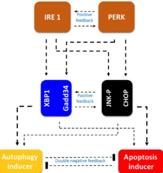
Since the cellular structure of vertebrates is so com- plex, and their regulatory networks require robust characteristics, therefore these above-mentioned multi- ple feedback loops must be achieved on various levels on ER stress response mechanism. It has recently revealed that the main transmembrane signal transduc- ers (i.e. PERK and IRE1) can promote both autop- hagy and apoptosis inducers with various strengths [29]; and a positive feedback loop between them was also suggested[29]. Novel experimental results suppose that a crosstalk can be definitively observed between the two branches of UPR (Fig.6).
It is well known that ATF6, the promoter of the third branch of UPR, can also induce both autophagy and apoptosis under various stress events in human cell lines [42,43]. Besides, it has been also shown that ATF6 can enhance CHOP transcription via binding to its promoter sequence. This result suggests that a crosstalk might be present between ATF6 and PERK pathways and connection between ATF6 and IRE1 pathways is also possible. However, many data are still lacking in the case of ATF6-dependent ER stress sen- sor mechanism; therefore, further studies are required to verify the presence of feedback loops in the control network.
According to the experimental data (see Table S1), here we show with a simple mathematical model that these multiple feedback loops are also present inside
Fig. 6.Regulatory crosstalk is present both inside and between the branches of UPR. The regulatory elements and their connections of life-and-death decision when the autophagy inducers, the apoptosis inducers and the ER stress sensors are grouped together in isolated yellow, red, orange, black and blue boxes, respectively. Dashed lines show how the molecules can influence each other, and thicker line assumes stronger effect.
Blocked end lines denote inhibition.
the PERK branches of UPR. We claim that both Gad- d34 and CHOP can enhance autophagy and apoptosis inducers with various strengths and a positive feedback loop between them is also suggested (Figs2–4). Inter- estingly, similar properties have been also discovered for XBP1 and JNK1-P, the main targets of IRE1 (see Tables S1 and S2). Although the spliced XBP1 is mainly an autophagy inducer[44] and JNK1-P is cru- cial for apoptosis induction [45], both proteins can control positively the other stress response mechanism, too [46,47]. Some data have already supposed that JNK1 has a positive effect on XBP1 (see Table S1);
however, the XBP1 -> JNK-P connection has not proved directly yet. Interestingly, both sustained XBP1 activity and the absence of XBP1 cause cell death (see Table S2). These results suggest that its regulatory role inside the IRE1 branch is similar to the key effect of Gadd34 inside the PERK branch. According to these above-mentioned experimental data, we suppose that positive feedback loop is present not only between the branches of UPR, but they can be observed inside the UPR branches between the key targets of ER stress sensors, too (Fig. 6). We assume that both Gadd34 – CHOP and XBP1 – JNK-P positive feedback loops are essential to generate a robust stress response mech- anism with respond to intolerable ER stress. ATF6 might also have downstream targets that control the ER stress response mechanism similarly. We claim that these connections might help to generate a robust life- and-death decision of the control network in any cir- cumstances. However, these connections require fur- ther experimental study in the near future.
ER stress is seriously involved in various human pathologies such as obesity, diabetes mellitus, several neurodegenerative diseases (i.e. Alzheimer’s disease, Parkinson’s disease, Huntington’s disease) and many others. Therefore, studying ER stress-related cellular life-and-death decision with systems biological meth- ods might have medical importance. Knowledge of the key regulatory motifs and molecules of this decision- making process might help us to enhance autophagy- dependent survival. For example, it is well known that intensive autophagy-dependent self-cannibalism might extend viability upon Huntington’s disease; meanwhile, inhibition of autophagy gets impaired the degradation of the huntingtin aggregates [48]. If we thoroughly explore the dynamic behaviour of the system; the exact regulatory connections, necessary to enhance autop- hagy-dependent survival, will be much more easily investigated in the future. In addition, a better under- standing of the dynamic behaviour of the control net- work can reduce the cost of the further experiments.
With this method, we will be also able to verify novel
drugs or natural compounds, which promote autop- hagy, and therefore, we can expand lifespan of people suffering from Huntington’s disease.
Our results confirm that this life-and-death decision is controlled at multiple levels of the control network during ER stress. This complex regulatory network guarantees the precise decision-making between autop- hagy-dependent survival and apoptotic cell death upon ER stress. With regard to the ER stress-dependent dis- eases, this knowledge might be used later to elaborate a precise medical treatment for the patient.
Acknowledgement
This work was supported by the Baron Munchausen Program of the Department of Medical Chemistry, Molecular Biology and Pathobiochemistry of Semmel- weis University, Budapest.
References
1 Labbadia J and Morimoto RI (2015) The biology of proteostasis in aging and disease.Ann Rev Biochem84, 435–464.
2 Yerbury JJ, Ooi L, Dillin A, Saunders DN, Hatters DM, Beart PM, Cashman NR, Wilson MR and Ecroyd H (2016) Walking the tightrope: proteostasis and neurodegenerative disease.J Neurochem137, 489–505.
3 Gorlach A, Klappa P and Kietzmann T (2006) The endoplasmic reticulum: folding, calcium homeostasis, signaling, and redox control.Antioxid Redox Signal8, 1391–1418.
4 Wickner W and Schekman R (2005) Protein translocation across biological membranes.Science310, 1452–1456.
5 Zhang K and Kaufman RJ (2006) Protein folding in the endoplasmic reticulum and the unfolded protein response. In Molecular Chaperones in Health and Disease. Handbook of Experimental Pharmacology, Vol 172(Starke K and Gaestel M, eds), pp. 69–91. Springer, Berlin, Heidelberg.
6 Malhi H and Kaufman RJ (2011) Endoplasmic reticulum stress in liver disease.J Hepatol54, 795–809.
7 Csala M, Margittai E and Banhegyi G (2010) Redox control of endoplasmic reticulum function.Antioxid Redox Signal13, 77–108.
8 Banhegyi G, Benedetti A, Csala M and Mandl J (2007) Stress on redox.FEBS Lett581, 3634–3640.
9 Tabas I and Ron D (2011) Integrating the mechanisms of apoptosis induced by endoplasmic reticulum stress.
Nat Cell Biol13, 184–90.
10 Banhegyi G, Baumeister P, Benedetti A, Dong D, Fu Y, Lee AS, Li J, Mao C, Margittai E, Ni Met al.
(2007) Endoplasmic reticulum stress.Ann N Y Acad Sci 1113, 58–71.
11 Bernales S, Papa FR and Walter P (2006) Intracellular signaling by the unfolded protein response.Annu Rev Cell Dev Biol22, 487–508.
12 Hetz C (2012) The unfolded protein response:
controlling cell fate decisions under ER stress and beyond.Nat Rev Mol Cell Biol13, 89–102.
13 Walter P and Ron D (2011) The unfolded protein response: from stress pathway to homeostatic regulation.Science334, 1081–1086.
14 Jiang HY and Wek RC (2005) Phosphorylation of the alpha-subunit of the eukaryotic initiation factor-2 (eIF2alpha) reduces protein synthesis and enhances apoptosis in response to proteasome inhibition.J Biol Chem280, 14189–14202.
15 Ogata M, Hino S, Saito A, Morikawa K, Kondo S, Kanemoto S, Murakami T, Taniguchi M, Tanii I, Yoshinaga Ket al. (2006) Autophagy is activated for cell survival after endoplasmic reticulum stress.Mol Cell Biol26, 9220–9231.
16 Hoyer-Hansen M and Jaattela M (2007) Connecting endoplasmic reticulum stress to autophagy by unfolded protein response and calcium.Cell Death Differ14, 1576–1582.
17 Maiuri MC, Zalckvar E, Kimchi A and Kroemer G (2007) Self-eating and self-killing: crosstalk between autophagy and apoptosis.Nat Rev Mol Cell Biol8, 741–752.
18 Levine B and Kroemer G (2008) Autophagy in the pathogenesis of disease.Cell132, 27–42.
19 Ravikumar B, Sarkar S, Davies JE, Futter M, Garcia-Arencibia M, Green-Thompson ZW, Jimenez- Sanchez M, Korolchuk VI, Lichtenberg M, Luo S et al. (2010) Regulation of mammalian autophagy in physiology and pathophysiology. Physiol Rev 90, 1383–1435.
20 Fulda S, Gorman AM, Hori O and Samali A (2010) Cellular stress responses: cell survival and cell death.Int J Cell Biol2010, 214074.
21 Saveljeva S, Mc Laughlin SL, Vandenabeele P, Samali A and Bertrand MJ (2015) Endoplasmic reticulum stress induces ligand-independent TNFR1-mediated necroptosis in L929 cells.Cell Death Dis6, e1587.
22 Riedl SJ and Shi Y (2004) Molecular mechanisms of caspase regulation during apoptosis.Nat Rev Mol Cell Biol5, 897–907.
23 Saelens X, Festjens N, Vande Walle L, van Gurp M, van Loo G and Vandenabeele P (2004) Toxic proteins released from mitochondria in cell death.Oncogene23, 2861–2874.
24 Gump JM and Thorburn A (2011) Autophagy and apoptosis: what is the connection?Trends Cell Biol21, 387–392.
25 Gordy C and He YW (2012) The crosstalk between autophagy and apoptosis: where does this lead?Protein Cell3, 17–27.
26 Holczer M, Marton M, Kurucz A, Banhegyi G and Kapuy O (2015) A comprehensive systems biological study of autophagy-apoptosis crosstalk during endoplasmic reticulum stress.Biomed Res Int2015, 319589.
27 Kapuy O, Vinod PK, Mandl J and Banhegyi G (2013) A cellular stress-directed bistable switch controls the crosstalk between autophagy and apoptosis.Mol Biosyst9, 296–306.
28 Taylor RC, Cullen SP and Martin SJ (2008) Apoptosis:
controlled demolition at the cellular level.Nat Rev Mol Cell Biol9, 231–241.
29 Marton M, Kurucz A, Lizak B, Margittai E, Banhegyi G and Kapuy O (2017) A systems biological view of life-and-death decision with respect to endoplasmic reticulum stress-the role of PERK pathway.Int J Mol Sci18, 58.https://doi.org/10.3390/ijms18010058.
30 Ma Y and Hendershot LM (2003) Delineation of a negative feedback regulatory loop that controls protein translation during endoplasmic reticulum stress.J Biol Chem278, 34864–34873.
31 Harding HP, Novoa I, Zhang Y, Zeng H, Wek R, Schapira M and Ron D (2000) Regulated translation initiation controls stress-induced gene expression in mammalian cells.Mol Cell6, 1099–1108.
32 McCullough KD, Martindale JL, Klotz LO, Aw TY and Holbrook NJ (2001) Gadd153 sensitizes cells to endoplasmic reticulum stress by down-regulating Bcl2 and perturbing the cellular redox state.Mol Cell Biol 21, 1249–1259.
33 B’Chir W, Maurin AC, Carraro V, Averous J, Jousse C, Muranishi Y, Parry L, Stepien G, Fafournoux P and Bruhat A (2013) The eIF2alpha/ATF4 pathway is essential for stress-induced autophagy gene expression.
Nucleic Acids Res41, 7683–7699.
34 Uddin MN, Ito S, Nishio N, Suganya T and Isobe K (2011) Gadd34 induces autophagy through the suppression of the mTOR pathway during starvation.
Biochem Biophys Res Commun407, 692–698.
35 Harding HP, Zhang Y, Bertolotti A, Zeng H and Ron D (2000) Perk is essential for translational regulation and cell survival during the unfolded protein response.
Mol Cell5, 897–904.
36 Marciniak SJ, Yun CY, Oyadomari S, Novoa I, Zhang Y, Jungreis R, Nagata K, Harding HP and Ron D (2004) CHOP induces death by promoting protein synthesis and oxidation in the stressed endoplasmic reticulum.Genes Dev18, 3066–3077.
37 Zinszner H, Kuroda M, Wang X, Batchvarova N, Lightfoot RT, Remotti H, Stevens JL and Ron D (1998) CHOP is implicated in programmed cell death in response to impaired function of the endoplasmic reticulum.Genes Dev12, 982–995.
38 Igase M, Okura T, Nakamura M, Takata Y, Kitami Y and Hiwada K (2001) Role of GADD153 (growth arrest-
and DNA damage-inducible gene 153) in vascular smooth muscle cell apoptosis.Clin Sci (Lond)100, 275–281.
39 Adler HT, Chinery R, Wu DY, Kussick SJ, Payne JM, Fornace AJ Jr and Tkachuk DC (1999) Leukemic HRX fusion proteins inhibit GADD34-induced apoptosis and associate with the GADD34 and hSNF5/INI1 proteins.
Mol Cell Biol19, 7050–7060.
40 Tyson JJ, Chen KC and Novak B (2003) Sniffers, buzzers, toggles and blinkers: dynamics of regulatory and signaling pathways in the cell.Curr Opin Cell Biol 15, 221–231.
41 Hollander MC, Zhan Q, Bae I and Fornace AJ Jr (1997) Mammalian GADD34, an apoptosis- and DNA damage-inducible gene.J Biol Chem272, 13731–13737.
42 Song S, Tan J, Miao Y, Li M and Zhang Q (2017) Crosstalk of autophagy and apoptosis: involvement of the dual role of autophagy under ER stress.J Cell Physiol232, 2977–2984.
43 Hillary RF and FitzGerald U (2018) A lifetime of stress: ATF6 in development and homeostasis.
J Biomed Sci25, 48.
44 Pehar M, Jonas MC, Hare TM and Puglielli L (2012) SLC33A1/AT-1 protein regulates the induction of autophagy downstream of IRE1/XBP1 pathway.J Biol Chem287, 29921–29930.
45 Chen Y and Brandizzi F (2013) IRE1: ER stress sensor and cell fate executor.Trends Cell Biol23, 547–555.
46 Wei Y, Pattingre S, Sinha S, Bassik M and Levine B (2008) JNK1-mediated phosphorylation of Bcl-2 regulates starvation-induced autophagy.Mol Cell30, 678–688.
47 Tian PG, Jiang ZX, Li JH, Zhou Z and Zhang QH (2015) Spliced XBP1 promotes macrophage survival and autophagy by interacting with Beclin-1.Biochem Biophys Res Commun463, 518–523.
48 Martin DD, Ladha S, Ehrnhoefer DE and Hayden MR (2015) Autophagy in Huntington disease and huntingtin in autophagy.Trends Neurosci38, 26–35.
Supporting information
Additional supporting information may be found online in the Supporting Information section at the end of the article.
Appendix S1. Mathematical codes for computational simulations.
Table S1. Collecting data from the literature about the most important regulatory connections between the elements of UPR and stress response mechanisms.
Table S2. Collecting data from the literature about the various mutant phenotypes of the elements of UPR upon ER stress.