Case Report
Surgical Solution for the Transformation of the Percutaneous Bone Anchored Hearing Aid to a Transcutaneous System in Complicated Cases
INTRODUCTION
Osseo-integrated implant systems have provided hearing rehabilitation in patients with conductive or mixed type hearing loss and single-sided sensorineural deafness [1, 2] since being introduced in 1977 [3]. Bone Anchored Hearing Aid system (BAHA®) is one of the most frequently applied device. The surgery of the Connect System with titanium abutment was based on the skin-flap formation and soft-tissue reduction [4-6] to avoid the peri-abutment pocket formation and soft-tissue hypertrophy, [5, 6] which has often resulted in the deterioration of sound processor usability [4, 7, 8]. In the new era of Connect Systems, minimal-incision-line technique with soft-tissue preservation [9-11], led to reduced postoperative complication rates [12]. Although hearing amplification of the Connect System was satisfactory, the possible complications (i.e. direct trauma or infection) and the patient’s aesthetic aversion [13-15] high- lighted the need for a more discreet system.
In the percutaneous Attract system, the skin over the implant is left intact; [16] thus, patients are more likely to accept the cosmetic results and the adverse events can be effectively prevented.
However, Connect System provides more favorable sound amplification—reported also in BAHA® guidelines and other clinical stud-
ies [17-20]—than the newly developed speech processors. Because of its aesthetic advantages and lower skin complication rates the
Attract System is preferred.
In certain cases, where the recurrent soft-tissue complications prohibit the use of the device, conversion of the percutaneous into a transcutaneous system should be considered.
The authors present their solution for such a conversion procedure.
CASE PRESENTATION
A 13-year-old child with bilateral Grade III microtia and complete atresia of the external ear canal had been aided with the BAHA® BI300 implant with titanium abutment and BP100 sound processor in 2010. According to the official surgical protocol, the soft Bone-anchored hearing aids (BAHAs) have been used for multiple types of hearing loss both in pediatric and adult cases. In the last decades, the percutaneous BAHA® Connect System (Cochlear Ltd., Sydney, Australia) was replaced by the fully implanted transcutaneous, magnet-based BAHA®
Attract System (Cochlear Ltd., Sydney, Australia). Since the implantable part of the Attract device is fully covered with soft tissue, skin complications that were frequently observed in the percutaneous system, could be eliminated. As an outcome of this important advantage of the Attract System, conversion of the percutaneous into a transcutaneous system should be considered. In the following methodology report, a possible surgical tech- nique is described. The method can easily be adopted to different conditions in which the replacement of the percutaneous device is necessary.
KEYWORDS: Bone-anchored device, reconstructive surgery, osseointegrated implants
Zsofia Bere , Gabor Vass , Adam Perenyi , Zoltan Tobias , Laszlo Rovo
Department of Oto-Rhino-Laryngology Head and Neck Surgery, University of Szeged, Szeged, Hungary
ORCID IDs of the authors: Z.B. 0000-0002-8342-1059; G.V. 0000-0003-0787-8679; A.P. 0000-0002-9162-6476; Z.T. 0000-0003-4871-9906; L.R. 0000- 0003-1782-1756.
Cite this article as: Bere Z, Vass G, Perenyi A, Tobias Z, Rovo L. Surgical Solution for the Transformation of the Percutaneous Bone Anchored Hearing Aid to a Transcutaneous System in Complicated Cases. J Int Adv Otol 2020; 16(3): 477-81.
tissue around the peri-implant area had been additionally reduced.
Two years after the surgery, the usability of the hearing aid gradually became degraded because of recurrent infection of the peri-implant soft tissue and granulation. As the child had minor signs of mental disorders, implant malfunction also led to underdevelopment in his education.
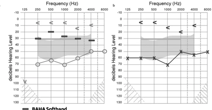
On admission to our department, an audiological follow-up test (Figure 1) was carried out with BAHA® Softband (BP 110 processor, Cochlear Ltd., Sydney, Australia), at which time he was unable to use his own device.
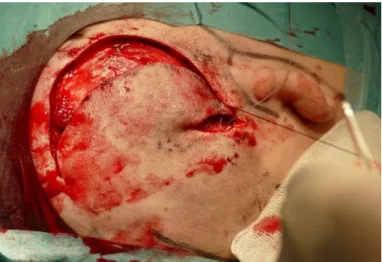
The BI300 implant and the abutment were placed 5 cm supero-pos- teriorly from the suspected position of the external auditory canal entrance. Peri-implant soft tissue had already been thinned around the abutment in a 1.5 cm×1.5 cm area (2 mm thickness). The adjacent tissue around the abutment was hypertrophic. Mild erythema and soreness were also present around the abutment; however, no mas- sive wound infection or discharge was observed (Figure 2).
On the basis of the above findings, we decided on surgery to convert the BAHA® Connect system to BAHA® Attract System. The patient ful- filled the audiology criteria for BAHA® Attract.
Figure 2. a, b. Immediate preoperative picture; bleeding granulation tissue and atrophic scabby skin around the abutment is visible (a). Schematic drawing of the temporal region (abutment, peri-implant scar tissue, and main arteries) (b).
a b
Figure 1. a, b. Preoperative pure tone audiometry results: panel a) indicates right side and panel b) indicates left side in dBHL. Air-bone gap indicates medi- um-profound conductive hearing loss in both sides, whereas bone conduction is relatively preserved. Because the child is right-handed, the right side had been previously aided with a BAHA® BIA300-BP 100 sound processor. Result of BAHA® Softband (BP110) test marked with a dark rectangle on the right panel.
a b
In the BAHA® Attract System, the implanted magnet must have a thick, well-vascularized soft-tissue coverage. Therefore, in the case of our patient, it was necessary to remove the scabby, reduced peri-im- plant skin (Figure 3) and the granulation tissue around the abutment.
The resultant tissue defect was closed by an occipitally pedicled tem- poroparietal galea-skin rotation flap. Preoperative ultrasonography was carried out to plan the best flap configuration (Figure 3).
The abutment was removed. and reconstructive flap was prepared and placed in position as shown in Figure 4.
Because skin incision lines and scar tissue above the Attract magnet should be avoided, the implant had to be placed in a more supe- ro-posterior position (Figure 5).
The Attract System was implanted into the bone in this new, opti- mal position. Perioperative parenteral antibiotic treatment was ad- ministered, and wound healing was unremarkable (Figure 6). The former BI300 implant was left in place because it was stable wing to osseo-integration and it did not interfere with the positioning of the new implant.
Sound processor fitting and audiological tests were performed after 5 weeks; the child was aided with BAHA® 5 Processor (Cochlear Ltd., Sydney, Australia). The result of pure tone audiometry correlated well with the result of the Softband test (Figure 7); there was no signifi- cant difference between thresholds at the different frequencies.
DISCUSSION
The main advantage of the BAHA® Attract System is that it allows for a thick soft-tissue coverage that protects the implanted magnet from any mechanical trauma. Because there is no direct connection between the skin surface and the underlying tissues through the abutment, the chance for any descending infection is negligible. The aesthetic benefits of the transcutaneous system are indisputable [16]. The recurrent soft-tissue complications of the Connect System may prohibit the proper use of the hearing aid, which may tend to result in impairment of hearing and communication development in child- Figure 3. a, b. Preoperative planning: continuous black line shows the main
artery branches. Area surrounded with red dash line represents scar tissue.
Blue dashed line is the plan for the skin flap and the white transparent area is the new position of the magnet. Dark blue dot represents the expected loca- tion of the ear canal for setting the new position of the implant (a). The main surgical steps: removal of scar tissue, flap formation and replacement of the abutment, and expected position of the magnet (b).
a b
Figure 6. Undisturbed wound healing 2 weeks postoperatively. Blue drawing shows the position of the magnet.
Figure 5. The implant and the attached magnet placed supero-posteriorly.
White arrow indicates the original implant position.
Figure 4. The occipitally pedicled temporoparietal galea-skin rotation flap af- ter removing scabby tissue.
Figure 7. a, b. Pre and postoperative audiological results with BAHA Softband (BP110) and BAHA® 5 processor, right ear implanted (panel a). Panel b indi- cates pure-tone audiometry of the left ear.
a b
hood and social separation in the elderly.[14,15] In such cases, instead of long-lasting, often ineffective conservative treatments the surgical conversion to Attract System is highly suggested.
One key point in the preoperative assessment of these conversion surgeries, in our opinion, is the prudent mapping of the vascular anatomy of the wide peri-implant area, which helps the surgeon plan for an adequate skin flap with a stable vascular pedicle. In or- der to securely cover the magnet, a relatively thick, well-vascularized tissue layer should be applied in cases where the resection of the damaged peri-implant soft tissue is excessive either because of the former soft-tissue reduction technique with consequent scar forma- tion, or infected/necrotic skin granulation. The other very important key point is the replacement of the implant into a more posterior po- sition to prevent any scar close to the rim of the magnet, keeping in mind that the new position of the implant should consider the rec- ommended distance from the external ear canal.
One report of such conversion surgery has already been published in the literature recently; [21] however, the unique aspects of our method are the following:
• We always consider the vascular anatomy of the region, and, instead of a simple excising-undermining-suturing technique, we apply a pedicled flap with a reliable supplying artery for the most optimal nutrition of tissues covering the implant.
• Scars of previous surgeries should always be removed—on the one hand, to avoid the increased risk of pressure skin necrosis due to the sparse vascularization of scars, and on the other hand, for a better aesthetic outcome.
• After excessive scar removal, large tissue defects can be replaced with a properly designed, well-vascularized flap.
• With our method, scars over the magnet and its rim could be completely avoided, which provides perfect docking of the sound processor and prevents secondary complications of the suture scar line caused by pressure or tension.
• Regarding the standard surgical technique of the Connect Sys- tem (i.e. the position of the abutment compared with the exter- nal ear canal) the occipitally based rotation flap could always be an adequate solution because of its standard anatomy and vascularization [22].
CONCLUSION
Our BAHA® Attract surgical conversion method provides a safe, tech- nically feasible solution for those patients who are not able to use their BAHA® Connect System. This case study demonstrates that all the disadvantages of the percutaneous implantation could be re- solved and that the surgical technique provided the more advanta- geous, aesthetically more favorable transcutaneous system, with fine linear scars covered completely by the hair and, most importantly, with satisfactory audiology results.
Informed Consent: Written informed consent was obtained from the patients who participated in this study.
Peer-review: Externally peer-reviewed.
Author Contributions: Concept – Z.B., L.R.; Design – Z.B., G.V.; Supervision – L.R.; Resource – Z .B.; Materials – Z.T.; Data Collection and/or Processing – G.V.,
A.P.; Analysis and/or Interpretation – Z.B., G.V.; Literature Search – Z.B., G.V.;
Writing – Z.B., G.V., A.P.; Critical Reviews – L.R.
Conflict of Interest: The authors have no conflict of interest to declare.
Financial Disclosure: The authors declared that this study has received no financial support.
REFERENCES
1. Gustafsson J. BC Drive performance vs. conventional bone conduction transducer. Cochlear Bone Anchored Solutions AB, 629908, 2015 Avail- able from: www.cochlear.com.
2. Voelker CC, Chole RA. Unilateral sensorineural hearing loss in adults:
Etiology and management. Seminars in Hearing 2010; 31: 313-25.
[Crossref]
3. Tjellström A, Granström G. Long-term follow-up with the bone-anchored hearing aid: a review of the first 100 patients between 1977 and 1985.
Ear Nose Throat J 1994; 73: 112-4. [Crossref]
4. Tjellström A, Lindström J, Hallén O, Albrektsson T, Brånemark PI. Osse- ointegrated titanium implants in the temporal bone. A clinical study on bone-anchored hearing aids. Am J Otol 1981; 2: 304-10.
5. Holgers KM, TjellströmA, Bjursten LM, Erlandsson BE. Soft tissue reac- tions around percutaneous implants: a clinical study of soft tissue con- ditions around skin penetrating titanium implants for bone-anchored hearing aids. Am J Otol 1988; 9: 56-9.
6. Tjellström A, Rosenhall U, Lindström J, Hallén O, Albrektsson T, Bråne- mark PI. Five-year experience with skin-penetrating bone-anchored implants in the temporal bone. Acta Otolaryngol 1983; 95: 568-75.
[Crossref]
7. Tjellström A, Granström G. How we do it: frequency of skin necrosis after BAHA surgery. Clin Otolaryngol 2006; 31: 216-20. [Crossref]
8. Van Rompaey V, Claes G, Verstraeten N, van Dinther J, ZarowskiA, Offe- ciers E, et al. Skin reactions following BAHA surgery using the skin flap dermatome technique. Eur Arch Otorhinolaryngol 2011; 268: 373-6.
[Crossref]
9. Hawley K, Haberkamp TJ. Osseointegrated hearing implant surgery: out- comes using a minimal soft tissue removal technique. Otolaryngol Head Neck Surg 2013; 148: 653-7. [Crossref]
10. Lanis A, Hultcrantz M. Percutaneous osseointegrated implant surgery without skin thinning in children: a retrospective case review. Otol Neu- rotol 2013; 34: 715-22. [Crossref]
11. Larsson A, Wigren S, Andersson M, Ekeroth G, Flynn M, Nannmark U. His- tologic evaluation of soft tissue integration of experimental abutments for bone anchored hearing implants using surgery without soft tissue reduction. Otol Neurotol 2012; 33: 1445-51. [Crossref]
12. Jarabin J, Bere Z, Hartmann P, Tóth F, Kiss JG, Rovó L. Laser-Doppler mi- crovascular measurements in the peri-implant areas of different osse- ointegrated bone conductor implant systems. Eur Arch Otorhinolaryn- gol 2015; 272: 3655-62. [Crossref]
13. Stalfors J, Tjellström A. Skin reactions after BAHA surgery: a comparison between the U-graft technique and the BAHA dermatome. Otol Neuro- tol 2008; 29: 1109-14. [Crossref]
14. Fontaine N, Hemar P, Schultz P, Debry C, Charpiot A. BAHA implant:
implantation technique and complications. Eur Ann Otorhinolaryngol Head Neck Dis 2014; 131: 69-74. [Crossref]
15. Ida JB, Mansfield S, Meinzen-Derr JK, Choo DI. Complications in pediatric osseointegrated implantation. Otolaryngol Head Neck Surg 2011; 144:
586-91. [Crossref]
16. Reinfeldt S, Håkansson B, Taghavi H, Eeg-Olofsson M. New developments in bone conduction hearing implants: a review. J Med Devices 2015; 8:
79-93. [Crossref]
17. Nelissen RC, Agterberg MJ, Hol MK, Snik AFM. Three-year experience with the Sophono in children with congenital conductive unilateral hearing loss: tolerability, audiometry, and sound localization compared to a
bone-anchored hearing aid. Eur Arch Otorhinolaryngol 2016; 273: 3149- 56. [Crossref]
18. Iseri M, Orhan KS, Tuncer U, Kara A, Durgut M, Guldiken Y, et al. Trans- cutaneous bone-anchored hearing aids versus percutaneous ones:
multicenter comparative clinical study. Otol Neurotol 2015; 36: 849-53.
[Crossref]
19. Hol MK, Nelissen RC, Agterberg MJ, Cremers CWRJ, Snik AFM. Compar- ison between a new implantable transcutaneous bone conductor and percutaneous bone-conduction hearing implant. Otol Neurotol 2013;
34: 1071-5. [Crossref]
20. Wigren S. Considerations for Transitioning from Baha Connect System to Baha Attract System. 2014 Available from: www.cochlear.com
21. Cedars E, Chan D, Lao A, Hardies L, Meyer A, Rosbe K. Conversion of tra- ditional osseointegrated bone-anchored hearing aids to the Baha(®) at- tract in four pediatric patients. Int J Pediatr Otorhinolaryngol 2016; 91:
37-42. [Crossref]
22. Perenyi A, Bere Z, Jarabin J, Sztano B, Kukla E, Bikhazi Z, Tiszlavicz L, Toth F, Kiss JG, Rovo L. Vascular mapping of the retroauricular skin - proposal for a posterior superior surgical incision for transcutaneous bone-conduction hearing implants. J Otolaryngol Head Neck Surg 2017; 46: 46. [Crossref]