Three dimensional tissue cultures and tissue engineering
Dr. Domokos Bartis – Dr.JuditPongrácz
Manifestation of Novel Social Challenges of the European Union in the Teaching Material of
Medical Biotechnology Master’s rogrammes at the University of Pécs and at the University of DebrecenP
Three dimensional tissue cultures and
tissue engineering
Dr. Domokos Bartis — Dr. Judit Pongrácz
“Manifestation of Novel Social Challenges of the European Union
in the Teaching Material of
Medical Biotechnology Master’s Programmes at the University of Pécs and at the University of Debrecen”
Identification number: TÁMOP-4.1.2-08/1/A-2009-0011
University of Pécs – Pécs, 2011
© Dr. Judit Pongrácz, 2011
The project is funded by the European Union and co-financed by the European Social Fund.
Manuscript completed: 11 November 2011
Editor in charge: University of Pécs Editor in charge: Dr. Judit Pongrácz, Rita Bognár
Technical editor: Zsolt Bencze, Veronika Csöngei and Szilvia Czulák Lector: Dr. György Miskei
Length: 133 pages
Identification number:
TÁMOP-4.1.2-08/1/A-2009-0011
3
Content
List of figures ... 5
I Three dimensional tissue cultures and tissue engineering – Basic principles ... 7
II Cells and tissue types in tissue engineering – Stem cells ... 11
III Bioreactors ... 25
IV Biomaterials ... 37
V Scaffold fabrication ... 45
VI Biocompatibility ... 53
VII Cell-scaffold interactions ... 55
VIII Biofactors ... 59
IX Controlled release ... 65
X Biosensors ... 73
XI Aggregation cultures ... 75
XII Tissue printing ... 81
XIII Tissue repair ... 85
XIV Commercial products ... 101
XV Clinical trials ... 111
XVI Research applications and drug testing ... 127
XVII Ethical issues ... 129
XVIII Economic significance ... 131
Recommended reading ... 133
Identification number:
TÁMOP-4.1.2-08/1/A-2009-0011
5
List of figures
Figure I-1: Basic principles of tissue engineering ... 8
Figure I-2: 2D tissue cultures ... 9
Figure I-3: 3D tissue cultures ... 10
Figure II-1: Stem cell types ... 11
Figure II-2: Types of stem cell replications I ... 12
Figure II-3: Types of stem cell replications II ... 13
Figure II-4: Epiblast stem cells ... 14
Figure II-5: Adult or somatic stem cells ... 15
Figure II-6: Hematopoietic stem cells (HSCs) ... 15
Figure II-7: Functional interdependency of bone marrow stem cells ... 17
Figure II-8: Ontogeny of tissue lineages in bone marrow ... 18
Figure II-9: Cord blood stem cells and foetal stem cells ... 19
Figure II-10: Stem cell populations in cord blood ... 19
Figure II-11: Isolation procedures of ASCs ... 20
Figure II-12: Immunophenotype of ASCs (Positive markers) ... 21
Figure II-13: Application of ESCs and ASCs ... 22
Figure III-1: Shear forces in dynamic fluids ... 28
Figure III-2: Spinner flask bioreactors ... 29
Figure III-3: Rotating wall bioreactors ... 30
Figure III-4: Compression bioreactors ... 32
Figure III-5: Flow perfusion bioreactors ... 34
Figure IV-1: Types of natural biomaterials ... 37
Figure IV-2: Types of synthetic biomaterials ... 41
6 The project is funded by the European Union and co-financed by the European Social Fund.
Figure V-1: Solvant casting – particulate leaching ... 47
Figure V-2: Electrospinning ... 49
Figure V-3: Selective laser sintering (SLS) ... 51
Figure VII-1: Cell line cells nestling on a scaffold ... 55
Figure VII-2: Primary SAEC in matrigel ... 55
Figure VIII-1: Signalling networks in tissue development and maintenance ... 59
Figure IX-1: Controlled release profiles in biodegradable systems ... 67
Figure X-1: Modeling tissue and biosensor ... 73
Figure XI-1: Cell adhesion ... 75
Figure XI-2: Microgravity culture (hanging-drop) I ... 77
Figure XI-3: Microgravity culture (hanging-drop) II ... 78
Figure XI-4: Synthetic cell aggregation ... 79
Figure XI-5: Biotinylated cell cross-linking ... 80
Figure XII-1: Cell clusters fuse into microtissues ... 81
Figure XII-2: The first tissue printer ... 82
Figure XIII-1: Bone marrow cells in cardiac repair, and skeletal myoblasts ... 91
Figure XIII-2: Concepts for tooth bioengineering ... 93
Figure XIII-3: Bioartificial kidney ... 98
Figure XIII-4: In vitro engineered murine kidney ... 99
Figure XIV-1: Integra® skin replacement ... 109
Figure XV-1: Strategies for cell based therapies for the lung ... 124
Figure XV-2: Complexity of the lung structure during development ... 125
Identification number:
TÁMOP-4.1.2-08/1/A-2009-0011
7
I Three dimensional tissue cultures and tissue engineering – Basic principles
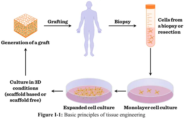
Tissue damages caused by mechanical injuries or diseases are frequent causes of morbidity and mortality. Tissue injuries are normally repaired by “built-in” regeneration mechanisms. However, if the tissue regeneration process malfunctions or the extent of the injury is too large, organ transplant can potentially be the only solution. Lack of transplantable organs when other therapies have all been exhausted adversely affects the quality and length of patients’ life, and is severe financial burden on the individual and society. Tissue injury associated diseases would become treatable using targeted tissue- regeneration or transplantation therapies. To provide tissues for therapy or for research to study tissue specific physiological mechanisms and diseases processes, the discipline of tissue engineering has evolved.
Originally, tissue engineering was categorized as a sub-field of engineering and bio-materials, but having grown in scale and significance tissue engineering has become a discipline of its own. To regenerate or even to re-create certain parts of the human body, tissue engineering uses combinations of various methods of cell culture, engineering, bio-materials and suitable biochemical and biophysical factors. While most definitions of tissue engineering cover a broad range of applications, in practice the term is closely associated with applications that repair or replace portions of whole tissues including bone, cartilage, blood vessels, skin, etc.
8 The project is funded by the European Union and co-financed by the European Social Fund.
Figure I-1: Basic principles of tissue engineering
Tissues created in vitro frequently originate from embryonic or adult cells.
Furthermore, in vitro generated tissues often need certain mechanical support and complex manipulation to achieve the required structural and physiological properties for proper functioning. To achieve complex tissue structures, conventional cell cultures (Figure I-2) where cells grow as monolayers in two-dimension (2D) can no longer be used.
Monolayer cell culture Expanded cell culture
Generation of a graft
Cells from a biopsy or resection
Grafting Biopsy
Culture in 3D conditions (scaffold based or
scaffold free)
Three dimensional tissue cultures and tissue engineering – Basic principles
Identification number:
TÁMOP-4.1.2-08/1/A-2009-0011
9 Figure I-2: 2D tissue cultures
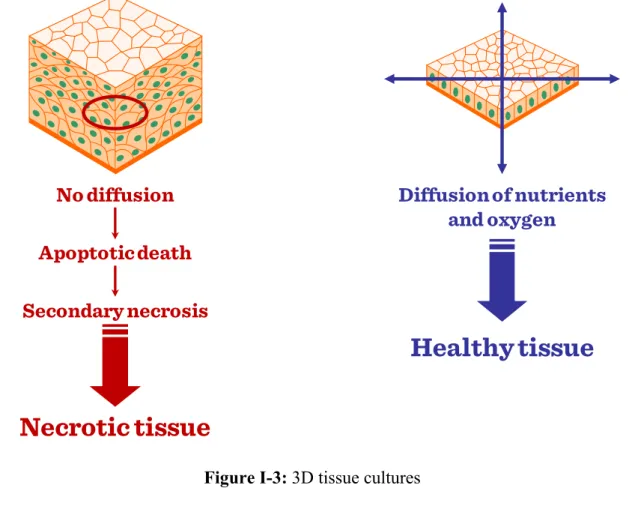
Particularly, as in monolayer cultures stretched cells form a single layer only network, that is incapable to perform complex functions. In tissue engineering, traditional cell culture technology is replaced by three-dimensional (3D) cell cultures (Figure I-3) where cultured cells assume a more natural morphology and physiology.
Adherent Non-Adherent
Suspension
Pelleting
Re-suspension
Seeding
Trypsin No trypsin
10 The project is funded by the European Union and co-financed by the European Social Fund.
Figure I-3: 3D tissue cultures
Various three dimensional tissue culture technologies have developed as tissue engineering gained impetus in medical research and therapy. Three dimensional culture technologies frequently apply various bio-materials where cells are provided with the necessary interactions to form the required tissue or organ.
If tissues are not needed immediately, both differentiated adult primary cells as well as adult and embryonic stem cells can be stored in liquid nitrogen, below -150°C.
The following chapters shall provide a brief overview of various aspects of tissue engineering including cell types, bio-materials, culture methods and application of engineered tissues. Clinical trials as well as ethical issues will also be discussed.
No diffusion Apoptotic death Secondary necrosis
Necrotic tissue
Diffusion of nutrients and oxygen
Healthy tissue
Identification number:
TÁMOP-4.1.2-08/1/A-2009-0011
11
II Cells and tissue types in tissue engineering – Stem cells
Various types of cells are used in tissue engineering that can be categorized in several ways. In broad terms, technologies in tissue engineering make use of both fully differentiated as well as progenitor cells at various stages of differentiation generally known as stem cells.
Stem cells are undifferentiated cells with the ability to divide in culture and give rise to different forms of specialized cells.
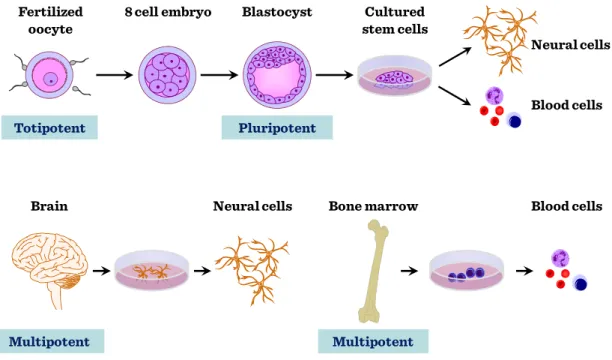
According to their source, stem cells are divided into two main groups: “adult”
and “embryonic” stem cells (Figure II-1).
Figure II-1: Stem cell types
While adult stem cells are multipotent, embryonic stem cells are mostly pluripotent except of cells in the earliest stages of embryonic development (fertilized
Neural cells
Blood cells Cultured
stem cells 8 cell embryo
Fertilized oocyte
Totipotent
Blastocyst
Pluripotent
Multipotent
Brain Neural cells Bone marrow Blood cells
Multipotent
12 The project is funded by the European Union and co-financed by the European Social Fund.
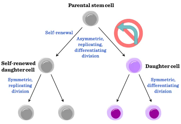
egg) that are totipotent. Stem cell niches where stem cells reside in an embryo or any given organ are defined as stem cell microenvironments. The fate of stem cells depends on the effect of the microenvironment. Stem cells – depending on the stimuli – have two main ways to replicate: either symmetrically resulting in two daughter cells with stem cell characteristics or asymmetrically yielding a daughter cell with stem and another daughter cell with differentiating cell characteristics (Figure II-2).
Figure II-2: Types of stem cell replications I
With the increasing number of divisions, the proliferation capacity of stem cells is decreasing favouring differentiated phenotypes (Figure II-3).
Asymmetric, replicating, differentiating
division
Symmetric, differentiating
division Symmetric,
replicating division
Parental stem cell
Daughter cell Self-renewed
daughter cell
Self-renewal
Cells and tissue types in tissue engineering – Stem cells
Identification number:
TÁMOP-4.1.2-08/1/A-2009-0011
13 Figure II-3: Types of stem cell replications II
As a result, continuous proliferation signals can lead to depletion of stem cell sources.
Characteristic stem cell types
Embryonic stem cells (ES). Although ethical debate is ongoing about using embryonic stem cells, embryonic stem cells are still being used in research for the repair of diseased or damaged tissues, or to grow new organs for therapy or for drug testing.
Especially, as rodent embryonic stem cells do not behave the same way as human embryonic stem cells and require different culture conditions to be maintained.
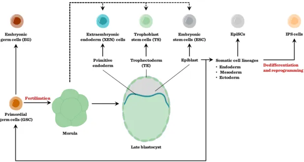
Recently, it has been discovered, that if stem cells are taken at a later stage not at the early blastocyst stage (3–4 days after conception), when the developing embryo implants into the uterus, epiblast – the innermost cell layer – stem cells form (Figure II-4), and these cells resemble human embryonic stem cells and had many of the same properties making it possible to use rodent rather than human cells in some research.
Proliferation capacity
Differentiation capacity
Terminally differentiated cells
Stem cell
Proliferating compartment
Asymmetric division Amplification of cell numbers Differentiation via progenitor
cells
14 The project is funded by the European Union and co-financed by the European Social Fund.
Figure II-4: Epiblast stem cells
Adult stem cells. Adult or somatic stem cells (ASC) (Figure II-5) are present in every organ including the bone marrow, skin, intestine, skeletal muscle, brain, etc.
(Figure II-6) where tissue specific microenvironments provide the necessary stem cell niches where stem cells reside and where the tissue gains its regenerative potential from.
Primordial germ cells (GSC)
Fertilization
Morula
Embryonic stem cells (ESC)
Epiblast
Late blastocyst
EpiSCs
Somatic cell lineages
•Endoderm
• Mesoderm
•Ectoderm
IPS cells
Dedifferentiation and reprogramming Embryonic
germ cells (EG)
Extraembryonic endoderm (XEN) cells
Primitive endoderm
Trophoblast stem cells (TS)
Trophectoderm (TE)
Cells and tissue types in tissue engineering – Stem cells
Identification number:
TÁMOP-4.1.2-08/1/A-2009-0011
15 Figure II-5: Adult or somatic stem cells
Figure II-6: Hematopoietic stem cells (HSCs)
Mature cells, when allowed to multiply in an incubator, ultimately lose their ability to function as differentiated cells. To proliferate, differentiate and function effectively in engineered tissues, cells must be easily procured and readily available;
The microenvironment where stem cells live are called stem cell niches:
Stem cells from fat
Peripheral
blood Bone marrow
Skeletal
muscle Placenta Hair follicle Cartilage Bone marrow Cartilage Skeletal muscle Bone marrow Skin
Muscle Nerve Tendon Smooth muscle Nerve Brain
Bone Blood cells Muscle Heart Cartilage Smooth muscle
Nerve Fat Bone Tendon Fat
Liver Fat Blood vessels
Bone marrow Cartilage Brain/nerve
Blood Heart All tissues
ASC in:
Prolymphocyte
Monocyte
Hematopoiesis in Humans Multipotential hematopoietic stem cell (Hemocytoblast)
Myeloblast Common myeloid
progenitor Common lymphoid
progenitor
Megakaryoblast
Promegakaryoblast
Megakaryocyte
Thrombocytes
Proerythroblast (Pronormblast)
Basophilic erythroblast
Polychromatic erythroblast
Orthochromatic erythroblast (Normoblast)
Polychromatic erythrocyte (Reticulocyte)
Erythrocyte
Mast cell Lymphoid
dendritic cell Myeloid
dendritic cell
Plasma cell Macrophage
Small lymphocyte Natural killer cell
T lymphocyte B lymphocyte Lymphoblast
B. promyelocyte
B. myelocyte
B. metamyelocyte
B. band
Basophil
N. promyelocyte
N. myelocyte
N. metamyelocyte
N. band
Neutrophil
E. promyelocyte
E. myelocyte
E. metamyelocyte
E. band
Eosinophil
Monoblast
Promonocyte
16 The project is funded by the European Union and co-financed by the European Social Fund.
they must multiply well without losing their potential to generate new functional tissue;
they should not be rejected by the recipient and not turn into cancer; and they must have the ability to survive in the low-oxygen environment normally associated with surgical implantation. Mature adult cells fail to meet many of these criteria. The oxygen demand of cells increases with their metabolic activity. After being expanded in the incubator for significant periods of time, they have a relatively high oxygen requirement and do not perform normally. A hepatocyte, for example, requires about 50 times more oxygen than a chondrocyte consequently much attention has turned to progenitor cells and stem cells. True stem cells can turn into any type of cell, while progenitor cells are more or less committed to becoming cell types of a particular tissue or organ. Somatic adult stem cells may actually represent progenitor cells in that they may turn into all the cells of a specific tissue. If somatic stem cells are intended to be used in regenerative therapy, healthy, autologous cells (one’s own cells) would be the best source for individual treatment as tissues generated for organ transplant from autologous cells would not trigger adverse immune reactions that could result in tissue rejection. Also, organs engineered from autologous cells would not carry the risk of pathogen transmission.
However, in genetic diseases suitable autologous cells are not available, while autologous cells from very ill (e.g. suffering from severe burns and require skin grafts) or elderly people may not have sufficient quantities of autologous cells to establish useful cell lines. Moreover, since this category of cells needs to be harvested from the patient, there are also some concerns related to the necessity of performing such surgical operations that might lead to donor site infection or chronic pain while the result might remain unpredictable. Also, autologous cells for most procedures must be purified and cultured following sample taking to increase numbers before they can be used: this takes time, so autologous solutions might be too slow for effective therapy.
Cells and tissue types in tissue engineering – Stem cells
Identification number:
TÁMOP-4.1.2-08/1/A-2009-0011
17 Recently there has been a trend towards the use of mesenchymal stem cells from bone marrow and fat. These cells can differentiate into a variety of tissue types, including bone, cartilage, fat, and nerve.
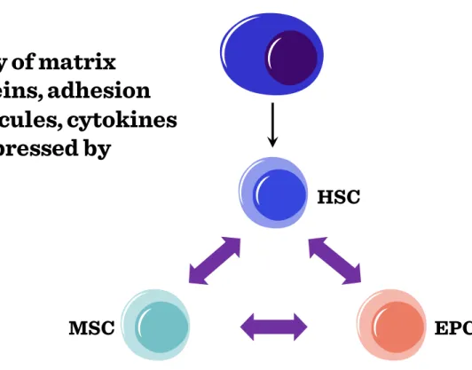
Bone marrow stem cells (MSCs). Bone marrow stem cells can be subdivided into bone marrow stromal (endothelial, mesenchymal) and haematopoietic lineages. All the specific progenitor types can be purified from the bone marrow based on their respective cell surface markers. In the bone marrow microenvironment, these stem cells show a functional interdependency (Figure II-7), secreting factors and providing the necessary cellular interactions to keep the stem cell niche suitable for all the progenitor cells.
Figure II-7: Functional interdependency of bone marrow stem cells
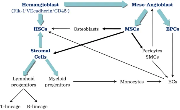
The ontogeny of tissue lineages in bone marrow is summarized in Figure II-8.
HSC
MSC EPC
Array of matrix
proteins, adhesion
molecules, cytokines
is expressed by
18 The project is funded by the European Union and co-financed by the European Social Fund.
Figure II-8: Ontogeny of tissue lineages in bone marrow
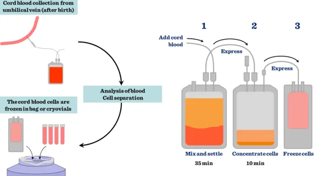
Cord blood stem cells. The largest source of stem cells for regenerative medicine is the umbilical cord stem cell pool. Based on the number of babies born yearly (approximately 130 million), this is not surprising. However, unless the cord blood is collected from the newborn to be used in potential diseases later on in his or her life – mainly childhood haematological disorders –, the application is not autologous, therefore cell types have to be matched just as in any other transplantation procedures.
Cord blood is collected at birth and processed immediately (Figure II-9) to purify stem cells based on their cell surface markers or simply just to be frozen with added DMSO in liquid nitrogen.
Pericytes SMCs
ECs
HSCs Osteoblasts MSCs EPCs
Stromal Cells
Lymphoid progenitors
Myeloid
progenitors Monocytes
T-lineage B-lineage
Meso-Angioblast Hemangioblast
(Flk-1+VEcadherin+CD45-)
Cells and tissue types in tissue engineering – Stem cells
Identification number:
TÁMOP-4.1.2-08/1/A-2009-0011
19 Figure II-9: Cord blood stem cells and foetal stem cells
Figure II-10: Stem cell populations in cord blood
In the past 36 years 10,000 patients were treated for more than 80 different diseases using cord blood stem cells. Ideally, cord blood banks should be set up with active links to international data bases where all the stored cells would be characterized with HLA typing and made available when needed. Unfortunately, storage of stem cells
Cord blood collection from umbilical vein (after birth)
The cord blood cells are frozen in bag or cryovials
Liquid nitrogen storage tank (-150°)
Analysis of blood Cell separation
Mix and settle Concentrate cells Freeze cells
35 min 10 min
Express
Express Add cord
blood
1 2 3
Adherent Pre-MSC MSC
Non-adherent CBE
Lin- CD133+ CD34+
Cord Blood
Bone marrow
Peripheral Blood
20 The project is funded by the European Union and co-financed by the European Social Fund.
is costly therefore this relatively simple source for progenitor cells is not used to its full potential.
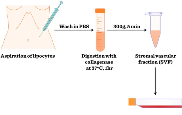
Adipose tissue derived stem cells. Recently there has been a trend to obtain mesenchymal stem cells from fat. Similarly to other differentiated tissue types, adipose tissues contain tissue specific stem cells. Adipose stem cells (ASC) can be easily isolated from adipose tissues (Figure II-11), and ASCs are multipotent, their immunophenotype is consistent (Figure II-12) and adipose stem cells are easily manipulated by genetic engineering.
Figure II-11: Isolation procedures of ASCs Digestion with
collagenase at 37oC, 1hr
Stromal vascular fraction (SVF) Wash in PBS
Aspiration of lipocytes
300g, 5 min
Cells and tissue types in tissue engineering – Stem cells
Identification number:
TÁMOP-4.1.2-08/1/A-2009-0011
21 Figure II-12: Immunophenotype of ASCs (Positive markers)
The differentiation potential of ASCs is also wide. By using the right combination of extracts and factors, ASCs can differentiate into cardio-myocytes, skeletal myocytes, chondrocytes, osteoblasts, neuronal, endodermal and ectodermal lineages.
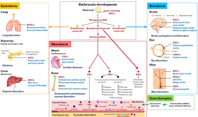
Application of stem cells (either embryonic or adult stem cells) (Figure II-13) is becoming broader since cellular manipulation can aid development of the required cell types.
Marker Name Marker Name
CD9 Tetraspan CD55 Decay accelerating factor
CD10 Common acute lymphocytic
leukemia antigen CD59 Protectin
CD13 Aminopeptidase CD71 Transferrin
CD29 β1-integrin CD73 5’-ectonucleotidase
CD34 Sialomucin CD90 Thy1
CD44 Hyaluronate receptor CD105 Endoglin
CD49d α4-integrin CD146 Muc-18
CD54 Intracellular adhesion
molecule CD166 Activated leukocyte cell
adhesion molecule HLA-ABC Histocompatibility locus
antigen-ABC a-SMA α-smooth muscle actin
22 The project is funded by the European Union and co-financed by the European Social Fund.
Figure II-13: Application of ESCs and ASCs
Cells can be reprogrammed by modification of their original gene expression patterns or cellular differentiation can be manipulated by applying directed changes into the cellular growth environment in forms of growth factors, cytokines, cellular interactions (e.g. feeder layer). Naturally, the right factors need to be identified prior to any attempts to grow specific tissue types for commercial use. In fact tight regulatory issues surround tissue engineering aimed at clinical application of laboratory generated tissues in regenerative medicine. Apart from ethical issues of using embryonic stem cells, and working with human subjects to purify adult stem cells, the purification of stem cells requires GLP (good laboratory practice) and GMP (good manufacturing practice) conditions that make the production of such tissues very expensive.
Differentiated cells. Previously, it was believed that many differentiated cells of adult human tissues have only a limited capacity to divide. In contrast, some almost differentiated cells have widely been used in clinical tissue engineering. The “almost”
status is frequently achieved by culturing purified differentiated cells in 2D culture
Vascular lumen
Vasculogenic zone Vascular disorders
Gene therapies
Genetically modified stem cell-based delivery Intravenous
injection
Mesoderm
Cardiac diseases CSCs Atria niche Apex niche
HSC
EPC MSC
Osteoblasts Chondrocytes
Adipocytes Myoblasts Common lymphoid
precursor
NK-cells T-lymphocytes
Dendritic cells B-lymphocytes Common myeloid
precursor
Macrophages Platelets Erythrocytes
Monocytes Granulocytes HSCs
Endosteum surface niche Microvasculature niche MSCs
Perivascular surface niche Bone
Hemopoietic and Immune system disorders
Endoderm
BASCs Bronchioalveolar duct junction niche Lung disorders
PSCs Pancreatic duct putative niche Pancreas
Insulin-secreting β-cells
Endocrine islets of Langerhans Exocrine
acini
Diabetes Liver
Hepatocytes
HDCs Bile duct (canal of Hering) Hepatic disorders
Lung
Ectoderm
NSCs Subventricular zone niche Hippocampus niche (dentate gyrus region) Neuron Astrocyte Oligodendrocyte
Brain and spinal cord disorders
Eye disorders RSCs Ciliary epithelium niche
CESCs Limbus niche Retina
Cornea
KSCs Basal layer niche bESCs eNCSCs Bulge niche SKPs Dermal papilla Skin disorders
Heart
Cardiomyocytes
Embryonic development
Inner cell mass ESCs Blastocyst
Pluripotent ESC
Mesodermal stem cell Endodermal
stem cell
Ectodermal stem cell Hemangioblast
HSC EPC
Vascular wall-resident stem cells EPCsandMSCs New endothelial cell
Bloodstream Macrophage
Platelets
Erythrocyte Monocyte
Eosinophil
NK-cell
T-lymphocyte Dendritic cell B-lymphocyte Basophil
Neutrophil
Media
Brain
Eye
Skin
Cells and tissue types in tissue engineering – Stem cells
Identification number:
TÁMOP-4.1.2-08/1/A-2009-0011
23 conditions where they lose their final differentiation characteristics. Among differentiated cells used in tissue engineering are notably fibroblasts, keratinocytes, osteoblasts, endothelial cells, chondrocytes, preadipocytes, and adipocytes. One of the most frequently used differentiated cell types are chondrocytes. In the following, chondrocytes shall serve as an example to describe the use of differentiated cells in tissue engineering applications.
Isolation of autologous chondrocytes for human use is invasive. It requires a biopsy from a non-weight-bearing surface of a joint or a painful rib biopsy. In addition, the ex vivo expansion of a clinically required number of chondrocytes from a small biopsy specimen, which may itself be diseased, is hindered by deleterious phenotypic changes in the chondrocyte. It is important that chondrocytes synthesize type II collagen, the primary component of the cross-banded collagen fibrils. The organization of these fibrils, into a tight meshwork that extends throughout the tissue, provides the tensile stiffness and strength of articular cartilage, and contributes to the cohesiveness of the tissue by mechanically entrapping the large proteoglycans. In growing individuals, the chondrocytes produce new tissue to expand and remodel the articular surface. With aging, the capacity of the cells to synthesize some types of proteoglycans and their response to stimuli, including growth factors, decrease. These age-related changes may limit the ability of the cells to maintain the tissue, and thereby contribute to the development of degeneration of the articular cartilage. For the organ repair or replacement process the in vitro amplified number of chondrocytes have to be grown in bioreactors to grow as weight bearing tissue. Although differentiated chondrocytes appear to be one of the best sources for engineered cartilage, to date, few tissue-engineered systems provide an autologous, minimally invasive, and easily customizable solution for the repair or augmentation of cartilage defects.
Identification number:
TÁMOP-4.1.2-08/1/A-2009-0011
25
III Bioreactors
One of the persistent problems of tissue engineering is the difficult production of considerable tissue mass. As engineered tissues normally lack blood vessels, it is difficult for cells in the inside of a larger tissue mass to obtain sufficient oxygen and nutrient supply to survive, and consequently to function properly.
Although self-assembly plays an important role in any tissue engineering methods, tissue growth and functional differentiation needs to be directed by the tissue engineeer. Creation of functional tissues and biological structures in vitro needs to observe some basic requirements for cellular survival, growth and differentiation. In general, the basic requirements include continuous supply of oxygen, correct pH, humidity, temperature, nutrients and osmotic pressure. If the structure of the tissue is important from the point of view of tissue function, suitable scaffolds are often needed.
Tissue engineered cultures present additional problems in maintaining culture conditions. In standard, simple maintenance cell cultures, molecular diffusion is often the sole means of nutrient and metabolite transport. However, as a culture becomes larger and more complex, for example in the case of engineered organs with larger tissue mass, other mechanisms must be employed to maintain the culture and to avoid cellular necrosis. It is important to create some sort of capillary networks within the tissue that allows relatively easy transport of nutrients and metabolites.
Similarly to directing growth and differentiation of stem cell cultures, complex tissue cultures also require added factors, hormones, metabolites or nutrients, chemical and physical stimuli to reach the desired functionality.
Just a couple of examples: chondrocytes for example respond to changes in oxygen tension as part of their normal development to adapt to hypoxia during skeletal
26 The project is funded by the European Union and co-financed by the European Social Fund.
development. Other cell types, such as endothelial cells, respond to shear stress from fluid flow, which endothelial cells encounter in blood vessels. Mechanical stimuli, such as pressure pulses seem to be beneficial to all kinds of cardiovascular tissue such as heart valves, blood vessels or pericardium. All the special requirements are intended to be resolved by the use of bioreactors.
A bioreactor in tissue engineering is a device that simulates a physiological environment in order to promote cell or tissue growth. A physiological environment can consist of many different parameters such as temperature and oxygen or carbon dioxide concentration, but can extend to all kinds of biological, chemical or mechanical stimuli.
Therefore, there are systems that may include the application of the required forces or stresses to the tissue two- or three-dimensional setups (e.g., flex and fluid shearing for heart valve growth). Several general-use and application-specific bioreactors are available commercially to aid research or commercial exploitation of engineered tissues.
Bioreactors, however, require a large number of cells to start off with.
Cells to be used in bioreactors
Static cell cultures are the most frequently applied cell culture methods. The maintenance is easy, cheap and no specialized laboratory equipment is needed. Petri dishes or disposable plastic tissue culture flasks are used most frequently. Basically 2 types of conventional tissue culture exist: Adherent cells which grow in monolayer cultures and non-adherent cells that grow in suspension cultures. In both case, conventional cell cultures are capable to maintain cells at relatively low densities. The main problem rises concerning static cell cultures when large numbers of cells are needed and conventional cultures need to be scaled-up to be added to bioreactors. In the conventional cultures nutrient supply is maintained by frequent and periodic change of
Bioreactors
Identification number:
TÁMOP-4.1.2-08/1/A-2009-0011
27 culture medium that is time consuming and involves a high risk of infection. The advantages of the dynamic cellular environment of bioreactors include the dynamic and continous supply of nutrients and oxigen. These features make the formation of larger 3D tissue structures possible. However, availability of nutrients and oxygen inside a 3D tissue construct is still problematic. While in 3D cell cultures direct cell-cell contacts enhance cellular communication, transport of oxygen, nutrients and metabolites is still a challenging issue. First, oxygen and nutrients should diffuse from the static medium to the surface cells. This concerns the oxygen content and the oxygen-carrying capacity of the medium. Diffusion from the surface cells to the deeper structures is also important.
Critical parameters are the porosity of the cultured cell/tissue construct.
Understandably, thickness of the tissue construct is a critical parameter. In static conditions tissue thichness should not exceed 100 μm. In the dynamic environment of bioreactors, this parameter can be increased several fold.
Bioreactor design requirements
Although bioreactors cannot recreate the physiological environment, they need to reproduce as many parameters as possible. Bioreactors need to maintain desired nutrient and gasconcentration in 3D constructs and to facilitate mass transport into and from 3D tissues. It is also important for bioreactors to improve even cellular distribution, which is also facilitated through the dynamic environment. Exposure of the construct to physical stimuli is also important during the engineering of load-bearing tissues, like cartilage, bone, tendon and muscle. Without mechanical stimuli these tissues cannot withstand physiological load and strain.
There is an additional critical parameter in dynamic tissue cultures: the shear force. Shear forces are particularly important, as cells are sensitive to shear stress,
28 The project is funded by the European Union and co-financed by the European Social Fund.
which may cause dedifferentiation, growth inhibition or apoptosis. Unfortunately, shear stress distribution is uneven in dynamic bioreactors. In dynamic bioreactors the highest stress is located around edges and sides of the moving vessel or around the moving edge of the stirrer and the edges of the scaffold which is static itself but the fluid is moving around it. (The measure unit of shear stress is the dyn/cm2. 1 dyn = 10 mN. The maximum shear stress for mammalian cells is 2.8 dyn/cm2, so in a well-designed bioreactor, shear stress values are well below this number) (Figure III-1).
Figure III-1: Shear forces in dynamic fluids
Perhaps the most difficult task is to provide real-time information about the structure of the forming 3D tissue. Normally the histological, cellular structure of the 3D construct can only be judged after the culturing period is completed.
In general, bioreactors should be as clear and simple as possible to make cleaning easy and to reduce the risk of infections. The assembly and disassembly of the device should be also simple and quick. It is extremely important that all parts of the bioreactor that comes into contact with the cell culture is made of biocompatible or bioinert materials. For example no chromium alloys or stainless steel should be used in biorecators. Also the material should withstand heat or alcohol sterilization and the
Shear stress measure unit:
dyn/cm2 1 dyn = 10mN
A shear stress,τis applied to the top of the square while the bottom is held in place.
l
Δ x
Bioreactors
Identification number:
TÁMOP-4.1.2-08/1/A-2009-0011
29 presence of the continuously humid atmosphere. The design should also ensure the proper embedding of instruments like the thermometer, pH meter, the pump or rotator motor, etc.
Main types of bioreactors
Spinner flask bioreactors (Figure III-2) are maybe the simplest and the most frequently used bioreactor types.
Figure III-2: Spinner flask bioreactors
Spinner bioreactor types mix the oxygen and nutrients throughout the medium and reduce the concentration boundary layer at the construct surface. In a spinner flask, scaffolds are suspended at the end of needles in a flask of culture media. A magnetic stirrer mixes the media and the scaffolds are fixed in place with respect to the moveing fluid. Flow across the surface of the scaffolds results in turbulences and flow
30 The project is funded by the European Union and co-financed by the European Social Fund.
instabilities caused by clumps of fluid particles that have a rotational structure superimposed on the mean linear motion of the fluid particles. Via these added fluid motions fluid transport to the centre of the scaffold is thought to be enhanced.
Typically, spinner flasks are around 120 ml in volume (although much larger flasks of up to 8 liters have also been used). The most frequent stirring speed is 50–80 rpm and generally 50% of the total medium is changed every two days. The efficiency of the enhancement of mass transport is indicated that cartilage constructs have been grown in spinner flasks to thicknesses of 0.5 mm compared to that of 100 μm in static cultures.
However, cell seeding efficiency is typically low in spinner flask bioreactors, this method usually fails to deliver homogeneous cell distribution throughout scaffolds and cells predominantly reside on the construct periphery.
The rotating wall bioreactor (Figure III-3) was originally developed by NASA.
Figure III-3: Rotating wall bioreactors
It was designed with a view to protect cell culture experiments from high forces during space shuttle take off and landing. The device has proved useful in tissue engineering laboratories on Earth too. In a rotating wall bioreactor, scaffolds are free to
Fc Fd
Fg
Bioreactors
Identification number:
TÁMOP-4.1.2-08/1/A-2009-0011
31 move in media in a vessel. A rotating wall vessel bioreactor consists of a cylindrical chamber in which the outer wall, inner wall, or both are capable of rotating at a constant angular speed. The vessel wall is then rotated at a speed such that a balance is reached between the downward gravitational force and the upward hydrodynamic drag force acting on each scaffold. The wall of the vessel rotates, providing an upward hydrodynamic drag force that balances with the downward gravitational force, resulting in the scaffold remaining suspended in the media. Dynamic laminar flow generated by a rotating fluid environment is an alternative and efficient way to reduce diffusional limitations of nutrients and wastes while producing low levels of shear compared to the stirring flask. Culture medium can be exchanged by stopping the rotation temporarily or by adding a fluid pump whereby media is constantly pumped through the vessel. Fluid transport is enhanced in a similar fashion to the mechanism in spinner flasks and the rotational devices also provide a more homogeneous cell distribution compared to static or spinner bioreactor cultures. Gas exchange occurs through a gas exchange membrane.
Typically, the bioreactor is rotated at speeds of 15–30 rpm. Cartilage tissue of 5 mm thickness has been grown in this type of bioreactor after seven months of culture. As tissue mass increases while cells grow in the bioreactor, the rotational speed must be increased in order to balance the gravitational force and to ensure that the scaffold remains in suspension.
Compression bioreactors (Figure III-4) are another widely used type of bioreactors.
32 The project is funded by the European Union and co-financed by the European Social Fund.
Figure III-4: Compression bioreactors
This class of bioreactor is generally used in cartilage engineering and can be designed so that both static and dynamic loading can be applied. In contrast to static loading that has a negative effect on cartilage formation dynamic loading is more beneficial and more representative of physiological tissue deposition. In general, compression bioreactors consist of a motor, a system providing linear motion and a controlling mechanism to provide different magnitudes and frequencies. A signal generator can be used to control the system including loading of cells while transformers can be used to measure the load response and imposed displacement. The load can be transferred to the cell-seeded constructs via flat platens which distribute the load evenly. However, in a device for stimulating multiple scaffolds simultaneously, care must be taken that the constructs are of similar height or the compressive strain applied will vary as the scaffold height does. Mass transfer is improved in dynamic compression bioreactors over static culture (as compression causes fluid flow in the
Head for dispensing pressure
Scaffold constructs
Bioreactors
Identification number:
TÁMOP-4.1.2-08/1/A-2009-0011
33 scaffold) which results in the improvement of the aggregate modulus of the resulting cartilage tissue to levels approaching those of native articular cartilage.
Tensile strain bioreactors have been used in an attempt to engineer a number of different types of tissues including tendon, ligament, bone, cartilage and cardiovascular tissue. Some designs are very similar to compression bioreactors, only differing in the way the force is transferred to the construct. Instead of flat platens as in a compression bioreactor, a way of clamping the scaffold into the device is needed so that a tensile force can be applied. Tensile strain has been used to differentiate mesechymal stem cells along the chondrogenic lineage. A multistation bioreactor was used in which cell- seeded collagen-glycosaminoglycan scaffolds were clamped and loaded in uniaxial tension. Alternatively, tensile strain can also be applied to a construct by attaching the construct to anchors on a rubber membrane and then deforming the membrane. This system has been used in the culture of bioartificial tendons with a resulting increase in Young’s modulus over non-loaded controls. (Young’s modulus is a numerical constant that was named after an 18th-century English physician and physicist. The constant describes the elastic properties of a solid material undergoing tension or compression forces in one direction only).
Culture using flow perfusion bioreactors (Figure III-5) has been shown to provide more homogeneous cell distribution throughout scaffolds.
34 The project is funded by the European Union and co-financed by the European Social Fund.
Figure III-5: Flow perfusion bioreactors
Collagen sponges have been seeded with bone marrow stromal cells and perfused with flow. This has resulted in greater cellularity throughout the scaffold in comparison to static controls, implying that better nutrient exchange occurs due to flow. Using a calcium phosphate scaffold, abundant extracellular matrix (ECM) with nodules of calcium phosphate was noted after 19 days in steady flow culture. In comparisons between flow perfusion, spinner flask and rotating wall bioreactors, flow perfusion bioreactors have proved to be the best for fluid transport. Using the same flow rate and the same scaffold type, while cell densities remained the same using all three bioreactors, the distribution of the cells changed dramatically depending on which bioreactor was used. Histological analysis showed that spinner flask and static culture resulted in the majority of viable cells being on the periphery of the scaffold. In contrast, the rotating wall vessel and flow perfusion bioreactor culture resulted in uniform cell distribution throughout the scaffolds. Flow perfusion bioreactors generally consist of a pump and a scaffold chamber joined together by tubing. A fluid pump is used to force media flow through the cell-seeded scaffold. The scaffold is placed in a
Scaffold constructs with seeded cells
Bioreactors
Identification number:
TÁMOP-4.1.2-08/1/A-2009-0011
35 chamber that is designed to direct flow through the interior of the scaffold. The scaffold is kept in position across the flow path of the device and media is perfused through the scaffold, thus enhancing fluid transport. Media can easily be replaced in the media reservoir. However, the effects of direct perfusion can be highly dependent on the medium flow rate. Therefore optimising a perfusion bioreactor for the engineering of a 3D tissue must address the careful balance between the mass transfer of nutrients and waste products to and from cells, the retention of newly synthesised ECM components within the construct and the fluid induced shear stresses within the scaffold pores.
Flow perfusion reactors in bone tissue engineering. Flow perfusion bioreactors have proved to be superior compared to rotating wall or spinner flask bioreactors to seed cells onto scaffolds. The expression levels of bone differentiation markers (namely Alkaline Phosphatase, Osteocalcin and the transcription factor Runx2) proved to be consistently higher in flow perfusion reactors than in any other type of bioreactors.
Additionally, the mineralization of the scaffolds is also higher. To avoid the disadvantageous effects of high shear stress, the flow rate in the reactor needs to be set carefully. Experiments demonstrated that intermittent dynamic flow is more favourable than steady speed flow in bone tissue engineering.
Two chamber bioreactor – The most current and revolutionary achievement in tissue engineering was the implantation of a tissue engineered trachea which was developed in a special two-chamber bioreactor. The external surface of the de- cellularized donor trachea was seeded with autologous chondrocytes differentiated from hemapoetic SCs of the recipient and airway epithelium was seeded to the inner surface in a rotation wall-like bioreactor. The application of two separate “chambers” allowed simultaneous culture of different cell types. The construct was then used for the surgical replacement of the narrowed trachea in a tuberculosis patient.
36 The project is funded by the European Union and co-financed by the European Social Fund.
Drawbacks to currently available bioreactors
Tissue engineering methodology is very labor intensive, specialized equipment and specially trained technicians are needed to perform the work. The current bioreactors are highly specialized devices which are difficult to assemble and disassemble. The cell output is low and the culturing times are long. Moreover, real-time monitoring of tissue structure and organization is not yet available. Problems with compression bioreactors involve mainly the mechanical parts, which are prone for leakage. Understandably, infection is a problem when such bioreactors are used. Added problem is the type of the scaffold to grow the cells on, as the applied scaffolds have to withstand mechanical stimulation, so strong scaffolds are needed, which may have longer degradation time once implanted. Naturally, this is not preferred. So a compromise has to be achieved between scaffold stiffness and resorption time.
Identification number:
TÁMOP-4.1.2-08/1/A-2009-0011
37
IV Biomaterials
The requirements for biomaterials used in tissue engineering are quite strictly defined.
Biocompatibility for example is high on the agenda, as scaffold and bioreactor materials have to be tissue friendly and not eliciting immunoresponse. Moreover, at best, the biomaterial should support cellular and tissue functions like adhesion, differentiation and proliferation via its special surface chemistry. Porosity is an important requirement concerning scaffolds. Generally the porosity should reach and even exceed 90% to allow even seeding of cells and to support vascular ingrowth after implantation.
Controlled biodegradation is also an important issue in some cases when the healthy tissue replaces the implanted biomaterial and the biomaterial gradually degrades in the body of the host. Biomaterials can be divided into natural and synthetic biomaterials.
Natural biomaterials
The advantages of natural biomaterials (Figure IV-1) are that they mostly come from an in vivo source therefore large quantities are constantly available at a reasonable price.
Figure IV-1: Types of natural biomaterials
Further advantages of natural biomaterials are that they already have binding sites for cells and adhesion molecules so the biocompatibility is not a major issue. However,
Proteins:
• Collagen
• Fibrin
• Silk
Polysaccharydes:
• Agarose
• Alginate
• Hyaluronic acid
• Chitosan
38 The project is funded by the European Union and co-financed by the European Social Fund.
there are also some disadvantages. Due to natural variability in the in vivo source, the lot-to-lot variability is always a concern. Additionally, potential impurities may also result in unwanted immune reactions, while their mechanical properties are also limited.
Collagen is the most frequently used and therefore the most studied biomaterial.
There are rich in vivo sources, as all connective tissues of animals are rich in this ubiquitous protein. Collagen has a fibrous structure, and its amino acid composition is unique. It provides binding sites for integrins called RGD sites (from the amino acid sequence arginine-glycine-aspartic acid). Collagen has a superior biocompatibility being a conserved protein. Additional benefit, that the immune system well tolerates collagen.
It is capable of supporting a large spectrum cellular differentiation types, therefore collagen is well preferred as scaffold.
Another, easily accessible type of scaffold is fibrinogen. Fibrinogen is obtained from (human) plasma. Although in its uncleaved form is a soluble protein, upon cleavage with thrombin fibrinogen sets as a gel and forms a 3D meshwork which is 100% biocompatible and physiological in wound healing. It is often used as a biological glue when cells to be seeded onto scaffolds (e.g. non-woven mesh or fleece or other porous materials) are suspended first in a fibrinogen-containing solution. Then the solution is applied onto the scaffold and upon the addition of thrombin, a hydrogel is formed which enhances the cells’ ability to attach to the 3D scaffold. Fibrin is also suitable for supporting ES cell differentiation as well as keeping differentiated cells in culture. Recent applications of fibrin include cardiovascular, cartilage, bone and neuronal tissue engineering.
Silk that is also used as scaffold material, is a protein produced within specialized glands of some arthropods. It has a special tertiary structure consisting of repeating amino acid motifs forming an overlapping beta-sheet structure giving the unique
Biomaterials
Identification number:
TÁMOP-4.1.2-08/1/A-2009-0011
39 sturdiness for this protein. There have been many industrial attempts to mimic the features of silk. As a result the availability of recombinant analogues is increasing.
The silkworm’s (Bombix mori) silk consists of two different protein components, namely Fibroin and Sericin. Sericin forms the outer layer on the fibroin core making it slippery and elastic. Fibroin that is biocompatible and possesses excellent mechanical properties is also used as tissue engineering scaffold. Its use is widespread in bone, cartilage and ligament engineering. Additionally, silk fibroin can be modified chemically, e.g. the attachment of RGD groups provides binding sites for osteoblasts and enhances Ca++ deposition and bone cell differentiation. Moreover, silk promotes more intensive chondrogenesis than collagen used as a scaffold material for cartilage engineering. The degradation rate of silk is very slow but finally bone tissue gradually replaces the silk scaffold.
Polysaccharide-based biomaterials are polymers consisting of sugar monomers.
Those used for tissue engineering purposes are of plant (seaweed) or animal origin.
Some polysaccharides may trigger unwanted immune reactions so a careful selection is advised considering polysaccharide scaffold materials. Polysaccharides are most frequently used as hydrogels, which per se form a 3D meshwork so they can provide a scaffold for seeded cells. These hydrogels are frequently used as injectons: they can be dispensed directly to the site of injury so it supports wound healing and also cell growth and differentiation.
Main source of agarose, another scaffold material for tissue engineering, are red algae and seaweed. Agarose is the most frequently used polysaccharide scaffold consisting of a galactose-based backbone. It is immunologically inert, so no immune response is triggered. One of its great advantages lies in its versatility: the stiffness and
40 The project is funded by the European Union and co-financed by the European Social Fund.
mechanical parameters can be easily manipulated of agarose gels. It has been used for scaffolding cartilage, heart, nerve tissues and it also supports stem cell differentiation.
Alginate is the polysaccharide component of the cell walls of brown algae. It is an acidic compound, so in tissue engineering various cationic alginate salts are used.
Sodium-alginate is a frequently used food additive (E-401) and its use is also widespread in gastronomy. Besides of gastronomic applications, sodium alginate is used in industry as a heavy metal-binding or fat-binding agent. The potassium salt is also used in the food industry as an emulsifier and stabilizer. In tissue engineering its calcium salt or Calcium-alginate has gained widespread application. Calcium alginate is a water-insoluble gel-like material, and it is generally used in the industry or laboratory for enzyme immobilization or encapsulation. In tissue engineering calcium-alginate proved to be useful for encapsulation of whole living cells, thus isolating them from the immune system preventing rejection after transplantation. In a clinical trial, calcium- alginate was used for the encapsulation of pancreatic islet cells which preserved their ability to produce insulin according to the needs of the host as the encapsulation prevented the immune reaction against the grafted cells.
Hyaluronan which is also termed as Hyaluronic acid is an animal-derived polysaccharide which is extensively used as a scaffold material in tissue engineering. It is a non-sulfatated Glucose-Amino-Glycan (GAG) molecule and exists as a major component of the ECM in hyalinic cartilage and skin but it is present in other organs as well. As a natural ECM component, multiple cell surface receptor binding and cell adhesion sites available on the macromolecular complex. Hyaluronan has an important role in wound healing and tissue repair. Moreover, it supports embryonic stem cell differentiation, survival and proliferation. Like other polysaccharides, hyaluronan is used as a gel in nerve, cartilage and skin tissue engineering.
Biomaterials
Identification number:
TÁMOP-4.1.2-08/1/A-2009-0011
41 Chitosan is derived from the deacetylation of chitin which is a strongly cationic polysaccharide which is the main component of the arthropod exoskeleton. Chitosan is commercially derived from sea-dwelling crustaceans and it is widely used for bandages and wound dressing, utilizing its ability to enhance blood clotting. Chitosan scaffolds are mainly used in bone tissue engineering, as chitosan supports osteoblast differentiation. Moreover, chitosan-calcium-phosphate composite scaffolds form a pliable, injectable hydrogel at slightly acidic pH. Upon transition to physiological pH, it gels thus anchoring osteocytes to the scaffold. Native and collagen-modified chitosan can be used for tissue engineering, as both forms support progenitor differentiation into osteoblasts.
Synthetic biomaterials
In tissue engineering a large scale of synthetic biomaterials (Figure IV-2) are used besides that of natural origin.
Figure IV-2: Types of synthetic biomaterials
Their main advantages over natural biomaterials are the high reproducibility, availability on demand and constant quality supporting industrial-scale production.
Moreover, by application of slight changes of production, an easy control of mechanical properties, degradation rate, shape, composition, etc. can be adjusted to current needs.
Organic polymers:
• PGA, PLA, PLGA
• PEG
• Peptides
Inorganic:
• Ceramic
• Metal
• Hydroxyapathite
42 The project is funded by the European Union and co-financed by the European Social Fund.
However, synthetic materials often lack sites for cell adhesion and the biocompatibility is frequently questionable. Biocompatibility and support of stem cell differentiation is not clear either, while immune reactions are also possible.
Poly (lactic-co-glycolic acid) PLGA is an FDA approved scaffold material.
PLGA is a mixed polymer consisting of lactic and glycolic acids in various ratios.
PLGA is a biodegradable material and its degradation rate can be modulated. It is one of the most frequently used biomaterials applied in neural tissue, bone and cartilage tissue engineering. PLGA is biocompatible, triggers no immune reaction and supports embryonic stem cell differentiation, proliferation and survival. However one has to consider that the degradation products are acidic, therefore may alter cell metabolism.
Poly (ethylene glycol) PEG is a commonly used biocompatible polymer. This molecule has amphophilic properties having a hydrophilic head and a variably long hydrophobic tail. The PEGylation of proteins is a commonly used pharmaceutical technology to modulate the degradation and absorbtion of bioactive proteins like interferons. The chemical modification of PEG is also available e.g. heparin, peptides, or RGD motifs. PEG is also a frequently used scaffold material in bone, cartilage, nerve, liver and vascular tissue engineering. Application of PEG-formed hydrogels is very versatile. The chemical nature, the extent of crosslinking, and the potential application of modifications, like RGD or other amino-acid groups, makes PEG widely used. PEG-based gels can be applied not only to entrap or anchor cells but also in storing and delivering bioactive molecules like BMP or TGFβ.
Peptide-based biomaterials consist of short amino acid sequences which usually are ampholitic thus making peptides capable of self-assembly. The application of these peptides allows the combination of the advantages of synthetic materials with that of natural scaffolds. Synthetic nature of the peptides eliminates batch-to-batch variability
Biomaterials
Identification number:
TÁMOP-4.1.2-08/1/A-2009-0011
43 and consistent purity and quality can be achieved. Moreover, known binding sites for cell-surface adhesion molecules can be included in the sequence. For example, IKVAV amino acid sequence is from laminin and it facilitates neurite outgrowth. RGD sequence is a binding site for integins which promotes cellular adherence and migration.
Ceramic-based biomaterials are used in bone tissue engineering only. These scaffolds are totally or partly inorganic. Mostly they are shaped with heat forming porous, brittle materials. For example, bioactive glass is slowly biodegradable. With an ion-exchange mechanism it slowly turns to natural hydroxyapathite via surface degradation. So bioglass is biocompatible and used as an implant material.
Hydroxyapatite is the inorganic compound of bone. Sometimes it is used as a completely inorganic, porous scaffold, more often it is combined with other biocompatible, organic polymers, like PLGA, collagen, or chitosan. This combination also enhances the drug delivery capacity of these scaffolds to enhance bone formation.
Metals are used as implant materials. Mostly alumina and titanium alloys are used because of their biocompatibility. The high durability of metals is needed where implants are subjected to extreme mechanical load, like articular prostheses, dental implants or heart valves. These metals are bioinert materials, however, sometimes metallic implants may cause immunological reactions like metal allergy in sensitive individuals. Also, as they are not biodegradable it is questionable whether metals can be listed amongst tissue enginnering scaffolds.