Molecular and linical asics of erontology
c b
g
Manifestation of Novel Social Challenges of the European Union in the Teaching Material of
Medical Biotechnology Master’s rogrammes at the University of Pécs and at the University of DebrecenP
Dr.KrisztiánKvell – Dr.JuditPongrácz – Dr.MiklósSzékely – Dr.MártaBalaskó – Dr.ErikaPétervári – Dr.GyulaBakó
Molecular and Clinical Basics of Gerontology
Dr. Krisztián Kvell – Dr. Judit Pongrácz
– Dr. Miklós Székely – Dr. Márta Balaskó – Dr. Erika Pétervári – Dr. Gyula Bakó
“Manifestation of Novel Social Challenges of the European Union
in the Teaching Material of
Medical Biotechnology Master’s Programmes at the University of Pécs and at the University of Debrecen”
Identification number: TÁMOP-4.1.2-08/1/A-2009-0011
University of Pécs – Pécs, 2011
© Dr. Krisztián Kvell, Dr. Judit Pongrácz, Dr. Miklós Székely, Dr. Márta Balaskó, Dr. Erika Pétervári, Dr. Gyula Bakó, 2011
The project is funded by the European Union and co-financed by the European Social Fund.
Editor in charge: University of Pécs
Editor in charge: Dr. Krisztián Kvell, Dr. Judit Pongrácz, Dr. Miklós Székely, Dr. Márta Balaskó, Dr. Erika Pétervári, Dr. Gyula Bakó, Rita Bognár
Technical editor: Zsolt Bencze, Veronika Csöngei and Szilvia Czulák Lector: Dr. György Miskei
Length: 215 pages
Identification number:
TÁMOP-4.1.2-08/1/A-2009-0011
3
Content
LIST OF FIGURES ... 9
I GERONTOLOGY ... 19
I.1 BASICS OF GERONTOLOGY, DEMOGRAPHIC DATA ... 19
I.1.1 Introduction, definitions ... 19
I.1.2 Population-wide aging ... 19
I.1.3 Chronological and biological age ... 22
I.1.4 Etiology of aging: genetic mechanisms and environmental factors ... 23
I.2 ADAPTATION AND VULNERABILITY, POLYMORBIDITY ... 25
I.2.1 Progressive deficit in adaptative homeostatic mechanisms in the course of aging ... 25
I.2.2 Polymorbidity in the elderly ... 30
I.3 NUTRITION, PHYSICAL STATUS, BODY COMPOSITION, SARCOPENIA ... 32
I.3.1 Introduction: age-related changes in body weight and body composition ... 32
I.3.2 Changes in fat mass (FM) and fat free mass (FFM) with age ... 33
I.3.3 The pathogenesis of sarcopenia ... 36
I.4 IMMOBILIZATION, PHYSICAL ACTIVITY, DISORDERS OF LOCOMOTOR ORGANS ... 39
I.4.1 The beneficial effects of physical exercise ... 39
4 The project is funded by the European Union and co-financed by the European Social Fund
I.4.2 Immobilization syndrome – chronic bedrest ... 40
I.4.3 Remobilization in the elderly ... 48
I.5 CHARACTERISTICS OF THE CARDIOVASCULAR SYSTEM, ABNORMALITIES AND DISEASES ... 50
I.5.1 Age-related alterations in the cardiovascular system ... 50
I.6 CHANGES OF THE RESPIRATORY SYSTEM, FREQUENT DISEASES ... 57
I.6.1 Age-related alterations in the chest and in the lungs ... 57
I.6.2 Age-related alterations in the airways ... 59
I.6.3 Abnormalities of other respiratory functions in the elderly ... 60
I.6.4 Diseases of the respiratory system with increased prevalence in old age-groups ... 60
I.7 CHANGES OF RENAL FUNCTION, ELECTROLYTE/WATER AND ACID/BASE HOMEOSTASIS ... 63
I.7.1 Aging vs. nephron dysfunctions ... 63
I.7.2 Aging vs. non-excretory kidney functions ... 68
I.7.3 Renal failure in the elderly ... 68
I.7.4 Urinary incontinence in the elderly ... 69
I.7.5 Electrolyte and water balance in the elderly ... 69
I.7.6 Aging vs. pH disturbances ... 71
I.8 CHANGES OF THE ENDOCRINE SYSTEM AND METABOLISM ... 74
I.8.1 Age-related alterations in the endocrine system ... 74
Content
Identification number:
TÁMOP-4.1.2-08/1/A-2009-0011
5 I.8.2 Functional abnormalities associated with endocrine
disorders in the elderly ... 77
I.8.3 Age-related alterations in intermediary metabolism ... 80
I.9 CHANGES OF THE GASTROINTESTINAL TRACT, ACUTE AND CHRONIC DISORDERS ... 82
I.9.1 Interaction with other systems ... 82
I.9.2 Common disorders in the upper gastrointestinal tract ... 83
I.9.3 Common disorders in the lower gastrointestinal tract ... 85
I.10 NEUROLOGICAL AND PSYCHOLOGICAL DISORDERS IN THE ELDERLY ... 90
I.10.1 Age-related alterations of the nervous system ... 90
I.10.2 Psychological disorders in the elderly ... 96
I.11 CARE OF ELDERLY PATIENT ... 99
I.11.1 Communication with the elderly patient ... 99
I.11.2 Eldercare systems ... 101
I.11.3 Polypharmacy (polypragmasia) in the elderly ... 102
I.12 SUCCESSFUL AGING ... 106
I.12.1 Factors influencing aging ... 106
II MOLECULAR GERONTOLOGY ... 113
II.1 BASICS OF MOLECULAR GERONTOLOGY ... 113
II.1.1 Basics ... 115
II.2 AGING THEORIES ... 118
6 The project is funded by the European Union and co-financed by the European Social Fund
II.2.1 Family tree of aging theories ... 118
II.2.2 Evolutionary theories, antagonistic pleiotropy ... 120
II.2.3 Programmed theories ... 121
II.2.4 Damage theories ... 122
II.3 MITOCHONDRIAL AGING ... 124
II.3.1 Mitochondria are vulnerable ... 125
II.3.2 Mitochondrial damage due to ROS, consequent senescence ... 127
II.3.3 Mitochondrial diseases ... 132
II.4 AGING AND GENE EXPRESSION ... 136
II.4.1 Telomere shortening ... 136
II.4.2 Telomere clock of aging ... 137
II.4.3 Telomerase ... 139
II.4.4 Antagonistic pleiotropy ... 142
II.5 GENETIC BACKGROUND OF LONGEVITY ... 144
II.5.1 Antagonistic pleiotropy and genetic programs ... 144
II.5.2 Centenarian studies ... 146
II.5.3 Longevity genes ... 148
II.6 CANCER AND TUMOR DEVELOPMENT, SENESCENCE AND CANCER, EPIDEMIOLOGY AND STATISTICS ... 155
II.6.1 Tumor suppressor genes ... 157
II.6.2 The ambivalent role of p53 ... 159
II.6.3 Antagonistic pleiotropy and tumor suppressor genes ... 162
Content
Identification number:
TÁMOP-4.1.2-08/1/A-2009-0011
7
II.6.4 Epidemiology and statistics ... 163
II.7 ALTERATIONS OF GENOME DUE TO AGING ... 166
II.7.1 Oxidative DNA damage and its repair ... 166
II.7.2 DNA damage and its repair in progeria ... 171
II.8 MOLECULAR / CELLULAR EFFECTS OF ACUTE AND CHRONIC STRESS ... 176
II.8.1 CR extends life-span ... 176
II.8.2 Reproducibility of CR effects ... 180
II.8.3 CR and antagonistic pleiotropy ... 180
II.9 METABOLISM AND LONGEVITY I ... 181
II.9.1 Antagonistic pleiotropy ... 181
II.9.2 Protein peroxidation, repair, associated diseases ... 181
II.9.3 PUFA controversy ... 184
II.10 METABOLISM AND LONGEVITY II. ... 186
II.10.1 Sirtuins as master regulators ... 186
II.10.2 Mammalian sirtuins ... 188
II.10.3 Functional listing of further mammalian sirtuins ... 191
II.10.4 Sirt1 mimetic compounds ... 193
II.11 SENESCENCE-RELATED INTERCELLULAR / INTRACELLULAR PATHOLOGIES ... 198
II.11.1 Lipofuscin or lysosomal waste ... 201
II.11.2 Amyloid aggregates ... 202
II.11.3 Proteasome function and senescence ... 205
8 The project is funded by the European Union and co-financed by the European Social Fund
II.12 MOLECULAR MECHANISMS OF INTERVENTIONS ... 212
II.12.1 Degree of life-extension, planned interventions ... 212
II.12.2 Limitations of SENS ... 213
II.13 RECOMMENDED LITERATURE ... 215
Identification number:
TÁMOP-4.1.2-08/1/A-2009-0011
9
List of figures
Figure I.1-1: Survival curves for different populations ... 20
Figure I.1-2: Age-specific death rates of Swedish females from 1751 to 1950 and for 1988 ... 20
Figure I.1-3: Expected life-span at birth in different European states ... 21
Figure I.1-4: Regional pattern of life expectancy in Germany: East-West difference (2003) ... 22
Figure I.2-1: Age-related changes in different functions ... 26
Figure I.2-2: Effect of 50 mmHg increment in systolic blood pressure on heart rate, cardiac index and stroke index in young and old rats ... 27
Figure I.2-3: Glucose tolerance tests (50 g glucose p.o.) in different age-groups ... 27
Figure I.2-4: Functional and reserve organ capacities in young and elderly ... 28
Figure I.2-5: Geriatric giants ... 30
Figure I.3-1: Between the ages of 20 and 70 – despite a stable, normal body weight – body composition is altered: fat mass increases (a 2 fold increase is still considered to be physiological) ... 33
Figure I.3-2: The pathogenesis and functional vs. metabolic consequences of sarcopenia ... 33
Figure I.3-3: Ectopic fat accumulation with aging: MAD cells are smaller and less insulin responsive than fully differentiated fat cells ... 34
Figure I.3-4: Fat (f), fat-free mass (ffm), and cell mass (cm) of males and females at various ages. (The number of subjects in each age-group is noted.) ... 35
Figure I.3-5: Body weight and adiposity index in rats ... 36
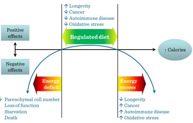
10 The project is funded by the European Union and co-financed by the European Social Fund Figure I.3-6: Hypothetical U-shaped curve over the spectrum of caloric intake
from insufficient to excessive calories, emphazing negative
physiological effects at both extremes and positive or hormetic effects within a range of normal (regulated) caloric intake ... 37 Figure I.4-1: Circulatory adaptation to chronic bed-rest ... 43 Figure I.4-2: Urinary loss of calcium and hydroxy-proline during chronic
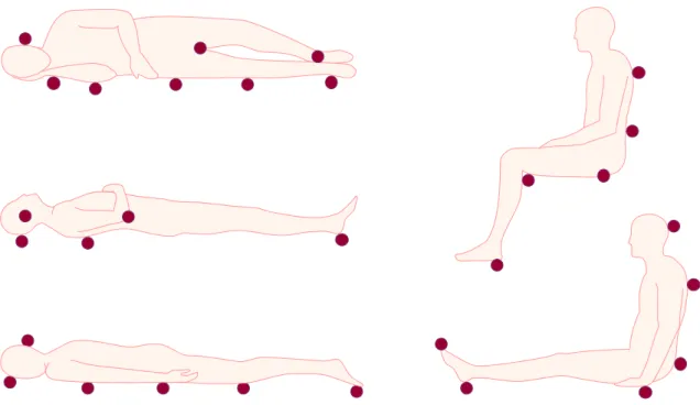
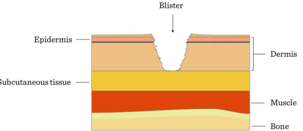
immobilization ... 44 Figure I.4-3: Typical points exposed to pressure in immobilization ... 46 Figure I.4-4: Stage I of pressure ulcer: lasting erythema on skin surface ... 46 Figure I.4-5: Stage II of pressure ulcer: superficial wound, which does not reach
subcutaneous tissues ... 47 Figure I.4-6: Stage III of pressure ulcer: deep wound affecting the subcutaneous
tissues (does not cross the fascia of the muscle) ... 47 Figure I.4-7: Stage IV of pressure ulcer: very deep wound, battering also the
muscles, bones, joints ... 48 Figure I.5-1: Age-related physiological changes in the heart ... 51 Figure I.5-2: Comparison between the early diastolic and atrial contribution to left
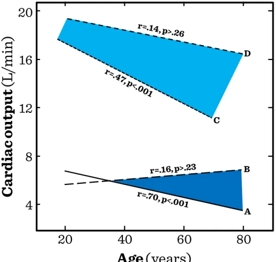
ventricular filling in persons of a broad age range ... 52 Figure I.5-3: Maximal heart rate vs. age ... 52 Figure I.5-4: Cardiac output measured at rest and at exhausting exercise (upright
position) vs. age ... 53 Figure I.5-5: Maximal oxygen consumption and endurance times according to
age. (data on trained and non-trained men) ... 53 Figure I.5-6: Mean aortic pressure and aortic pulse wave velocity vs. age in rural
and urban populations ... 54
List of figures
Identification number:
TÁMOP-4.1.2-08/1/A-2009-0011
11 Figure I.5-7: The interplay of vascular and adaptive cardiac changes during aging ... 54 Figure I.6-1: The thorax in the elderly ... 58 Figure I.6-2: Progressive loss of height from vertebral fractures causes a
protuberant abdomen and upper back curvature (“dowager’s hump”) ... 58 Figure I.6-3: Changes in airflow during aging (FEV1 = forced expiratory volume
in 1 second) ... 59 Figure I.7-1: Age-related reduction in the glomerular filtration rate. Because of
the loss of functioning nephrons, the progressive decrease of GFR
exceeds that of the renal blood flow ... 64 Figure I.7-2: Age-related changes in single nephron GFR (SNGFR) in% of total
nephrons ... 64 Figure I.7-3: Concentration and dilution are interconnected in different segments
of the nephron (purple arrows indicate active Na-reabsorption, the
green ones the passive water reabsorption) ... 65 Figure I.7-4: Changes of osmotic pressure and fluid volume along the nephron.
Without ADH, large volume of diluted urine, in case of high ADH levels small volume of concentrated urine is formed. The possible limits of dilution and concentration are determined in the loop of Henle. In case of hyposthenuria (interrupted line), the concentration- changes are moderate in the loop, and the renal concentration gradient decreases. ... 66 Figure I.7-5: Development of hyposthenuria, isosthenuria: less and less deviation
from the specific gravity of the filtrate during both concentration and
dilution ... 66
12 The project is funded by the European Union and co-financed by the European Social Fund Figure I.7-6: Age-dependence in the renal effect of ADH (the urine-plasma ratio
of inulin concentration characterizes the renal concentration process) ... 68
Figure I.8-1: Common endocrine alterations in elderly ... 77
Figure I.8-2: Premenopausal thermoregulation ... 78
Figure I.8-3: Peri/postmenopausal thermoregulation ... 78
Figure I.8-4: Benign prostatic hyperplasia ... 79
Figure I.8-5: Factors leading to frailty ... 80
Figure I.9-1: The most common gastric disorders in elderly ... 85
Figure I.9-2: Causes of lower GI tract bleeding ... 88
Figure I.10-1: Thrombolysis in stroke ... 93
Figure I.10-2: Neurotransmitter imbalance in Parkinson’s disease ... 94
Figure I.10-3: Prevalence of Alzheimer’s disease ... 96
Figure I.11-1: Illness-medication problems to which the elderly susceptible because of their medical problems ... 102
Figure I.12-1: Survival curves for male rats fed ad libitum or restricted to 60%: a 40% reduction of diet resulted in 48-months survival instead of the normal 30-months ... 107
Figure I.12-2: Factors influencing aging ... 108
Figure II.1-1: Correlation between body mass and lifespan ... 113
Figure II.1-2: Correlation of morbidity rates and age ... 114
Figure II.1-3: What is lifespan? ... 115
Figure II.1-4: Approaches in senescence research ... 116
Figure II.1-5: August Weismann’s concepts on aging ... 117
Figure II.1-6: Pioneers of aging research include ... 117
Figure II.2-1: The family tree of aging theories ... 118
List of figures
Identification number:
TÁMOP-4.1.2-08/1/A-2009-0011
13
Figure II.2-2: Major groups of aging theories ... 119
Figure II.2-3: Beyond molecular biology of aging ... 119
Figure II.2-4: Evolutionary theories of living and longevity ... 120
Figure II.2-5: Theory of antagonistic pleitropy ... 121
Figure II.2-6: Programmed theories ... 122
Figure II.2-7: General formulations ... 123
Figure II.2-8: Individual mechanisms ... 123
Figure II.3-1: Characteristics of mitochondria and mtDNA ... 124
Figure II.3-2: Mitochondrial DNA replication fork ... 125
Figure II.3-3: Genes encoded by mtDNA ... 126
Figure II.3-4: Reasons of mitochondrial vulnerability ... 126
Figure II.3-5: Reason and evidence of mitochondrial aging ... 127
Figure II.3-6: ROS and their major sources ... 128
Figure II.3-7: Mitochondrial oxygen radical theory of aging ... 128
Figure II.3-8: Mitochondrial ROS runaway ... 129
Figure II.3-9: Antioxidants ... 130
Figure II.3-10: Mitochondrial ROS production ... 131
Figure II.3-11: mtDNA oxidative damage ... 132
Figure II.3-12: mtDNA damage and hospital admission ... 133
Figure II.3-13: Mitochondrial apoptosis due to ex. stimulus ... 133
Figure II.3-14: Organ / tissue specific diseases of mt origin ... 134
Figure II.3-15: Mitochondrial diseases classified ... 134
Figure II.3-16: Diseases of mtDNA origin ... 135
Figure II.4-1: Telomeres as biological clocks ... 137
Figure II.4-2: Factors influencing telomere loss rate ... 137
14 The project is funded by the European Union and co-financed by the European Social Fund
Figure II.4-3: Further clocks ticking ... 138
Figure II.4-4: Telomere sequence and telomerase function ... 139
Figure II.4-5: Changes in telomere length ... 140
Figure II.4-6: Significance of telomere in cancer ... 140
Figure II.4-7: Cancer development and telomeres ... 141
Figure II.4-8: Acquiring immortality via telomerase ... 141
Figure II.4-9: Slowing, reversing telomere shortening ... 142
Figure II.4-10: Antagonistic pleiotropy – telomere length I ... 143
Figure II.4-11: Antagonistic pleiotropy – telomere length II ... 143
Figure II.5-1: Correlation between body mass and lifespan ... 144
Figure II.5-2: The family tree of aging theories ... 145
Figure II.5-3: Theory of antagonistic pleiotropy ... 145
Figure II.5-4: Centenarians ... 147
Figure II.5-5: Correlation of morbidity rates and age ... 147
Figure II.5-6: Molecular balance of aging and life-span ... 148
Figure II.5-7: Connection of metabolism and longevity ... 149
Figure II.5-8: Molecular pathways of aging and life-span ... 149
Figure II.5-9: Genes influencing longevity I ... 150
Figure II.5-10: Longevity genes across animal kingdom ... 150
Figure II.5-11: Genes affecting age-related diseases ... 151
Figure II.5-12: Genes influencing longevity II ... 152
Figure II.5-13: Genes influencing longevity III ... 153
Figure II.5-14: Aging genes conserved in animal kingdom ... 154
Figure II.6-1: DNA damage-triggered cell fate responses ... 155
Figure II.6-2: Molecular level senescence pathways ... 156
List of figures
Identification number:
TÁMOP-4.1.2-08/1/A-2009-0011
15
Figure II.6-3: Molecular level cell fate decisions ... 156
Figure II.6-4: Tumor suppressor genes ... 157
Figure II.6-5: Cancer stem cells escape routine elimination ... 158
Figure II.6-6: Malignant tumor escape mechanisms I ... 158
Figure II.6-7: Malignant tumor escape mechanisms II ... 159
Figure II.6-8: p53 has ambivalent talents I ... 160
Figure II.6-9: p53 has ambivalent talents II ... 161
Figure II.6-10: p53 polymorphisms in cancer and longevity ... 162
Figure II.6-11: Antagonistic pleitropy: p53 and p16 ... 163
Figure II.6-12: Cancer epidemiology worldwide ... 164
Figure II.6-13: Cancer statistics ... 165
Figure II.7-1: DNA damage: causes, results I ... 167
Figure II.7-2: Oxidative DNA damage ... 167
Figure II.7-3: DNA damage: causes, results II ... 168
Figure II.7-4: Non-oxidative DNA damage ... 168
Figure II.7-5: Non-oxidative protein damage ... 169
Figure II.7-6: Oxidative DNA damage repair types I ... 170
Figure II.7-7: Oxidative DNA damage repair types II ... 170
Figure II.7-8: Genes related to oxidative DNA damage repair ... 171
Figure II.7-9: Genomic instability in progeria types ... 172
Figure II.7-10: Oxidative DNA damage repair and aging ... 172
Figure II.7-11: Werner syndrome ... 173
Figure II.7-12: Cockayne syndrome ... 174
Figure II.7-13: Hutchinson-Guilford progeria syndrome ... 175
Figure II.8-1: CR increases life-span ... 177
16 The project is funded by the European Union and co-financed by the European Social Fund
Figure II.8-2: Lifespan increase due to CR ... 177
Figure II.8-3: CR extends life-span ... 178
Figure II.8-4: CR extends life-span via… ... 178
Figure II.8-5: Insulin / IGF signaling pathway ... 179
Figure II.8-6: Proof of GH / IGF signaling axis in aging ... 179
Figure II.9-1: Protein peroxidation ... 182
Figure II.9-2: Repair following protein peroxidation ... 183
Figure II.9-3: Protein peroxidation and diseases ... 184
Figure II.9-4: Lipid peroxidation ... 185
Figure II.9-5: PUFA controversy: AA and DHA ... 185
Figure II.10-1: The mechanism of action for sirtuins ... 187
Figure II.10-2: Features of Sir2 family ... 187
Figure II.10-3: Acetylation status and epigenetic silencing I ... 188
Figure II.10-4: Acetylation status and epigenetic silencing II ... 188
Figure II.10-5: Sirtuins as regulators for aging ... 189
Figure II.10-6: Sirt1 as regulator for aging I ... 190
Figure II.10-7: Sirt1 as regulator for aging II ... 190
Figure II.10-8: Sirt1 and stress resistance ... 191
Figure II.10-9: Properties of other mammalian sirtuins ... 192
Figure II.10-10: Redox signaling pathway ... 192
Figure II.10-11: TOR signaling pathway ... 193
Figure II.10-12: Resveratrol increases life-span ... 194
Figure II.10-13: Resveratrol ... 194
Figure II.10-14: Resveratrol / paclitaxel combination in cancer ... 195
Figure II.10-15: Mechanism of action for GH / IGF pathway ... 195
List of figures
Identification number:
TÁMOP-4.1.2-08/1/A-2009-0011
17
Figure II.10-16: Environmental effects in expected life-span ... 196
Figure II.10-17: Sirtuin switch in ad libitum and CR mice ... 196
Figure II.10-18: Sirt1 and CR ... 197
Figure II.11-1: Post-translational life of proteins ... 198
Figure II.11-2: Protein modifications due to stress ... 199
Figure II.11-3: Non-oxidative DNA damage ... 199
Figure II.11-4: Non-oxidative protein damage ... 200
Figure II.11-5: Modulation of non-oxidative protein damage ... 200
Figure II.11-6: Transcriptional and translational dysregulation in aging ... 201
Figure II.11-7: Lipufuscin, lysosomal waste ... 202
Figure II.11-8: Neuronal EC Aβ plaques and their effects ... 203
Figure II.11-9: Amyloid fibril development and growth ... 203
Figure II.11-10: Amyloid fibrils by AFM ... 204
Figure II.11-11: Amyloid deposits by histology ... 204
Figure II.11-12: Proteasome function ... 205
Figure II.11-13: Proteasome function in aging ... 206
Figure II.11-14: CR and non-oxidative protein damage ... 206
Figure II.11-15: Proteasome function in CR ... 207
Figure II.11-16: Proteasome function in immune senescence ... 207
Figure II.11-17: Proteasome function in neuronal senescence ... 208
Figure II.11-18: Autophagy and IC breakdown ... 209
Figure II.11-19: Prion protein conversion ... 209
Figure II.11-20: Histology in prion protein-related diseases ... 210
Figure II.11-21: Hutchinson-Guilford progeria syndrome ... 210
Figure II.11-22: Composition of nuclear envelope ... 211
18 The project is funded by the European Union and co-financed by the European Social Fund
Figure II.11-23: Nuclear envelope-related instability ... 211
Figure II.12-1: SENS ... 212
Figure II.12-2: SENS: planned interventions ... 213
Figure II.12-3: Limitations of SENS ... 214
Identification number:
TÁMOP-4.1.2-08/1/A-2009-0011
19
I Gerontology
I.1 Basics of gerontology, demographic data
I.1.1 Introduction, definitions
Gerontology (from Greek: Géron = “gray”, “old man”, logos = “study of”) is the study of the biological, psychological and social aspects of normal aging. It is distinct from geriatrics, which is the branch of medicine that studies the characteristic diseases of the elderly or age-related changes in diseases that already began in the young.
Biogerontology is a sub-field of gerontology studying the biological processes of aging.
It is composed of the interdisciplinary research on the causes, effects and mechanisms of biological aging, in order to achieve better understanding of human senescence. The huge increase in the elderly population in post-industrial Western nations has made biogerontology one of the most rapidly growing fields.
I.1.2 Population-wide aging
The worldwide prolongation of life expectancy has resulted in a rapid increase in the size of elderly populations (over the age of 65), both in absolute numbers and relative to the whole. Survival has increased with the passage of time since earlier historic periods (epochs) and shows larger improvements in more developed countries. Age-specific mortality has decreased. However, the age maximum ever achieved by human beings has not changed. (Figure I.1-1 and Figure I.1-2)
20 The project is funded by the European Union and co-financed by the European Social Fund Figure I.1-1: Survival curves for different populations
Figure I.1-2:
Age-specific death rates of Swedish females from 1751 to 1950 and for 1988
20 40 60 80
20 40 60 80
United States (1970)
1,100 BC Europe
15,000 yrs ago
Africa 50,000 yrs ago
Age (years)
P e rce n t su rv iv al
Death/1000/yearSweden (females)
Years
100 75
50 25
0 0.5
1 10 100 500
1751 - 1790 1851 - 1860 1901 - 1910 1920 - 1930 1941 - 1950 1988
Basics of gerontology, demographic data
Identification number:
TÁMOP-4.1.2-08/1/A-2009-0011
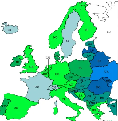
21 Regional differences in mortality or in life expectancy at birth are generally determined by the combination of a huge number of different factors. These differences are known for many regions but are usually compared among nations (Figure I.1-3).
One of the very few exceptions is Germany: here the survival conditions are not uniformly distributed over the whole national territory. The East-West differences are due to the special history of these two regions having belonged to completely different political and social regimes for several decades during the last century. The difference remains high despite the political reunion in 1990 (Figure I.1-4).
Figure I.1-3: Expected life-span at birth in different European states 64 – 67
67 – 70 70 – 73 73 – 75 75 – 78 78 – 80 80 – 82 82 – 84 84 – 86 86 – 88
IS
NO
RU
UA
MD RO
BY LV
TK BG MK CS HR
HU SK
LT EE
AL SL
CZ PL DK
PT IE
FI
UK BE
NL LU
DE
AT
IT
BA
ES GR
SE
FR CH
22 The project is funded by the European Union and co-financed by the European Social Fund Figure I.1-4:
Regional pattern of life expectancy in Germany: East-West difference (2003)
In Europe the number of people over 65 years will increase from 15.5% in 2000 to 24.3% in 2030. These demographic changes have major implications for health care, the labor force, welfare, insurance, and pensions.
I.1.3 Chronological and biological age
“How old would you feel if you did not know how old you were?”
These two numbers are not necessarily the same. The functional-biological age is determined by physiology rather than chronology. Factors include changes in the physical structure of the body as well as changes in the performance of motor skills and sensory perception.
life expectancy at birth (years)
males
life expectancy at birth (years)
females
<74 74-75 75-76 76-77
>77
<81 81-81.5 81.5-82 82-82.5
>82.5
Berlin
Frankfurt M.
Köln
München
100 km
Hamburg Hamburg
Berlin
Frankfurt M.
Köln
München
100 km
Basics of gerontology, demographic data
Identification number:
TÁMOP-4.1.2-08/1/A-2009-0011
23 I.1.4 Etiology of aging: genetic mechanisms and environmental factors Aging is a complex process that affects all living organisms. Animals living in the wild are less likely to live long enough to encounter aging, but interestingly all mammalian species (including humans) show very similar aging processes if kept under optimal conditions free from external risk factors like predators or famine. The aging process is multi-factorial, and no single factor has been identified which provides a satisfactory explanation of the phenomenon. During the process of aging, the organism accumulates damage to macromolecules of its own cells and tissues and to its organs. The maximum lifespan for humans is around 120 years, whereas the maximum lifespan of a mouse, commonly used in research as a model for aging, is about four years. Genetic differences between humans and mice that may account for these different rates of aging include efficiency of DNA repair, types and quantities of antioxidant enzymes, and different rates of free radical production.
Mutation rate in humans is about 1 per 107-11 base pair. Chromosome abnormalities, demethylation, as well as defects of protein synthesis also influence aging. The acceptable rate of mistakes (faulty aminoacids in the peptide chain) in protein synthesis is about 5/10.000 amino acids. Elongation factor-1 levels are also low in old populations, just as levels of some types of mRNA, e.g. mRNA for IL-1. The telomere is a region of repetitive DNA sequences at the end of chromosomes, which protects the end of the chromosome from damage during cell division. The telomere regions prevent the degradation of genes near the end of chromosomes by allowing for the inevitable shortening of the chromosome, which necessarily occurs during cell division. This telomere shortening mechanism normally allows cell lines only a fixed number of divisions. Animal studies suggest that this mechanism contributes to aging on a cellular level and sets a limit to lifespan. (It has been described that a certain cell
24 The project is funded by the European Union and co-financed by the European Social Fund type of a certain species is capable of only a certain number of cell divisions: Hayflick phenomenon.) Changing telomere lengths is usually associated with changing rate of senescence. This telomere shortening, however, might be a consequence of, and not a reason for aging.
Maximum lifespan of a species is determined by the rate of aging, inherent in its genes and also by environmental factors, i.e. high metabolic rate, free radical production, excessive caloric intake leading to high serum glucose level.
Further reading
Holliday R.: Understanding Ageing, Cambridge Univ. Press, Cambridge, New York, 1995
Arking R.: Biology of Aging. Sinauer Assoc.Inc, Sunderland, 1998
Merck Manual of Geriatrics, Eds.: M.H Beers, R. Berkow, MSD Labs, Merck & Co.
Inc., Rahway, N.J., 2000
Molecular and Cellular Gerontology. Eds.: Toussaint O., Osiewacz H., Lithgow G., Brack C., Annals NY Acad. Sci. Vol.908, New York, 2000
Healthy Aging for Functional Longevity – Molecular and Cellular Interactions in Senescence. Eds.: Sang Chul Park, Eun Seong Hwang, Hyun-Sook Kim, Woon- Yang Park, Annals NY Acad. Sci. Vol.928, New York, 2001
Adaptation and vulnerability, polymorbidity
Identification number:
TÁMOP-4.1.2-08/1/A-2009-0011
25
I.2 Adaptation and vulnerability, polymorbidity
I.2.1 Progressive deficit in adaptative homeostatic mechanisms in the course of aging
Deficient adaptive functions as well as morphological alterations contribute to age- related disorders observed in aged individuals. Young adults are characterized by maximal performance, maximal reserve capacity, optimal achievement of maximal performance via optimal utilization of capacities/ resources. In old age-groups maximal performance of various homeostatic systems decrease to a variable degree due to limited adaptive mechanisms.
Resting physiological normal values of young adults are easy to determine and they show small individual variability. In the course of aging individual variability expands to a great extent, with a small decline in successfully aging individuals and very pronounced falls in enhanced biological aging. Therefore, it is more difficult to determine the “normal range” of parameters of any given homeostatic parameter in older age-groups (Figure I.2-1). Additionally, different homeostatic systems may present different rates of decline within the same individual e.g. prematurely failing vision or graying hair may be observed in people with well-preserved cardio-respiratory fitness.
26 The project is funded by the European Union and co-financed by the European Social Fund Figure I.2-1: Age-related changes in different functions
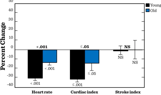
Although most homeostatic systems may function properly under resting conditions e.g. cardiac output, ventilation, or regulation of serum glucose levels, adaptation to enhanced demands (physical exercise) or to changes in external environmental factors (heat or cold) or those of the internal milieu is very limited (Figure I.2-2, Figure I.2-3).
Age (years)
H y p o th e ti c fu nc ti o n (% )
20 100
50
0 30 40 50 60 70 80 90
Adaptation and vulnerability, polymorbidity
Identification number:
TÁMOP-4.1.2-08/1/A-2009-0011
27 Figure I.2-2: Effect of 50 mmHg increment in systolic blood pressure on heart rate,
cardiac index and stroke index in young and old rats
Figure I.2-3: Glucose tolerance tests (50 g glucose p.o.) in different age-groups
In contrast to the maximal performance, maximal reserve capacity, optimal achievement of maximal performance via optimal utilisation of capacities observed in
Pe r c e n t C h a n g e
Young Old 50
10
-40 40 30 20
0 -10 -20 -30
Heart rate Cardiac index Stroke index
< .001 ≤.05 NS
NS NS
≤.05
≤.001
< .001
< .001
Time(min)
Blood sugar(mM)
0 5 6 7 8 9 10 11 12
20 30
40 50 60
70
Age(years)
20 0
60 90 120 150
* 50 g glucose p.o.
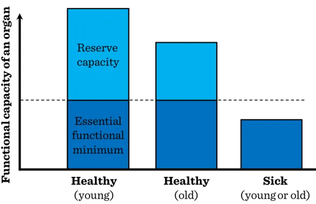
28 The project is funded by the European Union and co-financed by the European Social Fund young adults, reserve capacities of old individuals are significantly diminished (Figure I.2-4). Healthy old individuals may well be able to maintain the basic function of vital organ systems, such as the resting cardiac output (CO) of 5 L/min, their capacity to increase the CO in response to physical activity or in a hot or cold environment is very limited. Such limitations of reserve capacities make old individuals very vulnerable to environmental stress leading to decompensation of the circulation or to exacerbations of chronic diseases. Limited reserve capacities of numerous organ systems within the same individual will also aggravate complications of diseases, the symptoms of which may be ameliorated by the compensation of other organ systems, e.g. anemic tissue hypoxia will become more severe in old age-groups with limited capacity to maintain a hyperdynamic circulation.
Figure I.2-4: Functional and reserve organ capacities in young and elderly
Healthy (old)
F u n c ti ona l c a pa c ity of a n or g a n
Healthy (young)
Sick (young or old) Essential
functional
minimum
Reserve
capacity
Adaptation and vulnerability, polymorbidity
Identification number:
TÁMOP-4.1.2-08/1/A-2009-0011
29 In the elderly, a very delicate balance exists among different organ systems.
Disruption of homeostasis by any disease in previosly independent, functional elderly persons is likely to be expressed in the most vulnerable, most delicately balanced systems. Therefore, a disease in older persons manifests itself first as functional loss, usually in organ systems unrelated to the locus of illness. For example, it has long been recognized that disorders may present in a masked or apathetic form and inflammatory intra-abdominal disorders such as appendicitis may not evoke typical symptoms and signs. However, it is far less recognized that presenting symtoms in the elderly may be totally misleading with regard to the nature and the primary location of the disease process. For example, when the patient becomes confused, one thinks immediately of psychoactive drugs or disease processes primarily affecting the brain as a possible cause. In an elderly person, one must also consider such diversive factors as dehydration due to a wide variety of etiologies, infection, cardiac disorders, or intra-abdominal organ disease. In short, the diagnostic logic is different.
Geriatrics has described five entities, so-called geriatric giants that are the major categories of impairment appearing in elderly people leading to serious impairmnent of their quality of life. These include immobility (instability), incompetence (impaired intellect/memory), incontinence, impaired homeostasis, iatrogenic disorders (Figure I.2-5).
30 The project is funded by the European Union and co-financed by the European Social Fund Figure I.2-5: Geriatric giants
I.2.2 Polymorbidity in the elderly
Polymorbidity and related polypragmasia contribute to the development of iatrogenic disorders. The majority of elderly individuals suffer of a large number of chronic diseases affecting various homeostatic and organ systems. Age-related changes in body composition lead frequently to osteoporosis and sarcopenia, diminished insulin sensitivity in the elderly aggravated by age-dependent accumulation of fat mass frequently culminate in type 2 diabetes mellitus, long-term dust and or smoke-exposure result in chronic obstructive lung diseases or silicosis, progressive atherosclerosis will lead to infarctions or chronic atrophy of the myocardium or stroke, to name just a few.
For these diseases and abnormalities old individuals take a large number of drugs regularly.
Incompetence (Confusion) Immobility
(Falls) Incontinence
Iatrogenic disorders Impaired
homeostasis
Adaptation and vulnerability, polymorbidity
Identification number:
TÁMOP-4.1.2-08/1/A-2009-0011
31
Further reading
Geriatric Medicine. Eds.: C.K. Cassel C.K., D.E.Riesenberg, L.B.Sorensen, J.R.Walsh, Springer-Verlag, New York, Berlin, 1990.
Pharmacological Intervention in Aging and Age-associated Disorders. Eds.: K. Kitani, A. Aoba, S. Goto, Annals NY Acad. Sci. Vol. 786, New York, 1996.
R. Arking: Biology of Aging. Sinauer Assoc.Inc, Sunderland, 1998.
Merck Manual of Geriatrics, Eds.: M.H Beers, R. Berkow, MSD Labs, Merck & Co.
Inc., Rahway, N.J., 2000
E.R. Hajjar, A.C. Cafiero, J.T. Hanlon, Polypharmacy in elderly patients. Am. J.
Geriatr. Pharmacother. 5: 345-51. 2007.
32 The project is funded by the European Union and co-financed by the European Social Fund
I.3 Nutrition, physical status, body composition, sarcopenia
I.3.1 Introduction: age-related changes in body weight and body composition
Aging is accompanied by two major trends in the long-term regulation of energy balance: obesity of the middle-aged and late-appearing anorexia of aging often leading to senile cachexia and sarcopenia. Following a progressive increase in adiposity (Figure I.3-1) first a relative, then an absolute decrease in muscle mass is seen, pointing to the development of sarcopenia (decrease of muscle mass by more than 30%) in aged populations (above the age of 70). At the very end of the aging process adipose tissue is also lost in the process of cachexia. Both of these opposite disorders have enormous impact on the health status and life expectancy of those affected. Not only the consequences of obesity (metabolic syndrome) are serious, but the cachexia of old people as well: it causes muscle weakness, falls, frailty, functional and later cognitive disorders, a higher risk for decubitus (pressure ulcer) and hip fracture, impaired quality of life, a 3-4-fold increase in the risk of loss of self-reliance (expensive assisted living facilities in old age) and higher mortality. They are important especially in Hungary:
although the increase in the ratio of the extreme old population is not fast, in the old groups the rate of biological aging is faster, than in more developed countries. Both the weight gain of the middle aged and the sarcopenia of the elderly are multifactorial in their origin (Figure I.3-2).
Nutrition, physical status, body composition, sarcopenia
Identification number:
TÁMOP-4.1.2-08/1/A-2009-0011
33 Figure I.3-1: Between the ages of 20 and 70 – despite a stable, normal body weight – body composition is altered: fat mass increases (a 2 fold increase is still considered to
be physiological)
Figure I.3-2: The pathogenesis and functional vs. metabolic consequences of sarcopenia
I.3.2 Changes in fat mass (FM) and fat free mass (FFM) with age
Body weight increases gradually by 8-9 kg until 45-55 (this is predominantly an increase in FM with maintained muscle mass), then after a stagnation until the age of
Body Fat Ranges for Standard Adults 1
1 Based on NIH/WHO BMI Guidelines.
Underfat Healthy Overfat Obese
Underfat Healthy Overfat Obese
0% 10% 20% 30% 40%
Female20-39 Age40-59 60-79
Male20-39 Age40-59 60-79
SARCOPENIA Decreased
food intake
Impaired ADLS Malnutrition
Cytokines
Increased oxidative stress
Low testosterone, estrogen, GH, IGF-1
Decreased physical activity
GH= growth hormone;
IGF-1= insulin-like growth factor-1;
BMR= basal metabolic rate;
ADLS = activities of daily living
Decreased walking speed
Impaired
balance Osteoporosis Falls
Decreased BMR
Impaired thermoregulation
Decreased physical activity
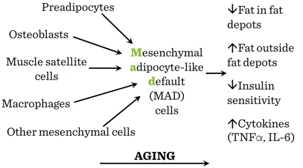
34 The project is funded by the European Union and co-financed by the European Social Fund 65-75, a decline (1-2 kg/decade) in all tissue types is seen without any apparent cause e.g. a slimming diet. During this period even the FM is decreased somewhat but the loss of muscle mass is dominant. In active athletes the body weight does not increase, the increase of FM with age is blunted (their body fat content is similar to that of young, lean, sedentary individuals). Intensive training decreases abdominal fat. Males have a tendency for visceral fat accumulation, after menopause females too. Fat accumulation does not only mean a simple growth of adipose tissue, but abnormal mesenchymal adipocyte-like default (MAD) cells appear among other cell types e.g. between muscle fibers, in the bone marrow (Figure I.3-3). Redistribution of lipid to extra-adipose sites with aging could result from loss of lipid storage capacity in fat depots (reduced fat cell size and function), altered fatty acid handling resulting in lipid accumulation, maldifferentiation of mesenchymal precursors into a partial adipocyte phenotype (due to falls of testosterone and IGF, elevated cytokine production and anorexia).
Figure I.3-3: Ectopic fat accumulation with aging: MAD cells are smaller and less insulin responsive than fully differentiated fat cells
Mesenchymal adipocyte-like
default (MAD) cells
Fat in fat depots
Fat outside fat depots
Insulin sensitivity
Cytokines (TNF α , IL-6) Preadipocytes
Osteoblasts Muscle satellite
cells Macrophages
Other mesenchymal cells
AGING
Nutrition, physical status, body composition, sarcopenia
Identification number:
TÁMOP-4.1.2-08/1/A-2009-0011
35 The FFM is stable until 40, then it decreases by about 3.5 kg or 3-4%/decade (Figure I.3-4). This change shows small individual differences, the rate of decrease is similar in athletes.
Figure I.3-4: Fat (f), fat-free mass (ffm), and cell mass (cm) of males and females at various ages. (The number of subjects in each age-group is noted.)
The water content of the body changes proportionally with FFM. Water content of the FFM is stable. Bone minerals also change proportionally with FFM. By 65 it decreases by 10-15%. In females the rate of decrease is enhanced after menopause. This dramatic fall can be prevented by estrogen supplemetation. In active athletes the rate of decrease is similar, but starts from a higher peak bone mass. Muscle mass and strength diminishes slowly until 50, then the rate is enhanced (sarcopenia). Between 30 and 80 there is a 30-40% decrease also in athletes. Especially the quick, dynamic contractions are impaired. The number of motoneurons/motor units fall. The production of muscle proteins decreases, especially that of type II fibers.
Fat , fat-free mass, and cell mass of males () and females () at various ages, values are given, and the number of subjects in each age group is noted.
27 58 33
37 42
18
89 33 44
72 54 13
18–25 25–35 35–45 45–55 55–65 65–85
Age(years)
Absolute weight(kg)
10 20 30 40 50 60
36 The project is funded by the European Union and co-financed by the European Social Fund I.3.3 The pathogenesis of sarcopenia
Weight loss seen in the elderly may be associated to anorexia of aging: between 20-70 years of age the basal metabolic rate decreases by less than 20%, on the other hand daily caloric intake decreases by as much as 35%. In the elderly, the etiology of poor nutrition includes social (such as poverty and isolation), psychological (especially depression) and physical factors (immobilization or missing teeth), abnormal states (heart failure, malignancies, GI abnormalities, chronic inflammation, infections, drugs, etc.). A large number of cases anorexia develops without any apparent reason (real age-related anorexia). The process is similar in all mammals thus the alterations of energy balance may be of regulatory origin (Figure I.3-5). Individual components of the regulatory systems (e.g. transmitters influencing feeding drive or satiety) may change according to different dynamics, that may explain the abnomalities of the energy balance in middle aged people and in the elderly.
Figure I.3-5: Body weight and adiposity index in rats Energy
deficit
Energy excess
Longevity
Cancer
Autoimmune disease
Oxidative stress Positive
effects
Negative effects
Longevity
Cancer
Autoimmune disease
Oxidative stress
Parenchymal cell number Loss of function
Starvation Death
Regulated diet
↑Calories
Nutrition, physical status, body composition, sarcopenia
Identification number:
TÁMOP-4.1.2-08/1/A-2009-0011
37 Both insufficient and excessive caloric intake exerts negative physiological effects (Figure I.3-6). It has been observed that among middle-aged and old people the severely undernourished persons showed higher mortality rates than the overweight ones. What is more, among the elderly a slight overweight of 10-20% indicated better survival. The higher body mass index among the elderly means a higher amount of muscle mass, that may reach sufficiently high levels to promote survival. To achieve such a higher body weight with acceptable muscle mass a diet relatively richer in proteins and special muscle (strength) training is suggested.
Figure I.3-6: Hypothetical U-shaped curve over the spectrum of caloric intake from insufficient to excessive calories, emphazing negative physiological effects at both extremes and positive or hormetic effects within a range of normal (regulated) caloric
intake
B o dy W e ig h t or A d ip osity I n dex
Age (mo)
3 12 18 24 30
100 200 300 400 500 600
Adiposity Index Body Weight
38 The project is funded by the European Union and co-financed by the European Social Fund
Further reading
Geriatric Medicine. Eds.: C.K. Cassel C.K., D.E.Riesenberg, L.B.Sorensen, J.R.Walsh, Springer-Verlag, New York, Berlin, 1990
Towards Prolongation of the Healthy Life Span. Eds.: D. Harman, R. Holliday, M.
Meydani, Annals NY Acad. Sci. Vol. 854, New York, 1998
Merck Manual of Geriatrics, Eds.: M.H Beers, R. Berkow, MSD Labs, Merck & Co.
Inc., Rahway, N.J., 2000
Healthy Aging for Functional Longevity – Molecular and Cellular Interactions in Senescence. Eds.: Sang Chul Park, Eun Seong Hwang, Hyun-Sook Kim, Woon- Yang Park, Annals NY Acad. Sci. Vol.928, New York, 2001
E. Pétervári, S. Soós, M. Székely, M. Balaskó: Alterations in the peptidergic regulation of energy balance in the course of aging. Curr. Protein Pept. Sci. (2011)
Immobilization, physical activity, disorders of locomotor organs
Identification number:
TÁMOP-4.1.2-08/1/A-2009-0011
39
I.4 Immobilization, physical activity, disorders of locomotor organs
I.4.1 The beneficial effects of physical exercise
In affluent societies the general level of physical activity progressively decreases. This is unfortunate, since physical activity has many advantages regarding physiological functions. It helps to reach or maintain normal, healthy body mass. In active athletes, for example, the usual body weight rise of middle-aged persons is absent. Regular sport improves body composition, it increases the amount of muscle mass (especially the amount of type-I, slow, red fibers), the ratio of active tissues, attenuates their age- dependent natural loss. Training programs (12 weeks – 3 times a week) may be able to increase the available muscle mass by 10% even in old age-groups. The greater ratio of active tissues enhances the basal metabolic rate. The trained muscle, during prolonged work (longer than 15-20 min) burns fat. On the surface of muscle fibers lipoprotein- lipase appears, which is able to release fatty acids from the circulating lipoproteins.
Regular physical activity suppresses the total-cholesterol level, but elevates the serum HDL-cholesterol concentration. Active skeletal muscles are able to take up glucose by an insulin-independent mechanism. Physical activity recruits GLUT 4 glucose transporter molecules to the surface of muscle cells, just as insulin does. Thus, regular activity decreases the insulin requirement, attenuates the strain on β-cells, and helps to prevent type 2 diabetes mellitus. The thermal adaptation capability improves. In working muscles epinephrine induces vasodilation, thereby it decreases total peripheral resistance, therefore regular exercise helps preventing the development of essential hypertension. Regular sports in children and young adults increases peak bone mass by
40 The project is funded by the European Union and co-financed by the European Social Fund applying traction forces on the bones (piezoelectric effect, activating bone formation).
The regular physical activity shifts bone metabolism to greater synthesis even at later ages. Regular sports can help in the prevention of osteoporosis, which would carry the dangers of vertebral compression, pathological fractures, fracture of the hip or the neck of the femur. Physical activity has an effect to decrease stress. It has no negative side- effects, and does not evoke pathological dependence, either. According to human surveys, regular physical activity can attenuate the appearance of depression and dementia, which often develop with advancing age. Data obtained in a mouse-model of experimental Alzheimer disease have shown that appropriate increase of physical activity favorably modifies the level of brain-derived neural growth factor (which is a contributing factor in hippocampal atrophy), and also the amyloid formation. A physically active lifestyle decreases the occurrence of certain cancers (colon, breast, uterus, esophagus, prostate) partly by maintaining normal body weight, partly through humoral factors. It influences insulin sensitivity, the level of insulin-like growth factors and estrogen.
I.4.2 Immobilization syndrome – chronic bedrest
In the course of some diseases a short-time bed rest has some advantages: this rest decreases the burden of the cardiovascular and respiratory systems. In febrile illnesses as part of sickness behavior, the patients do not have only fever, anorexia, decreased fluid intake, enhanced pain-sensation, lethargy, but they are also feeble and inactive, often somnolent. However, chronic bed-rest is harmful rather than advantageous. Some patients cannot avoid chronic immobilization. Loss of lower limbs, paralysis of the lower half of the body due to transversal lesion of the spinal cord or stroke, coma, extreme weakness, severe pains in the joints and severe chronic diseases (e.g. chronic
Immobilization, physical activity, disorders of locomotor organs
Identification number:
TÁMOP-4.1.2-08/1/A-2009-0011
41 heart failure, COPD), extreme obesity, rheumatic polymyalgy, hypothyroidism may also lead to immobilization. The incidence and the danger of immobilization are especially high in the elderly.
As many as 1/3 of older persons report yearly a fall or tendency to fall, which is the most common cause of accidents in people over 65 years of age and is the leading cause of mortality due to injury in that age-group. Complications include hip fractures, subdural hematomas and immobility. As a major public health problem, osteoporosis (metabolic bone disorder characterized by a gradual decline in absolute bone mass) also increases susceptibility to fractures especially in the vertebral bodies, the distal radius, and the proximal femur. Bone mass decreases from the age of 55 by around 1%/year in men and by 3-4%/year in women (peak bone mass is reached at 25-35 years of age, its value is higher in men). Inactivity, vitamin D and protein deficiency, hormonal factors (e.g. lower estrogen, secondary hyperparathyroidism, cortisol), alcohol, smoking and certain drugs may accelarate the age-related progressive reduction of bone mass.
Difficulty and unsteadiness in walking, with occasional falls, and stiffness with painful lower limbs are frequently reported by elderly patients and are often related to degenerative joint disease, rheumatoid arthritis or polymyalgia rheumatica.
Osteoarthritis is the most common form of joint disease and one of the leading causes of disability in persons above 65 years of age. As a person ages, the water content of the cartilage decreases as a result of a modification of proteoglycan content, thus causing the cartilage to be less resilient. Without the protective effects of the proteoglycans, the collagen fibers of the cartilage can become susceptible to degradation and thus exacerbate the degeneration. Cellular or matrix alterations in cartilage that occur with aging, obesity, trauma, endocrine diseases (e.g. diabetes mellitus) and primary disorders
42 The project is funded by the European Union and co-financed by the European Social Fund of the joint (e.g. inflammatory arthritis) predispose older persons to osteoarthritis characterized by progressive joint pain, limitation of movement and joint deformity.
Central and peripheral nervous system disorders (late stage of Parkinson disease or neuropathies) may present with motor symptoms. Cardiovascular, respiratory, endocrine, other systemic illnesses, or dementia, depression, isolation, fear of falling, anxiety, with fatigue and lack of motivation for activities of daily living, often limit exercise performance in the elderly, frequently without intrinsic muscle weakness.
Drugs, e.g. sedatives, narcotics (because of sedative effect), diuretics, antihypertensive medication (in the elderly these may cause orthostatic hypotension, dizziness) also enhance the danger of immobilization.
Consequences of a chronic bed-rest depend on the duration and level of inactivity. In prolonged supine position (as in weightlessness) the circulation is rearranged, on the short run the central blood volume increases, the perfusion and hydrostatic pressure decrease in the lower half of the body, the slightly higher preload and stroke volume lead to bradycardia, renal blood flow increases and slight polyuria develops. On the long run (weeks, months), the plasma volume and the efficacy of orthostatic reflexes (regulating blood pressure) decrease. When the patient is mobilized again, the low blood volume is not enough to maintain brain blood flow in an orthostatic position, therefore orthostatic hypotension develops, the patient is dizzy, eventually faints (Figure I.4-1).
Immobilization, physical activity, disorders of locomotor organs
Identification number:
TÁMOP-4.1.2-08/1/A-2009-0011
43 Figure I.4-1: Circulatory adaptation to chronic bed-rest
Muscle contractures develop (muscles and joints are less moveable). In case if the upper extremities are affected, the elderly patients lose the capability to eat alone, in case of lower extremities one contracture is enough to cause full immobility. A decrease of muscle mass can be observed already on the short-term, but upon a long-term bed- rest it is significantly enhanced. Immobilization greatly enhances the progression of pre- existing osteoporosis in elderly patients. The enhanced excretion of hydroxy-proline is a sign of increased muscle (protein) catabolism, while the Ca-excretion refers to bone absorption. (Figure I.4-2).
Earth gravity Normal condition
Zero gravity Acute exposure
Zero gravity Chronic exposure
Earth gravity Upon return Central venous
pool
44 The project is funded by the European Union and co-financed by the European Social Fund Figure I.4-2: Urinary loss of calcium and hydroxy-proline during chronic
immobilization
The maximal capacity for physical work will not be determined by the capacity of the cardiovascular system (as it normally happens), but by the exhaustion of the muscular system, or lack of local substrates (glycogen). Formation of red blood cells decreases, the low level of total ventilation also lessens the amount of oxygen carried by the arterial blood. The atrophied, deconditioned muscles of poor perfusion take up less oxygen from the blood. The decreased blood volume, decreased muscle tone and mass (decreased filling) and decreased baroreceptor response act to suppress stroke volume.
Following chronic bed-rest any physical activity evokes an exaggerated cardiovascular response, e.g. palpitation may appear already at work of low level (and low oxygen consumption). Due to the decreased venous return and hypovolemia the risk for deep venous thrombosis and pulmonary embolism is high. In the elderly population they enhance mortality by about 50%.
Time(weeks)
Control Bedrest
Calcium
Change in urinary calcium(mg/day)
-10 -5 0 5 10 15 20
150
100
50
0
-50
OH-Proline
Change in urinary hydroxyproline(mg/day)
20
10
0
-10
Immobilization, physical activity, disorders of locomotor organs
Identification number:
TÁMOP-4.1.2-08/1/A-2009-0011
45 The ventilation decreases, the V/Q mismatch becomes pronounced, and the activity of the immune system and the mucociliary clearance of the airways become insufficient. Elderly patients, if bed-ridden for only a couple of days, may develop congestive pneumonia. Surgical fixation of a fractured neck of femur is indicated mainly by faster mobilization and avoiding the pneumonia mortality induced by long (earlier advised for 9 weeks) bed-rest.
In chronic bed-rest the metabolic rate may be 20% lower than normally. The defense against either heat or cold is weaker. Immobilization decreases intestinal motility. The tendency for constipation increases significantly, even impactation may develop, eventually with consequent fecal incontinence.
The prevalence of pressure ulcer is 30% among elderly patients who are bed- ridden or have to stay in a wheel-chair for at least a week. At the points exposed to pressure the skin and the deeper tissues may be damaged in the course of prolonged sitting or lying (Figure I.4-3). Immobilization, fecal-, urinary-incontinence, hypoalbuminemia and shear stress due to the incompetent turning/moving of the patient contribute to the development of pressure ulcers (pressure ulcer staging: Figure I.4-4, Figure I.4-5, Figure I.4-6, Figure I.4-7). Decubitus causes 4-fold increase in the mortality rate of the patients (sepsis).
46 The project is funded by the European Union and co-financed by the European Social Fund Figure I.4-3: Typical points exposed to pressure in immobilization
Figure I.4-4: Stage I of pressure ulcer: lasting erythema on skin surface Epidermis
Subcutaneous tissue
Muscle Dermis
Bone Reddened area
Immobilization, physical activity, disorders of locomotor organs
Identification number:
TÁMOP-4.1.2-08/1/A-2009-0011
47 Figure I.4-5: Stage II of pressure ulcer: superficial wound, which does not reach
subcutaneous tissues
Figure I.4-6: Stage III of pressure ulcer: deep wound affecting the subcutaneous tissues (does not cross the fascia of the muscle)
Epidermis
Subcutaneous tissue
Muscle Dermis
Bone Blister
Epidermis
Subcutaneous tissue
Muscle Dermis
Bone Blister
48 The project is funded by the European Union and co-financed by the European Social Fund Figure I.4-7: Stage IV of pressure ulcer: very deep wound, battering also the muscles,
bones, joints
I.4.3 Remobilization in the elderly
Those people who are confined to stay inactive because of an acute disease or are bedridden due to chronic conditions are highly prone to lose their muscle mass and force very quickly. The proportion of the loss can even reach 1.5% per day. The loss is more pronounced in the muscles responsible for sitting up, standing up and standing straight, and therefore, these muscles are essential for everyday life. Gradual mobilization, passive movement and active exercising of joints on a regular basis, proper positioning of patient (prevention of pressure ulcers), replacement of fluids, optimal feeding, regular emptying of bladder, removal of catheter as soon as possible, cleaning of skin and active environment are important therapeutic measures. Certain specialists in geriatric medicine state that one day spent in bed can be compensated by a 2-week workout. Therefore, a personalized exercise program and care must be worked out for every hospitalized, chronically ill patient in order to maintain their physical activity. Maintenance of physical activity as long as possible in the elderly is essential via resistance training and daily activity- and work-oriented special exercises. The ideal frequency, intensity, duration and style of such physical activity have not been fully
Epidermis
Subcutaneous tissue
Muscle Dermis
Bone Blister