Effects of 17 β-estradiol
and glucagon-like peptide-1 on
gonadotropin-releasing hormone neurons in mice
PhD Dissertation
Flóra Bálint
Scientific advisors:
Imre Farkas PhD, Zsolt Liposits, MD, DSc
Pázmány Péter Catholic University Faculty of Information Technology and Bionics Roska Tamás Doctoral School of Sciences and Technology
Budapest, 2018
TABLE OF CONTENTS
LIST OF ABBREVIATIONS ... 4
INTRODUCTION ... 6
Regulation of the hypothalamo-pituitary-gonadal axis ... 6
Properties of the GnRH neuronal system ... 6
The gonadotropin-releasing hormone ... 7
The development, distribution and structural properties of GnRH neurons ... 8
The efferent projections of the GnRH neurons ... 10
Synaptic regulation of GnRH neurons ... 11
The main electrophysiological properties of GnRH neurons ... 12
GnRH neuron-related feedback mechanisms in reproduction ... 13
Classical and non-classical estrogen receptor signaling pathways ... 14
Influence of estrogen on functions of GnRH neurons ... 15
Metabolic signals ... 18
Glucagon-like peptide-1 signaling ... 19
The retrograde neurotransmission ... 21
The endocannabinoid system ... 21
The nitric oxide system ... 23
SPECIFIC AIMS ... 25
EXPERIMENTAL PROCEDURES ... 26
Experimental animals ... 26
Brain slice preparation and recording ... 26
Whole-cell patch clamp experiments ... 28
Loose-patch clamp experiments ... 30
Chemicals and reagents ... 31
Real-time PCR detection of Glp1r and Nos1 in GnRH neurons ... 32
Statistical analysis ... 33
RESULTS RELATED TO THE ESTRADIOL EFFECT DURING NEGATIVE FEEDBACK ... 35
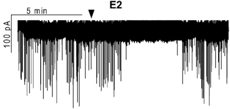
Estradiol significantly decreases the firing rate and frequency of spontaneous and
miniature postsynaptic currents in GnRH neurons of metestrous female mice ... 35
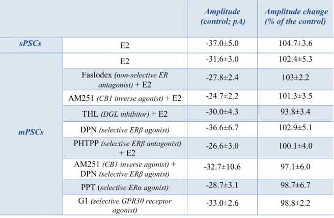
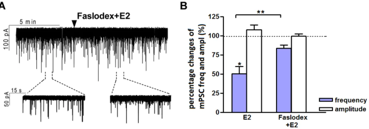
The direct rapid effect of estradiol requires estrogen receptor beta ... 37
Retrograde endocannabinoid signaling is involved in estradiol-triggered decrease of miniature postsynaptic currents... 40
RESULTS RELATED TO THE GLP-1 EFFECT IN GnRH NEURONS... 42
The GLP-1 agonist Exendin-4 increases the firing rate and the frequency of miniature postsynaptic currents of GnRH neurons via GLP-1 receptor ... 42
Nitric oxide and 2-arachidonoylglycerol signaling mechanisms are involved in the action of Exendin-4 on GnRH neurons ... 46
The retrograde 2-AG pathway is regulated by anandamide-TRPV1 signaling ... 50
DISCUSSION... 52
Estradiol suppresses the firing rate and frequency of postsynaptic currents in GnRH neurons in metestrous female mice ... 52
Estrogen receptor beta is required for the direct, rapid effect of estradiol on GnRH neurons ... 53
Retrograde endocannabinoid 2-AG signaling is involved in the estradiol-triggered reduction of miniature postsynaptic current frequency in GnRH neurons ... 54
GLP-1 is excitatory to GnRH neurons via GLP-1 receptor ... 57
Effect of GLP-1 is mediated partially by activation of the nitric oxide retrograde signaling on GnRH neurons ... 58
GLP-1 acts partially via retrograde endocannabinoid signaling pathway of GnRH neurons ... 59
The nitric oxide and endocannabinoid retrograde signaling pathways are simultaneously involved in the effect of GLP-1 ... 60
NEW SCIENTIFIC RESULTS ... 63
ACKNOWLEDGEMENTS ... 65
REFERENCES ... 66
BIBLIOGRAPHY ... 80
LIST OF ABBREVIATIONS
2-AG - 2-arachidonoylglycerol
aCSF - artificial cerebrospinal fluid
AEA - anandamide (N-arachidonoylethanolamide)
AM251 - N-(piperidin-1-yl)-5-(4-iodophenyl)-1-(2, 4-dichlorophenyl)- 4-methyl-1H-pyrazole-3-carboxamide
AMG9810 - (E)-3-(4-t-butylphenyl)-N-(2,3-dihydrobenzo[b][1,4] dioxin- 6-yl) acrylamide
AMPA -alpha-amino-3-hydroxy-5-methyl-4-isoxazole propionic acid
cAMP - cyclic adenosine monophosphate
CB1 - cannabinoid receptor type 1
CPTIO - 2-(4-Carboxyphenyl)-4,4,5,5-tetramethylimidazoline- 1-oxyl-3-oxide
CREB - cAMP response element binding protein
DAG - diacylglycerol
DGL - diacylglycerol lipase
DPN - diarylpropionitrile
E2 - 17β-estradiol
EGTA - ethylene-glycol-tetraacetic acid
ER - estrogen receptor
ERα - estrogen receptor alpha
ERβ - estrogen receptor beta
Ex4 - Exendin-4
FAAH - fatty acid amide hydrolase
FSH - follicle-stimulating hormone
G1 - [(3aR*,4S*,9bS*)-4-(6-Bromo-1,3-benzodioxol-5-yl)- 3a,4,5,9b-tetrahydro-3H-cyclopenta[c]quinolin-8-yl]-ethanon
GABA - gamma-aminobutyric acid
GABAA-R - gamma-aminobutyric acid receptor type A GDP-β-S - guanosine 5'-[β-thio] diphosphate
GFP - green fluorescent protein
GLP-1 - glucagon-like peptide-1
GLP-1R - glucagon-like peptide-1 receptor
GnRH - gonadotropin-releasing hormone
GPCR - G-protein-coupled receptor
GPR30 - G-protein-coupled receptor 30
GPR54 - G-protein-coupled receptor 54
GT1-7 - immortalized gonadotropin-releasing hormone neuron cell line HEPES -4-(2-hidroxietil)-1-piperazin-etánszulfonsav(4-(2-hydroxyethyl)-
1-piperazineethanesulfonic acid) HPG axis - hypothalamo-pituitary-gonadal axis
KA - kainate
KO - knockout
KP - kisspeptin
L-NAME - Nω-Nitro-L-arginine methyl ester hydrochloride
LH - luteinizing hormone
ME - median eminence
NPLA - Nω-Propyl-L-arginine hydrochloride
mGluR - metabotropic glutamate receptors
mPSC - miniature postsynaptic current
NMDA - N-methyl-d-aspartate
NO - nitric oxide
NOS - nitric oxide synthase
NST - nucleus of the solitary tract
OVLT - organum vasculosum of the lamina terminalis
PCR - polymerase chain reaction
PF3845 - N-3-Pyridinyl-4-[[3-[[5-(trifluoromethyl)-
2-pyridinyl]oxy]phenyl]methyl]-1-piperidinecarboxamide PHTPP - 4-[2-Phenyl-5,7-bis(trifluoromethyl)pyrazolo[1,5-a]pyrimidin-
3-yl] phenol
PPT - propylpyrazoletriol
PSC - postsynaptic current
RP3V - rostral periventricular area of the third ventricle
sGC - soluble guanylyl cyclase
sPSC - spontaneous postsynaptic current
THL - tetrahydrolipstatin
TRPV1 - transient receptor potential vanilloid 1
TTX - tetrodotoxin
vGlut - vesicular glutamate transporter
Vrest - resting membrane potential
INTRODUCTION
The survival of species depends on the capability of adaptation and success of reproduction.
Disorders affecting the units of the reproductive system can result in infertility. In mammals, reproduction is controlled by the hypothalamo-pituitary-gonadal axis (HPG axis) [1, 2]. Disorders affecting the reproductive axis result in infertility.
Regulation of the hypothalamo-pituitary-gonadal axis
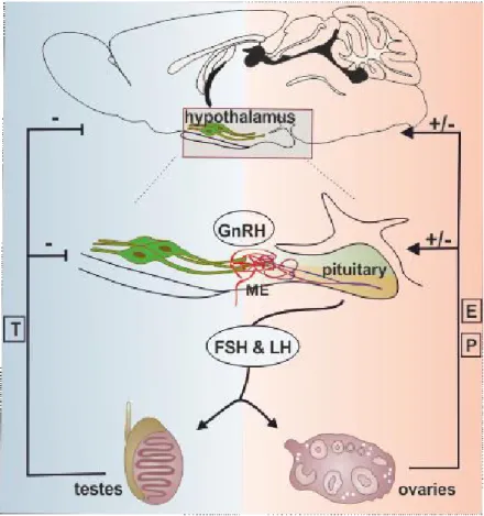
There are complex and precisely regulated interactions between the different units of the HPG axis via hormone messengers. The HPG axis controls the ovarian cycle, sexual development, maturation and aging [1-5]. Gonadotropin-releasing hormone (GnRH; also called luteinizing hormone- releasing hormone) synthesizing neurons of the hypothalamus form the key central elements of the HPG axis [1, 2, 6]. The hypophysiotropic axons of GnRH neurons release GnRH into the fenestrated capillaries of the hypophyseal portal circulation in a pulsatile manner [7]. GnRH neurohormone reaches its target cells in the anterior pituitary gland via the long portal veins. Here, the episodically released GnRH stimulates its receptors on the surface of the gonadotroph cells, that results in a rhythmic discharge of gonadotropins, the follicle-stimulating hormone (FSH) and luteinizing hormone (LH) [2, 8]. FSH and LH are released into the systemic circulation and act on the gonads:
FSH stimulates the maturation of ovarian follicles in females, the spermatogenesis in males and stimulates gonads to produce steroids. LH stimulates secretion of sex steroids from the gonads in both sexes. The gonadal steroids, in turn, exert negative and positive feedback actions on the hypothalamus and the pituitary, and regulate the synthesis of GnRH and the two gonadotropins [2, 9, 10] (Figure 1.). Gonadal steroid hormones also have a number of other important physiological effects upon almost all of the organs, including the brain.
Properties of the GnRH neuronal system
GnRH neurons play a decisive role in the regulation of the HPG axis and thus, in the control of reproduction. In this chapter, the properties and regulation of GnRH neurons will be discussed focusing on structural organization of the GnRH system, the operating signaling mechanisms of GnRH neurons and the molecular background of information processing.
Figure 1. Relationship between the regulatory units of the reproductive axis. Hypothalamic GnRH neurons release GnRH neurohormone into the hypophyseal portal circulation. Via this portal system GnRH reaches its target cells in the anterior pituitary and regulates the synthesis and secretion of the gonadotropins, such as follicle-stimulating hormone (FSH) and luteinizing hormone (LH). FSH and LH are released into the systemic circulation and act on the gonads to stimulate gonadal steroid secretion. Sexual steroid hormones, such as estrogen (E), progesterone (P), and testosterone (T) influence the hypothalamic and pituitary hormone secretions via feedback loops. Abbreviation: median eminence (ME).
The gonadotropin-releasing hormone
GnRH molecule was discovered in 1971 by Roger Guillemin and Andrew Schally [11]. Since the original discovery, different forms of GnRH have been found in vertebrates. GnRH-l is a decapeptide (pyroGlu-His-Trp-Ser-Tyr-Gly-Leu-Arg-Pro-Gly-NH2). The gene encoding GnRH1 is on chromosome 8. GnRH-1 is a neurohormone, the hormone itself is synthetized in specific neurons of olfactory placode origin and is released at their terminals. The hypophysiotropic GnRH-1
population regulates the synthesis and release of pituitary gonadotropins [12]. The GnRH-1 neurosecretory system and the structure of GnRH-1 are evolutionarily conserved in vertebrates. In all mammals the amino acid sequence of GnRH-1 is the same [12]. The scope of this dissertation has been the hypophysiotropic GnRH-1 neurons (hereafter GnRH) and their involvement in the regulation of the HPG axis.
There are two additional GnRH molecules that form distinct populations in the brains of several species. GnRH-2 and GnRH-3 differ at several amino acids from GnRH-1. GnRH-2 is often referred to as chicken GnRH and GnRH-2 neurons are present in the midbrain tegmental area [12]. GnRH- 3 can be found in the telencephalon of fish, amphibians and some mammals. This hormone is sometimes referred as salmon GnRH [12].
The development, distribution and structural properties of GnRH neurons
The GnRH neurons originate from the olfactory placode and migrate into the forebrain along the olfactory-vomeronasal nerves from the gestational day 11 in mice [13-16]. This phenomenon can be observed not only in rodents, but in humans [17], rhesus macaques [18], and chicken [19].
Disorders in the migration process can cause hypogonadotropic hypogonadism which is characterized by the lack of hypothalamic GnRH neurons in the forebrain leading to reproductive deficiencies. The human disease is called Kallmann syndrome, which often coincides with anosmia [20]. Migrating GnRH neurons settle down in the hypothalamic/median preoptic area by the time of birth [14]. According to the present literature there are approximately 800 GnRH neurons in the rodent brain, 1000-2000 in primates brain and their numbers are equal in both sexes [21-24].
Surprisingly, these small amount of GnRH neurons are sufficient to carry out the control of reproduction [25].
GnRH neurons have fusiform morphology. The perikarya of the cells are relatively small (10-15 µm). Being bipolar neurons GnRH cells have two processes emerging from the cell body: two dendrites or one dendrite and one axon. However, sometimes axon emanates from one of the main dendrites [26-28].
GnRH neurons form anatomical and functional networks. Communication among the cells is mainly achieved through axo-somatic and axo-dendritic connections [29], however other types of communication have also been observed. Some studies showed tight junctions between GT1-7 immortalized GnRH neurons [30]. The other connection type observed between GnRH nerve cells is formed by continuous intercellular bridges, where the cytoplasmic domains communicate with
each of them [31, 32]. These connections between GnRH neurons have a crucial role in the regulation of the synchronized operation of the system.
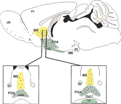
Figure 2. Distribution of the GnRH neurons in mice.
Sagittal (top) and coronal (bottom) views of the distribution of GnRH neuron cell bodies (black dots). These neurons located mainly in the diagonal band of Broca (dbb), medial septum (MS) and the preoptic area (POA). Abbreviations:
anterior commissure (ac); median eminence (ME); optic chiasm (oc); organum vasculosum of lamina terminalis (OVLT). The figure is based on the image of Dr. Michel Herde [33].
GnRH neurons are located in a relatively broad area in the brain. In rodents GnRH cells show a scattered distribution pattern, these neurons are located principally in the medial septum, the diagonal band of Broca, the medial and rostral preoptic area, in the vicinity of the organum vasculosum of the lamina terminalis (OVLT) [34, 35] (Figure 2.).
The efferent projections of the GnRH neurons
The major efferent targets of the GnRH neurons are two circumventricular organs: the median eminence (ME) and the organum vasculosum of lamina terminalis (OVLT) [15, 16]. The ME is a functional interface between the hypothalamus and the anterior pituitary gland. Hypothalamic neurons release peptides and small molecule neurotransmitters into portal vessels. The walls of the capillaries are fenestrated to ensure maximal hormone permeability, the axons of hypophysiotropic neurons terminate around this capillary network. The portal capillaries are reunited forming the long portal veins [36]. The ME has three parts: the ependymal layer, the internal zone and the external one. From 50% to 70% of all GnRH axons terminate in the external zone of the ME where GnRH is released into the portal capillaries in a pulsatile manner [7, 9, 37].
Besides the hypophysiotropic axon projections, GnRH fibers also innervate the OVLT. The OVLT is another sensory circumventricular organ of the brain, located along the ventral part of the anterior wall of the third ventricle [38]. It is made up of neurons, glial cells and ependymal cells. The ependymal cells form tight junction connections near their apical surfaces, which avert the free flow of the cerebrospinal fluid from the ventricle into the brain parenchyma. Being a circumventricular organ, OVLT contains a rich vascular network containing specialized fenestrated capillaries [38].
Multiple projections of GnRH neurons in the region of the OVLT and ME permit the sensing of peripheral signals due to the lack of the blood-brain barrier here [38].
There are less studied extrahypothalamic GnRH axon projections in addition to the above- mentioned ones in several areas of the brain. Although these cells make up 30 percent of all GnRH neurons, these non-hypophysiotropic GnRH cells and projections are rarely investigated [7]. A recent study showed that this kind of GnRH neurons communicate with approximately 50,000 neurons in 53 functionally diverse brain areas [39]. Moreover, a significant number of GnRH neurons are located in the medial septum and the olfactory tubercle, which innervate the olfactory bulb [35]. This suggests that the GnRH system may have a role in the transmission or modulation of olfactory stimuli, which is closely related to some reproductive functions and behavior [39].
Regarding the specific phenotype of GnRH efferent system, it has been shown that GnRH neurons express mRNA of vesicular glutamate transporter-2 (vGlut2) in rats [40]. This vesicular transporter is obligatory for accumulating glutamate into synaptic vesicles, thus it indicates that GnRH neurons are glutamatergic suggesting the possibility that GnRH neurons could release glutamate from their nerve terminals. Nevertheless, recently it has been also demonstrated that a subset of GnRH neurons is GABAergic in the mouse brain [41] indicating the chemical heterogeneity of GnRH neurons.
Synaptic regulation of GnRH neurons
One of the most significant regulatory neurotransmitters in the central nervous system, as well as in the hypothalamus, is γ-aminobutyric acid (GABA). GABA is the main neurotransmitter acting on GnRH neurons [42], as major proportion of synaptic contacts on GnRH neurons is GABAergic [43].
GnRH neurons express the ionotropic GABAA [44-46], and the metabotropic GABAB [47]
receptors. The observed GABAA receptor-mediated postsynaptic currents [45, 46] also confirm the GABAergic input to these neurons. Although GABA is usually an inhibitory neurotransmitter in the adult central nervous system, early data seemed controversial whether GABA stimulates [45, 48] or inhibits [49] GnRH neurons. Now, it has been widely accepted that most mature GnRH neurons are excited by GABA [50-52]. The intracellular chloride ion concentration is responsible for determining the polarity of GABA response [53]. The hyperpolarizing (generally inhibitory) action of GABA exists when intracellular chloride ion concentration is low whereas the depolarizing (generally excitatory) action of GABA occurs when intracellular chloride ion concentration is high, such as in GnRH neurons. The excitation by the GABAA receptor activation is due to the high intracellular chloride level of GnRH neurons. On one hand this high intracellular chloride level is due to the absence or low expression levels of the K-Cl cotransporter 1 in GnRH neurons. This cotransporter excludes chloride ion from the cytoplasm in neurons. On the other hand high expression of the Na-K-Cl cotransporter 2 in GnRH neurons is responsible for maintaining high chloride level in GnRH neurons, since this cotransporter mediates inward transport of chloride ion [45]. GABAA receptor is a ligand-gated chloride channel. Activation of this receptor leads to the opening of the channel and results in chloride ion efflux and neuronal depolarization in GnRH neurons.
As mentioned above GnRH neurons also express the metabotropic GABAB receptor [47, 54]. The functional GABAB receptor is a G-protein-coupled receptor linked to potassium channels or even calcium channels [55]. The receptor is a heterodimer formed by a GABA B1 and B2 subunit.
Activation of GABAB receptors triggers inhibitory postsynaptic currents leading to reduced neuronal excitability of GnRH neurons [55]. GABA exerts its inhibitory effect via GABAB receptors on GnRH neurons [47, 56].
The major GABAergic innervation of GnRH neurons is supposed to arise from local GABAergic interneurons [10]. GABAergic inputs play major roles in the mediation of metabolic [46], circadian [57] and estrogen [58] signals to the GnRH system [57].
Another major neurotransmitter in the afferent control of GnRH cells is glutamate [59]. The markers of glutamate-secreting nerve terminals are the vesicular glutamate transporters (vGlut1, vGlut2 and
vGlut3). GnRH neurons receive synapses from vGlut2 containing glutamatergic neurons and these inputs were observed mostly on the dendritic compartment of GnRH cells [59]. Consistent with this, GnRH neurons express all ionotropic glutamate receptors, the alpha-amino-3-hydroxy-5-methyl-4- isoxazole propionic acid/kainate (AMPA/KA) and N-methyl-d-aspartate (NMDA) receptors [44, 58, 60]. Whereas GABAA receptor mediated postsynaptic currents can be easily detected in all GnRH neurons, not all neurons exhibit postsynaptic currents mediated by these glutamate receptors [58, 60]. However, it should be noted that currents generated on the distal dendrites might be insufficient to be detected on the perikaryon [27], but may still have important role in the cell function. Whole-cell patch clamp studies reveal that glutamate transmission predominantly mediated by AMPA/KA receptors, but NMDA mediated postsynaptic currents were also observed in GnRH neurons [44, 58]. Activation of these ionotropic glutamate receptors contributes to the pulsatile [61] and also to the surge [62] release of GnRH.
In addition to the fast neurotransmission, slower glutamatergic neuromodulation is also present in GnRH neurons, for which metabotropic receptors are responsible. Metabotropic glutamate receptors (mGluRs) are G-protein-coupled receptors (GPCRs). These can be grouped into three groups (Group I, II and III). Group I mGluRs are localized postsynaptically on neurons or glial cells.
Activation of these receptors increases intracellular Ca2+ level. The presynaptically localized Group II/III mGluRs mediate feedback inhibition [63]. Bath application of Group II/III mGluR agonists decreased the frequency of GABAergic events on GnRH neurons. The mGluRs responsible for this synaptic inhibition are suggested to be located on the presynaptic GABA terminals and not on the GnRH neurons themselves [64].
The main electrophysiological properties of GnRH neurons
Our experiments were performed on GnRH-green fluorescent protein (GFP) transgenic mice, in which GnRH promoter is linked to a green fluorescent reporter molecule, allowing the microscopic detection of GnRH neurons in slices preparations [65], as the expression of the reporter is restricted to these neurons. Thus, GnRH neurons were visualized using fluorescence microscopy and the identified individual neurons were used for electrophysiological recordings and subsequent structural, molecular analysis. It is important to note that the main electrophysiological properties of GFP expressing GnRH neurons [44, 65], do not differ from the wild type cells using either brain slices [66], primary cell cultures [66, 67] or immortalized GnRH neuronal cell line (GT1) [68, 69].
Resting membrane potential (Vrest) ranges approximately -55 to -65 mV in GnRH-GFP cells [44, 65]. The input resistance shows the integrity of the cell, reflects the extent to which membrane
channels are open. Under physiological conditions the input resistance of GnRH neurons is high, approximately 1.60 GΩ [65]. This means that these cells have relatively few channels open at the resting membrane potential. The high input resistance also indicates that small currents can have a large impact on membrane potential.
GnRH neurons present periodic, spontaneous action potentials which could be blocked by tetrodotoxin (TTX) indicating that these are mediated by voltage-gated sodium channels [65]
because TTX can inhibit the firing of action potentials by binding to voltage-gated sodium channels.
The episodic, not continuous firing means that GnRH neurons showed quiescent periods with intermittent action potentials [65, 66]. The spontaneous action potentials were observed with an amplitude of >60 mV (76.9 ± 5.5 mV) [65].
Based on the firing patterns of adult GnRH neurons there are three different populations observed in acute brain slices. Cell-attached recordings revealed that majority of the GnRH neurons exhibit burst firing (∼65%), another population remains silent, and the last one is a smaller group exhibiting continuous activity. Note that, in vivo experiments showed that only ∼15% of GnRH neurons exhibit burst firing in mice [70]. These heterogenous firing patterns are observed in both gonadectomized and intact male and female mice. [66].
GnRH neuron-related feedback mechanisms in reproduction
The delicate balance of coordinated signals among the hypothalamus, pituitary gland and the gonads is strongly related to the precise function of GnRH neurons. The pattern of GnRH release forms the final output signal of the hypothalamus towards the pituitary gland. Thus, the secretion of gonadotropins (LH, FSH) from the anterior pituitary gland is influenced by hypothalamic GnRH pulse frequency and amplitude. GnRH pulses occur every 30-90 minutes and both the frequency and amplitude are crucial for normal gonadotropin release [9]. Low GnRH pulse frequency is required for FSH production and release, whereas high GnRH pulse frequency stimulates LH synthesis and release [9]. Furthermore, pulsatile GnRH secretion is indispensable to prevent the desensitization of GnRH receptors and thus to maintain the hormone sensitivity of neurons of the pituitary gland. Gonadotropins activate gonadal steroid hormone synthesis and these gonadal steroids exert negative (in both females and males) or positive (exclusively in females) feedback actions on the central components of the HPG axis (Figure 1.).
The negative feedback loop is the common regulatory mechanism in both sexes. In males sex steroids suppress GnRH neuron activity and GnRH release. After puberty, the testicular hormone genesis is continuous. FSH stimulates the spermatogenesis, while LH stimulates Leydig cells in
testes to produce testosterone. High levels of androgens exert a constant, direct/indirect inhibitory action on the hypothalamic GnRH neurons and pituitary via acting on the androgen receptors [71].
The GnRH and LH are secreted in pulsatile manner in males [72]. The orchidectomy of male animals and the testosterone treatment in females during the so-called critical postnatal period (postnatal day 5-10) has demonstrated that testosterone surge is responsible for the development of sexual dimorphism [73], including in brain structures. Consequently, testosterone and estrogen (which originates from testosterone by aromatase enzyme) have constant inhibitory effect on the reproductive axis through the androgen and estrogen receptors, respectively in adult rodents. The positive feedback does not occur in mature male animals.
Female animals have a cyclic pattern of reproductive function which is called estrus or ovarian cycle. A cycle lasts four days in mice and is characterized by four stages: proestrus, estrus, metestrus and diestrus. The pattern of GnRH and gonadotropin release and thus blood estrogen levels vary throughout the different stages. During the major part of the ovarian cycle (throughout the estrus- diestrus phase) the relatively low level of estrogen (approximately 10 pM) exerts negative feedback effect on the hypothalamus and pituitary. This means estrogen reduces GnRH pulse amplitude and frequency, leading to the suppression of LH release in the pituitary, thereby, repressing its own follicular synthesis [8, 74]. The negative feedback action of estrogen is modulated by peptides produced by the ovaries such as inhibin A, inhibin B, activin or follistatin [75, 76].
In proestrus, mature follicles show a dramatic increase in their estrogen secretion and a modest elevation in progesterone production. The response to this sustained elevating estradiol level is involved in the switch from negative to positive feedback in the hypothalamus. The positive feedback effect of estradiol initiates GnRH surge, a large increase in the volume of GnRH release [57, 77]. This can be explained by a fold elevation of amplitude and frequency of the GnRH secretory pulses [78]. Meanwhile, the responsiveness of the gonadotropin cells to GnRH increases [79] causing a surge in LH release from the anterior pituitary gland, initiating ovulation in the ovary [8, 77].
Classical and non-classical estrogen receptor signaling pathways
Estrogens in females are produced primarily by the ovaries, but the whole enzyme set is also expressed in neurons and astrocytes for estrogen synthesis in the central nervous system [80]. Sex steroids, including the three major natural estrogen – estrone (E1), estradiol (E2) and estriol (E3) – all derive from cholesterol through different steps of enzymatic reactions [80]. The major product from the whole biosynthesis process is the 17β-estradiol (E2) and it is the primary biologically
active and prevalent form of estrogen. E2 is produced by aromatase from testosterone or is converted from estrone at the end of the biosynthesis process [81]. E2 is one of the principal regulators of GnRH cells and acts as a classic, homeostatic feedback molecule between gonads and brain.
Estradiol is critical in controlling GnRH neurons to exhibit fluctuating patterns of biosynthetic and secretory activity [10].
The actions of estrogen on neurons are mediated by estrogen receptors (ER). Estradiol mainly interacts with two types of classical estrogen receptors, ERα and the ERβ, each encoded by a separate estrogen receptor gene (ESR) 1 and 2, respectively. These receptors belong to the superfamily of nuclear receptors [82, 83]. E2 exerts its effect through two major signaling pathways:
the genomic (classical, nuclear-initiated) signaling [84, 85] and the acute, non-genomic (non- classical, membrane-initiated) signaling [85-89]. In classical, genomic ER signaling pathway estrogen diffuses passively across the cell membrane then binds and activates ERs [82, 83].
Activation of ERs leads to a conformational change of the receptors and subsequent receptor dimerization. The two receptor types can form ERα (αα) or ERβ (ββ) homodimers or ERαβ (αβ) heterodimers. The dimeric receptors enter the nucleus and subsequently bind to the estrogen response element on the promoter regions of target genes leading to gene activation or repression [82, 83].
Additionally, estrogen is capable to bind and activate receptors associated with the plasma membrane, and thus exerts a rapid, direct effect (non-genomic, non-classical signaling), such as rapid increase in cAMP, or altered firing of neurons within seconds [85-89]. Receptors that are responsible for rapid action of estrogen are called extranuclear ERs. These ERs are associated with signaling complexes in the plasma membrane [90]. Most of the rapid effects of estrogen can be induced by selective ERα or ERβ agonists [91] or antagonized by the ER antagonists [92]. The rapid effects of estrogen are absent in ER mutant animals, since estrogen had no effect on an important target of the rapid estrogen action: the cyclic adenosine monophosphate (cAMP) response element binding protein (CREB) phosphorylation in double ERαβKO mice [93]. In addition to ERα and ERβ, other types of membrane associated receptors might also participate in the rapid estrogen effects, such as G-protein-coupled receptor 30 (GPR30) found in primate [94] and mouse [95]
GnRH neurons.
Influence of estrogen on functions of GnRH neurons
Rapid action of E2 effectively modulates neuronal functions. Several studies have reported the non- genomic effect of estradiol in GnRH neurons. Estradiol increased the phosphorylation of CREB
[89]. The action of estrogen was rapid (<15 min) on CREB phosphorylation, indicating a non- genomic mechanism [89]. It has also been demonstrated that CREB within GnRH neurons is an important target for estrogen negative feedback actions [96]. In addition, estradiol increased calcium oscillations [86] and potassium currents [97-99] in GnRH neurons. ERβ has a role to play in mediating acute estrogen actions. Moreover, an in vivo study revealed that GnRH neurons respond to estrogen in a rapid manner through an ERβ-dependent mechanism in the mouse [89].
Nevertheless, the role of ERβ in GnRH neuron function is far from the fully elucidated.
Regarding the rapid effect of low physiological estradiol levels, it has been demonstrated, that estradiol (10 pM) inhibited GnRH neurons firing in a rapid manner when the fast synaptic GABA and glutamate receptors were left intact [100]. This indicates the involvement of fast neurotransmission in the rapid effect of estradiol and suggests an effect upstream of GnRH neurons.
As mentioned above, E2 can inhibit (negative feedback) or stimulate (positive feedback) GnRH release, which depends on estradiol concentration and the physiological state of the body. Estradiol feedback mechanisms alter the synaptic transmission to GnRH neurons and their intrinsic excitability [45, 100, 101]. The way that estradiol changes GnRH neuron functions is different in the two feedback mechanisms. During positive feedback action high physiological (preovulatory) concentrations of estradiol (approximately 100-200 pM in rodents) has a great influence on GnRH neurons and pituitary gonadotrophs to generate the preovulatory LH surge [8, 10, 57, 77]. According to a popular hypothesis, positive steroid feedback is achieved via presynaptic interneurons that are estradiol-sensitive. Estradiol may act via various ERs, mainly the ERα and ERβ. These receptors can act either on the DNA as transcription factors [84, 85] or estradiol is able to initiate membrane- associated signaling cascades via membrane-associated receptors [85-89].
One of the estradiol-sensitive presynaptic systems acting upstream the GnRH neurons during positive feedback is the kisspeptin (KP) neuron system. A population of KP neurons form a compact nucleus in the rostral periventricular area of the third ventricle (RP3V) in the mouse brain. The RP3V population is proposed to mediate the positive feedback of estrogen [85]. This is indicated by the fact that administration of KP shows a profound increase in serum gonadotropin levels via the stimulation of the secretory activity of GnRH neurons [102]. KP is the endogenous ligand of G- protein coupled receptor 54 (GPR54) [103]. GPR54 is highly expressed in GnRH neurons [102].
Mutations of KP [104] and GPR54 [105] cause hypogonadotropic hypogonadism with partial or moderate puberty in humans, while over activation of this system causes puberty praecox [106].
The direct effect of KP-producing neurons on GnRH neurons is supported by numerous observations. It was shown that KP axons innervate the perikaryon and dendrites of GnRH neurons
[107] and they respond to KP with increased neuronal activity [108]. The RP3V population has role in estrogen positive feedback [85], as KP neurons that contact GnRH neurons have been shown to express ERα and these neurons in the RP3V release KP in response to estrogen action [85].
During negative feedback mechanism low physiological level of estradiol (~10 pM) represses gonadotropin secretory activity in females by inhibiting hypothalamic GnRH secretion [10, 100].
Changes in the function of GnRH neurons are thought to be mediated by estradiol-sensitive afferents at this stage of the cycle, and the population of KP neurons located in arcuate nucleus are defined as the main regulator of estrogen negative feedback [85, 109]. ERα was detected exclusively in various synaptic afferent systems such as KP neurons of arcuate nucleus regulating GnRH neurons, but importantly GnRH neurons themselves do not express this receptor type [109-114]. Thus, the regulation of the negative feedback is more complex, several brain regions, cell types, and estrogen receptors could be involved in suppressing the activity of GnRH neurons. The important fact that GnRH neurons express the ERβ [112, 115-117] as a direct target of estradiol feedback further increases complexity of this system.
Data in the literature seems to be controversial regarding the role of the ERβ in the negative feedback. Experiments from female mice of different ERβKO mutant mouse lines [118-120]
showed a range of reproductive phenotypes from mild subfertility [119] to complete infertility [120]. Examinations in global ERKO mice have shown the importance of ERα, and possibly ERβ, in reproductive regulation [113]. However, investigations in global knockout mice do not allow to make conclusions on a fine scale, and effects could be compensated by unknown mechanisms. In addition, neuron-specific deletion of ERα and ERβ in mice suggested that ERα seems to be essential for acute E2 negative feedback while ERβ particularly in GnRH neurons appears to be less critical [110]. These data suggest that ERβ may not be critical for central estradiol negative feedback of the HPG axis. In contrast, gonadotropin levels are less increased in ERα knockout versus the double (ERαβ) knockout mice, indicating that ERβ may still have a role in negative feedback of the HPG axis [121]. In addition, the exclusive role of the ERα in the negative feedback is questioned by a recent study in which KP-ERα knockout mice failed to show LH surges in response to estradiol but retained responsiveness to the negative feedback effects of estradiol [111]. Homozygous ERβKO female mice demonstrated subnormal fertility and had slightly elevated basal LH levels which suggests defective estrogen negative feedback [122]. These results indicate that estrogen negative feedback actions can be mediated by mechanisms that are independent of ERα and thus these other pathways may normally function as parts of the negative feedback mechanism. The fact that the ERβ is expressed in GnRH neurons suggests the physiological relevance and raises the possibility
of the direct role of the ERβ in feedback regulation. Thus, in this dissertation, I define one of the mechanisms present in negative feedback of E2.
Metabolic signals
Reproduction is an energy-demanding process which is related to the metabolic state of the body.
The reproductive success of an individual is tightly linked to the nutritional state. Both obesity and malnutrition have been reported to disrupt reproduction. This means, if the correct utilization of metabolic resources is not ensured (as in the case of anorexia nervosa), or on the contrary, if there is a constant energy surplus available (such as obesity) the body must serve its priorities to obtain the physiological state ensuring survival. For instance, neuronal activity or blood circulation cannot be compromised, whereas thermoregulation, or growth can be reduced in a somewhat wider scale in metabolic stress.
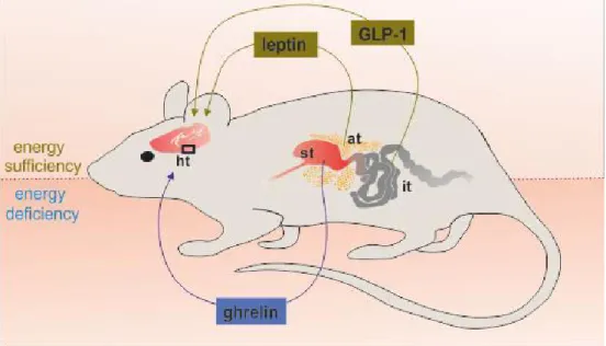
This reproductive-metabolic connection requires a coordinated action of many central and peripheral regulators. Changes in energy homeostasis trigger fluctuations in hormonal (for example leptin, insulin and ghrelin) and nutritional (for instance glucose, lipids) signals that feedback mainly to the brain regions which regulates metabolism and fertility. These actions modulate function of different levels of the HPG axis, enabling the close cooperation between the energy level and gonadal function [123] (Figure 3.). However, the effects of metabolic signals on GnRH neurons are mostly unknown.
The adipocyte hormone leptin is a signal of energy sufficiency, suppresses feeding and increases energy expenditure [124]. The central effect of leptin was demonstrated by the experiment in which leptin receptor was ablated in a forebrain specific manner and as a result of it, mice became obese and infertile [125]. However, it was thought that GnRH neurons may not be targeted directly by leptin, because there was no conclusive evidence for leptin receptor expression in GnRH neurons [125]. Nonetheless, there is also a study in the literature claiming that leptin may act directly on GnRH neurons to alter postsynaptic responsiveness to GABA [46]. Nevertheless, there is no doubt about the physiological importance of leptin in the regulation of reproductive functions.
Insulin is another factor which conveys information between metabolic and reproductive system.
Insulin is a pancreatic peptide hormone produced by beta cells of the pancreatic islets which modulates glucose homeostasis and body weight regulation. It has been shown, that neuron-specific insulin receptor-knockout mice exhibit hypogonadism [126]. Although insulin receptor is expressed in GnRH neurons, activation of it did not trigger any insulin-induced signal transduction pathway
such as phospho-Akt or phospho-extracellular-signal-regulated kinase 1/2 in GnRH neurons [127].
Another study has presented that specific deletion of insulin receptor in GnRH neurons did not modify puberty or fertility [128]. These data suggest that central insulin signaling on reproduction is not mediated directly via GnRH neurons.
In contrast to leptin and insulin, ghrelin is a signal of energy deficiency. Ghrelin is predominantly produced by the stomach but ghrelin-expressing neurons have also been detected in the central nervous system and specifically in the hypothalamus [129]. Several studies have demonstrated the negative effect of ghrelin at different regions of the HPG axis. It has been shown that ghrelin is able to suppress GnRH pulsatility and gonadotropin release [130]. First, ghrelin acts indirectly on GnRH neurons [131], on the other hand, our laboratory has provided evidence for direct inhibitory action of ghrelin on GnRH secreting neurons [132]. These studies clearly demonstrated that ghrelin plays a pivotal role in suppressing the reproductive axis during low energy conditions.
Glucagon-like peptide-1 signaling
Another candidate that responsible for transmitting metabolic information to GnRH neurons is the glucagon-like peptide-1 (GLP-1), which is one of the main target molecules of our study. Glucagon- like peptide-1 was originally described as a gut-derived peptide converted from the preproglucagon gene product and secreted from intestinal L-cells in response to food intake [133]. Being an incretin hormone GLP-1 is responsible for the control of insulin release following food intake from β cells of pancreatic islet in a glucose-dependent manner [134]. The mechanisms of GLP-1 controlling energy intake and nutrient assimilation are broad: enhances satiety, reduces food intake [135], inhibits gastric emptying [136], and increases insulin secretion in the presence of glucose [134].
GLP-1 exerts its biological effects by binding to GLP-1 receptor (GLP-1R), which is a member of the class B family of G-protein-coupled receptors [137]. Activation of this receptor is associated with increased intracellular calcium level, inhibition of voltage-dependent potassium currents and activation of gene expression through Erk1/2, protein kinase C, and phosphatidylinositol 3-kinase signaling pathways. GLP-1R signaling also triggers CREB phosphorylation [134]. GLP-1 and Exendin-4 (Ex4) a long lasting agonist of the GLP-1R can cross the blood-brain barrier [138], showing the ability to reach various control centers of homeostasis. The GLP-1 is produced not only in the periphery but also in neurons of the lower brain stem. These neurons are clustered in the nucleus of the solitary tract (NST) and the reticular nucleus of the medulla oblongata [139]. GLP-1 immunoreactive fibers and terminals were observed in various areas of the brain, for example hypothalamus, thalamus, septal regions, cortex and hindbrain (reviewed in: [139, 140]). GLP-1R is also widely expressed in numerous brain regions such as in neurons of the circumventricular organs,
amygdala, medulla oblongata, superior colliculus, NST, hippocampus, cortex [139], and in hypothalamic regulatory centers of glucose homeostasis [141] and feeding behavior [142].
In addition to modulating energy homeostasis, a large body of evidence indicates the regulatory influence of GLP-1 on reproduction. Intracerebroventricular administration of GLP-1 increased the plasma luteinizing hormone level of male rats, and concentration-dependent increase of GnRH was verified from cell clusters of immortalized GnRH-producing GT1–7 neurons [143]. GLP-1 doubled the amplitude of the preovulatory LH surge, changed the estradiol and progesterone levels leading to an increase in the number of mature Graafian follicles and corpora lutea in rats [144]. Experiments with male GLP-1R knockout mice showed reduced gonadal weights in males and delayed the onset of puberty in females [145].
Figure 3. Metabolic factors affecting the hypothalamic regulation of reproduction. Under energy deficiency, the secretion of ghrelin by the stomach increases, the serum levels of leptin and GLP-1 decreases. Under energy sufficiency the GLP-1 production from the intestine rises and the serum level of leptin also increases, but ghrelin production by stomach decreases. These changes affect the different regions of the hypothalamus leading to modulation in the regulation of reproduction. Abbreviations: adipose tissue (at); glucagon- like peptide-1 (GLP-1); hypothalamus (ht); intestine (it); stomach (st).
Since GnRH neurons are key regulators of the HPG axis, any GLP-1-induced alteration of the GnRH neuronal system may have a major impact on reproductive physiology (Figure 3.). Some of the intracellular elements of the GLP-1 activated pathway have already been identified [134]. Elevated cytoplasmic cAMP level in the GT1–7 cells has been proved [143], but the exact target and detailed
molecular mechanism involved in the downstream actions of GLP-1 in GnRH neurons have not been elucidated yet.
The retrograde neurotransmission
Although GABA is typical inhibitory neurotransmitter in the mature nervous system, GABA is excitatory on GnRH neurons via the ionotropic GABAA receptor [50-52]. The activity of GnRH neurons is also increased by the activation of ionotropic glutamate receptors [58, 60], therefore, these neurons need alternative mechanisms for their inhibitory regulation. Beside the inhibitory function of GABAB receptor, the retrograde endocannabinoid signaling can be a candidate to exert inhibitory tone on the excitatory afferents, since this machinery is one of the most widespread and efficient molecular pathway to control neurotransmitter release probability [146].
During a chemical synaptic transmission, a neurotransmitter is released from a presynaptic neuron and it diffuses to the postsynaptic neuron. Then the neurotransmitter binds to its receptor on the postsynaptic membrane and activates it. The postsynaptic neurons might synthetize and release diffusible messenger molecules from their postsynaptic dendrites or cell bodies back to the synaptic cleft. Next, the messenger travels “backwards” to the axon terminal of a presynaptic neuron, where it activates its receptors located in the membrane of the nerve terminals [146]. Activation of retrograde messenger receptors usually causes an alteration in synaptic transmitter release (Figure 4.) [146]. Retrograde signaling is known to play a role in long-term synaptic plasticity [146, 147].
In addition this mechanism has role on the short-term regulation of synaptic transmission [146, 147].
Mediators of the retrograde neurotransmission can be classified into different classes: molecules derived from lipids (endocannabinoids), gases (nitric oxide), conventional neurotransmitters (GABA), peptides (dynorphin), growth factors (brain-derived neurotrophic factor) [147]. Below, two of these retrograde molecules are described in more detail: the endocannabinoids and the nitric oxide.
The endocannabinoid system
The endocannabinoids are endogenous lipid-based messengers. 2-arachidonoylglycerol (2-AG) and anandamide (N-arachidonoyl ethanolamine, AEA) are the two most common endocannabinoids synthesized and released “on demand” by neurons in the brain. The endocannabinoid signaling system consists of two cannabinoid receptors, known as the cannabinoid type 1 and type 2 receptors (CB1 and CB2, respectively), their endogenous ligands (AEA, 2-AG) and the synthetizing and degrading enzymes that regulate the endocannabinoid synthesis and degradation [148]. Both
cannabinoid receptors are activated by all endocannabinoids and they are G-protein-coupled receptors. CB1 are abundant in the brain [149], while CB2 is mainly expressed in immune and blood cells, although it has been recently found in various brain areas.
The 2-AG and AEA both are arachidonic acid-containing lipid molecules generated from membrane glycerophospholipids, but their biosynthesis is different [148]. The depolarization of the postsynaptic cell - through different signaling pathways - leads to the activation of the phospholipase C and the generation of diacylglycerol (DAG) from the lipid phosphatidylinositol 4,5-bisphosphate.
Next, DAG is converted into 2-arachidonoylglycerol by DAG lipase (DGL) [147]. Anandamide synthetized together with other N-acylethanolamines in a two-step process of Ca2+-dependent N- acyltransferase and N-acylphosphatidylethanolamine-hydrolyzing phospholipase D activity [148].
Classical neurotransmitters and neuropeptides are stored in vesicles in the neurons. In contrast, endocannabinoids are not stored, but synthesized and released in situ from cells, followed by immediate action (including tonic one) as signaling molecules (Figure 4.).
Endogenous and exogenous cannabinoids (such as Δ-9-tetrahydrocannabinol, THC, the main psychoactive substance of the Cannabis sativa plant) known to modulate several endocrine functions under the control of the hypothalamus, and exert potent negative effects on reproduction in many species, like rodents, primates, and humans [150]. Endocannabinoid administration inhibited LH secretion from the adenohypophysis and reduced the concentration of sex steroids in the blood in both sexes [150]. Expression of CB1 receptors have been described in the hypothalamus, including the preoptic area [151], the main location of GnRH neurons [35].
Moreover, a previous study from our laboratory showed that the release of 2-AG from GnRH neurons caused a reduction in firing rate of GABAergic neurons and as a result a reduced GABAergic neurotransmission [152].
Various signals can modulate the endocannabinoid signaling in GnRH neurons. Contribution of endocannabinoids in GnRH neuron-GABAergic afferent local feedback circuits have been demonstrated and these local circuits can be altered by sex steroids [153]. This suggests the putative involvement of this retrograde signaling mechanism in the manifestation of feedback effects of estradiol on GnRH neurons. Endocannabinoids are also interplay with the modulation of other signals, such as metabolic factors. Farkas and colleagues for example showed that ghrelin decreased the activity of GnRH neurons in an endocannabinoid dependent manner [132, 154].
It has been shown that anandamide can also bind to and activate type-1 transient receptor potential vanilloid (TRPV1) channels in mammals [155]. The TRPV1 is a nonselective cation channel which is wildly expressed in the periphery and in the brain. There is also increasing evidence for the co-
localization of cannabinoid CB1 and TRPV1 [155]. Moreover, anandamide signaling modulates tonic 2-AG signaling via activation of TRPV1 receptors [156], thus, the TRPV1 plays a major role in controlling the endocannabinoid pathway.
The nitric oxide system
The free radical gas nitric oxide (NO) is another retrograde messenger in the central nervous system.
The NO is membrane permeant and cannot be stored in neurons, thus it is also synthetized “on demand” from L-arginine by nitric-oxide synthase (NOS) [157]. Increased intracellular calcium levels trigger a cascade of events leading to NOS activation and NO synthesis. Most of the retrograde messenger molecules act via membrane-bound receptors, but since this is a low molecule weight gas, the main target of the NO is the soluble guanylyl cyclase (sGC) located in the cytoplasm [157]. Guanylyl cyclase catalyzes the synthesis of cGMP from GTP which leads to the activation of cGMP-dependent protein kinases (Figure 4.). There are several data about the role of NO in the modulation of reproductive axis at various levels, for example NO synthesis increases with the follicular development, NO regulates the GnRH synthesis and NO has a modulatory effect on sexual behavior [158]. In the central nervous system NOS shows high expression, inter alia, in numerous hypothalamic nuclei (for example, suprachiasmatic nuclei, supraoptic nuclei and paraventricular nuclei) and also in the diagonal band of Broca in rats [159], where many GnRH neurons are located [35]. In rats, the LH surge was inhibited by the blockade of NO synthesis [160]. Similar to these experiments using hypothalamic fragments containing the median eminence showed that stimulation of NO release increased the release of GnRH [160]. An in vivo study also demonstrated the role of NO in the regulation of the hypothalamic centers of reproduction when a NOS inhibitor was infused into the preoptic region of female mice. The abolishment of NO synthesis and thus NO signaling disrupted the estrous cycle and eventually caused infertility [161]. These data suggest that NO plays a fundamental role in the regulation of GnRH neuron.
Figure 4. Schematic illustration of retrograde 2-AG and NO signaling pathways.
(A) The retrograde 2-AG endocannabinoid signaling. The release of neurotransmitter from the presynaptic neuron leads to the depolarization of the postsynaptic neuron which results in elevations in intracellular calcium levels through activation of ionotropic receptors, and/or voltage-gated calcium channels (VGCC). This leads to the activation of PLC, which converts the phospholipid precursor PIP2 into DAG. The DAG is metabolized to 2-AG by DGL. 2-AG moves across the synaptic cleft and activates the CB1 receptors thereby inhibiting the adenylyl cyclase (AC), then PKA which ultimately suppresses the probability of neurotransmitter release. (B) The retrograde NO signaling. The neurotransmitter release from the presynaptic neuron activates the postsynaptically located ionotropic receptors. This leads to calcium entry into the postsynaptic neuron, which activates neuronal nitric oxide synthase (nNOS) to produce NO from the NO precursor, L-arginine. The NO moves across the synaptic cleft and activates the soluble guanylyl cyclase (sGC), which activates PKG ultimately increasing the probability of neurotransmitter release. Abbreviations: 2 - arachidonoylglycerol (2-AG); adenylyl cyclase (AC); cannabinoid receptor type 1 (CB1); cyclic guanosine monophosphate (cGMP); diacyl glycerol (DAG);
diacylglycerol lipase (DGL); neuronal nitric oxide synthase (nNOS); nitric oxide (NO);
Phosphatidylinositol 4,5-bisphosphate (PIP2); protein kinase A (PKA); protein kinase G (PKG); phospholipase C (PLC); soluble guanylyl cyclase (sGC); volt age-gated calcium channels (VGCC).
SPECIFIC AIMS
The purpose of my doctoral thesis was to get a more accurate view about the operation of GnRH neurons using electrophysiological methods. In the first part of my work, I carried out detailed analyses to investigate the mechanisms of the negative estrogen feedback on GnRH neurons. To this end, the following essential questions have been raised and studied:
1. What is the effect of the estradiol on the function of GnRH neurons during the negative estradiol feedback period?
2. Which estrogen receptor is involved in the direct regulatory mechanism?
3. Does the retrograde endocannabinoid system play a role in the fast action of estradiol on GnRH neurons? If so, what are the molecular constituents and the presynaptic targets?
In the second part of the dissertation, I present my results about the regulatory role of the metabolic hormone glucagon-like peptide-1 (GLP-1). Earlier studies described the modulatory effect of this gut hormone on reproduction, although, targets and the involved molecular mechanisms have not been elucidated. Therefore, I sought the answers for the following questions:
1. Does GLP-1 directly affect the functions of GnRH neurons?
2. Which molecular pathways act downstream to the GLP-1 receptor in the GnRH neurons?
3. What sort of retrograde signaling mechanism relay the information to presynaptic regulators?
What are the intermediate components of this regulation?
EXPERIMENTAL PROCEDURES
All the following experiments were carried out with permissions from the Animal Welfare Committee of the Institute of Experimental Medicine Hungarian Academy of Sciences (Permission Number: A5769-01) and in accordance with legal requirements of the European Community (Decree86/609/EEC). All animal experimentation described here was conducted in accord with accepted standards of humane animal care and all efforts were made to minimize suffering.
Attention was paid to use only the number of animals necessary to produce reliable results.
Experimental animals
Experiments were performed using adult (postnatal day 50-100), gonadally intact, female or male mice from local colonies bred at the Medical Gene Technology Unit of the Institute of Experimental Medicine. All mice were housed in the same room under same environmental conditions: animals were kept in 12/12h light-dark cycle (lights on at 06:00 h) and temperature controlled environment (22±2°C), with standard rodent chow and tap water available ad libitum.
GnRH-green fluorescent protein (GnRH-GFP) transgenic mice (n=228) bred on a C57Bl/6J genetic background were used. In this transgenic animal model, a GnRH promoter segment drives selective GFP expression in about 90% of GnRH neurons [65]. Visualization of GnRH neurons using fluorescence allows the identification of individual GnRH neurons for electrophysiological recordings and subsequent morphological analysis.
In one part of the experiment series, the gonadal phase of the female animals was important. In mice the estrous cycle lasts four days and is characterized as: proestrus, estrus, metestrus, and diestrus.
These phases can be determined according to the cell types observed in the vaginal smear. Thus, the estrus cycle of mice was monitored by checking vaginal smears [162-164] and by visual observation of the vaginal opening using recently elaborated method [163, 164]. Metestrous mice were then chosen and used for testing how GnRH neurons react for the treatments during the negative estrogen feedback period.
Brain slice preparation and recording
Mice were decapitated in deep anesthesia by Isoflurane inhalation. All mice were sacrificed between 9 a.m. and 10 a.m. and all recordings performed between 11 a.m. and 4 p.m. period. After decapitation, brain was removed rapidly and immersed in ice cold sodium-free artificial cerebrospinal fluid (Na-free aCSF), which had been extensively saturated with carbogen gas, a mixture of 95% O2 and 5% CO2. Carbogen gas is indispensable to maintain oxygen saturation of
the solutions and the stability of pH value (pH 7). Sodium free solution is needed because the synaptic activity should be strongly reduced during slice preparation. In this solution, the low sodium concentration reduces presynaptic firing and glutamate release probability which otherwise would trigger sodium influx, water intake and subsequent swelling of cells leading to poor survival of neurons in the preparation. Thus, the composition of the solution helped to inhibit neuronal activity related to the extreme glutamate release and minimizing spontaneous activity and cell death.
The temperature around the freezing point (2-4 °C) of the solution also contributed to the survival of the neurons during the sectioning. The Na-free solution contained the following components (in mM): saccharose 205, KCl 2.5, NaHCO3 26, MgCl2 5, NaH2PO4 1.25, CaCl2 1, and glucose 10. The osmolarity of the solution was adjusted to 300 mOsm (Osmomat 3000, Gonotec GmbH, Germany).
Hypothalamic blocks were dissected and 250 μm-thick coronal slices containing the medial septum/preoptic area were prepared with a VT-1000S Vibratome (Leica GmBH, Germany) in ice- cold oxygenated Na-free aCSF. The slices were then transferred into normal aCSF (in mM): NaCl 130, KCl 3.5, NaH2PO4 1.25, MgSO4 1.2, CaCl2 2.5, NaHCO3 26, glucose 10, osmolarity adjusted to 300 mOsm saturated with carbogen gas and were incubated for 1 hour to be equilibrated.
Electrophysiological recordings were carried out at 33°C, during which the brain slices were oxygenated in aCSF with carbogen gas. Axopatch 200B patch clamp amplifier, Digidata-1322A data acquisition system, and pCLAMP 10.4 software (Molecular Devices Co., CA, US) were used for electrophysiological recordings. Cells were visualized with a BX51WI infrared-differential interference contrast microscope (Olympus Co., Japan) installed on an anti-vibration table (Supertech Kft, Hungary).
Patch electrodes (OD=1.5 mm, thin wall; Hilgenberg GmbH, Germany) were pulled with a Flaming- Brown P-97 puller (Sutter Instrument Co., CA). The resistance of the patch electrodes was 2–3 MΩ.
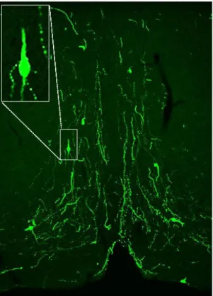
GnRH-GFP neurons in the close proximity of organum vasculosum of the lamina terminalis (OVLT, Bregma 0.49-0.85 mm [165]) were identified by brief illumination at 470 nm using an epifluorescent filter set, based on their green fluorescence, typical fusiform shape, and characteristic topography (Figure 5.) [65]. After control recordings (5 min), the slices were treated with various drugs (see below) and the recordings continued for a subsequent 10 min.
Figure 5. GnRH-GFP neurons and fibers in the organum vasculosum of the lamina terminalis. Visualization of GnRH neurons using fluorescence permits the identification of individual GnRH neurons for electrophysiological recordings and subsequent morphological analysis.
Courtesy of Dr. Csaba Vastagh, IEM, HAS Laboratory of Endocrine Neurobiology
Whole-cell patch clamp experiments
Currently, the most widely used method for studying the electrophysiological properties of biological membranes and the currents that flow through their ion channels is the patch clamp technique [166]. In various configurations, this technique permits experimenters to record and manipulate the currents that flow either through ion channels or those that flow across the whole plasma membrane. Patch clamp technique can even allow low noise measurements of the currents passing through a couple of ion channels, by isolating a small patch of the membrane, which sometimes can contain solely a single channel. Here, a high-resistance (“giga ohm”) seal is formed between the pipette and the membrane of the cell. In the experiments whole-cell configuration of
the patch clamp methods was used. This means that the membrane within the pipette is ruptured while the gigaseal is still maintained. The main advantage of this method is the ability to manipulate of ionic or other composition of the intracellular milieu to aid isolation and detection of conductances via specific ion channels.
During whole-cell patch clamp experiments spontaneous and miniature postsynaptic currents were measured. Spontaneous postsynaptic currents (sPSC) are currents generated via mainly by action potential dependent presynaptic release of neurotransmitters in the absence of experimental stimulation. Miniature postsynaptic currents (mPSC) are currents observed in the absence of presynaptic action potentials; they are thought to be the response that is elicited by random release of neurotransmitter vesicles.
The parameters of the measurements were the following: during sPSC and mPSC measurements in GnRH neurons the cells were voltage clamped at -70 mV holding potential. The voltage clamp technique allows to "clamp" the cell potential at a chosen value, make it possible to measure how much ionic current crosses through the membrane of the cell at any given voltage values. Before the recording, pipette offset potential, series resistance and capacitance were compensated. Cells with low holding current (<50 pA) and stable baseline were used exclusively. Input resistance, series resistance, and membrane capacity were also measured before each recording by using 5 mV hyperpolarizing pulses. To ensure consistent recording qualities, only cells with series resistance
<20 MΩ, input resistance >500 MΩ, and membrane capacity >10 pF were accepted. The intracellular pipette solution contained (in mM): HEPES 10, KCl 140, EGTA 5, CaCl2 0.1, Mg- ATP 4 and Na-GTP 0.4 (pH 7.3, osmolarity adjusted to 300 mOsm).
The postsynaptic current measurements were carried out with an initial control recording (5 min), then low physiological concentration of 17β-estradiol (E2, 10 pM), the GLP-1 analog Exendin-4 (1 µM), the NO-donor L-arginine (1 mM), the selective ERα agonist PPT (10 pM), the selective ERβ agonist DPN (10 pM) or the selective GPR30 receptor agonist G1 (10 pM) was added to the aCSF in a single bolus onto the slice in the recording chamber and the recording continued for a subsequent 10 min.
When the cannabinoid receptor type 1 inverse agonist AM251 (1 μM), the non-selective estrogen receptor antagonist Faslodex (1 µM), the ERβ antagonist PHTPP (1 µM), the NO-synthase (NOS) inhibitor L-NAME (100 μM), the GLP-1 receptor antagonist Exendin-3(9-39) (1 μM) or the nNOS inhibitor NPLA (1 μM) were used, they were added to the aCSF 10 min before starting the experiments and then they were continuously present in the aCSF during the electrophysiological recordings.
Intracellularly applied drugs, such as diacylglycerol lipase inhibitor tetrahydrolipstatin (THL, 10 μM), the membrane impermeable G-protein inhibitor GDP-β-S (2 mM), the membrane impermeable NO-scavenger CPTIO (1 mM), the transient receptor potential vanilloid 1 (TRPV1) antagonist AMG9810 (10 μM), NPLA (1 μM), or the anandamide-degrading enzyme fatty acid amide hydrolase (FAAH) inhibitor PF3845 (5 μM) were added directly to the intracellular pipette solution. To minimize the spill of the intracellularly applied drugs, the GnRH cells were approached rapidly (< 1 min), and the flow rate of aCSF was increased from 5–6 to 8–9 ml/min. Just before releasing the positive pressure in the pipette, the flow rate was restored to 5–6 ml/min to avoid any mechanical movement of the slice. After achieving whole-cell patch clamp configuration, we waited 15 min to reach equilibrium in the intracellular milieu before starting recording.
In the experiments where any spike-mediated release of substances was to be inhibited, firing was blocked by adding the voltage-sensitive Na-channel inhibitor TTX (660 nM) to the aCSF 10 min before mPSCs or Vrest were recorded. The mPSCs recordings conditions used in our experiments were related to the conditions in which GABAA-R activation occurs [46, 152], interestingly this GABAergic input via GABAA-R is excitatory on GnRH neurons [50, 167, 168]. Nevertheless, it is important to note that GABA inhibits GnRH neurons via GABAB-receptors [50, 169].
Resting potentials were recorded using current-clamp method. The current clamp technique records the membrane potential while injecting current into the cell through the recording electrode. This shows us the cell response when electric current enters the cell, therefore how neurons respond to substances that act by opening membrane ion channels. Vrest measurements were carried out at 0 pA holding current.
Loose-patch clamp experiments
In this type of recording, the pipette is pushed to the membrane not tightly but loosely without the formation of a tight gigaseal connection, and there is no direct exchange of cytoplasm and intracellular fluid. The action currents, which underlie action potential firing, can be recorded with this configuration. The advantage of the loose-patch technique is that the composition of the cytoplasm is not influenced, and the activity pattern of a cell can be observed for long time (even for hours) without changing the intracellular milieu. These experiments were carried out at 33 °C, pipette potential was set at 0 mV, pipette resistance 1–2 MΩ, and resistance of loose-patch seal varied between 7–40 MΩ. The composition of the pipette solution mimicking the extracellular milieu that contained the following (in mM): NaCl 150, KCl 3.5, CaCl2 2.5, MgCl2 1.3, HEPES 10, and glucose 10 (pH 7.3, osmolarity adjusted to 300 mOsm). Measurements were carried out with