Pázmány Péter Catholic University Faculty of Information Technology and Bionics Roska Tamás Doctoral School of Sciences and Technology
Erzsébet Hakkel
Regulation of energy homeostasis by hypothalamic circuits;
light- and electron microscopic studies in rodents
Ph.D Dissertation
Thesis Advisor:
Csaba Fekete D.Sc.
Budapest, 2017
1
“Research is to see what everybody else has seen, and to think what nobody else has thought.”
Albert Szent-Györgyi
2
Abstract
The incidence of obesity increased rapidly in the developed countries during the last decades.
Obesity has high impact on the population health and also on the health care cost. Therefore, understanding the regulatory mechanisms of the energy homeostasis has critical importance.
The goal of our studies was to better understand the regulation of the energy homeostasis by hypothalamic circuits. The hypothalamic paraventricular nucleus (PVN) is an important hypothalamic centre in the regulation of the energy homeostasis. Using electron microscopy, we demonstrated that the nitric oxide (NO) system is anatomically positioned to be utilized as both anterograde and retrograde transmitter system in the PVN. The association of the NO synthesizing enzyme, the neuronal nitric oxide synthase (nNOS), to the postsynaptic side of synapses formed by type 1 cannabinoid receptor (CB1)-containing axons in the parvocellular part of the PVN suggests that the NO and the endocannabinoid systems may interact in the regulation of presynaptic terminals of parvocellular neurons. Our in vivo studies revealed that both the NO and the endocannabinoid systems of the PVN are involved in the regulation of the energy homeostasis by neuropeptide Y (NPY) an orexigenic peptide of the hypothalamic arcuate nucleus (ARC). While inhibition of the NO system inhibits the effects of NPY on the food intake, inhibition of CB1 in the PVN markedly decreases the effect of NPY on the energy expenditure. The hypophysiotropic thyrotropin-releasing hormone (TRH)-synthesizing neurons of the PVN play important role in the regulation of energy expenditure by controlling the hormone synthesis of the thyroid gland. We demonstrated that the axon terminals of these neurons contain the thyroid hormone transporter monocarboxylate transporter 8 (MCT8) in the external zone of the median eminence (ME) where these terminals are closely associated with tanycytes, the cell type that expresses the thyroid hormone activating type 2 deiodinase suggesting that the TRH neurons accumulate the active thyroid hormone, T3, from the ME where T3 originates from the blood and from tanycytes.
The non-hypophysiotropic TRH neurons in the perifornical area/BNST region express a second anorexigenic peptide, the urocortin 3 (UCN3). We showed that these TRH/UCN3 neurons form symmetric type synaptic associations with the anorexigenic POMC neurons of the ARC raising the possibility that the TRH/UCN3 neurons regulate the food intake via the POMC neurons of the ARC.
In addition, we demonstrated that TRH-containing axons densely innervate the histaminergic neurons in all subnuclei of the tuberomammillary nucleus indicating that the histaminergic neurons may receive feeding related inputs from non-hypophysiotropic TRH populations.
3
Acknowledgements
Firstly, I would like to thank my scientific advisor Csaba Fekete for his guidance, patience and valuable support during my studies.
I am also grateful to Balázs Gereben, Imre Farkas and Zsolt Liposits for their help in my studies.
I thank my former and current colleagues Anett Szilvásy-Szabó, Andrea Kádár, Ágnes Simon, Andrea Juhász, Anikó Zeöld, Barbara Vida, Csaba Vastagh, Csilla Molnár, Enikő Kiss, Erik Hrabovszky, Flóra Bálint, Györgyi Zséli, Imre Kalló, Judit Szabon, Kata Skrapits, Mónika Tóth, Petra Mohácsik, Vera Maruzs, Zoltán Péterfi, Zsuzsa Beliczai and Zsuzsa Bardóczi.
I would also like to acknowledge the very important contribution of the co-authors of our papers Anna Sárvári, Gábor Wittmann, Kata Nagyunyomi-Sényi, László Barna, Masahiko Watanabe, Motokazu Uchigashima, Miklós Palkovits, Raphaël G. P. Denis, Ronald M.
Lechan, Serge Luquet, Tamás Füzesi, Ann Marie Zavacki, Rafael Arrojo e Drigo, Liping Dong, Beáta A. Borsay, László Herczeg, Antonio C. Bianco.
Furthermore, I am thankful to the Doctoral School; especially to Prof. Péter Szolgay for the opportunity to participate in the doctoral program. I thank Katinka Tivadarné Vida for her always kind help and patience to make the administrative side of life easier.
I am very grateful to my mother and my friends who always believed in me and supported me.
Finally, I am especially appreciated my husband András for all his love, patience, support and encouragement that gave me the strength to go on even in the hardest periods of this journey.
4
List of abbreviations
2-AG – 2-arachidonoylglycerol AGRP – agouti-related neuropeptide ARC – arcuate nucleus
BAT – brown adipose tissue BBB – blood-brain-barrier
BNST – bed nucleus of stria terminalis cAMP – cyclic adenosine monophosphate
CART – cocaine- and amphetamine-regulated transcript CB1 – type 1 cannabinoid receptor
CCK – cholecystocinin
CNS – central nervous system
CRFR2 – corticotropin-releasing factor receptor 2 CRH – corticotropin-releasing hormone
CSF – cerebrospinal fluid D2 – deiodinase 2 DAB – diaminobenzidine
DAGLα – diacylglycerol lipase alpha DMN – dorsomedial nucleus
eNOS – endothelial nitric oxide synthase GABA – gamma-aminobutyric acid GPCR – G-protein-coupled receptor H1R – histamine 1 receptor HDC – histidine decarbocylase
HPA – hypothalamus – pituitary – adrenal axis HPT – hypothalamus – pituitary – thyroid iNOS – inducible nitric oxide synthase IR – immunoreactive
KO – knock out
MAP2 – microtubule-associated protein 2 MC3R – melanocortin 3 receptor
MC4R – melanocortin 4 receptor ME – median eminence MR – Mammillary recess
5 mRNA – messenger ribonucleic acid MTC8 – monocarboxylase 8
NiDAB – nickel diaminobenzidine nNOS – neuronal nitric oxide synthase NO – nitric oxide
NOS – nitric oxide synthase NPY – neuropeptide Y
NTS – nucleus tractus solitary
OATP1C1 – organic anion-transporting polypeptide 1c1 PB – phosphate buffer solution
PBS – phosphate buffered saline PFA - paraformaldehyde
PHAL – Phaseolus vulgaris leucoagglutin PLCß – phospholipase C beta
POMC – proopiomelanocortin PPII – pyroglutamyl peptidase II PVN – paraventricular nucleus PYY – peptide YY
sGC – soluble guanylate cyclase SS – somatostatin
T3 – triiodothyronine
T4 – thyroxine
THß2 – thyroid hormone ß2 receptor TMN – tuberomammillary nucleus TRH – thyrotropin-releasing hormone TSH – thyroid-stimulating hormone UCN3 – urocortin 3
VGLUT1 – vesicular glutamate transporter 1 VGLUT2 – vesicular glutamate transporter 2
VIAAT – vesicular inhibitory amino acid transporter α-FMH – alpha-fluoro-methyl histidine
α-MSH – alpha melanocyte-stimulating hormone
6
Table of Contents
I. INTRODUCTION ... 11 Role of the hypothalamic arcuate nucleus (ARC) in the regulation of the energy I.1
homeostasis ... 11 Role of the PVN in the regulation of energy homeostasis ... 13 I.2
Retrograde transmitter systems in the parvocellular part of the PVN ... 14 I.3
Feedback regulation of the hypophysiotropic TRH neurons ... 16 I.4
Role of TRH neurons in the regulation of food intake ... 19 I.5
II. SPECIFIC AIMS ... 21 III. MATERIALS AND METHODS ... 22 Animals ... 22 III.1
Colchicine treatment ... 22 III.2
Fixation of animals for immunocytochemistry at light and electron microscopic levels 22 III.3
Tissue preparation for light microscopic immunohistochemistry ... 23 III.4
Tissue preparation for ultrastructural studies ... 24 III.5
Immunocytochemistry for ultrastructural localization of nNOS ... 26 III.6
Immunocytochemistry for ultrastructural localization of sGCα1 ... 27 III.7
Double-labeling immunocytochemistry for ultrastructural examination of the III.8
distribution of nNOS and CB1 ... 27 Quadruple-labeling immunofluorescence of the elements of the endocannabinoid and III.9
NO signaling systems and markers of glutamatergic and GABAergic neurons in the PVN ... 27 Light microscopic detection of MCT8 ... 28 III.10
Ultrastructural detection of MCT8-immunoreactivity in the rat ME ... 28 III.11
Double labeling immunofluorescence for MCT8 and TRH ... 28 III.12
Triple-labeling immunofluorescence for TRH, UCN3 and α-MSH or NPY ... 28 III.13
Double-labeling immunocytochemistry for ultrastructural examination of the UCN3-IR III.14
innervation of the α-MSH neurons in the ARC ... 29 Double labeling immunocytochemistry for TRH and histamine in the TMN ... 29 III.15
Double-labeling immunofluorescence for TRH and histamine in the TMN ... 30 III.16
7
Double-labeling immunocytochemistry for ultrastructural examination of the TRH-IR III.17
innervation of the histamine-IR neurons in the TMN ... 30 Image analyzes of light microscopic preparations ... 31 III.18
Embedding, sectioning and examination of preparations for electron microscopic III.19
studies ... 32 Specificity of antisera ... 32 III.20
Examination of the role of the endocannabinoid and NO transmitter systems in the III.21
mediation of the metabolic effects of NPY evoked in the PVN ... 33 Implantation of bilateral guide cannula in the PVN of mice ... 33 III.21.1
IntraPVN infusions in mice... 33 III.21.2
Measurement of metabolic parameters ... 34 III.21.3
Estimation of basal metabolism ... 35 III.21.4
Statistical analysis of the in vivo data ... 35 III.21.5
IV. RESULTS ... 36 Presence of the NO system and effect of the NO system on the parvocellular part of the IV.1
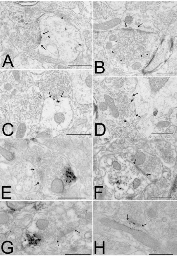
PVN ... 36 Elucidation of the ultrastructural localization of the elements of the NO transmitter system in the IV.1.1
PVN. ... 36 Anatomical relationship of the endocannabinoid and NO systems in the PVN ... 38 IV.1.2
Examination of the role of the endocannabinoid and the NO systems of the PVN in the mediation IV.1.3
of the NPY induced regulation of energy homeostasis... 40 Identification of the presence of MCT8 thyroid hormone transmitter in the axon IV.2
terminals of the hypophysiotropic TRH neurons. ... 47 TRH/UCN3 neurons of the perifornical area/BNST region innervate the POMC neurons IV.3
of the ARC ... 50 Relationship between TRH-IR axons and histaminergic neurons in the subnuclei of the IV.4
TMN ... 53 V. DISCUSSION ... 60 Anatomy of the NO system in the parvocellular part of the PVN and its potential V.1
interaction with the endocannabinoid system ... 60 The endocannabinoid and the NO systems of the PVN mediate different effects of NPY V.2
on the energy homeostasis ... 61 Presence of the MCT8 protein in axon terminals of the hypophysiotropic TRH axons in V.3
the ME of the rat... 63
8
TRH/UCN3 neurons of the perifornical area/BNST region innervate the α-MSH V.4
neurons of the ARC ... 64 Relationship between TRH-IR axons and histaminergic neurons in the subnuclei of the V.5
TMN ... 66 VI. NEW SCIENTIFIC RESULTS ... 69 [C1-C5] Thesis I ... 69 VI.1
[C1-C5] Thesis II ... 69 VI.2
[C1-C5] Thesis III ... 69 VI.3
[J1] Thesis IV ... 69 VI.4
[J2] Thesis V ... 70 VI.5
[J3] Thesis VI ... 70 VI.6
VII. AUTHOR’S JOURNAL PUBLICATIONS ... 71 VIII. AUTHOR’S CONFERENCE PUBLICATIONS ... 71 IX. AUTHOR’S OTHER PUBLICATIONS ... 72
9
List of figures
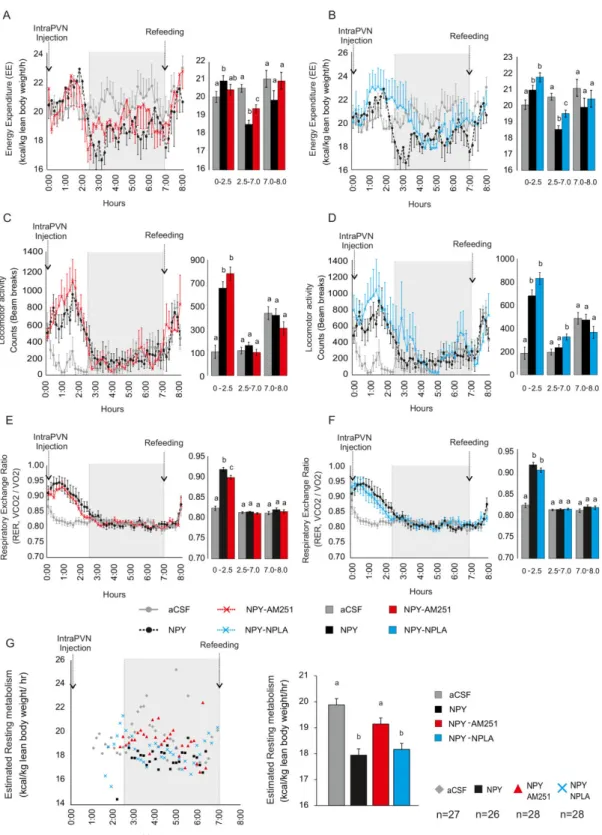
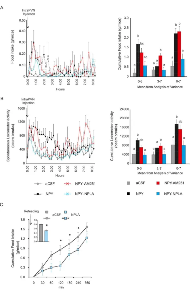
Figure 1 Schematic illustration of the effect of NPY on the parvocellular neurons in the hypothalamic PVN. ... 16 Figure 2 Schematic illustration of thyroid hormone negative feedback mechanism ... 18 Figure 3 Ultrastructural localization of the components of the NO system in the parvocellular part of the PVN in mice. ... 37 Figure 4 Association of the endocannabinoid and NO systems with the same synapses of parvocellular neurons in the PVN. ... 39 Figure 5 Effect of intraPVN co-administration of NPY with the CB1 antagonist, AM251, or the nNOS inhibitor, NPLA, on parameters of energy expenditure. ... 42 Figure 6 Food intake (A) and spontaneous activity (B) of mice injected intraPVN with aCSF (grey), NPY (black) alone, co-administrated with AM251 (red) or NPLA (blue). ... 45 Figure 7 MCT8 immunoreactivity in the rodent mediobasal hypothalamus. ... 48 Figure 8 Ultrastructure of MCT8 immunoreactive structures in the rat ME. ... 49 Figure 9 MCT8 immunoreactivity in axon varicosities of the rat hypophysiotropic TRH neurons. ... 49 Figure 10 UCN3-IR (green) and TRH-IR (red) innervation of the ARC in the rat. ... 51 Figure 11 Relationship of TRH/UCN3- containing axons on the NPY neurons in the ARC.... 51 Figure 12 Relationship of TRH/UCN3- containing axons and the α-MSH neurons in the ARC.
... 52 Figure 13 Distribution of the TRH-IR elements (black) and the histamine-IR neurons (brown) in the subnuclei of the TMN in four different rostrocaudal levels of the TMN. ... 54 Figure 14 Relationship of the TRH-IR (black) axon varicosities and the histamine-IR neurons (brown) in the 5 subnuclei of the TMN (E1-5). ... 55 Figure 15 TRH-IR (red) boutons innervate the histamine-IR neurons (green) (arrows) in the TMN. ... 56 Figure 16 Electron micrographs show synaptic associations (arrows) between histamine- IR neurons and TRH-IR terminals in the TMN. ... 58
10
List of tables
Table 1 Summary of the fixation methods and the number of used animals ... 23 Table 2 Summary of the primary and secondary antibodies used in light microscopic studies24 Table 3 Summary of the primary and secondary antibodies used in fluorescence microscopic studies ... 25 Table 4 Summary of the primary and secondary antibodies used in electron microscopic studies ... 26 Table 5 Summary of the antibodies ... 31 Table 6 Quantitative analysis of the juxtaposition of TRH-IR axon varicosities and histamine- IR neurons in the 5 TMN (E1-5). ... 59
11
I. Introduction
The obesity epidemic is one of the major health problem of our days [1]. More than 60% of the population is overweight or obese in the USA and in Europe [2]. Obesity is not only esthetical problem, but it is also major risk factor of devastating diseases like type 2 diabetes, cardiovascular diseases, cancer etc… [1]. Despite the very high impact on population health and healthcare cost, efficient, non-invasive and side effect free treatment is currently not available against obesity. Large pharmaceutical companies try to develop efficient anti-obesity drugs based on the currently available drug targets without major breakthrough indicating the necessity of the discovery of novel anti-obesity drug targets. Therefore, better understanding of the regulatory mechanisms of energy homeostasis has critical importance in the fight against obesity.
Role of the hypothalamic arcuate nucleus (ARC) in the regulation of the energy I.1
homeostasis
Information about the actual conditions of energy stores and about the consumed food is transmitted toward the central nervous system via peripheral nerves like the vagus nerve and the sensory fibers of sympathetic nerves and also by changes of the level of circulating hormones like leptin, ghrelin, cholecystokinin (CCK), peptide YY (PYY) and insulin [3]. This communication is critical for the maintenance of energy homeostasis [3]. Genetic alterations in these pathways cause obesity syndrome like in leptin or leptin receptor deficient animals or humans and in mice lacking insulin receptor in the brain [4-8].
A critical brain area that can sense these energy homeostasis related humoral signals is the ARC [3]. Ablation of the ARC by neonatal monosodium glutamate treatment induces obesity and leptin resistance [9].
At least two major energy homeostasis-related neuronal groups are located in the ARC [3].
There is a ventromedially located orexigenic neuronal population that expresses two potent orexigenic peptides, the neuropeptide Y (NPY) and the agouti-related protein (AGRP) [3].
These neurons also express the classical transmitter gamma-aminobutyric acid (GABA) [10]
that has been also shown to stimulate food intake [11]. NPY is one of the most potent orexigenic signals [3]. Central administration of NPY causes marked increase of food intake, weight gain and increased adiposity [12]. The effect of NPY on the weight gain, however, cannot be exclusively accounted to its effect on the food intake. NPY also has potent inhibitory effect on the energy expenditure [13]. NPY elicits these effect via the G protein coupled postsynaptic Y1 and Y5 receptors [12].
12
AGRP also increases the food intake and inhibits the energy expenditure [14]. The effect of AGRP on the energy homeostasis is mediated by two centrally expressed melanocortin receptors, the melanocortin 3 and 4 receptors (MC3R and MC4R). AGRP is the endogenous antagonist of these receptors [14]. Changes of energy availability regulate the expression of both peptides in the ARC neurons. While fasting stimulates the NPY and AGRP expression in these neurons, leptin administration inhibits the synthesis of the orexigenic peptides [3].
However, genetic ablation of NPY or AGRP has no major effect on the energy homeostasis;
the critical importance of the orexigenic ARC neurons has been demonstrated by the life threatening anorexia of mice after ablation of the NPY/AGRP neurons [15, 16].
The proopiomelanocortin (POMC) and cocaine- and amphetamine-regulated transcript- (CART) synthesizing neurons located in the lateral part of the ARC has opposite effect on the regulation of energy homeostasis [3]. The POMC derived α-melanocyte-stimulating hormone (α-MSH) is well known about its potent anorexigenic effect [3]. Central administration of α- MSH reduces food intake and simultaneously increases the energy expenditure [3]. The α- MSH exerts its effect as the agonist of the MC3R and MC4R [17]. CART also inhibits food intake and can completely block the NPY induced feeding response [18]. Currently, little information is available about the effect of CART on the energy expenditure. The receptor(s) of CART has not been identified yet.
The POMC/CART neurons are sensitive to the effects of peripheral energy homeostasis- related hormones, like leptin and insulin [3], but these neurons are regulated differently than the NPY/AGRP neurons. Fasting inhibits the POMC and CART synthesis, while leptin administration stimulates the expression of these genes [3]. Despite the presence of leptin receptor in these cells, indirect effect of leptin that is mediated by GABAergic neurons is also critical in the regulation of the POMC/CART neurons [19].
The importance of the POMC/CART neurons in the regulation of energy homeostasis was also demonstrated by genetic studies. Genetic ablation of the POMC or MC4R genes results in morbid obesity in mice [20, 21]. Mutations of the melanocortin pathway also cause obesity in humans. Indeed, mutations of this pathway are the most frequent reason of the human monogenic obesity syndromes [22]. The absence of CART has less profound effect [23]. The CART knock out mice develop only late onset obesity [23]. In humans, a single nucleotide polymorphism (A1475G) of the CART gene is associated with human obesity syndrome [23].
The two feeding related neuronal populations of the ARC sense and integrate the energy homeostasis related signals and transmit it toward the so-called second order feeding related neuronal populations, including the hypothalamic paraventricular nucleus (PVN), the
13
hypothalamic dorsomedial nucleus (DMN) and the histaminergic neurons of the tuberomammillary nucleus (TMN).
In addition to, the homeostatic regulation of the energy homeostasis, the feeding related neurons of the ARC are also involved in the regulation of food intake by adverse conditions like infection and stress [24, 25].
Role of the PVN in the regulation of energy homeostasis I.2
The PVN is a triangular shaped nucleus that is located on the two sides of the upper part of the third ventricle. It consists of magnocellular and parvocellular parts [26]. The oxytocin- and vasopressin-synthesizing neurons of the magnocellular part are involved in the regulation of the posterior pituitary function [26].
The parvocellular part can be further divided into five subdivisions: the anterior, periventricular, medial, ventral and lateral subdivisions and the dorsal cap [27]. The periventricular, and medial parvocellular subdivisions house both hypophysiotropic and non- hypophysiotropic neurons, while the other parvocellular subdivisions house only non- hypophysiotropic neurons.
The hypophysiotropic neurons project to the external zone of the median eminence where they secrete their hypophysiotropic hormones into the extracellular space around the fenestrated capillaries [26]. These hormones reach the anterior pituitary via the hypophyseal portal circulation and regulate the hormone production of this endocrine gland. There are three types of hypophysiotropic neurons in the parvocellular part of the PVN: the somatostatin-, the corticotropin-releasing hormone- (CRH) and the thyrotropin-releasing hormone- (TRH) synthesizing neurons. The somatostatin neurons inhibit the growth hormone synthesis of the pituitary, while the hypophysiotropic CRH and TRH neurons are the central regulators of the hypothalamic-pituitary-adrenal (HPA) and thyroid (HPT) axes, respectively. All of these neuroendocrine axes have major impact on the regulation of the energy homeostasis.
A large population of the non-hypophysiotropic neurons in the parvocellular part of the PVN regulates autonomic functions. These neurons project to the intermediolateral column of the spinal cord and to brainstem nuclei involved in the regulation of energy homeostasis like the nucleus tractus solitarii (NTS), the dorsal motor nucleus of vagus, the parabrachial nucleus and the catecholaminergic neurons of the ventral medulla [28]. Via these nuclei, the PVN multisynaptically linked to the pancreas, white and brown adipose tissue (BAT), liver and muscle [29-31]. Thus, these so-called preautonomic neurons of the PVN can regulate the energy homeostasis by controlling the lipid metabolism and storage, thermogenesis, gluconeogenesis and insulin synthesis [30].
14
However, the PVN receives energy homeostasis related inputs via multiple neuronal pathways and also via hormones, one of its most important energy homeostasis related input originates from the ARC [3]. Both the orexigenic and the anorexigenic neuronal groups densely innervate the neurons of the parvocellular part of the PVN [3]. In many instances, the same parvocellular neurons are innervated by both the orexigenic and anorexigenic ARC neurons [27].
The PVN is a critical mediator of the effects of ARC neurons on the energy homeostasis [3].
Focal administration of NPY into the PVN markedly increases food intake [13], increases the carbohydrate utilization [32], decreases the energy expenditure and the uncoupling protein 1 expression in the BAT [33, 34] and induces body weight gain [35].
Both postsynaptic NPY receptors, the Y1 and Y5, are expressed in the PVN [36], coupled to pertussis-toxin sensitive Gi/o proteins [37], and lead to the inhibition of cyclic adenosine monophosphate (cAMP) accumulation by inhibiting adenylate cyclase [38]. Some of the effects of NPY on energy expenditure are exerted through the regulation of TRH and CRH gene expression in the PVN via the modulation of the cAMP pathway [39-42]. NPY also has been shown to inhibit the GABAergic inputs of the parvocellular neurons of the PVN [43].
Similarly to NPY, intraPVN administration of AGRP also increases the food intake [44].
In contrast to the orexigenic peptides, α-MSH has potent anorexigenic effect when injected into the PVN [45]. Most of the effects of the α-MSH are mediated by the MC4R. The MC4R knockout (KO) mice are hyperphagic and have decreased energy expenditure [21]. Re- expression of MC4R exclusively in the PVN rescues the hyperphagic phenotype of the MC4R KO mice, but has only little effect on the energy expenditure [46] substantiating the importance of the PVN in the mediation of the effect of melanocortins on the food intake. α- MSH also regulates the HPA and HPT axes by stimulating the CRH and TRH gene expression in the PVN [47, 48].
Retrograde transmitter systems in the parvocellular part of the PVN I.3
Using patch clamp electrophysiology, our laboratory showed that NPY inhibits both the GABAergic and the glutamatergic inputs of the parvocellular neurons [49]. These effects were completely prevented by the intracellular administration of the calcium chelator drug BAPTA1 demonstrating that NPY inhibits the inputs of the parvocellular neurons by stimulating retrograde transmitter release of the target cells. [49]. The most widely utilized retrograde transmitter system in the brain is the endocannabinoid system [50]. In the central nervous system (CNS), the primary receptor of the endocannabinoid signaling system is the type 1
1 1,2-bis(o-aminophenoxy)ethane-N,N,N′,N′-tetraacetic acid
15
cannabinoid receptor (CB1) [50]. The two most abundant endogenous ligands of this receptor are the 2-arachidonoylglycerol (2-AG) and the anandamide [50]. 2-AG is synthesized by postsynaptic neurons in the perisynaptic region and acts on the CB1 located in the perisynaptic region of the presynaptic terminals [50]. Activation of the CB1 inhibits the activity of the presynaptic terminals [50]. An important regulator of the endocannabinoid synthesis is the synaptic activity. The synthesizing enzyme of 2-AG, the diacylglycerol lipase α (DAGLα), is activated in response to increased synaptic activity which effect is mediated by metabotropic receptors coupled to phospholipase C beta (PLCβ) like metabotropic glutamate receptor 1 and 5, muscarinic acetylcholine (mACh) receptor M1 and M3 [51].
Our laboratory has shown that CB1 is present in both the inhibitory and excitatory terminals innervating the parvocellular neurons in the PVN [52] and the endocannabinoid system has been shown to mediate the effects of ghrelin and the glucocorticoids on the parvocellular neurons of the PVN [53, 54].
We have found that inhibition of CB1 by AM2512 prevents the effect of NPY on the GABAergic input of parvocellular neurons [49]. However, the dose of AM251 that were sufficient to prevent the effect of ghrelin on the glutamatergic inputs of the parvocellular neurons [53], did not prevent the effects of NPY on the glutamatergic inputs [49] suggesting that other retrograde signaling system(s) is also involved in the mediation of the NPY induced effects (Fig. 1).
Nitric oxide (NO) is a gaseous transmitter [55]. NO is synthesized by a family of the NO synthesizing (NOS) enzymes: the neuronal NOS (nNOS), the inducible NOS (iNOS) and the endothelial NOS (eNOS) [55]. Among these enzymes, the nNOS is present in neurons [55].
The most sensitive receptor of NO is the soluble guanylate cyclase (sGC) [55]. In the hippocampus, both the nNOS and the sGC can be observed in both pre- and postsynaptic localization [56] suggesting that NO can serve as both anterograde and retrograde transmitter.
Electrophysiological experiments also provided evidence supporting the retrograde transmitter role of NO and the interaction of the endocannabinoid and NO signaling in the presynaptic plasticity in the hippocampal formation [57].
However, nNOS is also present in the PVN, little is known about the localization of the elements of NO signaling in this nucleus and it is also unknown whether NO is utilized as a retrograde transmitter in this nucleus [58].
2 biarylpyrazole cannabinoid receptor antagonist
16
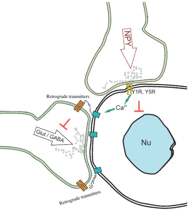
Figure 1 Schematic illustration of the effect of NPY on the parvocellular neurons in the hypothalamic PVN.
NPY acts via the Y1 and Y5 receptors in the parvocellular part of the hypothalamic PVN. The receptor activation inhibits gene transcription y decreasing the cAMP level, but at the same time, it can activate retrograde messengers systems via modulation of the intracellular Ca2+
levels. Thus, NPY can inhibit the synaptic inputs of the parvocellular neurons via retrograde messengers. Nu –nucleus, Glut – glutamatergic terminals, GABA – GABAergic terminals, NPY-neuropeptide Y, Y1R-NPY receptor type 1, Y5R- NPY receptor type 5
Feedback regulation of the hypophysiotropic TRH neurons I.4
One of the neuronal groups of the PVN that play critical role in the regulation of the energy homeostasis is the group of the hypophysiotropic TRH neurons [27]. These neurons control the hormone production of the thyroid gland through the regulation of the TSH secretion of
Retrograde tranmitters
17
the thyrotroph cells in the anterior pituitary [27]. The thyroid hormones are important regulators of the energy homeostasis [27]. In the absence of thyroid hormones, the basal metabolic rate is decreased by 30% and the cold induced thermogenesis is also absent in hypothyroid animals [27].
The main regulator of the HPT axis is the negative feedback effect of thyroid hormones that ensure the relatively stable circulating thyroid hormone levels [27]. Thus, when the peripheral levels of thyroid hormones are increased, the TRH synthesis is inhibited by thyroid hormones [27]. The hypophysiotropic TRH neurons contain the thyroid hormone β2 receptor (TRß2) that is essential for the feedback regulation of these cells [27]. In addition, implantation of T33 adjacent to the PVN inhibits the TRH expression on the side of the implantation [27].
However, restoration of the circulating T3 levels in hypothyroid animals without administration of the prohormone T44 is not sufficient to normalize the TRH expression in the PVN [27]. These data demonstrate that hypothalamic conversion of the prohormone T4 to its active form, T3, is necessary for the feedback regulation of the TRH neurons. In the hypothalamus, T4-T3 conversion is catalyzed by the type 2 deiodinase enzyme (D2) [59]. D2 activity or mRNA, however, is not present in the PVN where the hypophysiotropic TRH neurons reside [60]. D2 is synthesized in the hypothalamus by a special glial cell types, the tanycytes [60]. Tanycytes line the lateral wall and the floor of the third ventricle behind the optic chiasm. The long basal process of these cells projects to the hypothalamic dorsomedial and ventromedial nuclei, into the ARC and into the external zone of the median eminence (ME) [27]. Thus, the cell bodies of the TRH neurons are located relatively far from the thyroid hormone activating cells of the hypothalamus. In the external zone of the ME, however, end feet processes of the tanycytes and the axon terminals of the hypophysiotropic TRH neurons are closely associated raising the possibility that T3 released from the tanycytes may be taken up by the hypophysiotropic terminals and transported to the cell bodies of TRH neurons where T3 could bind to the nuclear TRβ2 [27]. The thyroid hormone transport is mediated by thyroid hormone transporters [27]. The main thyroid hormone transporters are the monocarboxylate transporter 8 (MCT8), organic anion-transporting polypeptide 1c1 (OATP1C1), Lat1 and Lat25[61]. The main thyroid hormone transporter of neurons is MCT8. The absence of MCT8 causes serious neurological symptom in humans and upregulation of the HPT axis in both humans and mice [62, 63] suggesting that MCT8 is involved in the feedback regulation of the HPT axis. However, the presence of MCT8 was demonstrated in tanycytes [64], data were not available about the presence of this transporter in hypophysiotropic axons. The presence of
3 triiodothyronine
4 thyroxine
5 heterodimeric large amino acid transporter 1 and 2
18
MCT8 in the axon terminals of the hypophysiotropic TRH neurons would suggest that the axon terminals of the hypophysiotropic TRH axons are able to take up T3 in the ME. The importance of this question is underlined by the different kinetics of the T4 and T3 transport through the blood brain barrier (BBB). T3 can far more efficiently pass through the BBB than T4 [27]. This is however not the case in the external zone of the ME which brain region is located outside of the BBB. Thus, the site of thyroid hormone uptake determines whether the hypophysiotropic TRH neurons can only sense the T3 that is activated within the BBB or these cells can sense a mixture of the T3 originating from the circulation and released by the tanycytes in the ME (Fig. 2).
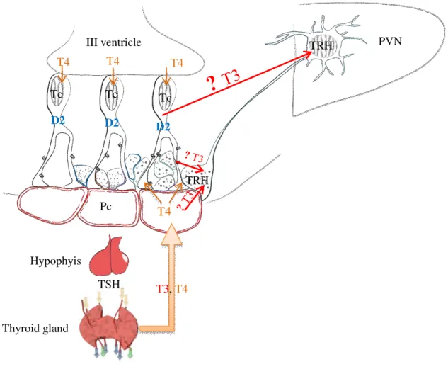
Figure 2 Schematic illustration of thyroid hormone negative feedback mechanism
Thyroid hormones exert negative feedback effect on the hypophysiotropic TRH neurons. This feedback mechanism requires central thyroid hormone activation. This T4 to T3 conversion is catalyzed by the type 2 deiodinase (D2) that is expressed by the tanycytes lining the wall of the third ventricle. It was not clear, how T3 released from tanycytes can reach the hypophysiotropic TRH neurons in the PVN. T3 may diffuse through the neuropil. An alternative hypothesis is that the axons of TRH neurons may take up the T3 in the external zone of the median eminence where the axons of hypophysiotropic TRH neurons and the endfeet processes of tanyctes are closely associated D2- type 2 deiodinase enzyme, ME –
Hypophyis
T4 Pc
III ventricle PVN
Tc Tc Tc
Thyroid gland
TRH
TRH
TSH
T4 T4 T4
D2 D2
D2
T3, T4
19
median eminence, PVN – paraventricular nucleus, TRH – thyrotropin-releasing-hormone, MCT8 – monocarboxylate transporter 8, T3 – triiodothyronine, T4 – thyroxine, TSH – thyroid-stimulating hormone, Pc – portal capillary
Role of TRH neurons in the regulation of food intake I.5
It was demonstrated decades ago that central administration of TRH decreases food intake and the time spent with feeding [65]. TRH can inhibit even the vigorous feeding when food is reintroduced after a period of fasting [65]. Despite the very robust anorexigenic effect of TRH, very little information is available which TRH cell population and where exerts this effect.
In addition to the very well-known hypophysiotropic TRH neurons in the medial and periventricular parvocellular subdivisions of the PVN, there are many non-hypophysiotropic TRH synthesizing neuronal groups in the brain including the TRH neurons in the anterior and lateral parvocellular parts and dorsal cap of the PVN and TRH neurons in the DMN, lateral hypothalamus, perifornical region, bed nucleus of stria terminalis (BNST) [66].
Our laboratory has shown [67] that a seemingly continuous population of TRH neurons in the perifornical region and BNST area expresses a second anorexigenic peptide, the urocortin 3 (UCN3) and demonstrated that these neurons project to the lateral part of the ARC. The presence of two anorexigenic peptides in the same neurons and the projection of these cells to the lateral part of the ARC where the anorexigenic POMC neurons are located raised the possibility that the TRH/UCN3 neurons of the perifornical area/BNST region could be involved in the regulation of food intake.
Another cell population that may be involved in the mediation of the anorexigenic effects of TRH is the histaminergic neurons of the TMN in the posterior hypothalamus. Similarly to TRH, central administration of histamine reduces food intake in a number of experimental models [68-70]. Furthermore, the absence of the histamine-synthesizing enzyme, the histidine decarboxylase (HDC), results in late onset obesity and hyperphagia [71] demonstrating the anorexigenic role of histamine.
Central administration of TRH not only decreases food intake in a dose-dependent manner, but also increases the concentration of histamine and t-methylhistamine (a major metabolite of neuronal histamine) in the TMN [72] suggesting that the histaminergic neurons may be involved in the mediation of TRH induced anorexia. This hypothesis is further supported by the data that the anorexic effects of TRH could be attenuated by pretreatment with the irreversible HDC inhibitor, α-fluoro-methyl histidine [72] and that TRH can excite the histaminergic neurons [73]. Based on these important functional data, we hypothesized that the identification of the sources of the TRH-containing inputs of the histaminergic neurons can be used to identify anorexigenic TRH cell populations. However, it was unknown, whether
20
TRH neurons innervate the histaminergic neurons and whether TRH neurons innervate the histaminergic neurons in all five subnuclei of the TMN or the communication of these two systems is localized to only certain TMN subnuclei. Therefore, the detailed description of the relationship of the TRH axons and the histaminergic neurons was necessary to provide anatomical data for later track tracing studies.
21
II. Specific aims
To better understand the hypothalamic network regulating energy homeostasis we:
1. Elucidated the ultrastructural localization of the elements of the NO transmitter system in the PVN.
2. Studied whether the NO and the endocannabinoid systems are associated to the same synapses of the parvocellular neurons of the PVN.
3. Examined the role of the endocannabinoid and the NO systems of the PVN in the mediation of the NPY induced regulation of energy homeostasis
4. Determined the presence of MCT8 thyroid hormone transmitter in the axon terminals of the hypophysiotropic TRH neurons.
5. Examined the role of TRH/UCN3 neurons of the perifornical area/BNST region in the regulation of the feeding related neuronal groups of the hypothalamic arcuate nucleus.
6. Studied the TRH-containing innervation of the histaminergic neurons in TMN.
22 III.
Materials and methods
Animals III.1
The experiments were carried on adult, male Wistar rats (Charles Rivers, Wilmington, MA), CD1 mice (Charles Rivers, Wilmington, MA) and MCT8 KO [74] mice housed under standard environmental conditions (light between 06:00 and 18:00 h, temperature 22±1 °C, rat chow and water ad libitum). The used animals are listed in the description of each experiment.
All experimental protocols were reviewed and approved by the Animal Welfare Committee at the Institute of Experimental Medicine of the Hungarian Academy of Sciences.
Colchicine treatment III.2
As peptides are rapidly transported into axons, immunocytochemistry can detect the axons of peptide producing neurons, but can only visualize the perikarya of only a small proportion of these cells. The visualization of peptide synthesizing perikarya can be facilitated by the central administration of the microtubules association inhibitor colchicine, which treatment prevents the axonal transport [75]. Therefore, colchicine-treatment of rats was performed in studies IV.3 and IV.4. Rats anaesthetized with a mixture of ketamine and xylazine (ketamine 50 mg/kg, xylazine 10 mg/kg body weight, ip.) were injected intracerebroventricularly with 100 µg colchicine in 5 µl 0.9% saline under stereotaxic control to facilitate the visualization of peptides in perikarya in the ARC and TMN. Twenty hours later, the animals were anaesthetized and perfused with fixative.
Fixation of animals for immunocytochemistry at light and electron III.3
microscopic levels
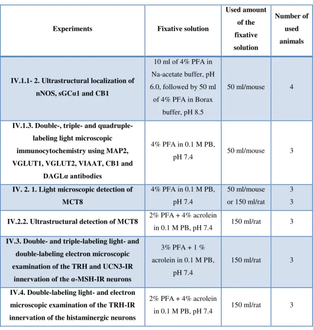
Under general anaesthesia (ketamine 50 mg/kg, xylazine 10 mg/kg body weight, ip.), the animals were perfused transcardially by 10 ml phosphate buffer saline (PBS) pH 7.5 followed by fixative solution. The different fixatives used in the studies are summarized in Table 1. For light microscopic studies, we used 4% PFA (pH 7.4) to perfuse the animals. This fixative is appropriate for light microscopic studies, and compatible for most antibodies, but it does not provide sufficient tissue preservation for ultrastructural studies. Three of the used antibodies, the sheep and mouse anti-TRH sera and the sheep anti-histamine serum require acrolein- containing fixative that is appropriate for both light- and electron microscopic studies. The rabbit anti-nNOS and the rabbit anti-sGCα sera were not compatible with strong fixatives like acrolein and glutaraldehyde that are routinely used for ultrastructural studies. Therefore, double pH fixatives were used for ultrastructural studies with these antibodies. First the animals were perfused with 4% PFA in sodium-acetate buffer pH 6.0, followed by 4% PFA in
23
borate buffer pH 8.5. This combination provides good ultrastructure and was compatible with these two antisera. After perfusion, the brains were rapidly removed and stored in 4%
paraformaldehyde (PFA) in 0.1M phosphate buffer (PB), pH 7.4 for 2 h for light microscopy or 24 h for electron microscopy. The different antibodies used in the studies are summarized in Table 2-4.
Table 1 Summary of the fixation methods and the number of used animals
Experiments Fixative solution
Used amount of the fixative solution
Number of used animals
IV.1.1- 2. Ultrastructural localization of nNOS, sGCα1 and CB1
10 ml of 4% PFA in Na-acetate buffer, pH 6.0, followed by 50 ml
of 4% PFA in Borax buffer, pH 8.5
50 ml/mouse 4
IV.1.3. Double-, triple- and quadruple- labeling light microscopic immunocytochemistry using MAP2, VGLUT1, VGLUT2, VIAAT, CB1 and
DAGLα antibodies
4% PFA in 0.1 M PB,
pH 7.4 50 ml/mouse 3
IV. 2. 1. Light microscopic detection of MCT8
4% PFA in 0.1 M PB, pH 7.4
50 ml/mouse or 150 ml/rat
3 3
IV.2.2. Ultrastructural detection of MCT8 2% PFA + 4% acrolein
in 0.1 M PB, pH 7.4 150 ml/rat 3 IV.3. Double- and triple-labeling light- and
double-labeling electron microscopic examination of the TRH and UCN3-IR
innervation of the α-MSH-IR neurons
3% PFA + 1 % acrolein in 0.1 M PB,
pH 7.4
150 ml/rat 3
IV.4. Double-labeling light- and electron microscopic examination of the TRH-IR innervation of the histaminergic neurons
2% PFA + 4% acrolein
in 0.1 M PB, pH 7.4 150 ml/rat 3
Tissue preparation for light microscopic immunohistochemistry III.4
The brains were cryoprotected in 30% sucrose in PBS at 4 °C overnight, then frozen in powdered dry ice. Serial, 25 μm thick coronal sections were cut on a freezing microtome
24
(Leica, Wetzlar, Germany), collected in cryoprotectant solution (30% ethylene glycol; 25%
glycerol; 0.05 M phosphate buffer (PB) and stored at −20 °C until use. The free aldehyde groups in acrolein fixed tissues can bind antibodies, therefore these aldehyde groups can cause high background signal [76]. To prevent this effect, acrolein fixed tissues were treated with 1% sodium borohydride in distilled water (DW) for 30 min. All tissues were treated with 0.5%
Triton X-100/0.5% H2O2 in PBS for 15 min to increase antibody penetration and reduce endogenous peroxidase activity. To limit the nonspecific antibody binding, the sections were treated with 2% normal horse serum (NHS) in PBS for 20 min.
Table 2 Summary of the primary and secondary antibodies used in light microscopic studies
Study
number Used primary antibodies and sources Dilution Secondary antibody
IV.2. rabbit anti-MCT8 (kind gift from Dr. TJ Visser Rotterdam, The Netherlands)
1:5000 – 10000
biotinylated donkey anti-rabbit IgG, 1:500; Jackson ImmunoResearch
IV.4.
sheep anti-TRH (#08W2) [67, 77] 1:50000 biotinylated donkey anti-sheep IgG, 1:500; Jackson ImmunoResearch sheep anti-histamine [78] 1:1000 biotinylated donkey anti-sheep IgG,
1:500; Jackson ImmunoResearch
Tissue preparation for ultrastructural studies III.5
After the perfusion with fixative, the brains were rapidly removed and postfixed in 4% PFA in 0.1M PB, pH 7.4 and overnight at 4°C. Serial, 25-50 μm thick, coronal sections were cut on a Leica VT 1000S vibratome (Leica Microsystems, Wetzlar, Germany) and collected in PBS.
Those sections which were fixed with acrolein containing fixative were treated with 1%
sodium borohydride in 0.1 M PB, pH 7.4, for 30 min. All sections were treated by 0.5% H2O2
in PBS for 15 min. The sections were cryoprotected in 15% sucrose in PBS for 15 min at room temperature (RT) and in 30% sucrose in PBS overnight at 4 °C. The sections were placed in a tinfoil dish or an Eppendorf tube and quickly frozen over liquid nitrogen, then thawed at RT.
This cycle was repeated three times to improve antibody penetration into the tissue. To reduce the nonspecific antibody binding, the sections were treated with 2% NHS in PBS for 20 min.
25
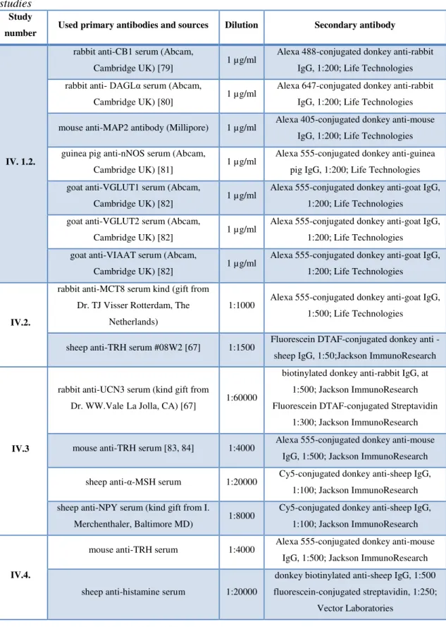
Table 3Summary of the primary and secondary antibodies used in fluorescence microscopic studies
Study
number Used primary antibodies and sources Dilution Secondary antibody
IV. 1.2.
rabbit anti-CB1 serum (Abcam,
Cambridge UK) [79] 1 µg/ml Alexa 488-conjugated donkey anti-rabbit IgG, 1:200; Life Technologies rabbit anti- DAGLα serum (Abcam,
Cambridge UK) [80] 1 µg/ml Alexa 647-conjugated donkey anti-rabbit IgG, 1:200; Life Technologies mouse anti-MAP2 antibody (Millipore) 1 µg/ml Alexa 405-conjugated donkey anti-mouse
IgG, 1:200; Life Technologies guinea pig anti-nNOS serum (Abcam,
Cambridge UK) [81] 1 µg/ml Alexa 555-conjugated donkey anti-guinea pig IgG, 1:200; Life Technologies goat anti-VGLUT1 serum (Abcam,
Cambridge UK) [82] 1 µg/ml Alexa 555-conjugated donkey anti-goat IgG, 1:200; Life Technologies
goat anti-VGLUT2 serum (Abcam,
Cambridge UK) [82] 1 µg/ml Alexa 555-conjugated donkey anti-goat IgG, 1:200; Life Technologies
goat anti-VIAAT serum (Abcam,
Cambridge UK) [82] 1 µg/ml Alexa 555-conjugated donkey anti-goat IgG, 1:200; Life Technologies
IV.2.
rabbit anti-MCT8 serum kind (gift from Dr. TJ Visser Rotterdam, The
Netherlands)
1:1000 Alexa 555-conjugated donkey anti-goat IgG, 1:500; Life Technologies
sheep anti-TRH serum #08W2 [67] 1:1500 Fluorescein DTAF-conjugated donkey anti - sheep IgG, 1:50;Jackson ImmunoResearch
IV.3
rabbit anti-UCN3 serum (kind gift from
Dr. WW.ValeLa Jolla, CA) [67] 1:60000
biotinylated donkey anti-rabbit IgG, at 1:500; Jackson ImmunoResearch Fluorescein DTAF-conjugated Streptavidin
1:300; Jackson ImmunoResearch mouse anti-TRH serum [83, 84] 1:4000 Alexa 555-conjugated donkey anti-mouse
IgG, 1:500; Jackson ImmunoResearch sheep anti-α-MSH serum 1:20000 Cy5-conjugated donkey anti-sheep IgG,
1:100; Jackson ImmunoResearch sheep anti-NPY serum (kind gift from I.
Merchenthaler, Baltimore MD) 1:8000 Cy5-conjugated donkey anti-sheep IgG, 1:100; Jackson ImmunoResearch
IV.4.
mouse anti-TRH serum 1:4000 Alexa 555-conjugated donkey anti-mouse IgG, 1:500; Jackson ImmunoResearch
sheep anti-histamine serum 1:20000
donkey biotinylated anti-sheep IgG, 1:500 fluorescein-conjugated streptavidin, 1:250;
Vector Laboratories
26
Table 4 Summary of the primary and secondary antibodies used in electron microscopic studies
Studies
number Used primary antibody and sources Dilution Secondary antibody
IV.1.1. rabbit anti-nNOS serum (Zymed
Laboratories, Waltham, MA) 1:200
donkey anti-rabbit IgG-conjugated with 0.8 nm colloidal gold, 1:100; Electron Microscopy
Sciences IV.1.1. rabbit anti-sGCα1 serum (Sigma
Aldrich St. Louis, MA) 1:4000 biotinylated donkey anti-rabbit IgG, 1:500;
Jackson Immunoresearch
IV.1.3.
rabbit anti-nNOS serum (Zymed
Laboratories, Waltham, MA) 1:200
donkey anti-rabbit IgG-conjugated with 0.8 nm colloidal gold, 1:100; Electron Microscopy
Sciences sheep anti-CB1 serum (kind gift from
Dr. Watanabe, Sapporo, Japan) 1:800 biotinylated donkey anti-sheep IgG, 1:500;
Jackson ImmunoResearch
IV.2.
rabbit anti-MCT8 serum (kind gift from Dr. TJ Visser, Rotterdam, The
Netherlands)
1:20000 biotinylated donkey anti-rabbit IgG, 1:500;
Jackson ImmunoResearch
IV.3.
sheep anti-α-MSH serum (kind gift
from Dr. JB Tatro, Boston, MA) 1:1000
donkey anti-sheep IgG-conjugated with 0.8 nm colloidal gold, 1:100; Electron Microscopy
Sciences rabbit anti-UCN3 serum (kind gift
from Dr. WW. Vale, La Jolla, CA) 1:1000 biotinylated donkey anti-rabbit IgG, 1:500;
Jackson ImmunoResearch
IV.4.
mouse anti-TRH serum 1:10000 biotinylated donkey anti-mouse IgG, 1:500;
Jackson ImmunoResearch
sheep anti- histamine serum 1:2000
donkey anti-sheep IgG-conjugated with 0.8 nm colloidal gold, 1:100; Electron Microscopy
Sciences
Immunocytochemistry for ultrastructural localization of nNOS III.6
Sections pretreated as described in III.5 were incubated in rabbit anti-nNOS serum (1:200, Zymed Laboratories, Waltham, MA) for 4 days at 4 °C. After rinsing in PBS and in 0.1% cold water fish gelatin (Aurion, Wageningen, Netherlands) /1% bovine serum albumin (BSA) in PBS, the sections were incubated in donkey anti-rabbit IgG-conjugated with 0.8 nm colloidal gold (Electron Microscopy Sciences, Fort Washington, PA) diluted at 1:100 in PBS containing 0.1% cold water fish gelatin and 1% BSA for 1 h. After washing, the sections were fixed in 1.25% glutaraldehyde (Electron Microscopy Sciences, Fort Washington, PA) in 0.1M PB for 10 min. The gold particles were silver intensified with the Aurion R-Gent SE-LM Kit (Aurion, Wageningen, The Netherlands) after rinsing in 0.2M sodium citrate, pH 7.5.
27
Immunocytochemistry for ultrastructural localization of sGCα1 III.7
Sections pretreated as described in III.5 were placed in rabbit anti-sGCα1 serum (1:4000, Sigma Aldrich, St. Louis, MO ) diluted in serum diluent for 4 days at 4 °C. After rinsing in PBS, the sections were incubated in biotinylated donkey anti-rabbit IgG diluted at 1:500 (Jackson Immunoresearch Lab, West Grove, PA) in serum diluent. After rinsing in PBS, and treated with avidin-biotin-complex (ABC elite; 1:1000 dilution; Vector laboratories, Burlingame, CA), the sGCα1-immunoreactivity was detected with NiDAB developer (0.05%
DAB, 0.15% nickel-ammonium-sulfate and 0.005% H2O2 in 0.05M Tris buffer pH 7.6). The immunoreaction product was silver-intensified by using Gallyas method [85].
Double-labeling immunocytochemistry for ultrastructural examination of the III.8
distribution of nNOS and CB1
Sections pretreated as described in III.5 were placed in a mixture of rabbit anti-nNOS serum (1:200) and sheep anti-CB1 serum (1:800, kind gift from Dr. Watanabe, Sapporo, Japan) for 4 days at 4°C. After rinsing in PBS and 0.1% cold water fish gelatin/1% BSA in PBS, they were incubated in a cocktail of donkey anti-rabbit IgG-conjugated with 0.8 nm colloidal gold (1:100) diluted at 1:100 and biotinylated donkey anti-sheep IgG (Jackson Immunoresearch Lab, West Grove, PA) diluted at 1:500 in PBS containing 0.1% cold water fish gelatin and 1%
BSA. After washing, the sections were fixed in 1.25% glutaraldehyde in 0.1M PB for 10 min.
The gold particles were silver intensified with the Aurion R-Gent SE-LM Kit after rinsing in 0.2M sodium citrate, pH 7.5, followed by treatment in ABC (1:1000). The CB1- immunoreactivity was detected with NiDAB developer.
Quadruple-labeling immunofluorescence of the elements of the endocannabinoid III.9
and NO signaling systems and markers of glutamatergic and GABAergic neurons in the PVN
Sections pretreated as described in III.4 were incubated in the mixture of primary antibodies overnight (1 µg/ml), and then in a mixture of fluorochrome-conjugated species-specific secondary antibodies for 2 h at (1:200; Life Technologies; Carlsbad, CA). The following primary antibodies were used: rabbit anti-CB1 (Abcam, Cambridge, UK) [79], rabbit anti- DAGLα (Abcam, Cambridge, UK) [80], mouse anti-MAP2 antibody (Millipore, Billerica, MA), guinea pig anti-nNOS (Abcam, Cambridge, UK) [81], goat anti-VGLUT1 (Abcam, Cambridge, UK) [82], goat anti-VGLUT2 (Abcam, Cambridge, UK) [82] and goat anti- VIAAT (Abcam, Cambridge, UK) [82] antibodies. PBS containing 0.1% Tween20 was used as dilution and as washing buffer.
28 Light microscopic detection of MCT8 III.10
Sections pretreated as described in III.4 containing the ME, were incubated in rabbit anti- MCT8 serum (rat and WT and MCT8 KO mouse tissues; 1:5000–10000; kind gift of Dr. TJ Visser Rotterdam, The Netherlands) for 2 days at 4°C. The sections were then incubated in biotinylated donkey anti-rabbit IgG (1:500) for 2 hours, followed by incubation in ABC (1:1000) for 1 hour. The peroxidase signal was visualized with a NiDAB developer. The resulted reaction product was silver-gold-intensified using the Gallyas method [86].
Ultrastructural detection of MCT8-immunoreactivity in the rat ME III.11
To study the cellular and subcellular distribution of MCT8 in the rat ME, sections pretreated as described in III.5 were incubated in the primary antibody (anti-MCT8 antiserum; 1:20000) for 36–48 h at 4°C, followed by biotinylated donkey anti-rabbit IgG (1:500) for 2 h and ABC (1:1000) for 1.5 h. The immunoreactive (IR) sites were visualized with NiDAB developer.
Finally the immunoreaction product was silver-gold intensified [86].
Double labeling immunofluorescence for MCT8 and TRH III.12
Sections pretreated as described in III.4 incubated in rabbit anti-MCT8 serum (1:1000, 48 h), and detected with Alexa 555-conjugated anti-rabbit IgG (1:500, 2 h, Thermo Fisher Scientific, Waltham, MA). Then, the sections were immersed in sheep anti-TRH serum (#08W2, 1:1500) followed by Fluorescein DTAF-conjugated donkey anti sheep IgG (1:50, 2h, Jackson ImmunoResearch, West Grove, PA).
Triple-labeling immunofluorescence for TRH, UCN3 and α-MSH or NPY III.13
One-in-four series of sections from each brain pretreated as described in III.4 were incubated in rabbit anti-UCN3 serum (kind gift from Dr. WW Vale, La Jolla, CA) at 1:60000 dilution, preabsorbed with 75 µg/ml rat CRF (corticotropin-releasing factor) (Bachem, Bubendorf, Switzerland), mouse anti-TRH serum [84] at 1:4000 and either sheep anti-α-MSH serum (kind gift from Dr. JB Tatro, Boston, MA) [78] at 1:20000 or sheep anti-NPY serum (kind gift from Dr. I. Merchenthaler, Baltimore MD) at 1:8000 for 2 days at 4oC. Then, sections were treated with biotinylated donkey anti-rabbit IgG (1:500) for 2 h, followed by the ABC (1:1000) for 2 h. After washes in PBS, sections were subjected to biotinylated tyramide signal amplification using the tyramide signal amplification (TSA) kit according to the manufacturer’s instructions (Life technologies, Carlsbad, CA). After further washes, the sections were incubated in a mixture of Fluorescein DTAF-conjugated Streptavidin (1:300, Jackson ImmunoResearch, West Grove, PA), Alexa 555-conjugated donkey anti-mouse IgG (1:500, Jackson
29
ImmunoResearch, West Grove, PA) and Cy5-conjugated donkey anti-sheep IgG (1:100, Jackson ImmunoResearch, West Grove, PA) for 2 h.
Double-labeling immunocytochemistry for ultrastructural examination of the III.14
UCN3-IR innervation of the α-MSH neurons in the ARC
Sections pretreated as described in III.5 were placed in a mixture of sheep anti-α-MSH serum (1:1000) and rabbit anti-UCN3 serum (1:1000) preabsorbed with 75 µg/ml rat CRF for 4 days at 4 °C. After rinsing in PBS and in 0.1% cold water fish gelatin/1% BSA in PBS, the sections were incubated in donkey anti-sheep IgG-conjugated with 0.8 nm colloidal gold (Electron Microscopy Sciences, Fort Washington, PA) diluted at 1:100 and biotinylated donkey anti- rabbit IgG diluted at 1:500 in PBS containing 0.1% cold water fish gelatin and 1% BSA. After washing, the sections were fixed in 1.25% glutaraldehyde in 0.1M PB for 10 min at RT. After further rinsing in PBS, the sections were washed in Aurion ECS buffer (1:10, Aurion, Wageningen, The Netherlands) diluted in DW. The gold particles were silver intensified with the Aurion R-Gent SE-LM Kit after rinsing in 0.2M sodium citrate, pH 7.5. After treatment in ABC (1:1000), the UCN3-immunoreactivity was detected with NiDAB developer.
Double labeling immunocytochemistry for TRH and histamine in the TMN III.15
Coronal sections through the posterior hypothalamus were pretreated as described above in III.4 and then were incubated in sheep TRH antiserum [67, 83] at 1:50000 dilution in PBS containing 2% NHS and 0.2% sodium azide for 2 days at 4 °C. After rinses in PBS, the sections were incubated in biotinylated donkey anti-sheep IgG for 2 h (1:500; Jackson ImmunoResearch, West Grove, PA) followed by treatment in ABC (1:1000) in 0.05M Tris buffer for 1 h at RT. The immunoreaction was developed with NiDAB developer. The chromogen was then further intensified by a silver intensification technique to yield a black precipitate [87] After visualization of TRH, the sections were incubated in sheep antiserum to histamine [78] at 1:1000 dilution in antiserum diluent for 2 days at 4 °C, followed by treatment in biotinylated donkey anti-sheep IgG (1:500) and in ABC (1:1000). The immunolabeling was visualized by DAB developer (0.025% DAB/0.0036% H2O2 in 0.05M Tris buffer pH 7.6) to yield a brown reaction product. Using the silver intensified NiDAB and DAB fluorochromes sequentially, two antibodies raised the same species can be used for innervation studies without cross-reaction [78, 88] because the use of low pH gold chloride solution during the silver intensification procedure elutes the antigens from the sections [89]
and the black silver precipitate completely fills the profiles and thereby obscures any potential, brown, DAB precipitate. Thus, the TRH-IR fibers were labeled by black, silver-intensified Ni-
30
DAB, and the histamine-IR neurons were labeled with brown DAB, which chromogens could be easily distinguished in the same section.
Double-labeling immunofluorescence for TRH and histamine in the TMN III.16
To facilitate the quantification of the TRH-IR innervation of the histaminergic neurons, confocal microscopic analyses of double-labeled immunofluorescent sections was performed.
Pretreated sections, as described in III.4, containing the TMN were incubated in a mixture of mouse anti-TRH serum [90] at 1:4000 dilution and sheep anti-histamine serum (1:20000) for 2 days at 4 °C. After washing in PBS, the sections were immersed in a mixture of Alexa 555- conjugated donkey anti-mouse IgG (1:500, Jackson ImmunoResearch, West Grove, PA) and biotinylated donkey anti-sheep IgG (1:500) for 2 h at room temperature. This was followed by treatment in ABC (1:1000) diluted in 0.05M Tris buffer for 1 h at room temperature. The sections were then rinsed in PBS and the immunoreaction product was amplified by TSA kit according to the manufacturer’s instructions. After further rinses, the sections were incubated in Fluorescein DTAF-conjugated Streptavidin (1:250) for 1 h.
Double-labeling immunocytochemistry for ultrastructural examination of the III.17
TRH-IR innervation of the histamine-IR neurons in the TMN
Sections pretreated as described in III.5 were incubated in mouse anti-TRH serum (1:10000) for 4 days at 4 °C, followed by biotinylated donkey anti-mouse IgG (1:500) for 20 h at 4 °C and ABC (1:1000) for 1 h at RT. Immunoreactivity was detected with DAB developer. The sections were then placed into sheep anti-histamine serum (1:250) for 2 days at 4 °C and after rinsing in PBS and in 0.1% cold water fish gelatin/1% BSA in PBS, the sections were incubated in donkey anti-sheep IgG-conjugated with 0.8 nm colloidal gold diluted at 1:100 in PBS containing 0.1% cold water fish gelatin and 1% BSA. The sections were washed in the same diluent and PBS, followed by a 10 min treatment in 1.25% glutaraldehyde in PBS. After rinsing in Aurion ECS buffer (1:10), the gold particles were silver intensified with the R-Gent SE-LM kit [91].