Molecular and Clinical Basics of Gerontology
Dr.. Krisztián Kvell, University of Pécs Medical School Dr.. Judit Pongrácz, University of Pécs Medical School Dr.. Miklós Székely, University of Pécs Medical School Dr.. Márta Balaskó, University of Pécs Medical School Dr.. Erika Pétervári, University of Pécs Medical School
Dr.. Gyula Bakó, University of Debrecen
technicaleditor: Zsolt Bencze, Veronika Csöngei, Szilvia Czulák
Editor in charge: Dr.. Krisztián Kvell, Dr.. Judit Pongrácz, Dr.. Miklós Székely,
Dr.. Márta Balaskó, Dr.. Gyula Bakó, Rita Bognár
Molecular and Clinical Basics of Gerontology
by Dr.. Krisztián Kvell, Dr.. Judit Pongrácz, Dr.. Miklós Székely, Dr.. Márta Balaskó, Dr.. Erika Pétervári, and Dr.. Gyula Bakó
technicaleditor: Zsolt Bencze, Veronika Csöngei, Szilvia Czulák
Editor in charge: Dr.. Krisztián Kvell, Dr.. Judit Pongrácz, Dr.. Miklós Székely, Dr.. Márta Balaskó, Dr.. Gyula Bakó, Rita Bognár
Publication date 2011
Copyright © 2011 University of Pécs
Copyright 2011, Dr. Krisztián Kvell, Dr. Judit Pongrácz, Dr. Miklós Székely, Dr. Márta Balaskó, Dr. Erika Pétervári, Dr. Gyula Bakó
Table of Contents
1. Gerontology ... 1
1. Basics of gerontology, demographic data ... 1
1.1. Introduction, definitions ... 1
1.2. Population-wide aging ... 1
1.3. Chronological and biological age ... 4
1.4. Etiology of aging: genetic mechanisms and environmental factors ... 4
2. Adaptation and vulnerability, polymorbidity ... 4
2.1. Progressive deficit in adaptative homeostatic mechanisms in the course of aging ... 5
2.2. Polymorbidity in the elderly ... 8
3. Nutrition, physical status, body composition, sarcopenia ... 8
3.1. Introduction: age-related changes in body weight and body composition ... 8
3.2. Changes in fat mass (FM) and fat free mass (FFM) with age ... 10
3.3. The pathogenesis of sarcopenia ... 11
4. Immobilization, physical activity, disorders of locomotor organs ... 13
4.1. The beneficial effects of physical exercise ... 13
4.2. Immobilization syndrome – chronic bedrest ... 14
4.3. Remobilization in the elderly ... 18
5. Characteristics of the cardiovascular system, abnormalities and diseases ... 19
5.1. Age-related alterations in the cardiovascular system ... 19
6. Changes of the respiratory system, frequent diseases ... 23
6.1. Age-related alterations in the chest and in the lungs ... 23
6.2. Age-related alterations in the airways ... 25
6.3. Abnormalities of other respiratory functions in the elderly ... 26
6.4. Diseases of the respiratory system with increased prevalence in old age-groups ... 26
7. Changes of renal function, electrolyte/water and acid/base homeostasis ... 27
7.1. Aging vs. nephron dysfunctions ... 27
7.2. Aging vs. non-excretory kidney functions ... 31
7.3. Renal failure in the elderly ... 31
7.4. Urinary incontinence in the elderly ... 31
7.5. Electrolyte and water balance in the elderly ... 32
7.6. Aging vs. pH disturbances ... 32
8. Changes of the endocrine system and metabolism ... 33
8.1. Age-related alterations in the endocrine system ... 33
8.1.1. Sex hormones ... 33
8.1.2. Synchropause ... 33
8.1.3. The growth hormone (GH), insulin-like growth factor (IGF) system ... 34
8.1.4. Adrenal cortex ... 34
8.1.5. Thyroid gland ... 34
8.2. Functional abnormalities associated with endocrine disorders in the elderly ... 35
8.2.1. Thermoregulation – hot flashes ... 35
8.2.2. Benign prostate hyperplasia ... 36
8.2.3. Frailty ... 37
8.3. Age-related alterations in intermediary metabolism ... 37
8.3.1. Carbohydrate metabolism ... 37
8.3.2. Lipid metabolism ... 37
8.3.3. Purine metabolism ... 37
9. Changes of the gastrointestinal tract, acute and chronic disorders ... 38
9.1. Interaction with other systems ... 38
9.2. Common disorders in the upper gastrointestinal tract ... 38
9.3. Common disorders in the lower gastrointestinal tract ... 39
10. Neurological and psychological disorders in the elderly ... 41
10.1. Age-related alterations of the nervous system ... 41
10.1.1. Stroke ... 42
10.1.2. Neurodegenerative disorders affecting motor functions: Parkinson’s disease 43 10.1.3. Dementias, Alzheimer’s disease ... 44
10.2. Psychological disorders in the elderly ... 45
10.2.1. Delirium ... 45
10.2.2. Affective disorders, depression ... 45
11. Care of elderly patient ... 46
11.1. Communication with the elderly patient ... 46
11.2. Eldercare systems ... 46
11.3. Polypharmacy (polypragmasia) in the elderly ... 47
12. Successful aging ... 48
12.1. Factors influencing aging ... 49
2. Molecular gerontology ... 52
1. Basics of molecular gerontology ... 52
1.1. Basics ... 53
2. Aging theories ... 55
2.1. Family tree of aging theories ... 55
2.2. Evolutionary theories, antagonistic pleiotropy ... 56
2.3. Programmed theories ... 57
2.4. Damage theories ... 58
3. Mitochondrial aging ... 59
3.1. Mitochondria are vulnerable ... 60
3.2. Mitochondrial damage due to ROS, consequent senescence ... 62
3.3. Mitochondrial diseases ... 66
4. Aging and gene expression ... 68
4.1. Telomere shortening ... 68
4.2. Telomere clock of aging ... 69
4.3. Telomerase ... 70
4.4. Antagonistic pleiotropy ... 73
5. Genetic background of longevity ... 74
5.1. Antagonistic pleiotropy and genetic programs ... 74
5.2. Centenarian studies ... 75
5.3. Longevity genes ... 76
6. Cancer and tumor development, senescence and cancer, epidemiology and statistics ... 82
6.1. Tumor suppressor genes ... 83
6.2. The ambivalent role of p53 ... 85
6.3. Antagonistic pleiotropy and tumor suppressor genes ... 87
6.4. Epidemiology and statistics ... 88
7. Alterations of genome due to aging ... 89
7.1. Oxidative DNA damage and its repair ... 90
7.2. DNA damage and its repair in progeria ... 94
8. Molecular / cellular effects of acute and chronic stress ... 98
8.1. CR extends life-span ... 98
8.2. Reproducibility of CR effects ... 101
8.3. CR and antagonistic pleiotropy ... 101
9. Metabolism and longevity I ... 101
9.1. Antagonistic pleiotropy ... 101
9.2. Protein peroxidation, repair, associated diseases ... 102
9.3. PUFA controversy ... 104
10. Metabolism and longevity II. ... 104
10.1. Sirtuins as master regulators ... 105
10.2. Mammalian sirtuins ... 107
10.3. Functional listing of further mammalian sirtuins ... 109
10.4. Sirt1 mimetic compounds ... 110
11. Senescence-related intercellular / intracellular pathologies ... 114
11.1. Lipofuscin or lysosomal waste ... 117
11.2. Amyloid aggregates ... 118
11.3. Proteasome function and senescence ... 121
12. Molecular mechanisms of interventions ... 127
12.1. Degree of life-extension, planned interventions ... 128
12.2. Limitations of SENS ... 128
13. Recommended literature ... 129
Chapter 1. Gerontology
1. Basics of gerontology, demographic data
1.1. Introduction, definitions
Gerontology (from Greek: Géron = “gray”, “old man”, logos = “study of”) is the study of the biological, psychological and social aspects of normal aging. It is distinct from geriatrics, which is the branch of medicine that studies the characteristic diseases of the elderly or age-related changes in diseases that already began in the young. Biogerontology is a sub-field of gerontology studying the biological processes of aging. It is composed of the interdisciplinary research on the causes, effects and mechanisms of biological aging, in order to achieve better understanding of human senescence. The huge increase in the elderly population in post-industrial Western nations has made biogerontology one of the most rapidly growing fields.
1.2. Population-wide aging
The worldwide prolongation of life expectancy has resulted in a rapid increase in the size of elderly populations (over the age of 65), both in absolute numbers and relative to the whole. Survival has increased with the passage of time since earlier historic periods (epochs) and shows larger improvements in more developed countries.
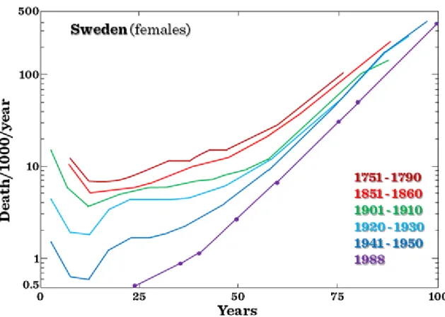
Age-specific mortality has decreased. However, the age maximum ever achieved by human beings has not changed. (Figure I.1-1 and Figure I.1-2)
Figure I.1-1: Survival curves for different populations
Figure I.1-2:Age-specific death rates of Swedish females from 1751 to 1950 and for 1988
Regional differences in mortality or in life expectancy at birth are generally determined by the combination of a huge number of different factors. These differences are known for many regions but are usually compared among nations (Figure I.1-3). One of the very few exceptions is Germany: here the survival conditions are not uniformly distributed over the whole national territory. The East-West differences are due to the special history of these two regions having belonged to completely different political and social regimes for several decades during the last century. The difference remains high despite the political reunion in 1990 (Figure I.1-4).
Figure I.1-3: Expected life-span at birth in different European states
Figure I.1-4:Regional pattern of life expectancy in Germany: East-West difference (2003)
In Europe the number of people over 65 years will increase from 15.5% in 2000 to 24.3% in 2030. These demographic changes have major implications for health care, the labor force, welfare, insurance, and pensions.
1.3. Chronological and biological age
“How old would you feel if you did not know how old you were?”
These two numbers are not necessarily the same. The functional-biological age is determined by physiology rather than chronology. Factors include changes in the physical structure of the body as well as changes in the performance of motor skills and sensory perception.
1.4. Etiology of aging: genetic mechanisms and environmental factors
Aging is a complex process that affects all living organisms. Animals living in the wild are less likely to live long enough to encounter aging, but interestingly all mammalian species (including humans) show very similar aging processes if kept under optimal conditions free from external risk factors like predators or famine. The aging process is multi-factorial, and no single factor has been identified which provides a satisfactory explanation of the phenomenon. During the process of aging, the organism accumulates damage to macromolecules of its own cells and tissues and to its organs. The maximum lifespan for humans is around 120 years, whereas the maximum lifespan of a mouse, commonly used in research as a model for aging, is about four years. Genetic differences between humans and mice that may account for these different rates of aging include efficiency of DNA repair, types and quantities of antioxidant enzymes, and different rates of free radical production.
Mutation rate in humans is about 1 per 107-11 base pair. Chromosome abnormalities, demethylation, as well as defects of protein synthesis also influence aging. The acceptable rate of mistakes (faulty aminoacids in the peptide chain) in protein synthesis is about 5/10.000 amino acids. Elongation factor-1 levels are also low in old populations, just as levels of some types of mRNA, e.g. mRNA for IL-1. The telomere is a region of repetitive DNA sequences at the end of chromosomes, which protects the end of the chromosome from damage during cell division. The telomere regions prevent the degradation of genes near the end of chromosomes by allowing for the inevitable shortening of the chromosome, which necessarily occurs during cell division. This telomere shortening mechanism normally allows cell lines only a fixed number of divisions. Animal studies suggest that this mechanism contributes to aging on a cellular level and sets a limit to lifespan. (It has been described that a certain cell type of a certain species is capable of only a certain number of cell divisions: Hayflick phenomenon.) Changing telomere lengths is usually associated with changing rate of senescence. This telomere shortening, however, might be a consequence of, and not a reason for aging.
Maximum lifespan of a species is determined by the rate of aging, inherent in its genes and also by environmental factors, i.e. high metabolic rate, free radical production, excessive caloric intake leading to high serum glucose level.
Further reading
Holliday R.: Understanding Ageing, Cambridge Univ. Press, Cambridge, New York, 1995 Arking R.: Biology of Aging. Sinauer Assoc.Inc, Sunderland, 1998
Merck Manual of Geriatrics, Eds.: M.H Beers, R. Berkow, MSD Labs, Merck & Co. Inc., Rahway, N.J., 2000
Molecular and Cellular Gerontology. Eds.: Toussaint O., Osiewacz H., Lithgow G., Brack C., Annals NY Acad.
Sci. Vol.908, New York, 2000
Healthy Aging for Functional Longevity – Molecular and Cellular Interactions in Senescence. Eds.: Sang Chul Park, Eun Seong Hwang, Hyun-Sook Kim, Woon-Yang Park, Annals NY Acad. Sci. Vol.928, New York, 2001
2. Adaptation and vulnerability, polymorbidity
2.1. Progressive deficit in adaptative homeostatic mechanisms in the course of aging
Deficient adaptive functions as well as morphological alterations contribute to age-related disorders observed in aged individuals. Young adults are characterized by maximal performance, maximal reserve capacity, optimal achievement of maximal performance via optimal utilization of capacities/ resources. In old age-groups maximal performance of various homeostatic systems decrease to a variable degree due to limited adaptive mechanisms.
Resting physiological normal values of young adults are easy to determine and they show small individual variability. In the course of aging individual variability expands to a great extent, with a small decline in successfully aging individuals and very pronounced falls in enhanced biological aging. Therefore, it is more difficult to determine the “normal range” of parameters of any given homeostatic parameter in older age-groups (Figure I.2-1). Additionally, different homeostatic systems may present different rates of decline within the same individual e.g. prematurely failing vision or graying hair may be observed in people with well-preserved cardio-respiratory fitness.
Figure I.2-1: Age-related changes in different functions
Although most homeostatic systems may function properly under resting conditions e.g. cardiac output, ventilation, or regulation of serum glucose levels, adaptation to enhanced demands (physical exercise) or to changes in external environmental factors (heat or cold) or those of the internal milieu is very limited (Figure I.2-2, Figure I.2-3).
Figure I.2-2: Effect of 50 mmHg increment in systolic blood pressure on heart rate, cardiac index and stroke index in young and old rats
Figure I.2-3: Glucose tolerance tests (50 g glucose p.o.) in different age-groups
In contrast to the maximal performance, maximal reserve capacity, optimal achievement of maximal performance via optimal utilisation of capacities observed in young adults, reserve capacities of old individuals are significantly diminished (Figure I.2-4). Healthy old individuals may well be able to maintain the basic function of vital organ systems, such as the resting cardiac output (CO) of 5 L/min, their capacity to increase the CO in response to physical activity or in a hot or cold environment is very limited. Such limitations of reserve
capacities make old individuals very vulnerable to environmental stress leading to decompensation of the circulation or to exacerbations of chronic diseases. Limited reserve capacities of numerous organ systems within the same individual will also aggravate complications of diseases, the symptoms of which may be ameliorated by the compensation of other organ systems, e.g. anemic tissue hypoxia will become more severe in old age- groups with limited capacity to maintain a hyperdynamic circulation.
Figure I.2-4: Functional and reserve organ capacities in young and elderly
In the elderly, a very delicate balance exists among different organ systems. Disruption of homeostasis by any disease in previosly independent, functional elderly persons is likely to be expressed in the most vulnerable, most delicately balanced systems. Therefore, a disease in older persons manifests itself first as functional loss, usually in organ systems unrelated to the locus of illness. For example, it has long been recognized that disorders may present in a masked or apathetic form and inflammatory intra-abdominal disorders such as appendicitis may not evoke typical symptoms and signs. However, it is far less recognized that presenting symtoms in the elderly may be totally misleading with regard to the nature and the primary location of the disease process. For example, when the patient becomes confused, one thinks immediately of psychoactive drugs or disease processes primarily affecting the brain as a possible cause. In an elderly person, one must also consider such diversive factors as dehydration due to a wide variety of etiologies, infection, cardiac disorders, or intra-abdominal organ disease. In short, the diagnostic logic is different.
Geriatrics has described five entities, so-called geriatric giants that are the major categories of impairment appearing in elderly people leading to serious impairmnent of their quality of life. These include immobility (instability), incompetence (impaired intellect/memory), incontinence, impaired homeostasis, iatrogenic disorders (Figure I.2-5).
Figure I.2-5: Geriatric giants
2.2. Polymorbidity in the elderly
Polymorbidity and related polypragmasia contribute to the development of iatrogenic disorders. The majority of elderly individuals suffer of a large number of chronic diseases affecting various homeostatic and organ systems. Age-related changes in body composition lead frequently to osteoporosis and sarcopenia, diminished insulin sensitivity in the elderly aggravated by age-dependent accumulation of fat mass frequently culminate in type 2 diabetes mellitus, long-term dust and or smoke-exposure result in chronic obstructive lung diseases or silicosis, progressive atherosclerosis will lead to infarctions or chronic atrophy of the myocardium or stroke, to name just a few. For these diseases and abnormalities old individuals take a large number of drugs regularly.
Further reading
Geriatric Medicine. Eds.: C.K. Cassel C.K., D.E.Riesenberg, L.B.Sorensen, J.R.Walsh, Springer-Verlag, New York, Berlin, 1990.
Pharmacological Intervention in Aging and Age-associated Disorders. Eds.: K. Kitani, A. Aoba, S. Goto, Annals NY Acad. Sci. Vol. 786, New York, 1996.
R. Arking: Biology of Aging. Sinauer Assoc.Inc, Sunderland, 1998.
Merck Manual of Geriatrics, Eds.: M.H Beers, R. Berkow, MSD Labs, Merck & Co. Inc., Rahway, N.J., 2000
E.R. Hajjar, A.C. Cafiero, J.T. Hanlon, Polypharmacy in elderly patients. Am. J. Geriatr. Pharmacother. 5: 345- 51. 2007.
3. Nutrition, physical status, body composition, sarcopenia
3.1. Introduction: age-related changes in body weight and body
composition
Aging is accompanied by two major trends in the long-term regulation of energy balance: obesity of the middle- aged and late-appearing anorexia of aging often leading to senile cachexia and sarcopenia. Following a progressive increase in adiposity (Figure I.3-1) first a relative, then an absolute decrease in muscle mass is seen, pointing to the development of sarcopenia (decrease of muscle mass by more than 30%) in aged populations (above the age of 70). At the very end of the aging process adipose tissue is also lost in the process of cachexia.
Both of these opposite disorders have enormous impact on the health status and life expectancy of those affected. Not only the consequences of obesity (metabolic syndrome) are serious, but the cachexia of old people as well: it causes muscle weakness, falls, frailty, functional and later cognitive disorders, a higher risk for decubitus (pressure ulcer) and hip fracture, impaired quality of life, a 3-4-fold increase in the risk of loss of self- reliance (expensive assisted living facilities in old age) and higher mortality. They are important especially in Hungary: although the increase in the ratio of the extreme old population is not fast, in the old groups the rate of biological aging is faster, than in more developed countries. Both the weight gain of the middle aged and the sarcopenia of the elderly are multifactorial in their origin (Figure I.3-2).
Figure I.3-1: Between the ages of 20 and 70 – despite a stable, normal body weight – body composition is altered: fat mass increases (a 2 fold increase is still considered to be physiological)
Figure I.3-2: The pathogenesis and functional vs. metabolic consequences of sarcopenia
3.2. Changes in fat mass (FM) and fat free mass (FFM) with age
Body weight increases gradually by 8-9 kg until 45-55 (this is predominantly an increase in FM with maintained muscle mass), then after a stagnation until the age of 65-75, a decline (1-2 kg/decade) in all tissue types is seen without any apparent cause e.g. a slimming diet. During this period even the FM is decreased somewhat but the loss of muscle mass is dominant. In active athletes the body weight does not increase, the increase of FM with age is blunted (their body fat content is similar to that of young, lean, sedentary individuals). Intensive training decreases abdominal fat. Males have a tendency for visceral fat accumulation, after menopause females too. Fat accumulation does not only mean a simple growth of adipose tissue, but abnormal mesenchymal adipocyte-like default (MAD) cells appear among other cell types e.g. between muscle fibers, in the bone marrow (Figure I.3- 3). Redistribution of lipid to extra-adipose sites with aging could result from loss of lipid storage capacity in fat depots (reduced fat cell size and function), altered fatty acid handling resulting in lipid accumulation, maldifferentiation of mesenchymal precursors into a partial adipocyte phenotype (due to falls of testosterone and IGF, elevated cytokine production and anorexia).
Figure I.3-3: Ectopic fat accumulation with aging: MAD cells are smaller and less insulin responsive than fully differentiated fat cells
The FFM is stable until 40, then it decreases by about 3.5 kg or 3-4%/decade (Figure I.3-4). This change shows small individual differences, the rate of decrease is similar in athletes.
Figure I.3-4: Fat (f), fat-free mass (ffm), and cell mass (cm) of males and females at various ages. (The number of subjects in each age-group is noted.)
The water content of the body changes proportionally with FFM. Water content of the FFM is stable. Bone minerals also change proportionally with FFM. By 65 it decreases by 10-15%. In females the rate of decrease is enhanced after menopause. This dramatic fall can be prevented by estrogen supplemetation. In active athletes the rate of decrease is similar, but starts from a higher peak bone mass. Muscle mass and strength diminishes slowly until 50, then the rate is enhanced (sarcopenia). Between 30 and 80 there is a 30-40% decrease also in athletes. Especially the quick, dynamic contractions are impaired. The number of motoneurons/motor units fall.
The production of muscle proteins decreases, especially that of type II fibers.
3.3. The pathogenesis of sarcopenia
Weight loss seen in the elderly may be associated to anorexia of aging: between 20-70 years of age the basal metabolic rate decreases by less than 20%, on the other hand daily caloric intake decreases by as much as 35%.
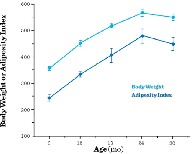
In the elderly, the etiology of poor nutrition includes social (such as poverty and isolation), psychological (especially depression) and physical factors (immobilization or missing teeth), abnormal states (heart failure, malignancies, GI abnormalities, chronic inflammation, infections, drugs, etc.). A large number of cases anorexia develops without any apparent reason (real age-related anorexia). The process is similar in all mammals thus the alterations of energy balance may be of regulatory origin (Figure I.3-5). Individual components of the regulatory systems (e.g. transmitters influencing feeding drive or satiety) may change according to different dynamics, that may explain the abnomalities of the energy balance in middle aged people and in the elderly.
Figure I.3-5: Body weight and adiposity index in rats
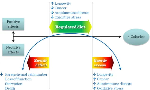
Both insufficient and excessive caloric intake exerts negative physiological effects (Figure I.3-6). It has been observed that among middle-aged and old people the severely undernourished persons showed higher mortality rates than the overweight ones. What is more, among the elderly a slight overweight of 10-20% indicated better survival. The higher body mass index among the elderly means a higher amount of muscle mass, that may reach sufficiently high levels to promote survival. To achieve such a higher body weight with acceptable muscle mass a diet relatively richer in proteins and special muscle (strength) training is suggested.
Figure I.3-6: Hypothetical U-shaped curve over the spectrum of caloric intake from insufficient to excessive calories, emphazing negative physiological effects at both extremes and positive or hormetic effects within a range of normal (regulated) caloric intake
Further reading
Geriatric Medicine. Eds.: C.K. Cassel C.K., D.E.Riesenberg, L.B.Sorensen, J.R.Walsh, Springer-Verlag, New York, Berlin, 1990
Towards Prolongation of the Healthy Life Span. Eds.: D. Harman, R. Holliday, M. Meydani, Annals NY Acad.
Sci. Vol. 854, New York, 1998
Merck Manual of Geriatrics, Eds.: M.H Beers, R. Berkow, MSD Labs, Merck & Co. Inc., Rahway, N.J., 2000
Healthy Aging for Functional Longevity – Molecular and Cellular Interactions in Senescence. Eds.: Sang Chul Park, Eun Seong Hwang, Hyun-Sook Kim, Woon-Yang Park, Annals NY Acad. Sci. Vol.928, New York, 2001 E. Pétervári, S. Soós, M. Székely, M. Balaskó: Alterations in the peptidergic regulation of energy balance in the course of aging. Curr. Protein Pept. Sci. (2011)
4. Immobilization, physical activity, disorders of locomotor organs
4.1. The beneficial effects of physical exercise
In affluent societies the general level of physical activity progressively decreases. This is unfortunate, since physical activity has many advantages regarding physiological functions. It helps to reach or maintain normal, healthy body mass. In active athletes, for example, the usual body weight rise of middle-aged persons is absent.
Regular sport improves body composition, it increases the amount of muscle mass (especially the amount of
type-I, slow, red fibers), the ratio of active tissues, attenuates their age-dependent natural loss. Training programs (12 weeks – 3 times a week) may be able to increase the available muscle mass by 10% even in old age-groups. The greater ratio of active tissues enhances the basal metabolic rate. The trained muscle, during prolonged work (longer than 15-20 min) burns fat. On the surface of muscle fibers lipoprotein-lipase appears, which is able to release fatty acids from the circulating lipoproteins. Regular physical activity suppresses the total-cholesterol level, but elevates the serum HDL-cholesterol concentration. Active skeletal muscles are able to take up glucose by an insulin-independent mechanism. Physical activity recruits GLUT 4 glucose transporter molecules to the surface of muscle cells, just as insulin does. Thus, regular activity decreases the insulin requirement, attenuates the strain on β-cells, and helps to prevent type 2 diabetes mellitus. The thermal adaptation capability improves. In working muscles epinephrine induces vasodilation, thereby it decreases total peripheral resistance, therefore regular exercise helps preventing the development of essential hypertension.
Regular sports in children and young adults increases peak bone mass by applying traction forces on the bones (piezoelectric effect, activating bone formation). The regular physical activity shifts bone metabolism to greater synthesis even at later ages. Regular sports can help in the prevention of osteoporosis, which would carry the dangers of vertebral compression, pathological fractures, fracture of the hip or the neck of the femur. Physical activity has an effect to decrease stress. It has no negative side-effects, and does not evoke pathological dependence, either. According to human surveys, regular physical activity can attenuate the appearance of depression and dementia, which often develop with advancing age. Data obtained in a mouse-model of experimental Alzheimer disease have shown that appropriate increase of physical activity favorably modifies the level of brain-derived neural growth factor (which is a contributing factor in hippocampal atrophy), and also the amyloid formation. A physically active lifestyle decreases the occurrence of certain cancers (colon, breast, uterus, esophagus, prostate) partly by maintaining normal body weight, partly through humoral factors. It influences insulin sensitivity, the level of insulin-like growth factors and estrogen.
4.2. Immobilization syndrome – chronic bedrest
In the course of some diseases a short-time bed rest has some advantages: this rest decreases the burden of the cardiovascular and respiratory systems. In febrile illnesses as part of sickness behavior, the patients do not have only fever, anorexia, decreased fluid intake, enhanced pain-sensation, lethargy, but they are also feeble and inactive, often somnolent. However, chronic bed-rest is harmful rather than advantageous. Some patients cannot avoid chronic immobilization. Loss of lower limbs, paralysis of the lower half of the body due to transversal lesion of the spinal cord or stroke, coma, extreme weakness, severe pains in the joints and severe chronic diseases (e.g. chronic heart failure, COPD), extreme obesity, rheumatic polymyalgy, hypothyroidism may also lead to immobilization. The incidence and the danger of immobilization are especially high in the elderly.
As many as 1/3 of older persons report yearly a fall or tendency to fall, which is the most common cause of accidents in people over 65 years of age and is the leading cause of mortality due to injury in that age-group.
Complications include hip fractures, subdural hematomas and immobility. As a major public health problem, osteoporosis (metabolic bone disorder characterized by a gradual decline in absolute bone mass) also increases susceptibility to fractures especially in the vertebral bodies, the distal radius, and the proximal femur. Bone mass decreases from the age of 55 by around 1%/year in men and by 3-4%/year in women (peak bone mass is reached at 25-35 years of age, its value is higher in men). Inactivity, vitamin D and protein deficiency, hormonal factors (e.g. lower estrogen, secondary hyperparathyroidism, cortisol), alcohol, smoking and certain drugs may accelarate the age-related progressive reduction of bone mass.
Difficulty and unsteadiness in walking, with occasional falls, and stiffness with painful lower limbs are frequently reported by elderly patients and are often related to degenerative joint disease, rheumatoid arthritis or polymyalgia rheumatica. Osteoarthritis is the most common form of joint disease and one of the leading causes of disability in persons above 65 years of age. As a person ages, the water content of the cartilage decreases as a result of a modification of proteoglycan content, thus causing the cartilage to be less resilient. Without the protective effects of the proteoglycans, the collagen fibers of the cartilage can become susceptible to degradation and thus exacerbate the degeneration. Cellular or matrix alterations in cartilage that occur with aging, obesity, trauma, endocrine diseases (e.g. diabetes mellitus) and primary disorders of the joint (e.g.
inflammatory arthritis) predispose older persons to osteoarthritis characterized by progressive joint pain, limitation of movement and joint deformity.
Central and peripheral nervous system disorders (late stage of Parkinson disease or neuropathies) may present with motor symptoms. Cardiovascular, respiratory, endocrine, other systemic illnesses, or dementia, depression, isolation, fear of falling, anxiety, with fatigue and lack of motivation for activities of daily living, often limit exercise performance in the elderly, frequently without intrinsic muscle weakness. Drugs, e.g. sedatives,
narcotics (because of sedative effect), diuretics, antihypertensive medication (in the elderly these may cause orthostatic hypotension, dizziness) also enhance the danger of immobilization.
Consequences of a chronic bed-rest depend on the duration and level of inactivity. In prolonged supine position (as in weightlessness) the circulation is rearranged, on the short run the central blood volume increases, the perfusion and hydrostatic pressure decrease in the lower half of the body, the slightly higher preload and stroke volume lead to bradycardia, renal blood flow increases and slight polyuria develops. On the long run (weeks, months), the plasma volume and the efficacy of orthostatic reflexes (regulating blood pressure) decrease. When the patient is mobilized again, the low blood volume is not enough to maintain brain blood flow in an orthostatic position, therefore orthostatic hypotension develops, the patient is dizzy, eventually faints (Figure I.4-1).
Figure I.4-1: Circulatory adaptation to chronic bed-rest
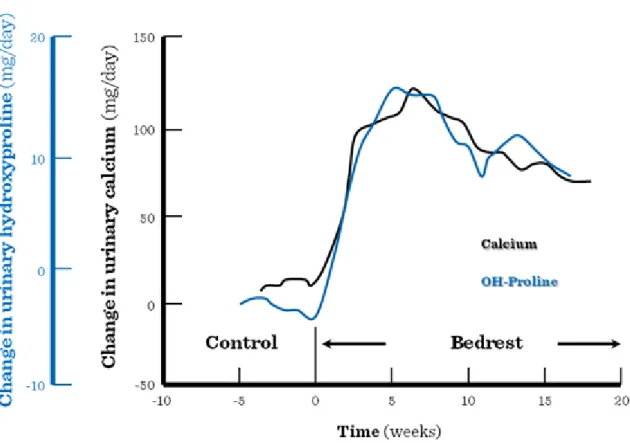
Muscle contractures develop (muscles and joints are less moveable). In case if the upper extremities are affected, the elderly patients lose the capability to eat alone, in case of lower extremities one contracture is enough to cause full immobility. A decrease of muscle mass can be observed already on the short-term, but upon a long-term bed-rest it is significantly enhanced. Immobilization greatly enhances the progression of pre- existing osteoporosis in elderly patients. The enhanced excretion of hydroxy-proline is a sign of increased muscle (protein) catabolism, while the Ca-excretion refers to bone absorption. (Figure I.4-2).
Figure I.4-2: Urinary loss of calcium and hydroxy-proline during chronic immobilization
The maximal capacity for physical work will not be determined by the capacity of the cardiovascular system (as it normally happens), but by the exhaustion of the muscular system, or lack of local substrates (glycogen).
Formation of red blood cells decreases, the low level of total ventilation also lessens the amount of oxygen carried by the arterial blood. The atrophied, deconditioned muscles of poor perfusion take up less oxygen from the blood. The decreased blood volume, decreased muscle tone and mass (decreased filling) and decreased baroreceptor response act to suppress stroke volume. Following chronic bed-rest any physical activity evokes an exaggerated cardiovascular response, e.g. palpitation may appear already at work of low level (and low oxygen consumption). Due to the decreased venous return and hypovolemia the risk for deep venous thrombosis and pulmonary embolism is high. In the elderly population they enhance mortality by about 50%.
The ventilation decreases, the V/Q mismatch becomes pronounced, and the activity of the immune system and the mucociliary clearance of the airways become insufficient. Elderly patients, if bed-ridden for only a couple of days, may develop congestive pneumonia. Surgical fixation of a fractured neck of femur is indicated mainly by faster mobilization and avoiding the pneumonia mortality induced by long (earlier advised for 9 weeks) bed- rest.
In chronic bed-rest the metabolic rate may be 20% lower than normally. The defense against either heat or cold is weaker. Immobilization decreases intestinal motility. The tendency for constipation increases significantly, even impactation may develop, eventually with consequent fecal incontinence.
The prevalence of pressure ulcer is 30% among elderly patients who are bed-ridden or have to stay in a wheel- chair for at least a week. At the points exposed to pressure the skin and the deeper tissues may be damaged in the course of prolonged sitting or lying (Figure I.4-3). Immobilization, fecal-, urinary-incontinence, hypoalbuminemia and shear stress due to the incompetent turning/moving of the patient contribute to the development of pressure ulcers (pressure ulcer staging: Figure I.4-4, Figure I.4-5, Figure I.4-6, Figure I.4-7).
Decubitus causes 4-fold increase in the mortality rate of the patients (sepsis).
Figure I.4-3: Typical points exposed to pressure in immobilization
Figure I.4-4: Stage I of pressure ulcer: lasting erythema on skin surface
Figure I.4-5: Stage II of pressure ulcer: superficial wound, which does not reach subcutaneous tissues
Figure I.4-6: Stage III of pressure ulcer: deep wound affecting the subcutaneous tissues (does not cross the fascia of the muscle)
Figure I.4-7: Stage IV of pressure ulcer: very deep wound, battering also the muscles, bones, joints
4.3. Remobilization in the elderly
Those people who are confined to stay inactive because of an acute disease or are bedridden due to chronic conditions are highly prone to lose their muscle mass and force very quickly. The proportion of the loss can even reach 1.5% per day. The loss is more pronounced in the muscles responsible for sitting up, standing up and standing straight, and therefore, these muscles are essential for everyday life. Gradual mobilization, passive movement and active exercising of joints on a regular basis, proper positioning of patient (prevention of pressure ulcers), replacement of fluids, optimal feeding, regular emptying of bladder, removal of catheter as soon as possible, cleaning of skin and active environment are important therapeutic measures. Certain specialists in geriatric medicine state that one day spent in bed can be compensated by a 2-week workout. Therefore, a personalized exercise program and care must be worked out for every hospitalized, chronically ill patient in order to maintain their physical activity. Maintenance of physical activity as long as possible in the elderly is essential via resistance training and daily activity- and work-oriented special exercises. The ideal frequency, intensity, duration and style of such physical activity have not been fully defined yet. According to the current recommendations, 30-60 min fast walking repeated 3-4 times a week is the most suitable workout during which the pace is slowed down for 5 minutes in every ten minutes.
Further reading
Geriatric Medicine. Eds.: C.K. Cassel C.K., D.E.Riesenberg, L.B.Sorensen, J.R.Walsh, Springer-Verlag, New York, Berlin, 1990
Merck Manual of Geriatrics, Eds.: M.H Beers, R. Berkow, MSD Labs, Merck & Co. Inc., Rahway, N.J., 2000
Healthy Aging for Functional Longevity – Molecular and Cellular Interactions in Senescence. Eds.: Sang Chul Park, Eun Seong Hwang, Hyun-Sook Kim, Woon-Yang Park, Annals NY Acad. Sci. Vol.928, New York, 2001 Hazzard’s Geriatric Medicine and Gerontology (6th ed.), Eds.: J. Halter, J. Ouslander, M. Tinetti, S. Studenski, K. High, S. Asthana, W. Hazzard, McGraw-Hill, 2009.
5. Characteristics of the cardiovascular system, abnormalities and diseases
5.1. Age-related alterations in the cardiovascular system
Aging affects the heart and cardiac functions profoundly. The most widespread morphological changes include dilation of the atria, especially that of the left atrium (even in healthy individuals). The structure of the ventricular myocardium exhibits characteristic proliferation of connective tissue, decrease in cardiomyocyte numbers and enlargement of the remaining myocardial cells with decreased contractility and impaired compliance of the ventricles. Depending on the accompanying diseases the ventricles may show overall hypertrophy, maintained mass or dilation with thinner myocardial wall.
The size of the aging heart shows characteristic changes during the cardiac cycle. Under resting conditions the heart of young adults becomes smaller during systole due to a 15-20% shortening of ventricular fibers. Physical exercise induces significantly stronger contraction that results in an even smaller size of the heart at the peak of contraction compared to contractions at rest. No significant end-diastolic dilation of the ventricle is observed in the young (except in cases of extreme strain). The function of the heart at rest is similar in older individuals.
However, during moderate physical activity a significant adaptive end-diastolic dilation is observed indicating activation of the Frank-Starling mechanism. Due to the limited contractility, in the elderly it is necessary to increase the end-diastolic volume with consequent pronounced increase in end-diastolic pressure (since the ventricular compliance is reduced) that leads to significant venous stagnation (dyspnea and systemic congestive edema formation in the lower limbs) in older individuals (Figure I.5-1). The end-systolic volume during exercise also exceeds that seen in the young.
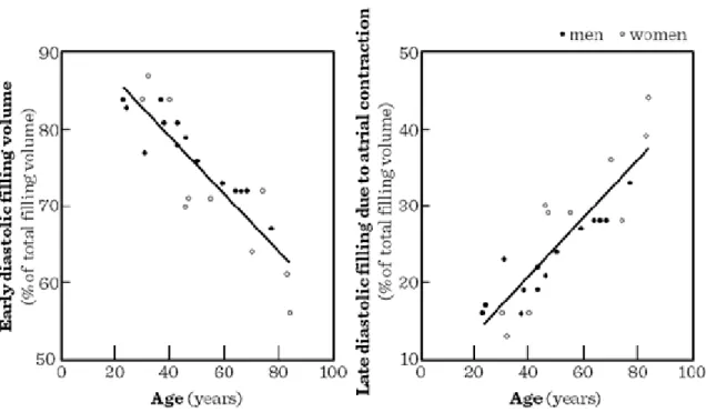
Regarding diastolic filling, the early diastolic function (active distension of the ventricles) shows diminishing significance, while late diastolic functions (dependent on atrial contractions) reach higher significance (Figure I.5-2). Functionally, the maximal heart rate achived by the heart is also diminishing with advancing age (Figure I.5-3). All these alterations in cardiac functions are reflected by the age-related decline both in resting and maximal cardiac output observed in humans (Figure I.5-4). Similar age-related patterns may be observed in maximal oxygen consumption and endurance time during physical exercise, as in healthy individuals, cardiac functions, rather than respiratory ones limit the maximal potential intensity and duration of physical exercise (Figure I.5-5).
Figure I.5-1: Age-related physiological changes in the heart
Figure I.5-2: Comparison between the early diastolic and atrial contribution to left ventricular filling in persons of a broad age range
Figure I.5-3: Maximal heart rate vs. age
Figure I.5-4: Cardiac output measured at rest and at exhausting exercise (upright position) vs. age
Figure I.5-5: Maximal oxygen consumption and endurance times according to age. (data on trained and non- trained men)
Figure I.5-6: Mean aortic pressure and aortic pulse wave velocity vs. age in rural and urban populations
Figure I.5-7: The interplay of vascular and adaptive cardiac changes during aging
Age-associated abnormalities of the vascular system also contribute to the decline in cardiovascular functions in the elderly. Atherosclerosis of the small and large vessels grow more progressive with age. As a result, total peripheral vascular resistance increases with age. To this increase, frequent adaptive activation of the sympathetic nervous system also contributes sigificantly, made necessary (sometimes even at rest) by reduced contractility. The wall of large vessels especially that of the aorta grows progressively rigid and distended. Thus, aortic elastic properties essential in maintaining optimal diastolic flow and pressure become severly impaired.
As a result, systolic pressure rises excessively and diastolic pressure drops abnormally (impairing coronary perfusion pressure). Aortic pulse wave velocity increases significantly with age (Figure I.5-6) causing abnormal, harmful wave reflections within the circulatory system impairing coronary blood flow further. Figure I.5-7 summarizes the complex system of cardiac and vascular changes during aging.
Further reading
Geriatric Medicine. Eds.: C.K. Cassel C.K., D.E.Riesenberg, L.B.Sorensen, J.R.Walsh, Springer-Verlag, New York, Berlin, 1990
Handbook of Physiology (Section 11): Aging. Ed.: E.J. Masoro, Oxford University Press, New York, Oxford, 1995.
Merck Manual of Geriatrics, Eds.: M.H Beers, R. Berkow, MSD Labs, Merck & Co. Inc., Rahway, N.J., 2000
T. Hagen: Mechanisms of Cardiovascular Aging, Volume 11 (Advances in Cell Aging and Gerontology).
Elsevier, Amsterdam, 2002.
Physiological Basis fof Aging and Geriatrics. Ed.: P.S. Timiras, INFRMA-HC, 2007.
Hazzard’s Geriatric Medicine and Gerontology (6th ed.), Eds.: J. Halter, J. Ouslander, M. Tinetti, S. Studenski, K. High, S. Asthana, W. Hazzard, McGraw-Hill, 2009.
6. Changes of the respiratory system, frequent diseases
6.1. Age-related alterations in the chest and in the lungs
The lungs (via their large respiratory surface of 70-90 m2) are exposed to damaging effects of the environment all through life. These effects lead to morphological as well as functional abnormalities in the respiratory system.
Mechanics of breathing involve the compliance of the lungs, that of the chest and the activity of respiratory muscles. With age, the elastic recoil of the lungs diminishes due to progressive destruction of elastic fibers and remodeling of the parenchyma induced by inflammatory processes upon prior activation induced by harmful
environmental stimuli and/or age-related mechanisms. Alveolar airspace enlargement is also associated with this process. The chest becomes more rigid at the same time, the respiratory muscles grow weaker from the age of 55 resulting in a progressive increase in the functional residual capacity (the amount of air in the lungs at the end of a normal expiration). Total lung capacity (TLC, the amount of air in the lungs at the end of maximal inspiration) may also increase in healthy old individuals (Figure I.6-1) leading to the development of aging- associated emphysema and barrel chest.
Even more frequently osteoporosis induces the compression of the vertebrae, that enhances the dorsal kyphosis (“dowager’s hump”, Figure I.6-2). Consequent severe reduction of the TLC indicates a restrictive ventilatory disorder in the elderly.
Figure I.6-1: The thorax in the elderly
Figure I.6-2: Progressive loss of height from vertebral fractures causes a protuberant abdomen and upper back curvature (“dowager’s hump”)
6.2. Age-related alterations in the airways
Cumulative effects of inflammatory processes activated by noxious agents throughout life induce progressive airway inflammation and an increase in airway resistance that is different from classical chronic obstructive pulmonary diseases (COPD), although the prevalance of the latter also increases in the course of aging.
Additionally, age-associated remodeling of the lung parenchyma, reduction of elastic fibers (that are attached to the walls of small airways, anchoring them to neighbouring structures and keeping them from collapsing during expiration) makes small airways increasingly prone for collapse during expiration. Based on these mechanisms, forced expiratory volume in one second (FEV1) shows an age-related decline throughout life (Figure I.6-3).
Smoking enhances this decline significantly in susceptible individuals.
Figure I.6-3: Changes in airflow during aging (FEV1 = forced expiratory volume in 1 second)
6.3. Abnormalities of other respiratory functions in the elderly
Ventilation/perfusion ratio of young adults aprroaches the optimal value of 1.0. In the elderly, obstruction of small airways with maintained perfusion of hypoventilated alveolar regions enhance dead space ventilation.
Diffusion capacity also declines by about 0.5%/year, due to destruction of interalveolar septa with consequently diminished respiratory surface and fibrosis-induced thickening of the diffusion membrane.
Regulation of respiratory functions also show characteristic age-related alterations. Diminishing responsiveness of the respiratory center to hypercapnia and hypoxia-induced stimuli are thought to be responsible for the small but steady reduction in arterial partial oxygen pressure observed in the course of aging.
6.4. Diseases of the respiratory system with increased prevalence in old age-groups
Due to prolonged exposure to cigarette smoke, to occupational dust and/or gas exposure or age-related suppression in protective antiprotease alpha1-antitrypsin activity, COPD develops with increasing frequency and severity in the elderly. The majority of the patients suffer from chronic bronchitis, a small but significant minority develop different types of emphysema. COPD is currently the 5th most frequent cause of death, but with the present trends it will advance to 3rd place in 15 years.
Bronchial asthma begins typically in children and young adults, but aging induces characteristic changes in this disease group. The previously reversible airway obstruction becomes increasingly irreversible. Thus, the difference between chronic bronchitis and bronchial asthma diminishes with age.
Pneumonias develop with increasing frequency in older individuals due to suppression of immune defence mechanisms in the lungs. The symptoms of pneumonia are not always specific: in the elderly incontinence or confusion may be the dominant sign. A majority (70%) of lethal pneumonias occur in old individuals.
Tuberculosis (TBC) also affects the elderly more frequently, even reactivation of long-healed TBC may be observed in old age-groups.
As a result of the above mentioned respiratory disorders, many elderly persons (about 5% of the population above 50 years of age) suffer from chronic respiratory failure.
The highest prevalence of lung tumors is also found in aged populations. In addition to life-long accumulation of the consequences of harmful stimuli, diminished airflow has also been shown to contribute indepently to cancer risks.
Aging aggravates various predisposing factors to pulmonary embolism. Immobilisation, visceral obesity, varicose veins, hemoconcentration, polyglobulia induced by chronic hypoxia, etc. increase the risk of deep venous thrombosis and consequent pulmonary embolism. Difficulties in diagnosis make this abnormality one of the frequent causes of death in the elderly.
Further reading
Geriatric Medicine. Eds.: C.K. Cassel C.K., D.E.Riesenberg, L.B.Sorensen, J.R.Walsh, Springer-Verlag, New York, Berlin, 1990
Handbook of Physiology (Section 11): Aging. Ed.: E.J. Masoro, Oxford University Press, New York, Oxford, 1995.
Merck Manual of Geriatrics, Eds.: M.H Beers, R. Berkow, MSD Labs, Merck & Co. Inc., Rahway, N.J., 2000
Physiological Basis fof Aging and Geriatrics. Ed.: P.S. Timiras, INFRMA-HC, 2007.
Hazzard’s Geriatric Medicine and Gerontology (6th ed.), Eds.: J. Halter, J. Ouslander, M. Tinetti, S. Studenski, K. High, S. Asthana, W. Hazzard, McGraw-Hill, 2009.
7. Changes of renal function, electrolyte/water and acid/base homeostasis
7.1. Aging vs. nephron dysfunctions
In the elderly renal mass, renal blood flow, the number of functioning nephrons decrease leading to both glomerular and tubular dysfunctions. The glomerular filtration rate (GFR) also decreases progressively with age (Figure I.7-1). By the age of 80, GFR may decrease to 50%, this results in a tendency for azotemia due to fall of kidney perfusion (thirst, heat, cardiac output redistribution e.g. heart failure), but often without proportional rise in serum creatinine level (less muscle lost). In earlier stage of the chronic renal insufficiency the hyperfiltrating nephrons may compensate the hypofiltrating ones and maintain the total GFR at a normal level, but later the hyperfiltrating glomeruli may be destroyed due to high glomerular filtration pressure (Figure I.7-2). The glomeruli become more and more sclerotic, the basement membrane gets thicker (degeneration) leading to proteinuria (even in nonhypertensive, nondiabetic elderly). High protein intake and hyperproteinemia are associated with hyperfiltration and promote progression of glomerulosclerosis.
Figure I.7-1: Age-related reduction in the glomerular filtration rate. Because of the loss of functioning nephrons, the progressive decrease of GFR exceeds that of the renal blood flow
Figure I.7-2: Age-related changes in single nephron GFR (SNGFR) in% of total nephrons
The impaired tubular function (decrease in the function of the thick ascending limb of the loop of Henle where the reabsorption of Na-K-Cl without water takes place and impairment of the corticomedullary osmotic concentration gradient, (Figure I.7-3 and Figure I.7-4) leads to hyposthenuria (Figure I.7-5). A disturbance of concentrating ability may be regarded as a defect of water-retention, while a limitation of diluting ability may be regarded as a defect of water-excretion. Limited capacity of either process can cause severe clinical consequences.
Figure I.7-3: Concentration and dilution are interconnected in different segments of the nephron (purple arrows indicate active Na-reabsorption, the green ones the passive water reabsorption)
Figure I.7-4: Changes of osmotic pressure and fluid volume along the nephron. Without ADH, large volume of diluted urine, in case of high ADH levels small volume of concentrated urine is formed. The possible limits of dilution and concentration are determined in the loop of Henle. In case of hyposthenuria (interrupted line), the concentration-changes are moderate in the loop, and the renal concentration gradient decreases.
Figure I.7-5: Development of hyposthenuria, isosthenuria: less and less deviation from the specific gravity of the filtrate during both concentration and dilution
Although the ADH production may be maintained or even increased in elderly, in response to ADH the increase in the specific gravity of the urine is diminished due to decreased nephron numbers and dysfunctional receptors (Figure I.7-6). This may lead to water loss and hypertonicity. On the other hand, suppression of ADH is delayed, therefore hypotonicity (hyponatremia) may also develop, e.g. water intake (exceeding the decreased excretion capacity) may lead to “water intoxication”. Tubular effect of aldosterone is also impaired, but there is a tendency for K-loss and hypokalemia due to frequently occuring secondary hyperaldosteronisms in the elderly.
Glucose reabsorbing proximal tubular cells still function, therefore glucosuria in old people does not reflect serum glucose level dependably. Decreased kidney perfusion (frequently occuring in elderly e.g. due to circulatory redistribution in heart failure, exsiccosis) and impaired tubular excretion of substances enhance the risk for drug intoxication. The dose of drugs that are eliminated through the kidney has to be decreased.
Figure I.7-6: Age-dependence in the renal effect of ADH (the urine-plasma ratio of inulin concentration characterizes the renal concentration process)
7.2. Aging vs. non-excretory kidney functions
Defective renal regulation of blood pressure in aged persons enhances the tendency for hypertension (sclerosis of a. renalis and atrophy of renal parenchyma may lead to renovascular and renoparenchymal hypertension), but frequently occuring hypovolemia may cause hypotension. Deficient erythropoietin production (due to reduced renal parenchyma and gonadal hormone secretion) leads to anemia, decreased renal formation of active vitamin D to bone abnormalities (senile osteoporosis).
7.3. Renal failure in the elderly
Besides age-related renal changes (decrease of renal blood flow, GFR, and of ability to concentrate or to dilute urine), diabetes mellitus, hepatic cirrhosis, congestive heart failure, drugs may increase the incidence of acute renal failure induced by acute tubular necrosis in the eldely. Chronic ischemic renal disease and progressive damage of the renal parenchyma lead to chronic renal failure. Diabetes mellitus, hypertension, hyperlipidemia and obesity are the most important risk factors. The most common indication of dialysis due to chronic renal failure is diabetic nephropathy (35-40%). With higher capacity of dialysis, the age-related limits of dialysis have faded away. Among the dialyzed there are less candidates for transplantation due to co-morbidity.
7.4. Urinary incontinence in the elderly
In the elderly, the muscles of the urinary bladder and pelvic floor tend to weaken, the capacity of the bladder reduces which leads to frequent urination. It is often accompanied by incontinence, i.e. involuntary loss of urine.
In functional incontinence, the patient is not able to control his bladder due to altered circumstances (e.g.
disability, impaired vision, dementia, bigger amount of urine induced by diuretics, diabetes mellitus). Urethral sphincter insufficiency due to weakness of pelvic floor musculature, obesity, prolapsed uterus, atrophic vaginitis, bladder hernia result in involuntary loss of urine upon elevated intra-abdominal pressure (stress incontinence). Overflow incontinence is an unexpected urine loss from the overfilled bladder (urinary retention) e.g. due to benign prostatic hyperplasia, weakness of muscles of the bladder. In cystitis urge incontinence may occur (sudden, unexpected urge to void after certain stimuli). Urinary infections in the elderly often appear with
symptoms of impaired physical and/or mental status. Sepsis can develop quickly and atypically. The treatment of a urosepsis is extremely difficult.
7.5. Electrolyte and water balance in the elderly
In elderly persons the spontaneous water intake decreases. Their regulation is insufficient e.g. their thirst sensation is impaired. Following water deprivation fluid replacement is slower and incomplete. (In old animals the angiotensin II-induced water intake is smaller than that seen in young animals. The dypsogenic effect of ADH is weak.) Upon water deprivation or salt and water loss, severe hypovolemia and hypertonicity develops.
This can also contribute to the development of orthostatic hypotension in the elderly. Salt/water loss, diuretic therapy, inappropriate excess of ADH (e.g. operation, pain), water intake (exceeding the decreased excretion capacity) cause dangerous hypotonicity. Hypotonicity may lead to cerebral edema, nausea, convulsions, muscle cramps, 6-8 times higher all-cause mortality. On the other hand, upon salt and/or water load a fast elevation of blood pressure can also be observed. Too fast fluid replacement in exsiccosis may result in acute heart failure and pulmonary edema.
Besides age-related changes of renal structure and blood flow, altered responsiveness to hormones plays a role in impaired salt and water balance. The same decrease in plasma volume elicits a smaller RAAS (renin- angiotensin-aldosterone system) activation than in young individuals. The effects of aldosterone, angiotensin, or ADH are diminished compared to those in young adults. Elderly patients cannot properly protect themselves against salt/water overload either. Suppression of baseline RAAS or ADH activity is delayed; activation of natriuretic factors is inefficient (atriopeptin level is high, but effects are blunted).
Overdose of drugs containing potassium, renal failure, cell lysis, use of potassium sparing diuretics in renal failure, side-effect of NSAIDs and hypoaldosteronism are most common causes of hyperkalemia in the elderly.
High potassium level results in fatigue, muscle weakness, paresthesias in the lower limbs, metabolic acidosis, changes in the mental status, bradycardia and conduction blocks. Hypokalemia appears usually as a result of insufficient potassium intake, increased loss due to diuresis, vomiting, primary or secondary hyperaldosteronism (e.g. edema). Its clinical signs are muscle weakness, muscle cramps, sleepiness, changes in the mental status, metabolic alkalosis, as a potential complication paralytic ileus or ventricular fibrillation may occur.
7.6. Aging vs. pH disturbances
The normal pH value does not change with age, but aging-associated alterations in its regulation may contribute to development of disturbances in acid-base homeostasis. Diabetic ketoacidosis, lactic acidosis, decreased erythropoietin production (anemia), salicylate-toxicosis, diarrhea, renal failure, renal tubular acidosis are the most frequent causes of metabolic acidosis in the elderly. Compensation by hyperventilation is weaker, because of the decreased sensitivity of the respiratory center (for CO2, hypoxia and H+). The aging kidney shows an impaired reaction to acidosis, therefore, it takes longer to normalize pH.
Vomiting, secondary hyperaldosteronism (e.g. chronic congestive heart failure with edema), diuretic-induced hypokalemia and secondary hyperaldosteronism (aggravating already existing secondary hyperaldosteronism of patients with heart failure) may cause metabolic alkalosis in the elderly. Hypokalemia promotes alkalosis by both internal K+-balance (cellular H+/K+ exchange) and external K+-balance (bicarbonate reabsorption in the proximal tubules, in the distal tubules Na+/H+ exchange is emphasized).
Respiratory acidosis may also occur in the elderly. The respiratory center is less sensitive to hypercapnia and to impulses originating from hypoxia (by the age of 70, the sensitivity to hypoxia decreases by 50%, to hypercapnia by 40-50%; arterial pO2 decreases by 0.3% per year). Medications decreasing the sensitivity of the respiratory center, as well as decreased vital capacity and FEV1, decreased chest wall compliance (kyphoscoliosis, obesity), neuromuscular diseases can worsen the respiratory function. Chronic bronchitis is more frequent in older individuals (impaired mucociliary clearance, longer exposition time to environmental pollutants, smoking).
Hypoxia, sepsis, pulmonary embolism, heart failure (enhanced sympathetic tone), liver failure (NH3 accumulation), mild salicylate-toxicosis (regular use of NSAIDs for pain), common situations with anxiety are common causes of respiratory alkalosis in the elderly.
In the elderly mixed acid-base disturbances are also very common. In acute respiratory insufficiency (e.g.
pneumonia) combined with heart failure respiratory acidosis is mixed with metabolic acidosis. In serious heart
failure a decreased tissue perfusion leads to lactate (metabolic) acidosis, but diuretic therapy influences the balance towards metabolic alkalosis.
Compensatory capacity of both the kidneys and the lungs is narrowed. In respiratory acidosis, oxygen therapy may be needed. Its danger: due to decreased CO2-sensitivity hypoxia regulates ventilation – oxygen therapy may result in hypoventilation, further CO2 accumulation and CO2 coma. Assisted ventilation may be necessary.
Further reading
Geriatric Medicine. Eds.: C.K. Cassel C.K., D.E.Riesenberg, L.B.Sorensen, J.R.Walsh, Springer-Verlag, New York, Berlin, 1990
Handbook of Physiology (Section 11): Aging. Ed.: E.J. Masoro, Oxford University Press, New York, Oxford, 1995.
Merck Manual of Geriatrics, Eds.: M.H Beers, R. Berkow, MSD Labs, Merck & Co. Inc., Rahway, N.J., 2000 Physiological Basis fof Aging and Geriatrics. Ed.: P.S. Timiras, INFRMA-HC, 2007.
Hazzard’s Geriatric Medicine and Gerontology (6th ed.), Eds.: J. Halter, J. Ouslander, M. Tinetti, S. Studenski, K. High, S. Asthana, W. Hazzard, McGraw-Hill, 2009.
8. Changes of the endocrine system and metabolism
8.1. Age-related alterations in the endocrine system
An important role of the aging endocrine system is widely assumed in the background of various age-related alterations, e.g. in body composition, in essential organ functions, in affective disorders of the elderly, etc.
(Figure I.8-1). Responsiveness to hypothalamic releasing factors or that of pituitary troph-hormones have been shown to decrease with age. Many hormones have age-dependent normal values. Frequent attempts to use hormone replacement to delay or reverse aging have been made.
8.1.1. Sex hormones
The most spectacular age-related alterations may be observed in the field of sex hormones. Menopause, the sudden decline in estrogen and inhibin levels in females around 50 years of age, that lead to a rise in follicle- stimulating and luteinizing hormones (FSH and LH, respectively) has been associated with hot flashes, osteoporosis, autonomic and emotional dysfunctions. Andropause, the slow and progressive suppression of testosterone may play a role e.g. in the osteoporosis and sarcopenia of the elderly. In males and females alike diminished production of weak androgens, such as dehydro-epiandrosterone (DHEA) associated with
“adrenopause”, (the failing activity of the adrenal cortex) is likely to contribute to bone resorption and loss of muscle mass/strength. Hormone replacement therapies in the field of sex steroids have been shown to prevent certain age-related dysfunctions and related symptoms.
8.1.2. Synchropause
Healthy, young individuals (humans and mammals) show a characteristic daily pattern called circadian rhythm regarding body temperature, activity, blood pressure (BP), endocrine functions (e.g. release of GH, ACTH, etc.), sleep, etc. In the elderly, such circadian rhythmicity becomes disturbed, most frequently affecting sleep, activity and blood pressure. Disturbances of sleep frequently appear as advanced sleep-phase syndrome (due to an early onset of sleep around 6-8 p.m., the patient wakes up very early in the morning between 3-5 a.m.). Sleep disturbances may lead to non-dipper blood pressure pattern (night-time BP is higher and not lower than the day- time level). In animal studies the otherwise strict circadian rhythm of food intake was also altered (food intake of old rodents was not restricted to the night). Although, the pathogenesis is unknown, decline in melatonin production of the pineal gland is assumed. Low day-time activity, prolonged daily bed-rest may also play a role in the development. Repeated use of sleeping pills may even further aggravate the disorder. Whereas there is no cure for aging-associated disturbances of circadian rhythm, benefits were shown using some therapeutic measures, e.g. bright light therapy in the morning, behavior and chronoterapy (adjusting activity/light and avoiding coffee/nicotine and other stimulation before desired sleeping time), a significantly increased level of
day-time physical activity (e.g. a fitness training program for 3 months) or melatonin administration in the evening.
8.1.3. The growth hormone (GH), insulin-like growth factor (IGF) system
Aging is associated with declines in spontaneous overnight GH-secretion, a reduced GH amplitude and low serum IGF-I levels. Changes in body composition with age are similar to those observed in patients with the adult GH-deficiency syndrome. Administration of GH to the latter group of patients has significantly improved body composition, muscle strength, functional performance and quality of life.
8.1.4. Adrenal cortex
Following the second and third decade of life, there is a continuous decline of adrenal androgen production and the term “adrenopause” has been coined to reflect this.
Adrenal androgens (DHEA and DHEA-S), are the most abundant steroid hormones in the human body, yet we know little about the function of these hormones. Studies utilizing the supplementation of DHEA in autoimmune diseases and Addison's disease provided promising results, demonstrating clear benefits. The issue of replacing DHEA in elderly remains controversial with some reports demonstrating conflicting results. Elderly men with a physiological decline of DHEA did not benefit from DHEA replacement in contrast to women with adrenal failure.
8.1.5. Thyroid gland
Goiter with one or more nodules means the most common endocrine abnormality in the elderly. Its incidence increases proportionally with age. Hyper- or hypothyroidism also occurs more frequently in older populations.
Hyperthyroidism is associated with toxic adenoma or toxic multinodular goiter in iodine-deficient regions, whereas Graves-Basedow disease is the most common form observed in regions with optimal iodine intake. In the elderly, hyperthyroidism does not show those spectacular symptoms characterizing young adults suffering of the disease. No sympathetic hyperactivity is observed, on the other hand, weight loss, weakness, cardiological complications (palpitation, arrhythmias, atrial fibrillation) dominate the clinical picture.
Hypothyroidism is frequently overlooked, its symptoms are often considered to be signs of “age-related decline”. The patients report weakness, somnolence, slow reactions, sensitivity to cold, loss of memory, constipation. Hypercholesterolemia is a frequent finding. Upon diagnosis and treatment, the symptoms are reversible.