CHARACTERISING HUMAN PLURIPOTENT STEM CELLS-DERIVED ENDOTHELIAL CELLS
PhD thesis
dr. Edit Gara
Doctoral School of Basic Medicine Semmelweis University
Supervisor: Dr. Gábor Földes PhD
Official reviewers: Dr. Melinda Pirity PhD Dr. János Réthelyi PhD
Head of the Final Examination Committee:
Dr. Péter Ferdinandy PhD DSc MBA Members of the Final Examination Committee:
Dr. Lívia Jánoskuti PhD Dr. Ágota Apáti PhD
Budapest
2015
1
TABLE OF CONTENTS
1. Abbreviations 4
2. Introduction 8
2.1. Current applications and future challenges in cell therapy 8
2.2. Types of stem cells 14
2.2.1. Circulating progenitor cells 14
2.2.2. Mesenchymal stem cells 15
2.2.3. Cardiac stem cells 18
2.2.4. Embryonic stem cells 19
2.2.5. Induced pluripotent stem cells 21 2.3. Cardiovascular derivatives of stem cells, preclinical and early
clinical results 25
2.3.1. Clinical studies investigating endothelial progenitor cells 25 2.3.2. Clinical results with mesenchymal stem cells for cardiac
regeneration 26
2.3.3. Clinical studies with cardiac stem cells for cardiac
regeneration 29
2.3.4. Preclinical and early clinical studies with embryonic stem
cells in the cardiovascular field 30
2.3.5. Preclinical results and future challenges with induced
pluripotent stem cells 33
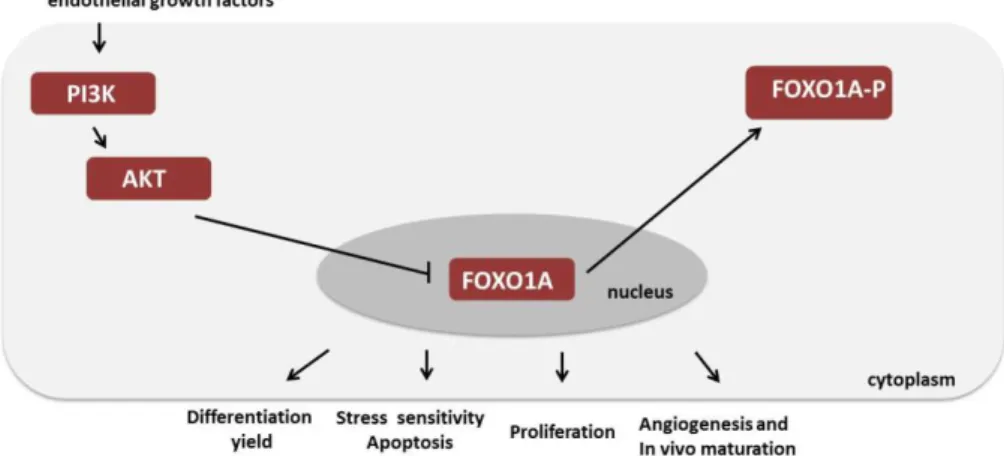
2.4. Tissue engineering for cardiovascular repair 35 2.5. Endothelial function and differentiation 38 2.6. Phosphatidylinositol 3-kinase (PI3K)-Forkhead box O
transcription factor 1A (FOXO1A) signalling pathway 41
3. Aims 43
4. Materials and methods 44
4.1. Human pluripotent stem cell culture 44
4.2. Generating human induced pluripotent stem cells 45
4.3. Characterising pluripotency 46
4.4. Endothelial differentiation 47
4.4.1. Flow cytometry and cell sorting 49
2
4.5. Endothelial cell culture and cell isolation techniques 51 4.5.1. Isolation and culture of blood outgrowth endothelial
cells 51
4.5.2. Isolation of human umbilical vein endothelial cells 52
4.6. Three-dimensional culture system 52
4.6.1. Bioscaffolds 52
4.6.2. Bioreactor system 54
4.7. Endothelial characterisation 54
4.7.1. Immunocytochemistry 54
4.7.2. Immunohistochemistry 55
4.7.3. Matrigel tube formation assay 57
4.7.4. Assessment of endothelial alignment under sheer stress 57
4.7.5. Ac-LDL uptake 58
4.7.6. Gene expression analysis, PCR 58
4.7.7. Proteome profiling assay 59
4.7.8. ELISA measurements 60
4.8. Modulation of PI3K/FOXO1A signalling pathway in hESC-EC 60 4.8.1. Cell treatments with PI3K inhibitor, FOXO1A small
interfering RNA, and FOXO1A plasmid 60 4.8.2. Measurement of cell viability 61
4.8.3. Matrigel tube formation assay 61
4.8.4. PI3K/Akt PCR array 61
4.9. Antiplatelet functional assay 62
4.9.1. Preparation of platelet rich plasma 63
4.10. In vivo experiments 63
4.11. Statistical analysis 64
4.12. Ethical considerations 64
5. Results 66
5.1. Characterisation of human pluripotent stem cells 66 5.2. Reprogramming human adult fibroblasts to pluripotent state 67 5.3. Characteristics of human pluripotent stem cells-derived
endothelial cells 70
3
5.4. Role of differentiation conditions on endothelial marker genes and arterial and venous endothelial subpopulations 80 5.5. Role of PI3K/Akt/FOXO1A pathway on endothelial
differentiation 82
5.6. Modulation of PI3K/FOXO1A signalling pathway in sorted
hESC-EC 85
5.7. Differentiation conditions and PI3K/FOXO1A pathway
modulate angiogenic activity of hESC-EC 86 5.8. Effects of PI3K/FOXO1A pathway on arterial,
venous and general endothelial gene expressions 91 5.9. Effects of PI3K/FOXO1A pathway on hESC-EC
proliferation 92
5.10. Effects of PI3K/FOXO1A pathway on hESC-EC viability 93 5.11. Modulation of endothelial gene expressions and
anticlotting function in 3D cultures 94
5.12. Transplantation of hESC-EC, hiPSC-EC and HUVEC in vivo 96
6. Discussion 98
6.1. Endothelial characteristics and functional activity 100
6.1.1. Proteomics 100
6.1.2. Anticlotting propertis of hESC-EC and hiPSC-EC 101 6.2. Arterial and venous endothelial subpopulations 102 6.3. Regulatory mechanisms of endothelial differentiation, behaviour
and specifications via the PI3K/FOXO1A signalling pathways 104
6.4. Limitations 108
7. Conclusions 109
8. Summary 111
9. Reference list 113
10. Publications 138
11. Acknowledgements 140
4 1. Abbreviations
6MWT six-minute walk test
ACh acetylcholine
ACEi angiotensin converting enzyme inhibitor Ac-LDL acetylated low density lipoprotein
AE adverse event
Akt protein kinase B
ALS amyotrophic lateral sclerosis
BB beta receptor blocker
BM bone marrow
BNP B-type natriuretic peptide
BOEC Blood outgrowth endothelial cells cDNA complementary deoxyribonucleic acid CGMP current good manufacturing practice
CM cardiomyocytes
CSC cardiac stem cells
DAPI 4', 6-diamidino-2-phenylindole - dihydrochloride
DCM dilated cardiomyopathy
DLL4 Delta-like ligand-4 DMSO dimethyl-sulfoxide
EB embryoid body
EC endothelial cells
EBM endothelial basal medium ECM extracellular matrix
EGM endothelial growth medium EHT engineered heart tissue
ELISA enzyme-linked immunosorbent assay ET-1 endothelin-1
5
eNOS endothelial nitric oxide synthase EPC endothelial progenitor cells ERC endometrial regenerative cells FACS fluorescence-activated cell sorting FDA Food and Drug Administration
FOXO1A Forkhead box O transcription factor 1A FBS foetal bovine serum
GABA gamma amino butyric acid
GAPDH glyceraldehyde 3-phosphate dehydrogenase GVHD graft-versus-host disease
HAEC human aortic endothelial cells
HCAEC human coronary arterial endothelial cells hESC human embryonic stem cells
hESC-EC human embryonic stem cells -derived endothelial cells hESC-CM human embryonic stem cells-derived cardiomyocytes HFpEF heart failure with preserved ejection fraction
hiPSC human induced pluripotent stem cells
hiPSC-EC human induced pluripotent stem cells-derived endothelial cells HLA human leucocyte antigen
HOCM hypertrophic obstructive cardiomyopathy hPSC human pluripotent stem cells
HUVEC human umbilical cord endothelial cells I-DCM ischemic dilatative cardiomyopathy iNOS inducible nitric oxide synthase ITGB1 integrin beta-1
IVF in vitro fertilisation
LVEF left ventricular ejection fraction
MI myocardial infarction
6
MLHFQ Minnesota living with heart failure questionnaire MRI magnetic resonance imaging
MSC mesenchymal stem cells
MS multiple sclerosis
NA not applicable
NEC non-enzyme control
nNOS neural nitric oxide synthase NOS nitric oxide synthese NTC non-templates control PBS phosphate buffered saline
PCI percutaneous coronary intervention PCR polymerase chain reaction
PDGFRA platelet-derived growth factor receptor alpha PI3K phosphatidylinositol-4, 5-bisphosphate 3-kinase PIK3R1 phosphoinositide-3-kinase regulatory subunit 1 beta PIK3R2 phosphoinositide-3-kinase regulatory subunit 2 beta PPP platelet poor plasma
PRKCA protein kinase C alpha PRKCB protein kinase C beta PRKCZ protein kinase C zeta PRP platelet rich plasma PSC pluripotent stem cells
PSC-EC pluripotent stem cell-derived endothelial cells PTEN phosphatase and tensin homolog
qRT-PCR quantitative real-time polymerase chain reaction rh-bFGF recombinant human basal fibroblast growth factor rh-TGFβ recombinant human transforming growth factor beta RNA ribonucleic acid
7 siRNA small interfering RNA SNP sodium nitroprusside
SPECT single-photon emission computed tomography STEMI ST segment elevation myocardial infarction VEGF vascular endothelial growth factor
vWF von Willebrand factor WBC white blood cells WSS wall sheer stress
8 2. Introduction
2.1. Current applications and future challenges in cell therapy
Cell therapy is the focus of medical research for developing novel therapeutic strategies. Degenerative diseases affect large number of patients placing high burden on health and social care systems. Many patients suffer from severe neurodegenerative diseases (e.g. Parkinson‟s disease, amyotrophic lateral sclerosis (ALS), and multiple sclerosis (MS)) and ischemic cardiovascular events (stroke or myocardial infarction (MI)).
Cardiovascular diseases are the leading cause of death in industrialised countries.
Pathophysiology of these diseases involves definitive necrosis of cells with a crucial role in the physiology of the human body (circulation, central or peripheral nervous system). The regenerative capacity of neurons is limited, similarly as cardiomyocytes [1, 2]. Many patients suffer severe decrease in quality of life due to a lack of definitive treatment.
Insulin-dependent diabetes mellitus may develop in very young children and so far no definite therapy exists; treatment options focus on substitution of pancreatic β cell product, insulin. Preclinical research and early cell therapy trials are underway in these medical conditions. Regenerative medicine and tissue engineering may establish definitive treatments to cure these conditions.
Cell therapy is being routinely used in special clinical conditions. Bone marrow transplantation is performed frequently in haemato-oncological diseases, e.g. leukaemia and lymphomas. It is carried out through both autologous and allogeneic route.
In severe graft-versus-host disease (GVHD) mesenchymal stem cells (MSC)-derivatives are transplanted if the condition is resistant to steroid shot therapy. The Remestemcel-L® (Prochymal, Osiris) product is available in the US and Canada, and was approved by the FDA. Remestemcel-L® contains MSC from healthy adults, and applied intravenously in GVHD. Remestemcel-L® is a novel, of-the-shelf available cell therapy [3].
Umbilical cord blood contains multipotent stem cells (mononuclear haematopoietic stem cells and mesenchymal stromal cells), similar to MSC. Several commercial companies are offering collection and bio-banking of umbilical cord blood after birth. The treatment costs can be extremely high. These companies promote umbilical blood bio-banking as a procedure that could provide life-saving therapy in a theoretical future malignant haemato- oncological disease. However, the literature is lacking in data on autologous
9
transplantation of these precursor cells in malignant conditions. There is growing evidence supporting the theory that life-threatening hemato-oncological diseases are clonal diseases.
This means that the multipotent precursor itself may carry malignant mutations. Albeit, collection of umbilical cord blood cells may serve public health purposes, HLA typization may enhance their widespread use. In the USA five umbilical cord blood products have received FDA approval so far for hemato-oncological therapeutic use. In Hungary, two allogeneic transplantations were performed using umbilical cord blood-derived precursor cells [4].
In recent years, new stem cell-derivatives have already been tested in various clinical conditions. Pluripotent stem cells show magnificent promises for tissue regeneration.
Pieces of the mosaic in their future therapeutic palette involve recovery of motoneuron function after spinal cord injury or in neurodegenerative diseases, developing insulin producing β cells and regenerating the failing human heart.
Geron (Asterias Biotherapeutics) has initiated the first-in-human clinical trial with human embryonic stem cells (hESC) (NCT01217008) [5]. The phase I trial investigated the safety of hESC-derived oligodendrocytes implantation after spinal cord injury [6]. Administration of the stem cell product required continuous immunosuppression, as hESC-derivatives were allogenic. The trial recruited a small number of patients (n=10) due to large financial burden; the trial was terminated early for the same reason. Published safety results have not reported any surgical or neurologic adverse events (AE). Most AEs were as a result from immunosuppressive medication.
Another novel breaktrough in cell therapy was the development on donor type 0, Rh negative red blood cells from human pluripotent stem cells. A research group at Glasgow University has been working on this project [7]. Fulfilling the transfusion need with healthy blood donors is limited in many countries in the EU. The recently developed type 0, Rh negative red blood cells promise blood available off-the-shelf for transfusion, and researchers plan first-in-human transplantation in 2020 in the UK. The hESC-derived red blood cells function similarly to those isolated from bone marrow [8, 9]. However, directing the enucleation of cells and haemoglobin switch may be challenging during differentiation process. Large-scale engineering also promotes further development in differentiation procedure [10]. Immunological consideration is required, as these red blood cells are derived from hESC and may initiate immunological responses during allogeneic
10
transplantation. Human ESC derivatives may possess unexpected immunological properties [10-12].
Another field of medicine where cell therapy paves the way with novel therapeutic products, is ophthalmology. Holoclar contains limbal epithelial stem cells. These cells are isolated from the edge of the cornea and are responsible for corneal regeneration. Holoclar is transplanted autologous after chemical injury of the cornea, authorised by EU regulations. Pluripotent stem cell-derivatives are being used in several ophthalmological clinical trials. The eye is thought to be an immunologically special and protected organ in the human body. Human ESC derivatives transplanted into the macular region of retina are feasible without rejection or tumour formation complications. There are many clinical trials underway, collecting human samples for the generation of human induced pluripotent stem cells (hiPSC) in retinoblastoma (NCT02193724). Other trials are investigating putative beneficial effects of cell therapy in bestrophinopathy (NCT02162953), age-related macular degeneration (NCT01674829), macular dystrophy (NCT01469832) and myopic macular degeneration (NCT02122159).
Many clinical trials are enrolling patients in other special medical conditions (type 1 diabetes mellitus (NCT02084407), ALS (NCT00801333), ataxia teleangiectasia (NCT02246491), Barth syndrome (NCT01625663)) to collect human samples (especially fibroblasts) for hiPSC generation. Patients-derived hiPSC provide chance for investigating pathophysiology of diseases in vitro. Furthermore hiPSC offer personalised drug testing in vitro. The first clinical trial with hiPSC has already started in Japan [13]. Elderly patients, suffering from age-related macular degeneration receive autologous hiPSC-derived retinal pigment epithelial implantation. The hiPSC-derived retinal epithelial cells are grown in cell sheets to enable ophthalmic surgical procedures. The preclinical animal experiments demonstrated no tumorigenic potential of hiPSC-derived retinal pigment epithelial implantation on the long run [10, 14]. Altough first patient has already been treated without any significant adverse events so far [13] the trial was recenlty suspended. Single- nucleotid variations and copy-number variations were found in hiPSC of the second patient. These genomic abnormalities were not detected in the original fibroblasts. Impacct of these findings on the future of hiPSC clinical trials is not yet clear [15].
Following the early steps of cell therapy the clinical use still faces many challenges and open questions. My PhD thesis focuses on cardiovascular derivatives of human pluripotent
11
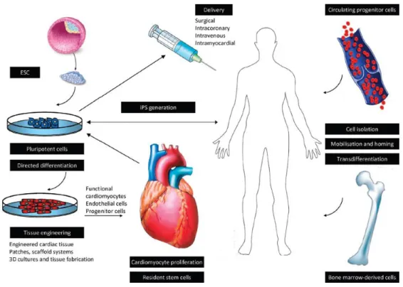
stem cells, especially on endothelial cells. Preclinical results and early clinical trials with cell therapy in cardiovascular field will be discussed in details later in section 2.3. Figure 1.
summarizes cell therapy approaches in regenerative cardiac medicine.
Figure 1. Experimental approaches to cell therapy in heart failure Figure shows experimental approaches, cell types and cell delivery routes for regenerative purposes in heart failure (Figure is from Kosztin/Gara et al. Stem cell therapy to treat heart failure Elsevier 2015, Reference Module in Biomedical Sciences)
The key aims of cardiovascular cell therapy are:
1) to prevent myocardial loss during myocardial tissue injury (ischaemic attacks, cardiomyopathy, valve diseases, etc.);
2) to decrease the ratio of definite necrosis after ischemic events and salvage stunned myocardium;
3) to decrease the rate of remodelling, and enhance reverse remodelling after myocardial injury;
4) to support angiogenesis during and after ischemic attack;
5) and to regenerate myocardial tissue in severe heart failure from different aetiologies.
12
Regarding cell therapy and translational research in the cardiovascular field many questions remain open:
1) Which is the ideal cell type for cell therapy?
Circulating progenitor cells, multipotent stem cells, cardiac progenitor cells and pluripotent stem cell-derivatives are candidates.
2) How to perform the cell implantation?
Applications: direct surgical implantation, surgical cardiac patches, local intra- myocardial injection, intracoronary injection, coronary sinus injection, systemic intravenous injection, use of bioscaffolds for cell engineering and implantation.
3) What is the ideal number of cells for implantation?
Early clinical studies lack valuable information about the required amount of implanted cells. To build up a human adult heart, ~ 6 billion cells may be needed.
The left ventricle is made up from ~ 4x10⁹ cardiomyocytes (2x10⁶/g of tissue, the whole heart is ~300g) [16]. In a large MI ~ 109 cardiomyocytes suffer irreversible damage. State-of-the-art clinical imaging systems (e.g. SPECT-CT) may estimate the number of cells needed for regeneration before performing transplantation.
Considering personalised regenerative therapy with hiPSC-derivatives, the amount of injected cell should also be adjusted individually.
4) How to track injected cells after implantation to enable ability of following their fate in vivo?
The vast amount of transplanted cells seem to be washed out from the target tissue via venous and lymphatic circulation and imaging their homing in vivo is still a challenge. In most preclinical studies, the data are lacking about the amount of cells engrafted into the myocardium. Furthermore, no information exists about what happens to non-hosting cells. As clinical trials typically follow up the patients for a short period (two years), long term data is missing on attainable tumour formation or pro-carcinogenic considerations.
13 5) What is the ideal timing for cell implantation?
Clinical studies are inconclusive regarding the timing of cell therapy. Scientific knowledge is lacking regarding the best timing for anticipated beneficial regenerative effects. Some trials aim to treat in the acute phase of cardiovascular events, e.g. within one week after myocardial infarction. Others aim regeneration late after the myocardial event, e.g. in end-stage heart failure.
14 2.2. Types of stem cells
2.2.1. Circulating progenitor cells
In cardiovascular diseases the pathological steps are endothelial dysfunction and damage. Circulating endothelial progenitor cells (EPC) are in the focus of research for over a decade, providing possible source for therapeutic re-endothelisation and endothelial repair [17]. Furthermore, EPC were nominated to be biomarkers for cardiovascular risks and disease progression [18]. Many studies have also considered EPC to be putative biomarkers for pathological conditions [19]. The amount of circulating EPC is proposed to be biomarker for the progression of atherosclerosis and for coronary plaque characteristics [20, 21]. After promising early studies, recently published data have been controversial regarding the role of EPC as biomarkers. Data are also diverse regarding therapeutic considerations [17].
EPC were first characterised in 1997 [22] and early studies demonstrated them to be bone marrow derived, CD34-positive cells. Despite great efforts, no clear definition is available on these progenitor cells. In fact, data are confusing on titles and subgroups of EPC, naming them such as “circulating angiogenic cells”, “endothelial colony forming cells” and
“colony forming unit cells” [17]. EPC are mainly characterised by the presence of cell surface markers: CD34, VEGFR2, CD133, ICAM-1 and E-Cadherin, with flow cytometry.
Isolation protocols are also available that group EPC to early and late outgrowth population [19, 23, 24]. In 2004 EPCs have been therefore redefined [25]. Population of cells from human peripheral blood were expanded in vitro, which expressed CD31 and vWF. In addition, these cells showed cobblestone morphology in culture which is characteristic for endothelial cells. It was proved that these human cells have the capacity to form chimeric human-murine vessels when implanted subcutaneously into mice [25].
Their role in adult organism was thought to be the enhancement of angiogenesis in adults [26]. Indeed, a number of clinical studies in humans show the activation or injection of these cells. Whether these cells contribute directly to new vessel formation or simply support that by cytokine release and the rate of beneficial effects remains unclear [19].
In a prominent phase II, randomised clinical trial patients with type 2 diabetes mellitus and hyperlipidaemia discontinued statin treatment for a short period and meanwhile changes in
15
EPC levels and angiogenic function were measured [27]. The results were impressive, as large increases were detected in the levels of circulating EPC and in their functional activity, after 5 days of statin discontinuation [27]. The data suggest that EPC may have an important role in atherosclerotic disease progression and therapeutic efforts for endothelial repair and re-endothelisation [27]. However, more investigations are needed to understand exact function of EPC and their role in vascular biology.
2.2.2. Mesenchymal stem cells
MSC are either derived from the bone marrow or from peripheral organs. MSC were first described in 1970 as nucleated cells in the bone marrow and were known to be responsible for steady-state circumstances in the bone marrow niche [28].
MSC are able to differentiate into multi-lineage derivatives and are able to perform self- renewal. Thus, they were named as mesenchymal stem cells, albeit recent publications rather call them mesenchymal stromal cells [29, 30]. The early clinical trials used many different types of MSC-like cells which were not precisely characterized. To enhance standardised characteristics of MSC in translational research and early phase clinical trials, strict requirements were set up for MSC definition [31]. In 2006, the International Society for Cellular Therapy published MSC definitions:
Adherence to plastic surface in standard culture conditions
Expression of the following cell surface markers: CD73, CD90, and CD105
Absence of the following cell surface markers: CD34, CD45, HLA-DR, CD14 or CD11b and CD79a or CD19
Ability to differentiate to osteoblasts, adipocytes, and chondroblasts in vitro [31].
Sources of MSC isolation and collection are the bone marrow and peripheral organs. These include adipose tissue and many other organs like the liver, placenta, gut, lung, heart and even amniotic fluid, dental pulp, periodontal ligament or Wharton jelly of the umbilical cord. Many studies use adipose tissue-derived MSC, as their collection is easier and more feasible. Wharton jelly-derived MSC were also studied in many trials [32]. MSC from different sources possess many differences in genotype and phenotype. MSC derived from
16
the amniotic fluid express pluripotency marker SSEA4. MSC-like cells isolated from heart tissue express myogenic markers and tend to be myogenic precursor cells [33].
Despite the improvement of protocols, the isolation and expansion of MSC are still with low efficacy. For clinical therapeutic use the isolation and expansion requires therefore better and CGMP competent protocols. From 10ml of bone marrow aspirate 50-400 x 106 MSC can be isolated. Huge variation exists in elderly population and in case of co- morbidities.
MSC are elongated, fibroblast like cells which grow on plastic in MSC medium. MSC can differentiate into chondrocytes, osteoblast, myocyte precursor cells and adipocytes in vitro, although the differentiation rate is low. Beside direct use of cells for therapy, studies also focus on the indirect anti-inflammatory and paracrine effects of MSC. The favourable effect of MSC for therapeutic purposes may be their anti-inflammatory and anti-fibrotic role. These cells have been shown to secrete wide range of proteins which would promote regenerative angiogenesis and stimulate endogenous cardiac regeneration in vivo [33]. In post-MI failing myocardium, two major types of macrophages are present in the injured myocardium. Type M1is responsible for the production of pro-inflammatory proteins, debris degradation and apoptosis. Type M2 has anti-inflammatory role and supports early angiogenesis. After MI, in the presence of MSC, the number of type M2 macrophages increase [34, 35]. MSC have cardioprotective effects and can decrease size of scar after MI. For this, they may enhance the survival rate in stunned myocardium and decrease necrosis. Detailed studies showed, cardiac cytoprotection occurs by modulating key signalling pathways, which include the Wnt, IGF1-NF-kB, Akt1 and TGFβ pathways [36- 39]. For cardiac regeneration and repair, MSC share pleiotropic mechanisms of action (Table 1.) [8]. However, limitations and controversial effects of MSC reside in formation of mesoderm-derived ectopic cells and enhancement in fibrosis may progress in vivo [40].
Clinical trials with MSC in cardiovascular field are discussed in details in section 2.3.2.
17
Table 1. Mechanisms of action of mesenchymal stem cells in cardiac regeneration Table shows putative effects and mechanisms of action of mesenchymal stem cells in cardiac regeneration (WBC: white blood cells, ECM: extracellular matrix)
Effect Mechanism
Immunomodulation Supressing WBC, enhancing M2 type macrophages Antifibrosis Modulation of ECM niche by paracrine effects Pro-angiogenesis Secretion of pro-angiogenic proteins and molecules Pro-neomyogenesis Activation of cardiac resident stem cells and progenitors Cardiac cytoprotection Modulating signalling pathways, anti-apoptotic effects
Direct cellular effects Differentiation to cardiac myogenic precursor cells
18 2.2.3. Cardiac stem cells
Resident, adult stem cells occur in many organs and tissues of the human body. The main role of adult stem cells may be their participation in physiological turn-over of tissues as well as support of endogenous regeneration after tissue injury. In the cardiovascular system, endothelial and cardiac progenitor cells stand for adult stem cells. Adult, resident stem cells (CSC) in the myocardium are clonogenic, have the ability for self-renewal and may differentiate to any cardiovascular cell type (cardiomyocytes, endothelial cells and smooth muscle cells) [41, 42]. However, their exact role in cardiac regeneration and their potential to improve decreased cardiac function is still controversial. Scientific knowledge is diverse regarding the role of CSC in the physiologic turnover of the myocardium [41, 43-45]. Some resident CSC may re-enter cell-cycle and replace damaged or dysfunctional cardiomyocytes. The main characteristics of CSC cells are c-kit positivity; further characteristics can include positivity for islet-1, Nkx-2.5 and MEF-2c (cardiac progenitor markers). C-kit is a tyrosine kinase receptor (also known as CD117), which binds SCF (stem cell factor) and involved in many important signalling pathways, including the PI3K and ERK, mainly responsible for cell proliferation and migration [46-48]. The origin of c- kit positive CSC in the adult heart is not clear. Some studies showed their origin may be the cardiomyocytes and epicardial cells of the heart and CSC develop by dedifferentiation and epicardial-mesenchymal transdifferentiation [49, 50]. Other groups showed that CSC may arise from circulating progenitor cells and home to the adult heart [51, 52]. Further studies report that CSC preserved stem cell niche during embryonic cardiomyogenesis [53, 54]. Many studies have been investigating potential role of CSC in cardiac regeneration and repair. Similarly to MSC, CSC have multimodal mechanisms to enhance cardiac repair [53]. Their paracrine effects incluede secretion of pro-angiogenic, anti-apoptotic factors and supporting angiogenesis after ischemic injury. The direct cellular effects contain differentiation to early/late cardiac progenitors or cardiomyocytes (most controversial field).
Preclinical results and clinical trials investigating CSC in cardiovascular field are discussed in details in section 2.3.3.
19 2.2.4. Embryonic stem cells
Human ESC were first established in 1998, by James Thomson et al., in Wisconsin [55]. Inner cell mass of the blastocyst were isolated and embryonic stem cells were plated.
Thomson used five blastocysts remaining from in vitro fertilisation (IVF) procedure, fourteen samples from inner cell masses were isolated and the first five hESC lines were established. Today over 1000 hESC lines exist [56, 57]. Ethical regulations are diverse regarding the development and use for research purposes of hESC (detailed in section 4.12.). Early protocols for inner cell mass isolation from the blastocyst resulted in the destruction of the embryo. Mechanical isolation, surgical isolation and even laser techniques have been developed [58-60]. Recent isolation procedures are enable to rescue the embryo, thus ethical concerns may be reduced [61]. Human ESC lines were successfully developed from single blastomeres by routine biopsies performed for preimplantation genetic testing during IVF [62-64]. These derivation protocols are facing a number of challenges, regarding the distinct phenotype and genotype of the developed hESC lines [65].
Human ESC can expand unlimitedly with asymmetric self-renewal while preserving normal karyotype and pluripotent state. During directed differentiation, hESC are able to give rise to all three germ layers (ectoderm, endoderm and mesoderm) and for germline, except extra-embryonic tissues of the placenta (trophoectoderm) (Figure 2.). Human ESC are able to differentiate into all tissue and cells in human organism: ectoderm derived neurons [66], Schwann cells [67], melanocytes [68], skin cells [69]; endoderm derived hepatocytes [70], intestinal cells [71], pancreatic cells [72], pneumocytes [73]; mesoderm derived blood cells [74], fibroblasts [75], adipocytes [76], smooth muscle cells [77], cardiomyocytes [78], endothelial cells [79], etc. Upon in vivo transplantation of hESC they spontaneously form teratomas. For in vitro colony formation, hESC require special cell culture conditions. Early protocols included xenogenic feeder cells and growth factors;
later xeno-free feeder culture conditions developed. Recently hESC have been grown on ECM components in xeno-free conditions (eg. laminin, fibronectin, vitronectin, Matrigel) in commercially available predefined media for pluripotent cells [65]. Human ESC are characterized by cell surface markers and transcription factors associated with pluripotency. Briefly, these pluripotency markers are TRA1-60/81, TRA2-49/54, CD-
20
9/24/29/49f/324/338 and SSEA-1/3/4/5 (cell surface proteins) and transcription factors Oct-4, Nanog, Stat-3, FoxD3, c-Myc and Sox-2 [65]. Translational studies with hESC in the cardiovascular field are discussed in section 2.3.4. in details.
Figure 2. Human embryonic stem cells and derivatives Figure shows origin of human embryonic stem cells, these pluripotent stem cells are able to differentiate into any cell types of the three germ layers and germline (original figure from Edit Gara)
21 2.2.5. Induced pluripotent stem cells
In 2006 Japanese researcher Shinya Yamanaka, and his group successfully developed mouse pluripotent stem cells from adult somatic cells, by using genetic reprogramming [80]. The first hiPSC line was developed in 2007, parallel by two independent research groups: Yamanaka et al. and Thomson et al. [81, 82]. The idea of nuclear transfer itself, existed earlier, notably Dolly the sheep was born through the nuclear transfer cloning method [83]. Forced expression of specific genes in differentiated somatic cells has been shown to reverse the developmental program in adult somatic cells; this process is called reprogramming. When using reprogramming method, nuclear transfer or oocyte manipulation is not necessary to induce genome modification. During reprogramming, genes responsible for pluripotency are inserted into the genome of adult somatic cells by transfection. These pluripotency genes are named Yamanaka factors or master genes: Oct-3/4, Sox-2, Klf-4 and c-Myc [80]. Oct-3 and 4 play a key role in early embryonic development and in maintaining pluripotent fate is stem cells. Sox-2 is responsible for continuous self-renewal in pluripotent stem cells. Klf-4 is also involved in maintenance of pluripotency, proliferation and cell survival. Myc is known to be an oncogenic regulator, tumour suppressor and is affected in almost all human cancers. Myc plays a crucial role in physiologic operation of cell cycle and proliferation [84].
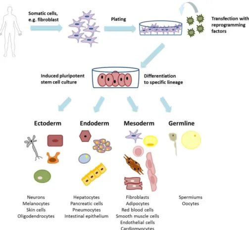
Human iPSC promise wide range of new possibilities in basic research, translational medicine and therapy. Human iPSC offer specific cell types for modelling diseases, testing drug toxicology and developing drugs in vitro, even in a patient-specific manner. Human iPSC have similar characteristics to those of embryonic stem cells, as they express same pluripotency markers and can differentiate into all three germ lines [85] (Figure 3.).
Indeed, undifferentiated hiPSC were shown to form teratomas in vitro [80]. Human iPSC may later be the most efficient and feasible option for tissue engineering therapeutic perspectives. Ongoing preclinical studies with hiPSC will be discussed in details in section 2.3.5.
22
Figure 3. Induced pluripotent stem cells and derivatives Figure shows the derivation of human induced pluripotent stem cells. These pluripotent stem cells are able to differentiate to any cell types of the three germ layers and germline (original figure from Edit Gara) Due to the great translational and clinical potential, hiPSC are in the focus of basic research. The reprogramming method itself determines the fate and phenotype of the developed pluripotent cells, also the donor cell line influences reprogramming. Several reprogramming techniques were established in the last decade with a large variability in safety and efficacy profile [86]. First techniques used fibroblast as somatic cells, however keratinocytes, lymphocytes, hepatocytes, neurons, or multipotent stem cells are also good sources for reprogramming [86]. Regarding availability and reprogramming efficiency, the most commonly used somatic cell type is still adult fibroblast [86]. Reprogramming methods mainly differ in the route of delivery of pluripotency genes. First protocols used retroviral, lentiviral or adenoviral vectors to integrate pluripotency genes into the genome of targeted cells. These methods may not be feasible for clinical use, as viral vectors mean infection of the cells with viruses and integration of viral genes into the human genome.
Despite their risks integrating viral vector methods have high reprogramming efficacy. On the other hand, use of adenoviral vectors has very low transfection efficiency, although
23
adenoviruses do not integrate into human genome. To overcome feasibility issues, non- integrating viral methods have been developed, including the Sendai virus as a vector [87].
Sendai virus technique provides a safer and more efficient production of hiPSC, using RNA-based transfection of pluripotency genes. A special group of DNA-based integration technique is called the Sleeping Beauty transposon system [88]. Sleeping Beauty is a non- viral carrier system of the genetic information that can insert a gene into human chromosomes. The future clinical use of Sleeping Beauty transposon system, would be much safer than those vector systems that are viral-dependent. Ivics at al. found that the chromosomal distribution of Sleeping Beauty transposon insertions is random in the human genome. They also proved that insertions could occur at each chromosome. Sleeping Beauty transposons could be inserted at specific regions of the human genome, by modifying the insertion [89]. Furthermore, the third generation of virus-free/DNA-free iPSC was generated by direct delivery of small molecules, proteins and antibodies that remodel cellular epigenetic pattern [85, 90, 91]. Transgene-free non-integrating methods were developed, e.g. piggyBac system. The piggyBac transposon system enables insertion and then directed excision of a naked DNA segment from the genome, thus the constant modification of the human genome is avoided [88]. The transfection with a piggyBac transposon is completely under control, as the promoter region is induced by an antibiotic enhancer (e.g. doxycycline). Finally, modified messenger RNA delivery and microRNA- based transfection methods also exist. These viral-free/DNA-free, non-integrating methods partially eliminated the risk of modifying the human genome.
Recent studies have investigated the epigenetic effects of reprogramming and genomic stability of established hiPSC [92]. The genetic manipulation during hiPSC may be a critical issue in clinical scenario. Human iPSC seems to be superior among other pluripotent and multipotent stem cell lines, thus assessing and developing the safest and most efficient reprogramming method may be a critical issue for future therapeutic use.
Short summary of reprogramming methods is shown in Figure 4.
24
Figure 4. Advantages and disadvantages of reprogramming methods Figure shows main types of reporgramming methods to develop human induced pluripotent stem cells from adult, somatic cells. Advantages and disadvantages of main reprogramming methods are listed in figure (+ advantage, - disadvantage, original figure from Edit Gara)
25
2.3. Cardiovascular derivatives of stem cells, preclinical and early clinical results
2.3.1. Clinical studies investigating endothelial progenitor cells
After characterising EPC, translational research moved toward cardiac regeneration. First clinical trials assessed safety and feasibility of their activation in vivo and injection after exogenous boost [93-95].Small clinical trials reported beneficial results and improvement in hemodynamic parameters. Injection of EPC into the injured myocardium resulted in increased left ventricular ejection fraction (LVEF) and inhibition of fibrosis after myocardial infarction [94, 96, 97]. To enhance stent function, anti-CD34 antibodies were applied onto coronary stents. The bioengineered stents can capture EPC, thus enhance endothelisation and defeat late in-stent restenosis. In BOne MarrOw Transplant to enhance ST-elevation infarct regeneration (BOOST, NCT00224536) clinical trial patients received bone marrow-derived mononuclear cells (hematopoietic precursor).
The autologous precursor cells were harvested and injected at the same day, after suffering ST segment elevation myocardial infarction, with preserved LVEF. The study reported no significant adverse event and significant increase in LVEF was found in the treatment group, after 6 months follow-up.
At the Heart and Vascular Centre, Semmelweis University, Budapest, the Cxcr4 AnTagonism for Cell mobilization and Healing in AMI (CATCH-AMI, NCT01905475) trial is investigating effects of endothelial progenitor/stem cells in acute ST segment elevation myocardial infarction (STEMI). I am taking part in this trial as a sub- investigator. This is a phase IIa, double-blind, placebo-controlled, randomised and multi- centre study of POL6326, a CXCR4 antagonist study drug. The objective of this study is to mobilise circulating endothelial progenitor cells by modulating the CXCR4/CXCL12 axis.
Modulation of CXCR4/CXCL12 pathway results in homing of EPC into the injured myocardial tissues. The study drug inhibits CXCR4, thus mobilisation of EPC and their homing into the injured myocardium can occur. Enrolled patients received the study drug intravenously on day 5 and day 7 after STEMI. The primary end-point of the study is increase in LVEF at 4- and 12-month follow-up. The secondary end-point includes detailed hemodynamic parameters, mortality parameters and analysis of the level of activated EPC.
Fluorescence cell sorting method is performed in a core lab, evaluating the exact amount of
26
activated CD31 positive, CD34 positive EPC. Outcome of this and other similar trials will reveal if EPC possess efficient regenerative capacity for cardiac repair. Mobilisation, characterisation and collection of EPC must improve to use them for regenerative cardiac repair.
2.3.2. Clinical results with mesenchymal stem cells for cardiac regeneration
The fate and characteristics of MSC were discussed in details in section 2.2.2. This section will introduce the clinical data of MSC treatment in cardiovascular diseases. As MSC have good availability and safety profile, most of the current clinical trials were performed with these cell types. Unfortunately, some of these clinical trials reported false results to enhance beneficial outcome. Comprehensive results of meta-analyses showed modest increases in cardiac functional parameters (e.g. LVEF, six-minute walk test (6MWT)). However, these significant beneficial effects diminished in longer-term follow up [98].
Major difficulty when comparing clinical results from MSC trials is the diversity in study designs. In most studies, intracoronary injections were performed after acute MI, using varying numbers of MSC (between 2x10⁶ and 2500x106). However, these clinical trials can be described by their procedural heterogeneity, and a lack of standardization [99]. Some studies reported beneficial results, like a study from Lunde et al., which showed improvement in echocardiography, SPECT and MRI parameters after 6 months follow-up [100, 101]. The Reinfusion of Enriched Progenitor Cells And Infarct Remodelling in Acute Myocardial Infarction (REPAIR-AMI, NCT00711542) trial recruited a large number of patients (240) with acute phase STEMI. This study was a phase II, randomised, placebo controlled, and double-blinded one. Patients received autologous intracoronary injections of bone-marrow derived MSC (50ml) vs. placebo medium. Detailed coronary artery hemodynamics were measured to assess coronary microvascular function. At four months after MSC implantation, patients showed significant improvement in LVEF compared to placebo (+5.5 vs +3.0%, absolute difference +2.5%). However, detailed analysis revealed that substantial treatment effects of MSC were limited to areas with the greatest damage or extent of scarring [102].
27
Further studies investigated the effects of MSC implantation in stable coronary artery diseases. These studies showed improvement in left ventricular function, perfusion and relief of angina pectoris [94]. The Transplantation Of Progenitor Cells And Recovery of Left Ventricular Function in Patients with non-ischemic Dilatative CardioMyopathy (TOPCARE-DCM, NCT00284713) trial studied effects of MSC transplantation in non- ischemic heart failure. Patients with severe, idiopathic and non-ischemic heart failure received autologous implantation of bone marrow-derived progenitor cells into the LAD.
MSC implantation was found to be safe and feasible in non-ischemic etiology.
Furthermore, improvement was noted in clinical parameters such as left ventricular volume, LVEF and decrease in serum BNP levels.
In contrast to beneficial study results, some others published minor or no improvement in clinical parameters after MSC implantation. A study from Janssens and colleagues found no benefit in LVEF, but a significant reduction in infarct size and improved in regional left ventricular function [103].
Special type of MSC, the endometrial regenerative cells (ERC) is under investigation in congestive heart failure. Patients with NYHA III-IV functional status and decreased LVEF (<40%) receive ERC implantation via the coronary sinus. The study involves three cohort of cell dose: 50-100-200 million. The interim analysis provided beneficial safety results of ERC delivery [104].
In the recent years meta-analyses published large cohort of results with MSC. These trials [105-108] involving altogether 999 patients with acute MI or chronic ischaemic cardiomyopathy showed that transplantation of MSC improved LVEF by 5.4%, decreased infarct scar size by 5.5% and lowered left ventricular end-systolic volume by 4.8 ml [108].
Other meta-analyses [109] indicated that MSC treatment is beneficial; however, the typical modest increase in ejection fraction is of uncertain clinical significance. Recent meta- analysis from Gyöngyösi et al. suggested neutral effects of MSC treatment, especially in longer-term follow-up [98]. It was shown that intracoronary or direct intramyocardial delivery route may be the most beneficial. Other delivery routes (coronary sinus, systemic venous or surgical) have proven less success. The analysed data displayed an overall 0.9- 6.1% improvement in LVEF; nevertheless these improvements diminished in half of the trials on the longer term follow-up. By now, arguments are clear that MSC are unable to transdifferentiate into cardiomyocytes [110]. Their beneficial effects may derive from
28
immunomodulation, pro-angiogenic, antiapoptotic and anti-fibrotic effects. Table 2.
summarizes MSC advantages and disadvantages for cardiac regeneration. Preclinical and translational studies must improve to enhance MSC use for cardiac regeneration. State-of- the-art studies suggest utilizing MSC products, which may be responsible for their regenerative effects, e.g. transplantation of MSC-derived exosomes and microvesicles only [111, 112].
Table 2. Characteristics of mesenchymal stem cells in cardiac regeneration Table shows advantages and disadvantages of mesenchymal stem cells concerning cardiac regeneration (MSC: mesenchymal stem cells)
Advantages of MSC Disadvantages of MSC
Easy to collect, isolate and expand Exact mechanism of action is unclear Low immunogenicity True myocardial differentiation
is lacking
Multipotent Large heterogeneity
Beneficial safety reports from previous studies
Diverse study designs aggravate clinical interpretation
While unmodified MSC have shown some promise in cardiac repair, angiogenic and myogenic effect of these cells might be enhanced by phenotypic modification. To facilitate a standardized performance of MSC used for cardiac repair, a group at Mayo Clinic led by André Terzic has used proteomic and genomic analysis to identify critical factors in the pathways regulating cardiac differentiation. Their approach has yielded a suite of molecules that drive cells in vitro into cardiopoiesis. A company, Cardio3 Biosciences, (currently Celyad) completed a clinical study (NCT00810238) to apply this technology for the development of the cardiac cell lineage, named: C3BS-CQR-1 (C-Cure®) to treat heart failure, reporting a significant improvement of LVEF associated with reduction in LV end- systolic volume and improvement in the 6MWT [113]. The larger randomized study (CHART-1, NCT01768702) is currently recruiting patients with ischemic heart failure.
This study is running at the Heart and Vascular Centre, Semmelweis University, I am currently working as a sub-investigator in the trial. Patients are treated with autologous bone-marrow-derived MSC (C3BS-CQR-1) product). MSC are collected from the iliac crest via posterior crista biopsy performed by haematologists. A large volume of BM is
29
harvested (65-85ml) in order to collect a sufficient quantity of MSC. Harvested BM is shipped to a core laboratory, where MSC are isolated. Cells are treated with a special cocktail containing growth factors to enhance development of cardiomyocyte progenitors.
The differentiation procedure takes up to 7-15 weeks. When the differentiation part is completed, autologous cells are shipped back to local hospital sites, where C3BS-CQR-1 is injected intra-myocardially via femoral artery catheterisation. C3BS-CQR-1 are expanded into cell numbers of 600x106 and injected into 20 sites of the left ventricular wall. The primary end-point is set up from a composite involving number of hospitalisations due to worsening heart failure, changes in Minnesota living with heart failure questionnaire (MLHFQ) (improvement at least ten points) and 6MWT (increase at least 40m), changes in LV parameters (15ml absolute change), and LVEF (4% absolute improvement). Secondary end-points include efficacy and safety measurements. Safety parameters include the number and cause of deaths and hospital re-admissions, the number of cardiac transplantations and myocardial infarctions and the number of strokes. Efficacy parameters entail time to all-cause mortality, time to worsening of heart failure and time to aborted sudden death. After promising results from C-Cure®, CHART-1 gives hope for beneficial outcome.
2.3.3. Clinical studies with cardiac stem cells for cardiac regeneration
Characteristics of cardiac stem cells are discussed in section 2.2.3. Here the preclinical results and clinical trials with CSC are discussed. The injection of human cardiosphere-derived cells into injured myocardium showed some benefit in animal models mainly by improving left ventricular function [114, 115]. The cardiospheres are obtained from myocardial biopsy and can give rise to cardiomyocytes, endothelial cells and smooth muscle cells. Human cardiospheres exhibit significant proliferation and differentiation capacity [114]. Isolated cell populations can be differentiated into spontaneously beating aggregates of cardiomyocytes [115]. In mouse model for myocardial infarction epicardium derived CSC could restore cardiac function, by reducing dilatation of the heart chambers and increasing ejection fraction [116].
The CArdiosphere-Derived aUtologous stemCElls to reverse ventricUlar dySfunction (CADUCEUS, NCT00893360) trial is a completed phase I safety study, investigating the
30
effects of CSC in ischemic heart failure. During the study procedure by Marban et al. from the Johns Hopkins Hospital, small biopsy samples were taken from recruited patients‟
myocardium. Later 12.5x 10⁶ or 25x 106 of autologous CSC were injected intracoronary.
Results reported that CSC inplantation is safe after myocardial infarction [117].
Unexpected beneficial results showed in addition to decreased scar size, the increased amount of viable myocardium as measured by cardiac MRI [117].
Therapeutic perspectives of CSC are also investigated in rare and congenital cardiac diseases. The Transcoronary Infusion of CArdiac Progenitor Cells in Patients with Single Ventricle Physiology (TICAP, NCT01273857) and Cardiac Progenitor Cell Infusion to Treat Univentricular Heart disease (PERSEUS, NCT01829750) trials study the effects of cardiac progenitor cells in congenital heart diseases. In TICAP, cardiospehere-derived cells are implanted intracoronary, one month after Norwood, Glenn or Fontan procedures.
Patients receive 0.3 million/kg autologous CSC. In phase I designed TICAP trial, safety results enhanced the running of phase II PERSEUS trial.
2.3.4. Preclinical and early clinical studies with embryonic stem cells in the cardiovascular field
Human ESC have emerged as one of the most promising sources of new cardiac cells for transplantation because of their capacity to efficiently undergo directed differentiation into genuine cardiomyocytes and supportive endothelial cells. A number of groups have successfully isolated cardiomyocytes or cardiac progenitor cells from differentiating ESC cultures [118, 119]. These in vitro derived cardiomyocytes have been characterized extensively. Structural, electrophysiological and contractility studies indicated that ESC-derived cardiomyocytes exhibit a phenotype reminiscent of foetal, rather than adult cardiomyocytes. In the animal transplantation models of cardiac disease, use of ESC-derived cardiomyocytes has resulted in a significant improvement in ventricular function and structure (Table 3.). The cells appear to form gap junctions with host cardiac tissue; however, formation of protective fibrotic tissue around the grafts can interfere with complete electrophysiological coupling [120]. The beneficial effects in MI have been reported one month after transplantation [121].
31
Table 3. Preclinical studies on embryonic stem cell derivatives in myocardial infarction models Table shows details of preclinical studies with human embryonic stem cells regarding cell number, animal models, timing and follow-up (Table is adapted from Kosztin/Gara et al. Stem cell therapy to treat heart failure Elsevier 2015, Reference Module in Biomedical Sciences)
Cell type Number of injected cells
Animal model
Timing Follow-up Ref.
ESC (cardiac committed)
5x10⁷ sheep 14 days after MI 4 weeks [122]
hESC-CM 0.03-0.1x10⁶ mouse Day of MI 3-4 weeks [123]
hESC-CM 106 mouse Day of MI 12 weeks [120]
hESC-CM 107 rat 4 days after MI 4 weeks [121]
hESC-CM 108 guinea pig 10 days after MI 4 weeks [124]
hESC-CM 109 monkey 14 days after MI 3 months [125]
Murry et al. evaluated the effects of human cardiomyocyte implantation in non-human primate model of myocardial infarction. In their study design Macaca nemestrina non- human primates suffered myocardial infarction via balloon inflation in LAD. Two weeks later, hESC-derived cardiomyocytes were injected transepicardially into the myocardial scar and surrounding tissue. Animals received immunosuppression to avoid rejection of human cells. The injected CM cells expressed eGFP construct. This trial was the first to prove feasible and successful delivery of large human grafts in MI model. After follow-up for 14-84 days electromechanical coupling have been verified. Furthermore, GFP-positive human cells were successfully seeded among endothelial cells from host primate, suggesting enhancement in angiogenesis. Recently this study has used the largest number of injected cells. During the implantation procedure one billion hESC-CM were injected into the peri-infarct zone of the hearts. The commendable results postulate for further evaluation of post-transplantation expansion, maturation, survival, and long-term effects of grafted ESC-derived cardiomyocytes. In Murry‟s large animal model non-fatal, sustained ventricular and supraventricular tachyarrhythmias were observed after the implantation of cells. These pro-arrhythmogenic effects were not developed in small animal models, emphasizing importance of large animal studies [125, 126]. However, these cells have not been tested therapeutically since the problems of immunosuppression and the risk of
32
teratomas from residual undifferentiated ESC remain. The latest preclinical data suggest that co-transplantation of hESC-derived cardiovascular cells with MSC may enhance the beneficial effects of hESC derivatives. MSC may support the engraftment and survival of implanted hESC derivatives via anti-inflammatory and immunosuppressive effects [127].
The first clinical trials in cardiovascular disease are just launched. Menasche and colleagues have differentiated a population of CD15 and Isl1 positive cardiac progenitors from hESC. The Transplantation of Human embryonic Stem Cell-derived Progenitors in Severe Heart Failure (ESCORT, NCT02057900) phase I trial recruits patients with ischaemic heart failure with indication for coronary-artery bypass graft or valvular surgery.
Eligible patients have NYHA III-IV functional status and decrease LVEF (<35%). During the operation a fibrin-based cardiac patch, seeded with CD15/Isl1 double-positive cardiopoietic cells, are placed epicardially onto the infarcted area with a pericardial flap [32, 128].
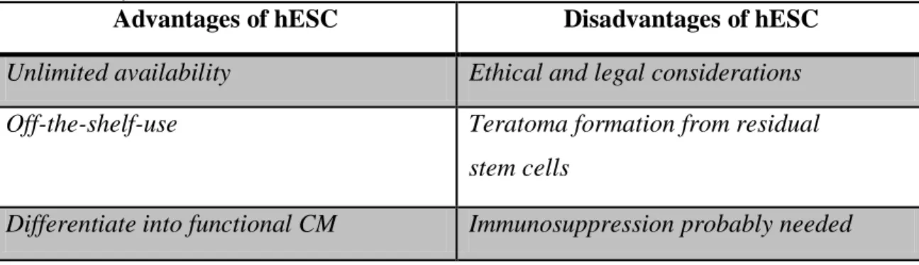
Table 4. shows the advantages and disadvantages of hESC for regenerative purposes.
Table 4. Advantages and disadvantages of hESC for cell therapy Table lists advantages and disadvantages of human embryonic stem cells for cell therapy in heart failure (hESC:
human embryonic stem cells)
Advantages of hESC Disadvantages of hESC Unlimited availability Ethical and legal considerations Off-the-shelf-use Teratoma formation from residual
stem cells
Differentiate into functional CM Immunosuppression probably needed
33
2.3.5. Preclinical results and future challenges with induced pluripotent stem cells
Induced pluripotent stem cell-derivatives promise considerable benefits for disease modelling and drug testing in vitro. As hiPSC are generally derived from adult somatic cells, new cell lineages may be patient specific. Thus, personalised drug testing and therapeutic strategies can be established. Commercially available hiPSC-derived cardiovascular cells, especially cardiomyocytes can be used in in vitro assays for testing cardiotoxicity, contractile properties, drug-induced early afterdepolarization or QT prolongation. First cardiac studies on hiPSC described long QT syndromes and catecholaminergic ventricular tachycardias [129-131]. Inherited disorders, like arrhythmogenic right ventricular cardiomyopathy or Brugada syndrome were also investigated by using hiPSC [132, 133].
Beside disease modelling in vitro, hiPSC provide platform for cardiovascular tissue engineering and cell therapy. A group developed differentiation methods to engineer cardiac tissue cell sheet, which contains cardiomyocytes, endothelial cells and pericytes [134]. This preclinical study involved implantation of hiPSC-derived cardiac cell sheets into rat model of myocardial infarction. The cell sheets were cultured on special thermo- sensitive polymers, and then the biopolymers were detached without enzymatic digestion.
This method is promising, as non-enzymatic protocols avoid modification of cell phenotype or loss of cell number during enzymatic digestion. Results of this preclinical trial are in progress.
Human iPSC give opportunity to model cardiovascular diseases in vitro, to investigate inherited cardiovascular conditions in a dish, to assess cardiovascular drug toxicity and to move towards therapeutic options and cardiovascular tisseue engineering; even individually. A recent clinical trial, the Individualized Early Risk Assessment for Heart Disease (IndivuHeart, NCT02417311) clinical trial is recruiting patients from June 2015.
The study is aimed to characterise in details phenotype of specific cardiac pathologies in engineered heart tissue (EHT) setting, in vitro. EHT enables high-throughput, comprehensive studies on hiPSC-CM, in 3D culture platforms [135, 136]. Fourty healthy volunteers, ten patients with DCM, ten patients with HOCM and ten patients with HFpEF will be recruited. Skin biopsies will be taken and EHT will be developed patient- specifically. The study design aims to characterise hiPSC-EHT function in basal and in
34
hemodynamic stress conditions. Furthermore, this study aims to define disease specific phenotype in vitro, to study pathophysiology steps of DCM and HOCM in EHT. These patient specific EHT constructs provide platform for individualised drug testing both on acute applications and long-term drug administrations.
Tracking cells via cell therapy and studying fate of the transplanted cell is one of the major challenges in preclinical trials with hiPSC. At Stanford University, Wu and colleagues work on hiPSC-derived cardiomyocytes to provide high scale results regarding prevention of donor cell loss during cell therapy [135, 137]. They aimed to establish high-throughput imaging techniques to track cells after implantation.
Advantages and disadvantages of hiPSC for therapeutic purposes are shown in Table 5.
Advantages may translate into safety, cost-effectiveness and can terminate the ethical debates related to harvesting of stem cells from different sources. Immunocompatibility issues may be reduced with iPSC derivatives because the beginning cell line (e.g. skin fibroblasts) can be obtained patient-specificially. Albeit, the logistics of using them therapeutically may not be as simple as hoped [138]. Large-animal models using monkeys, dogs and pigs were demonstrated their feasibility and superiority to other pluripotent stem cells (ESC), due to low immunological complications with intramyocardial injection of autologous cells.
Efforts are now focused on the development of large hiPSC banks for a variety of diseases.
Banking these cells, similarly to widespread umbilical cord blood banking, may serve public health advantages in the future. Characterizing the human leukocyte antigen (HLA) pattern of the banked cells may further enhance their future clinical use.
35
Table 5. Advantages and disadvantages of hiPSC for cell therapy Table lists advantages and disadvantages of human induced pluripotent stem cells for cell therapy in heart failure (hiPSC: human induced pluripotent stem cells)
Advantages of hiPSC Disadvantages of hiPSC Patient specific therapy Genome instability
during and after reprogramming Patient specific disease modelling Epigenetic modulations
during reprogramming
Patient specific drug testing Development of product takes time Immunosuppression possibly not needed Teratoma formation from residual cells
2.4. Tissue engineering for cardiovascular repair
In recent years many novel bioengineering methods and perspectives have been born, enabling cardiovascular tissue engineering. As cardiovascular diseases are the leading cause of death in industrialised countries, replacement of damaged cardiovascular tissue is in the focus of research. The number of cadaveric donor organs for cardiac transplantation is limited. In case of vascular diseases such as aortic aneurysm and aortic dissection, artificial tissue vascular grafts are available, but the number of biological donor vascular grafts is also limited. Thus, cardiovascular tissue engineering paves the way for novel therapeutic options concerning cardiovascular tissue regeneration. For cardiovascular tissue engineering, pluripotent stem cell derivatives and ECM components offer promising sources. Beside early clinical trials involving cell therapy in vivo, tissue engineering in vitro was also in focus in recent years. In vitro engineered cardiovascular tissues are developed for transplantation, to fill the gap between the availability of donor organs and their unmet need.
Tissue engineering requires special cell culture methods in vitro. To develop large number of cardiovascular cells, bioreactor systems seem to be ideal for scale-up. For tissue engineering purposes at least 106-109 cells (cardiomyocytes, endothelial cells and smooth muscle cells) are needed. Simple cell culture methods may be inefficient for such large number of cells. Bioreactor systems are capable for developing large capacity cultures with or without ECM components. Indeed, bioreactors provide cell culture techniques which