Development/Plasticity/Repair
Arginine Methyltransferase PRMT8 Provides Cellular Stress Tolerance in Aging Motoneurons
X Zoltan Simandi,
1,2Krisztian Pajer,
3Katalin Karolyi,
1XTatiana Sieler,
1X Lu-Lin Jiang,
4X Zsuzsanna Kolostyak,
2X Zsanett Sari,
2Zoltan Fekecs,
3Attila Pap,
2XAndreas Patsalos,
2Gerardo Alvarado Contreras,
5XBalint Reho,
6Zoltan Papp,
5X Xiufang Guo,
7X Attila Horvath,
2Greta Kiss,
8Zsolt Keresztessy,
2Gyo¨rgy Va´mosi,
6XJames Hickman,
7X Huaxi Xu,
4X Dorothee Dormann,
9,10X Tibor Hortobagyi,
11X Miklos Antal,
8,12Antal No´gra´di,
3* and XLaszlo Nagy
1,2,13*
1Sanford Burnham Prebys Medical Discovery Institute, Orlando, Florida 32827,2Department of Biochemistry and Molecular Biology, Faculty of Medicine, University of Debrecen, Debrecen, Hungary, HU 4032,3Department of Anatomy, Histology and Embryology, University of Szeged, Szeged, Hungary, HU 6720,4Neuroscience Initiative, Sanford Burnham Prebys Medical Discovery Institute, La Jolla, California 92037,5Division of Clinical Physiology, Institute of Cardiology, Faculty of Medicine, University of Debrecen, Debrecen, Hungary, HU 4032,6Department of Biophysics and Cell Biology, Faculty of Medicine, University of Debrecen, Debrecen, Hungary, HU 4032,7NanoScience Technology Center, University of Central Florida, Orlando, Florida 32816,
8Department of Anatomy, Faculty of Medicine, University of Debrecen, Debrecen, Hungary, HU 4032,9BioMedical Center, Ludwig-Maximilians-University Munich, Planegg-Martinsried, Germany 80539,10Munich Cluster for Systems Neurology (SyNergy), Munich, Germany 80539,11HAS-UD Cerebrovascular and Neurodegenerative Research Group, Department of Neurology and Neuropathology, University of Debrecen, Debrecen, Hungary, HU 4032,12HAS-UD Neuroscience Research Group, University of Debrecen, Debrecen, Hungary, HU 4032, and13HAS-UD Momentum Immunogenomics Research Group, University of Debrecen, Debrecen, Hungary, HU 4032
Aging contributes to cellular stress and neurodegeneration. Our understanding is limited regarding the tissue-restricted mechanisms providing protection in postmitotic cells throughout life. Here, we show that spinal cord motoneurons exhibit a high abundance of asymmetric dimethyl arginines (ADMAs) and the presence of this posttranslational modification provides protection against environ- mental stress. We identify protein arginine methyltransferase 8 (PRMT8) as a tissue-restricted enzyme responsible for proper ADMA level in postmitotic neurons. Male PRMT8 knock-out mice display decreased muscle strength with aging due to premature destabilization of neuromuscular junctions. Mechanistically, inhibition of methyltransferase activity or loss of PRMT8 results in accumulation of unrepaired DNA double-stranded breaks and decrease in the cAMP response-element-binding protein 1 (CREB1) level. As a conse- quence, the expression of CREB1-mediated prosurvival and regeneration-associated immediate early genes is dysregulated in aging PRMT8 knock-out mice. The uncovered role of PRMT8 represents a novel mechanism of stress tolerance in long-lived postmitotic neurons and identifies PRMT8 as a tissue-specific therapeutic target in the prevention of motoneuron degeneration.
Key words: ADMA; aging; CREB1; motoneuron; neurodegeneration; PRMT8
Introduction
The rate of cellular aging and the appearance of age-related pathol- ogies are modulated by highly conserved stress response and repair
pathways (Haigis and Yankner, 2010). Accumulating evidence indi- cates that the longevity of a cell or organism depends on its ability to properly regulate signaling pathways that counteract perturbations, such as protein misfolding, DNA damage, or oxidative stress (Kour-
Received Nov. 29, 2017; revised June 22, 2018; accepted June 25, 2018.
Author contributions: Z. Simandi and L.N. edited the paper; Z. Simandi, K.P., and L.N. designed research;
Z. Simandi, K.P., K.K., T.S., L.-L.J., Z. Kolostyak, Z. Sari, Z.F., A. Pap, A. Patsalos, G.A.C., B.R., X.G., and G.K. performed
research; Z.P., Z. Keresztessy, J.H., H.X., D.D., M.A., and A.N. contributed unpublished reagents/analytic tools;
Z. Simandi, K.P., A.H., G.V., T.H., M.A., A.N., and L.N. analyzed data; Z. Simandi, A.N., and L.N. wrote the paper.
Significance Statement
Although most of the cells in our body have a very short lifespan, postmitotic neurons must survive for many decades. Longevity
of a cell within the organism depends on its ability to properly regulate signaling pathways that counteract perturbations, such as
DNA damage, oxidative stress, or protein misfolding. Here, we provide evidence that tissue-specific regulators of stress tolerance
exist in postmitotic neurons. Specifically, we identify protein arginine methyltransferase 8 (PRMT8) as a cell-type-restricted
arginine methyltransferase in spinal cord motoneurons (MNs). PRMT8-dependent arginine methylation is required for neuro-
protection against age-related increased of cellular stress. Tissue-restricted expression and the enzymatic activity of PRMT8 make
it an attractive target for drug development to delay the onset of neurodegenerative disorders.
tis and Tavernarakis, 2011). However, stress responses must funda- mentally differ between proliferating cells, which can be easily replaced, and cells that are terminally differentiated and nonreplace- able, such as postmitotic neurons. In proliferating cells, the risk of propagating cellular defects is great, which can be potentially threat- ening the survival of the entire organism. Therefore, elimination of damaged proliferating cells is the most straightforward solution. In contrast, in long-lived neurons, this could damage the integrity of the complex neural network, so alternative mechanisms need to be used to properly maintain the homeostasis of these cells (Herrup and Yang, 2007; Kole et al., 2013). Identifying and understanding molec- ular mechanisms regulating cellular stress resistance in postmitotic neurons is important for defining novel therapeutic targets in late- onset neurodegenerative diseases.
Posttranslational modifications (PTMs), such as methylation, acetylation, and phosphorylation are crucial for balanced func- tions of stress response pathways and dysregulated PTMs can potentially lead to a pathological state (Lake and Bedford, 2007;
Dantuma and van Attikum, 2016). Arginine is a positively charged residue that is often found in protein motifs, including the RGG/RG motifs (Thandapani et al., 2013). The RGG se- quences within these motifs are substrate recognition sites for type I and type II protein arginine methyltransferases (PRMTs) catalyzing monomethylation and asymmetric or symmetric dim- ethylation of arginine residues, respectively (Bedford and Clarke, 2009; Blanc and Richard, 2017). In the last decade, hundreds of methylarginine proteins have been detected by mass spectrome- try and other proteomic and molecular techniques (Hart-Smith et al., 2012; Bremang et al., 2013; Guo et al., 2014; Auburger et al., 2016). These methyltransferase substrate proteins are involved in transcriptional regulation (TAF15, CRTC2), pre-mRNA splicing (FUS, G3BP1, hnRNP A1, DDX3X, SAM68), mRNA translation (EIF3, EIF4), DNA-damage response (MRE11, 53BP1, FUS), and regulation of apoptosis (PABP1). Prior
in vitrostudies estab- lished that asymmetric arginine dimethylation of amyotrophic lateral sclerosis (ALS)-related FUS, TAF15, hnRNPA1, and meth- ylation of many others is required for adequate stress response (Liu and Dreyfuss, 1995; Tradewell et al., 2012; Yamaguchi and Kitajo, 2012; Shorter and Taylor, 2013). Other studies implicated the involvement of arginine methylation in DNA damage re- sponse through the methylation of MRE11 and 53BP1 (Boisvert et al., 2005a,b). Together, these results strongly indicate a regula- tory role for asymmetric arginine methylation in the regulation of stress response of neurons and the pathogenesis of neurodegen- erative diseases.
Thus far, 11 members of this enzyme family have been iden- tified (Bedford and Clarke, 2009; Blanc and Richard, 2017).
PRMT8 is a unique member of the family because it shows highly tissue-specific expression by being restricted to the CNS (Taneda et al., 2007; Kousaka et al., 2009). PRMT8 has been shown to act as a posttranslational modifier of various proteins (Kim et al., 2008; Pahlich et al., 2008). Similar to PRMT1, its closest paralog, PRMT8, is involved in the epigenetic control of gene expression and normal function of neurons (Simandi et al., 2015). However, the
in vivobiological role of PRMT8 in the CNS and the mecha- nisms resulting in neural defects remain largely unknown except for very recent studies that described the role of PRMT8 in Pur- kinje cells (Kim et al., 2015) and excitatory synaptic function (Penney et al., 2017).
Here, we show that asymmetric dimethyl arginine (ADMA) level declines during embryonic development in the mouse.
Strikingly, choline acetyltransferase (ChAT)⫹ MNs selectively maintain high ADMA level in the adult spinal cord. Fused in sarcoma (FUS), a prominent arginine methyltransferase target, shows similar cell-type-restricted expression. Inhibition of meth- yltransferases results in accumulation of DNA double-stranded breaks (DSBs), altered FUS kinetics at DNA DSBs, and overall a more vulnerable cellular state and decreased cellular viability. We show that, among the asymmetric arginine methyltransferases, PRMT8 is selectively expressed in the spinal cord MNs. Loss of PRMT8 results in a progressive decrease in muscle strength due to the dysfunction and gradual loss of MNs in aging animals. The per- sistent stress in the absence of methyltransferase activity leads to decreased cAMP response-element-binding protein 1 (CREB1) level and insufficient activation of prosurvival and regeneration gene net- work in response to aging-related oxidative and ER stress. This work is a proof-of-concept showing that ADMA has a nonproliferation- associated, cell-type-restricted role and is required for proper stress response in postmitotic neurons. Based on our findings, PRMT8 should be considered as a novel component of neural stress resis- tance and a target for future drug discovery in neurodegenerative diseases.
Materials and Methods
Cell culture. Undifferentiated NSC34 cells were maintained in prolifera- tion media (DMEM, 10% FBS, 1% antibiotics). For differentiation, cells were seeded at a concentration of 5000 cells/cm2and, 24 h after plating, medium was changed to differentiation medium (1:1 DMEM/F12, 1%
FBS, 1% nonessential amino acid, and 1Mall-trans retinoic acid). Cells were allowed to differentiate up to 4 d and differentiation medium was changed on the second day. Differentiated cells were treated with stress inducers for 3 h before RNA isolation and 12 h for immunoblot analysis.
Endoplasmic Reticulum (ER) stress was induced by thapsigargin (Milli- pore) in a 0.2M(cell viability assay) or 1M(gene expression, protein analysis) final concentration. Sodium arsenite (Sigma-Aldrich) was used in a 0.1M(cell viability assay) or 10Mconcentration (gene expression, protein analysis) to induce oxidative stress. ADOX (Sigma-Aldrich) was used in a 40Mfinal concentration. Human induced pluripotent stem cells (iPSCs) were differentiated to motoneurons (MNs) stress as de- scribed previously (Das et al., 2007).
Gene-silencing assays. siRNA constructs were purchased from Sigma- Aldrich for targeting the mouse Creb1 (Mm02_00289365) and Prmt8 (Mm01_00128129). MISSION siRNA fluorescent universal negative control #1 (Sigma-Aldrich) was used as a control. NSC34 cells were plated and differentiated 2 d before transfection. Transfection of NSC34 cells was performed using Lipofectamine 3000 transfection reagent (Thermo Fisher Scientific) according to the manufacturer’s instructions.
Mice. The PRMT8-null mouse line was generated by the European Conditional Mouse Mutagenesis Program and Knockout Mouse Project (EUCOMM/KOMP) and Sanger Mouse Genetics Project (EPD0105_1_A03) and was purchased from the European Mouse Mutant Archive (EM:04479). For genotyping of PRMT8 knock-out mice, tail DNA
*L.N. and A.N. share senior authorship.
This work was supported by the research program GINOP-2.3.2-15/2016-00043 (T.H.), KTIA_13_NAP-A_II/7 (T.H.), GINOP 2.3.2-15-2016-00034 (A.N.), KTIA_NAP_13-1-2013-0001 (M.A.), and MTA- TKI 242 (M.A.). L.N. is supported by grants from the Hungarian Scientific Research Fund (OTKA 116855, 124298, 126885) and TAMOP 422_2012_0023 VE´D-ELEM implemented through the New Hungary Development Plan cofinanced by the Euro- pean Social Fund and the European Regional Development Fund and the Hungarian Brain Research Program (Grant KTIA_13_NAP-A-I/9). D.D. is supported by the Deutsche Forschungsgemeinschaft, within the framework of Munich Cluster Systems Neurology (EXC 1010 SyNergy) and the Emmy Noether program (DO 1804/1-1). H.X. is supported by the National Institutes of Health (Grants R21 AG048519, R01 AG021173, R01 AG038710, R01 AG044420, R01 NS046673, RF1 AG056130, and RF1 AG056114), the Tanz Family Fund, and Cure Alzheimer’s Fund. We thank Drs.
Randy Kaufman, Barbara Ranscht (Sanford Burnham Prebys Medical Discovery Institute), Peter Somogyi (University of Oxford), and members of the Nagy laboratory for discussions and comments on the manuscript and Mrs. Heja Aga and Dr. Andras Szabo for technical assistance with imunohistochemistry.
The authors declare no competing financial interests.
Correspondence should be addressed to Laszlo Nagy, Sanford Burnham Prebys Medical Discovery Institute, 6400 Sanger Road, Orlando, FL 32827. E-mail:lnagy@jhmi.edu.
DOI:10.1523/JNEUROSCI.3389-17.2018
Copyright © 2018 the authors 0270-6474/18/387684-18$15.00/0
was extracted and multiplex allele-specific PCR was performed using the following PCR primers: forward (P1), 5⬘-CCTGGCACTTTGAGGTGTTG- 3⬘, and reverse (P2), 5⬘-GTCTGATGGAATGGGCCTG-3⬘, which generated a 380 bp product for the Prmt8 wild-type allele; and reverse (P3), 5⬘- TCGTGGTATCGTTATGCGCC-3⬘, which generated a 252 bp product for the Prmt8 knock-out allele. Male mice were used for the experiments.
Onset ages of the disease in G93A SOD1-transgenic mice were deter- mined as the time when the animals reached peak body weight and the end stage of the disease were determined when the animals could not right themselves within 10 s when placed on their side, typically⬃12–15 weeks of age.
Animals that underwent surgical maneuvers (both recovery and ter- minal surgeries) were anesthetized with ketamine–xylazine combined intramuscular anesthesia as follows: ketamine hydcrochloride (Ketavet;
Pharmacia & Upjohn) at 110 mg/kg body weight and xylazine (Rompun;
Bayer) at 12 mg/kg body weight.
Grip strength,Rotarod,and limb clasping. Phenotyping characteriza- tion was performed in the Department of Behavioral Neurobiology (In- stitute of Experimental Medicine of the Hungarian Academy of Sciences, Budapest, Hungary). For grip strength measurement, animals were held by the base of the tail and allowed to grasp a steel grip gauge (2 mm) with their forepaws. Then, the mice were gently pulled away from the grip gauges in a steady fashion until the grip was released. The force exerted at the gauge at the time of grip release is determined by the mouse itself and is recorded as the grip force. At each time point, forelimb forces were the average of five measurements.
For the accelerating Rotarod test, the rotating rod apparatus (ITC Life Science) was used to measure the motor coordination and ability of mice to improve motor skill performance with training. Mice were placed on a rod and the rod was accelerated from 0 to 45 rpm in 2 min. The time periods that individual mice spent on the rod without falling were re- corded. Three trials with a 30 min intertrial interval were performed every day for 3 d. Hindlimb position forelimb clasping was observed and scored as described byGuyenet et al. (2010).
Muscle tension recording. Animals that were 3, 6, and 12 months old were randomly selected for tension recording (n⫽5 in both the control and the knock-out groups) and anesthetized with ketamine–xylazine at the 3, 6, and 12 months survival period and the tibialis anterior (TA) and extensor digitorum longus (EDL) muscles of hindlimb were prepared for tension recording. The distal tendons were dissected free and attached to strain gauges and the exposed parts of the muscles were kept moist with Krebs’ saline solution. Isometric contractions were then elicited from the muscles by stimulating the common peroneal nerve with bipolar elec- trodes. The length of each muscle was adjusted so as to produce the maximum twitch tension. Single twitch and tetanic (40 –100 Hz) con- tractions were displayed and recorded on a computer; all the additional recording hardware and software were developed by Supertech (Kelle´nyi system). Maximum tetanic tension was achieved at a stimulation fre- quency of⬃100 Hz. An estimate of the numbers of motor axons supply- ing the muscles was obtained by stimulating the common peroneal nerve with stimuli of increasing intensity and recording the stepwise incre- ments of twitch contractions.
Force measurements in single myocyte preparations. Force development during isometric contractions was measured in fast skeletal muscle fibers from EDL muscles of 3- and 12-month-old control and PRMT8-null mice. Permeabilized EDL myocytes were mounted between two thin needles with silicone adhesive and viewed under an inverted microscope.
One needle was attached to a force transducer and the other to an elec- tromagnetic motor. The measurements were performed at 15°C and the average sarcomere length was adjusted to 2.3m. The generation of total isometric force production was measured at saturating Ca2⫹concentra- tion (pCa 4.75). When a steady force level was reached, myocyte length was reduced by 20% within 2 ms and then quickly restretched. As a result, the force first dropped from the peak isometric level to zero (difference⫽ maximal Ca2⫹-activated force) and then started to redevelop. After 8 s of force redevelopment, the myocyte was transferred into the relaxing solution.
Maximal Ca2⫹-activated force was normalized to cross-sectional area.
Muscle histochemistry. Motor end plates and their axons were visual- ized by using a combined silver impregnation and acetylcholine esterase histochemistry as described previously (Namba et al., 1967).
Retrograde labeling. The operations were performed under deep ket- amine–xylazine anesthesia as described above using sterile procedures.
On the left side, the sciatic nerve was sectioned and the proximal stump of the nerve was covered with a few Fast Blue crystals. Five animals per group were used (from 6- and 12-month-old control and the knock-out groups). Four days after the application of this fluorescent dye, the ani- mals were reanesthetized and perfused transcardially with 4% parafor- maldehyde in 0.1 mol/L phosphate buffer, pH 7.4.
Quantification of motor fibers of the L4 ventral root. Semithin sections were cut from L4 ventral root (n⫽5 in each group). Remnants of fixative were carefully washed out from the root and the tissue was treated in 1%
OsO4 (Agar Scientific) in PBS for 1 h, dehydrated in a graded ethanol series and propylene oxide, and then embedded in Durcupan (Fluka).
Semithin sections (0.5m) were cut from the L4 ventral root on a Leica Ultracut-R ultramicrotome. Images of the whole cross-sectional area of the nerve were taken with an Olympus DP70 camera attached to an Olympus BX-50 microscope. Myelinated fibers were counted with the aid of ImageJ image analysis software. Myelinated axons displaying path- ological alterations were not included in the axon counts.
Cell counts and quantification of neuromuscular junction (NMJ) patho- logical alterations. The numbers of retrograde labeled cells and/or anti-vesicular acetylcholine transporter (VAChT)-positive cells were de- termined on 25-m-thick serial cryostat sections. To avoid double counting of neurons present in consecutive sections, the retrograde la- beled neurons and VAChT⫹MNs were mapped with the aid of an Olym- pus drawing tube and their locations compared with those of the labeled neurons in the previous section (No´gra´di et al., 2007;Pinte´r et al., 2010).
To analyze the denervated NMJ structures, every third longitudinal section of TA and EDL muscles processed for a combined silver–acetyl- cholinesterase histochemistry were mounted onto gelatin-coated glass slides. Morphological characterization of NMJ changes was scored based according to recent studies (Bruneteau et al., 2015) and (Valdez et al., 2010). Normal NMJs have a normal postsynaptic apparatus and a termi- nal arborization of the axon on the presynaptic side. Fragmented NMJs are marked by their structure broken up into three or more fragments.
Hypertrophic NMJs were characterized by enlarged postsynaptic ele- ments. Flattened NMJs were faintly visible and the area of NMJ was flattened and considerably decreased. The unclassified group of NMJs contains those motor end plates that could not be classified into any of the above groups.
In intact muscles, subterminal axons running to a NMJ do not divide.
Sprouts develop when the axon supplying a motor end plate splits before innervating the NMJ (preterminal sprout) or beyond the NMJ (ultrater- minal sprout) in any direction. Preterminal and ultraterminal sproutings were determined in 6- and 12-month-old PRMT8-null mice by counting the sprouts in every third section containing the innervations zone of TA and EDL muscles.
Electron microscopy. Experiments were performed on 3- and 12- month-old PRMT8 knock-out and wild-type mice. The animals were deeply anesthetized with sodium pentobarbital (50 mg/kg, i.p.) and tran- scardially perfused first with Tyrode’s solution (oxygenated with a mix- ture of 95% O2, 5% CO2), followed by a fixative containing 2.5%
glutaraldehyde, 0.5% paraformaldehyde, and 0.2% picric acid dissolved in 0.1Mphosphate buffer (PB, pH 7.4). The L3–L5 segments of the spinal cord, the L4 ventral root, the sciatic and phrenic nerves, and the TA and EDL muscles were removed and postfixed in the same fixative for 1–2 h.
Following extensive washes in 0.1Mphosphate buffer, the tissue blocks were treated with 1% OsO4for 1 h and then dehydrated and embedded into Durcupan ACM resin (catalog #44610-1EA; Sigma-Aldrich). Ultra- thin sections were cut, collected on Formwar-coated single-slot grids, and counterstained with uranyl acetate and lead citrate. Sections were investigated in JEOL JEM 1010 transmission electron microscope and photographed with an Olympus Veleta slow scan cooled CCD camera.
Images were stored in an IBM PC and processed with Adobe Photoshop CS5 software (SciRes_000161).
PRMT8 antibodies. Cellular localization of endogenous PRMT8 has been debated due to the lack of a specific antibody (Lee et al., 2005;
Kousaka et al., 2009). In this work, we used three antibodies. P90742 PP1/PP13 rabbit polyclonal antibodies were developed by Cell Signaling Technology and used for immunoblotting only.
We also developed a new antibody for immunohistochemical detec- tion of PRMT8. Briefly, the synthetic gene encoding for the N-terminal amino acids (Ala2-Glu55, AENAVESTEVSSAPPQPPQPVIPAKPVQ CVHHVSTQPSCPGRGKMSKLLNPEE, mPRMT8-54) was subcloned into pUC57-Simple vector and then further subcloned into pGEX-6P-1 vector (Sigma-Aldrich). pGEX-6P-1-mPRMT8-54 was transformed into Rosetta (DE3) pLysS (Novagen) and transformants were tested for GST- mPRMT8-54 overexpression. The fusion protein was purified via GST affinity chromatography and a subsequent desalting step on Bio-Rad PROFINIA Automated Affinity Purification System. PreScission Pro- tease (GE Healthcare), provided as 2 units/l in storage buffer (50 mM
Tris-HCl, 150 mMNaCl, 10 mMEDTA, 1 mMDTT, pH 8.0, 20% glyc- erol), was used to digest GST-mPRMT8-54 (Mr32 kDa). Purity and homogeneity of the mPRMT8-54 peptide preparations was investigated using SDS-PAGE analysis with Coomassie staining. Immunization of two rabbits (rabbit 1341032 and 134033) for mPRMT8 polyclonal anti- body development against the N-terminal Ala2-Glu55 peptide was per- formed by Covalab. Further details are available upon request. To verify the specificity of the antibody, we tested the antibody on mouse brain and spinal cord sections obtained from wild-type and PRMT8 knock-out animals. We could detect strong nuclear localization of PRMT8 in the cortex and in Purkinje cells, confirming essentially a previous report documenting PRMT8 mRNA distribution in these tissues (Taneda et al., 2007).
Immunostaining and confocal microscopy. The mice were killed under deep ketamine–xylazine anesthesia (ketamine hydrochloride, 110 mg/kg body weight; xylazine [Rompun] 12 mg/kg body weight) and perfused tran- scardially with 4% paraformaldehyde in PBS. The spinal cords were dis- sected and the lumbar segments (L4) were identified using the ribs and vertebrae as a guide. Spinal cords, L4 spinal nerves, and phrenic nerves were removed, along with the TA and EDL muscles. Serial transverse sections (25
m thick) were cut from the spinal cords on a cryostat (Leica CM 1850) and mounted onto gelatin-coated glass slides, which were placed into a⫺20°C fridge until further use. Nonspecific binding sites were subsequently blocked with 1% solution (v/w) made of skimmed milk powder. Spinal cord cross- sections were incubated overnight at 4°C with anti-PRMT8 (noncommer- cial, see “PRMT8 antibody” section, VAChT; Synaptic Systems), anti-ChAT (Merck-Millipore, ab-144p) anti-MBP (sc-13914; Santa Cruz Biotechnol- ogy), anti-asymmetric di-methyl arginine motif (13522S; Cell Signaling Technology), anti-GFAP (ab4674; Abcam), and anti-␥H2AX (AF2288;
R&D Systems) antibodies. The antigen–antibody reactions were visualized through the use of various fluorescent-conjugated Alexa Fluor secondary antibodies for 1 h at room temperature. Mounting medium containing DAPI (H1500, Vectashield; Vector Laboratories) was used before covering slides. Fluorescence signals were detected in an Olympus BX50 epifluores- cence microscope equipped with a DP70 digital camera (Olympus) or in an Olympus Fluoview FV10c-W3 compact confocal microscope. Quantifica- tion of immunohistochemistry results was performed by ImageJ.
Immunoblotting. Total extracts of the spinal cords or NSC34 cells were prepared by homogenization in RIPA buffer containing protease inhib- itors. Then, 20g of protein extract was separated by SDS-PAGE in 4 –20% gel (Bio-Rad) and then transferred to Immobilon-P Transfer Membrane (Millipore). Membranes were probed with anti-BIP (C50B12; Cell Signaling Technology), anti-␥H2AX (AF2288; R&D Sys- tems), anti-asymmetric di-methyl arginine motif (13522S; Cell Signaling Technology), anti-GFAP (ab4674; Abcam), anti-ChAT (NBP1-30052;
Novus), anti-FUS (IHC-00074; Bethyl), anti-GAPDH (sc-32233; Santa Cruz Biotechnology), anti-MBP (sc-13914; Santa Cruz Biotechnology), anti-␣Tubulin (sc-8035; Santa Cruz Biotechnology), anti-CREB1 (06- 863; Millipore), anti-FLAG (F1804; Sigma-Aldrich), and anti-GFP (ab290; Abcam) antibodies according to the manufacturer‘s recommen- dations. Methylated anti-FUS antibodies were described previously (Sua´rez-Calvet et al., 2016). Quantification of immunoblot results were performed by ImageJ.
qRT-PCR. qRT-PCR was performed using real-time PCR equipment (Bio-Rad CFX384 Touch). Gene expression was quantified by the com- parative CTmethod and normalized to36b4orPpiaexpression. Values are expressed as mean⫾SD of the mean. GraphPad Prism version 5.02 was used for data interpretation. The sequences of the primers are avail- able upon request.
RNA sequencing (RNA-Seq). The RNA-Seq library was prepared from five animals per genetic group by using a TruSeq RNA Sample Prepara- tion Kit (Illumina) according to the manufacturer‘s protocol. Illumina RNA-Seq was performed using standard procedures at the University of Debrecen.
RNA-Seq samples were analyzed using an in-house pipeline. Briefly, the 50 bp raw single-end reads were aligned using TopHat (Trapnell et al., 2013) to the mm10 genome assembly (GRCm38) and only the uniquely mapped reads were kept using the “⫺max multihits 1” option;
otherwise, the default parameters were used. Samtools version 1.0 was used for indexing the alignment files. Coverage density tracks (wig files) for RNA-Seq data were generated by igvtools with the “count” command and then converted into tdf files using the “toTDF” option. The aligned reads were assembled by Cufflinks using default parameters. Genes with RPKMⱖ1 (at least in one sample) were considered to be expressed. Heat maps were drawn with the R package pheatmap. Statistically significant difference was considered as FDR⬍0.1 and FC⬎ ⫽1.5 using Cufflinks.
The raw sequence data have been submitted to the NCBI SRA database (BioProject: PRJNA359090).
ChIP. ChIP-qPCR experiments were performed as described previ- ously (Barish et al., 2012) with minor modifications. Briefly, cells were cross-linked in two steps: (1) disuccinimidylglutarate (DSG) for 30 min and then (2) 1% methanol-free ultrapure formaldehyde for 10 min at room temperature. Glycine was added for 5 min at a 125 mMfinal con- centration. After fixation, chromatin was sonicated with Diagenode Bioraptor to generate 200 –1000 bp fragments. Chromatin was immuno- precipitated with antibodies against preimmune IgG (12-370; Millipore) and CREB1 (06 – 863; Millipore). The eluted DNA was purified (Min- Elute; Qiagen) and quantified with Qubit fluorometer (Invitrogen). A more detailed description is available upon request.
ChIP-Seq analysis. ChIP-Seq analysis was performed as described pre- viously (Simandi et al., 2016). Briefly, primary analysis of the ChIP-Seq raw reads was performed using a ChIP-Seq analyze command line pipe- line (Barta, 2011). The Burrows-Wheeler Alignment Tool (Li and Durbin, 2009) was used to align the reads to mm10 genome assembly (GRCm38) with default parameters. MACS2 (Zhang et al., 2008) was used for predicting transcription factor peaks (q-valueⱕ0.01). Artifacts were removed using the ENCODE blacklist (ENCODE Project Consor- tium, 2012). Genome coverage files (bedgraph files) for visualization were generated by makeUCSCfile.pl and then converted into tdf files using igvtools with “toTDF” option.De novomotif discovery was per- formed on the 100 bp vicinity of the peak summits using findMotifs- Genome.pl with options “⫺length⫺len 10,12,14,16” and “⫺size 200”
on the repeat-masked mouse genome (mm10r) from HOMER. Integra- tive Genomics Viewer (IGV2.3, Broad Institute) was used for data brows- ing (Thorvaldsdo´ttir et al., 2013) and creating representative snapshots.
Cellular viability assay. Cells were plated at a density of 5000 cells per well on 96-well plates, differentiated in the presence of retinoic acid for 2 d, and then treated with thapsigargin for 24 h. Cell numbers were determined by MTT-based colorimetric assay (Roche). Results are rep- resented relative to the control conditions.
DNA damage repair assay. HeLa-GFP-FUS cells were a kind gift from Ina Poser. Cells were maintained in DMEM supplemented with 10% FBS and 400 g/ml G418. For micro-irradiation experiments, cells were plated onto 8-well Ibidi chambered coverslips 2 d before measurements at a density of 15,000 cells in 300l of phenol red free DMEM per well.
Medium was changed for DMEM containing 40MADOX. Micro- irradiation studies were performed on a Zeiss LSM 880 confocal laser scanning microscope. Briefly, 20 min before the irradiation, cells were presensitized with 2g/ml HOECHST 33258 dye. A 150⫻1 pixel (step size: 70 nm) ROI inside the cell nucleus was selected and micro- irradiated by a 405 nm diode laser at 100% intensity (32W at the
objective) using the bleach function of ZEN software with 8 iterations and 33s pixel dwell time, resulting in a total delivered energy of 8.44 nJ/pixel. Cells were scanned before and immediately after micro- irradiation and then a time series of images was recorded with 30 s inter- vals for 5 min. We used FiJi Software for subsequent data analysis. The fluorescence intensity ratio characterizing the enrichment of GFP-FUS in the micro-irradiated ROI was calculated by determining the average flu- orescence intensity per pixel for GFP-FUS in the ROI and dividing this value with the average pixel intensity detected in the whole cell.
Statistical analysis. Results are presented as the mean⫾SEM. Com- parisons between two groups were performed using Student’sttest for unpaired data. Ap-value of⬍0.05 was considered significant. All data were analyzed using GraphPad Prism software.
Study approval. All animal study protocols were approved by the An- imal Care and Protection Committee at the Universities of Debrecen and Szeged and were performed in accordance with the European Commu- nity Council Directives.
Results
ADMA level is restricted to postmitotic spinal cord MNs
Biological functions of arginine methylation have been previously studied mostly in the context of aberrant cellular proliferation, in- cluding many types of cancers (for review, see Wei et al., 2014; Pou- lard et al., 2016). Therefore, the function of arginine methylation and the enzymes producing it remain largely uncharted in terminally differentiated, postmitotic cells. Comparing total levels of asymmet- ric and symmetric arginine dimethylation of highly proliferating embryonic stem cells (ESCs) versus brain or spinal cord tissues derived from 2-week-old (juvenile), 1-month-old (early adult), and 3- or 6-month-old (adult) mice revealed that the level of ADMA, catalyzed by type I methyltransferases, was greatly reduced during early embryonic development and further decreased in the transi- tion from juvenile to adult stage (Fig. 1A). FUS, a well established substrate of asymmetric arginine methyltransferases (Dormann et al., 2012; Scaramuzzino et al., 2013; Scekic-Zahirovic et al., 2017), also showed reduction in its arginine methylation level as detected by a methylated FUS-specific antibody (Dormann et al., 2012) (Fig.
1A). These changes in the ADMA and Met FUS levels were more pronounced in the spinal cord (Fig. 1A). In contrast, symmetric arginine dimethylation, detected by SYM11 antibody, was relatively constant between the compared stages (Fig. 1A).
Further immunohistochemistry analysis showed that, whereas many cells, including GFAP⫹ astrocytes, show strong staining for ADMA in the spinal cord of 2-week-old and 1-month-old mice;
ADMA signal become restricted to very few cells by 3 months of age (Fig. 1B). These results suggest that the low abundance of total ADMA in adult mice is not due to uniformly reduced levels of asym- metrically dimethylated proteins in the various cell types. In fact, ADMA signals detected in total spinal cord lysates appears to be derived mostly from ChAT⫹ MNs (Fig. 1C). These postmitotic cells, representing only a small percentage of all the spinal cord cells, showed strong ADMA staining in their nuclei (Fig. 1C). Strikingly, methylated FUS also showed similar neuron-restricted expression in the spinal cord of 6-month-old mice (Fig. 1C).
We hypothesized that spinal cord ADMA level would likely de- crease in certain neurodegenerative diseases such as ALS simply due to the gradual decrease in the number of ADMA⫹ MNs with the disease progression. To test this, we compared ADMA levels in spi- nal cord samples of control and mutant G93A-SOD1 transgenic mice and found that ADMA level was indeed decreased in the end stage (Fig. 1D), further confirming that MNs could be the primary source of ADMA detected in total tissue lysates.
Inhibition of methyltransferase activity induces
accumulation of unrepaired DNA DSBs and decreases cell viability
The striking cell-type-restricted distribution of ADMA in adult spinal cord raised the question of whether the residual ADMA is required for viability of MNs and their response to external stim- uli. To address this question, we used retinoic acid-differentiated NSC34 MN cells and treated them with previously described meth- yltransferase inhibitor ADOX. Untreated NSC34 cells showed high ADMA level, which was completely diminished by ADOX treatment (Fig. 2A). In parallel, ADOX-dependent inhibition of methyltrans- ferase activity resulted in gradual accumulation of BIP (marker of ER stress),
␥H2AX (marker of unrepaired DNA DSBs), and cleavedform of Caspase-3 (marker of apoptosis), implicating methyl- ation in the control of ER stress and in the maintenance of genome and cellular integrity (Fig. 2
A,B). Consistent withthis, cellular viability and stress resistance was also decreased in ADOX–treated cells (Fig. 2C).
Previous studies extensively documented the role of arginine methyltransferases in gene expression regulation in various cel- lular contexts (Lee and Stallcup, 2009). As part of the normal stress response upon brain or spinal cord injury, the activation of immediate early genes (IEGs) (e.g.,
Ier2,Fos,Myc,Egr1,Atf3) hasa pivotal role (Minatohara et al., 2015). Dysregulated control of these genes has been linked to decreased synaptic plasticity of neurons in Alzheimer’s disease, ALS, and frontotemporal de- mentia (FTD) (Lederer et al., 2007; Seijffers et al., 2014; Minato- hara et al., 2015), suggesting a general involvement of these genes in neurodegenerative diseases. To test the role of arginine meth- ylation in transcriptional response of MNs, we pretreated differ- entiated NSC34 cells with ADOX and then treated the cells with thapsigargin or sodium arsenite for 3 h to elicit ER stress or oxi- dative stress, respectively. Pretreatment with methyltransferase inhibitor reduced the stress-mediated induction of many IEGs (e.g.,
Ier2,Egr1,Atf3), as well as other stress-activated gene tran-scription (e.g.,
Hspa1b), indicating a broad dysregulation of thestress-induced neuroprotective gene networks (Fig. 2D).
Overall, these results demonstrate the spatiotemporal pres- ence of ADMA in the developing spinal cord and cell-type- restricted occurrence in the adult MNs. Considering the general effect of ADOX on methylation processes, these results also pro- vide hints regarding the potential role of arginine methylation in the regulation of cellular stress tolerance.
PRMT8 is a tissue-specific arginine methyltransferase in spinal cord MNs
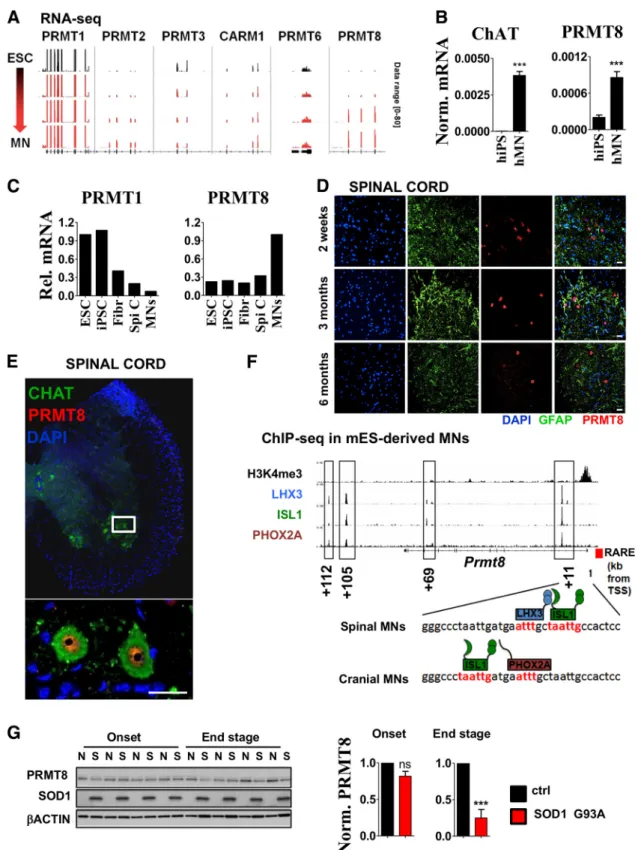
To mechanistically characterize the role of asymmetric arginine
dimethylation in MNs, we decided to identify the arginine meth-
yltransferases expressed in MNs. First, we compared the expres-
sion of known PRMTs during MN differentiation. Reanalysis of
transcriptomic data obtained during direct reprogramming of
mouse embryonic stem cells to MNs (Mazzoni et al., 2013) re-
vealed the high expression of
Prmt1at each stage (Fig. 3A). Inter-
estingly, its closest homolog,
Prmt8(Lee et al., 2005), showed
robust induction in the differentiating MNs (Fig. 3A). Human
PRMT8 expression showed similar induction along the differen-
tiation of human iPSCs to MNs (Fig. 3B). In addition, reanalysis
of gene expression data from Ho et al. (2016) and comparison of
PRMT8 expression in total human spinal cord versus LCM-isolated
MNs confirmed the relative enrichment of PRMT8 in MNs over
other cell types of the human spinal cord (Fig. 3C), whereas PRMT1
expression was more robust in undifferentiated human ESCs. Over-
all, these findings, partially based on previously published human
Figure 1. Asymmetric arginine dimethylation is highly abundant in postmitotic spinal cord MNs.A, Immunoblot analysis of FUS, methylated FUS, symmetric (SYM11) and asymmetric (ADMA) arginine dimethylated protein levels in ESCs versus spinal cord samples derived from 2-week-old (2w) to 6-month-old (6m) mice. Representative result of at least three independent immunoblot analyses and its quantification are shown. Note that FUS in ESCs show slightly different molecular weight.B, Coimmunostaining of mouse spinal cord cross sections derived from 2-week-old, 1-month-old, and 3-month-old animal for ADMA-containing proteins and GFAP (astrocyte marker). Magnification of a region marked by yellow star on the section of the 1-month-old sample is shown below the panel. Representative results of at least three independent analyses and their quantification are shown. Scale bar, 25m.C, Coimmunostaining of spinal cord cross sections derived from 6-month-old mouse for ADMA-containing proteins, methylated FUS (Met FUS), and ChAT (MN marker). Representative results of at least three independent analyses are shown. Scale bar, 25
m.D, Immunoblot analysis of ADMA levels in G93A SOD1 transgenic (S) and control (N) mice (n⫽4 in each group). Onset ages of the disease were determined as the time when the animals reached peak body weight and the end stage of the disease were determined when the animals could not right themselves within 10 s when placed on their side (typically 12–15 weeks). Data are presented as mean⫾SEM. **pⱕ0.01 (Student’s t-test).
and mouse transcriptomic data, led us to focus on PRMT8 and further investigate its role in the CNS for two reasons: (1) cell- restricted expression of
Prmt8indicated a unique role of this arginine methyltransferase in adult spinal cord MNs and (2) cell- type-selective expression of
Prmt8makes it a potentially exploit- able target for therapeutic interventions.
To characterize the cellular localization and distribution of PRMT8 protein, we developed an antibody against mouse PRMT8 (see Materials and Methods for more details). PRMT8 expression was restricted to a subset of cells in the spinal cord at all studied ages (2 weeks to 12 months) (Fig. 3D,E). PRMT8 costaining with ChAT showed the presence of PRMT8 in mouse spinal cord MNs and revealed its predominantly nuclear localiza- tion (Fig. 3E).
Next, we sought to understand the regulation of cell-type- restricted PRMT8. MN cell identity and cell-type-specific gene expression appears to be strictly coordinated by lineage-specific transcription factors [e.g., by LHX3, ISL1 (spinal MNs) or LHX3 and PHOX2A (cranial MNs) Sockanathan and Jessell, 1998; Maz- zoni et al., 2013]. Analysis of ChIP-Seq data obtained from ESC- derived MNs (Mazzoni et al., 2013) and from our previous report (Simandi et al., 2015) revealed that PRMT8 is likely under the
control of RAR:RXR, LHX3, PHOX2A, and ISL1, providing the basis of its cell-type-specific expression (Fig. 3F ).
Cell type-restricted expression of PRMT8 led us to investigate the consequences of disease-related loss of MNs to the expression of PRMT8. Using the same sample set as shown in Figure 1D, we found that the PRMT8 level significantly decreased in the end stage, further supporting the notion that predominantly MNs express PRMT8 (Fig. 3G).
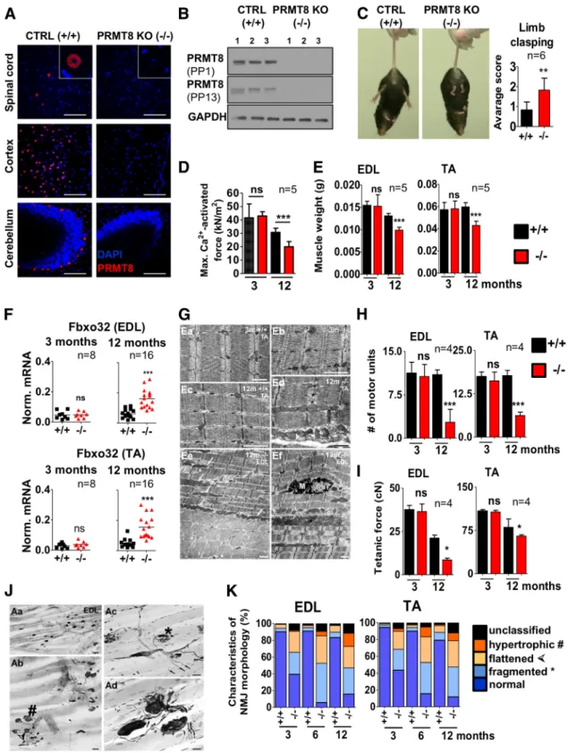
PRMT8-null animals show progressive muscle atrophy and NMJ fragmentation
To elucidate the role of PRMT8
in vivoin spinal cord MNs, we used a knock-out line generated by the European Conditional Mouse Mutagenesis Program and Knock-out Mouse Project (EUCOMM/KOMP). As described recently, these animals are viable, suggesting that PRMT8 per se is not required for the de- velopment of the CNS (Kim et al., 2015; Simandi et al., 2015). We validated the knock-out by PRMT8 immunostainings of the spi- nal cord and various brain regions (Fig. 4A) and also by Western blot (Fig. 4B). To assess the motor function of mice, we used 3-, 6-, and 12- to 15-month-old littermate control and PRMT8-null animals. A limb-clasping test revealed that PRMT8-null animals
Figure 2. Inhibition of methyltransferase activity induces accumulation of unrepaired DNA DSBs.A, Immunoblot analysis of BIP (ER stress), cleaved CASP3 (apoptosis), FUS/Met FUS, ADMA, and␥H2AX (DNA DSB) in differentiated NSC34 cells treated with the methyltransferase inhibitor ADOX (40M) for the indicated times. Representative results of at least three independent immunoblot analyses and their quantification are shown.B, Immunostaining of␥H2AX in cells treated with ADOX for 24 h (40M). Representative cells were chosen to show at high magnification. Scale bar, 5m.C, Cellular viability of NSC34 cells measured by MTT assay. Differentiating cells were pretreated with ADOX for 24 h (40m) at day 3 and then subjected to thapsigargin (ER stress, 0.2M) for 24 h. Averages of eight biological replicates are shown.D, qRT-PCR analysis of gene expression in differentiated NSC34 cells pretreated with ADOX for 24 h (40m) from day 3 and then subjected to thapsigargin (ER stress) or sodium arsenite (oxidative stress) for 3 h at day 4. Averages of three biological replicates are shown. Data are presented as mean⫾SEM. ns, Nonsignificant, *pⱕ0.05,
**pⱕ0.01, ***pⱕ0.001 (Student’sttest).
Figure 3. PRMT8 is a tissue-specific arginine methyltransferase in spinal cord MNs.A, IGV genome browser view of RNA-Seq data comparing expression levels of type I arginine methyltransferases along ESC to MN differentiation. RNA-Seq experiment was performed byMazzoni et al. (2013)and data have been reanalyzed to compare expression levels of type I PRMTs. Data range used for visualization was set to [0 – 80] for each track.B, CHAT and PRMT8 expression in human iPSCs vs iPSC-derived MNs as measured by qRT-PCR. Averages of three biological replicates are shown.
C, Relative expression levels of PRMT1 and PRMT8 in human ESCs and iPSCs versus spinal cord and LCM-captured postmortem human MNs. Raw data were obtained fromHo et al. (2016). Fibr, Fibroblast; Spi C, spinal cord.D, Coimmunostaining of spinal cord cross-sections derived from 2-week-old, 3-month-old, and 6-month-old mice for PRMT8 and GFAP (astrocyte marker). Represen- tative results of at least three independent analyses are shown. Scale bar, 25m.E, Immunostaining of spinal cord cross sections derived from 6-month-old mouse for PRMT8 and MN marker ChAT.
Scale bar, 25m.F, Occupancy of LHX3, ISL1, and PHOX2A transcription factors in the proximity ofPrmt8encoding gene as detected by ChIP-Seq. Primary ChIP-Seq data were obtained fromMazzoni et al. (2013). Previously described (Simandi et al., 2015) retinoic acid response element (RARE) in the proximity of the transcription start site (TSS) is also marked with a red box.G, Immunoblot analysis of PRMT8 in G93A SOD1 transgenic (S) and control (N) mice (n⫽4 in each group). Samples are identical to the ones shown inFigure 1D. Quantification of relative PRMT8 levels in the onset and end stages are shown. Data are presented as mean⫾SEM. ns, Nonsignificant, *pⱕ0.05, **pⱕ0.01, ***pⱕ0.001 (Student’sttest).
Figure 4. Aged PRMT8-null mice develop muscle weakness and progressive fragmentation of NMJs.A, Immunostaining of spinal cord (anterior horn), cortex, and cerebellum isolated from control and PRMT8-null mice for PRMT8. Scale bars, 100m.B, Immunoblot analysis of PRMT8 in brain samples of control and PRMT8 knock-out mice. Further details on antibodies PP1 and PP13 are presented in the Materials and Methods.C, Control animals at 15 months of age have normal limb reflexes when suspended by the tail, whereas age-matched PRMT8-null animals show an abnormal and weak limb clasping (n⫽6 in each group). Scoring is based onGuyenet et al. (2010). See also Movies 1 (ctrl) and 2 (PRMT8 knock-out).D, Comparison of maximal Ca2⫹-activated force levels of single isolated EDL myocytes. Force development during isometric contractions was measured in fast skeletal muscle fibers from EDL muscles of 3- and 12-month-old control and PRMT8-null mice (n⫽5 in each group).E, Muscle weight loss in TA and EDL muscles of aging animals (n⫽5 in each group). Note the significant muscle weight loss in 12-month-old PRMT8-null animals compared with their age-matched controls.F, qRT-PCR analysis of muscle atrophy markerFbxo32. Total RNA was extracted from TA and EDL muscles of 3- and 12-month-old control and PRMT8-null mice (n⫽ 8 –16 in each group).G, Electron microscopic ultrastructural analysis of 3- and 12-month-old control and PRMT8-null TA and EDL muscles. Shown are normal structure of a control (Ea) and PRMT8-null (Eb) TA muscles; also shown is a TA muscle sample taken from the 12-month-old control animals (Ec). Note that there is no obvious difference compared with 3-months samples (Ea).
Ed is a TA muscle sample taken from 12-month-old PRMT8-null animals. Note the obvious morphological changes including uneven sizes and a fuzzy appearance of the cross-striation pattern of the sarcomers, enlarged mitochondria, and occasional loss of Z discs. The presumed location of a missing Z disc is marked by asterisks. Ee, Ef, Disintegrated structure of EDL muscles taken from 12-month-old PRMT8-null animals. Note the enlarged mitochondria (arrows), uneven staining density of fibers, and splitting myofibrils (asterisks) in Ee. Ef shows a (Figure legend continues.)
show decreased motor performance, which was more pronounced in 12-month-old mice (Fig. 4C, Movies 1 and 2). Muscle force mea- surement on isolated muscle fibers revealed that 12-month-old PRMT8-null animals exhibit decreased muscle strength [30.7
⫾3.1 kN/m
2(control) vs 20.6
⫾1.9 kN/m
2(PRMT8-null); Fig. 4D].
Wet muscle weights of control versus PRMT8-null animals indicated an accelerated muscle weight loss in the knock-out mice (Fig. 4E). Accordingly, transcriptional levels of
Fbxo32(Atrogin-1) (a muscle atrophy-induced gene) (Bonaldo and San- dri, 2013) showed a remarkably elevated level in 12-month-old PRMT8-null animals compared with controls (Fig. 4F ), suggest-
ing an extensive atrophy in the muscle of PRMT8-null mice dur- ing aging.
Next, we used electron microscopy to evaluate morphological changes of the muscle structure. No differences were identified in young animals; however, lack of PRMT8 resulted in ultrastruc- tural changes, including disorganized sarcomer structure, un- even size and staining density of individual fibers and myofibrils, enlarged mitochondria, and occasional loss of Z discs and ap- pearance of splitting myofibrils in 12-month-old animals (Fig.
4G). Numbers of motor units were significantly lower in 12- month-old PRMT8-null mice (Fig. 4H ). Determination of motor units belonging to control and PRMT8-null TA and EDL muscles revealed a significant age-dependent decrease in PRMT8-null an- imals (EDL: 10.6
⫾1 vs 2.75
⫾1.1, TA: 16.25
⫾1.2 vs 6.25
⫾0.5 in 3 in and 12-month-old animals, respectively, see Fig. 4H ), whereas the number of motor units remained constant in the 12-month-old control animals. Smaller numbers of motor units produced smaller tetanic forces in 12-month-old PRMT8-null animals compared with their age-matched controls (PRMT8- null EDL: 36.5
⫾4.3 cN vs 8.6
⫾1.0cN, wild-type EDL: 37.6
⫾2.4cN vs 21.2
⫾1.7 cN in 3- and 12-month-old animals, respec- tively, Fig. 4I), especially in EDL muscles. A marked reduction of tetanic forces was also observed in aging control animals and this change is likely to be attributable to the aging processes of the muscle fibers as reported previously (Nair, 2005).
Because
Prmt8is not expressed in the muscle, the observed age-dependent muscle phenotype is most likely neurogenic due to loss of PRMT8 in the MNs and develops on the basis of reduced neuromuscular activity (Bu¨tikofer et al., 2011). To test this hy- pothesis, we next analyzed the number and morphology of motor end plates in TA and EDL muscles. Several morphological char-
4(Figure legend continued.) macrophage (M) among the myofibrils of a disorganized muscle fiber. Scale bars, 1m.H, Number of motor units as determined by counting the recruited individual twitch contractions elicited by increasing stimulation of the supplying nerve (n⫽4 in each group). Note the dramatic loss of functional motor units in both TA and EDL muscles of PRMT8-null animals.I, Smaller numbers of motor units produced smaller maximum tetanic forces (cN) in muscles of aging PRMT8-null animals, especially in the EDL muscle (n⫽4 in each group).J, Histological analysis of NMJs in EDL muscles of 12-month-old animals. Aa, Distribu- tion of motor end plates and axons in the innervation zone of an intact EDL muscle. Ab, Various pathological forms of degenerating NMJs in an EDL muscle. Double arrowhead, right upper corner: intact-looking NMJ. Hatchmark indicates hypertrophic NMJs; asterisks, fragmented NMJs; and arrowheads flattened NMJs. Ac, Higher-magnification view of two fragmented NMJs (asterisks) in an EDL muscle. Small arrows point to axons with bulb-like endings, which do not belong to motor end plates. Ad, Various forms of sprouting in EDL muscles. Arrows indicate preterminal sprouting and arrowheads ultraterminal sprouting. Scale bars: Aa, 50m; Ab, 25
m; Ac, 20m; Ad, 10m.K, Quantification of NMJ morphology data.y-axis represents the percentage of NMJs appearing either pretzel-like (i.e., normal) and pathological NMJs. Note the high proportion of pathological NMJs in PRMT8-null animals, grouped as fragmented, hyper- trophic, or flattened. Morphological characterization of NMJ changes was scored based accord- ing to recent studies (Valdez et al., 2010;Bruneteau et al., 2015). Data are presented as mean⫾ SEM. ns, Nonsignificant, *pⱕ0.05, **pⱕ0.01, ***pⱕ0.001 (Student’sttest).
Movie 1. Control animals at 15 months of age have normal limb reflexes when suspended by the tail.
Movie 2. PRMT8-null animals show abnormal and weak limb clasping.
acteristics of NMJs from control and PRMT8-null mice were assessed. In wild-type animals clusters of NMJs exhibited typical pretzel-like appearance and they were supplied by regularly ar- ranged bundles of motor axons (Fig. 4J, Aa). In contrast, NMJs of PRMT8-null mice showed various pathological alterations, in- cluding hypertrophy, fragmentation, and flattened morphology (Fig. 4J, Ab,Ac). More than 80% of the NMJs in 12-month-old knock-out animals were found to be pathological, whereas only up to 20% of NMJs were affected in age-matched control animals (Fig. 4K ).
Impaired axonal transport and premature accumulation of aging pigments in MNs of PRMT8 knock-out animals
We next performed histological analysis of MNs in the lumbar spinal cord using VAChT-specific antibody. The numbers of motor neurons (VAChT⫹) in the lumbar ventral horn (L4 – L5) supplying the sciatic nerve decreased in PRMT8-null ani- mals by 16.8% and 20.7% in 6- and 12-month-old animals compared with their age-matched controls, respectively (Fig.
5A). Retrograde labeling of the same motor pool with Fast Blue also revealed a mild decrease (17.4% and 26.8% in 6- and
Figure 5. Age-dependent dysfunction of axonal transport and decreased MN numbers in PRMT8-null mice.A, Left, Representative images taken from cross-sections ofL4 spinal cord segments showing Fast Blue-labeled and VAChT⫹MNs of a control and PRMT8-null animals at 6 and 12 months of age. Right, Quantification of Fast Blue-labeled and VAChT⫹MNs (n⫽4 in each group).Note the moderate loss of VAChT⫹(surviving) MNs and the more severe loss of capacity to retrogradely transport Fast Blue (neurons with lost or degenerating axons) in these neurons. Scale bar, 100m.B, Immunoblot analysis of spinal cord samples taken from 12-month-old mice for ADMA (n⫽3 in each group).C, Electron micrographs of MNs from the L4 spinal segment of 12-month-old control and PRMT8-null mice. MNs show normal ultrastructural morphology but lipofuscin granules (some of the labeled with arrowheads) are much more numerous in the PRMT8-null MNs. Scale bars: (A): 5m; (B, C): 1m; (D–F): 2m.D, Quantification of autofluorescent signals detected in spinal cord samples of 12-month-old control and PRMT8 knock-out mice. Representative results of at least three independent analyses are shown. Data are presented as mean⫾SEM. *pⱕ0.05, **pⱕ0.01 (Student’st-test).
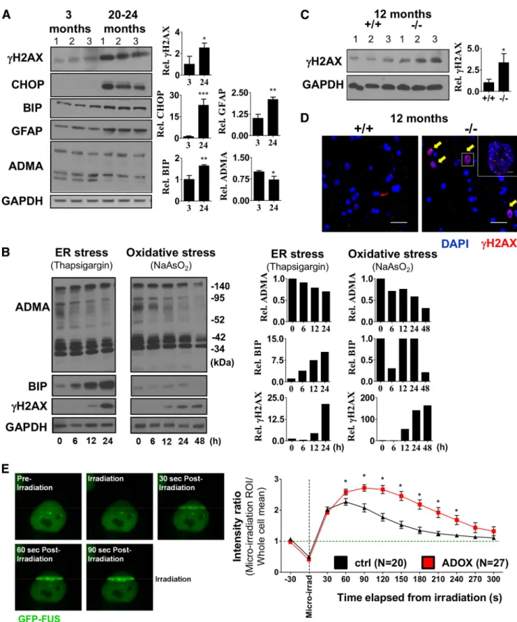
Figure 6. Age-dependent accumulation of unrepaired DNA DSBs in MNs of PRMT8-null mice.A, Immunoblot analysis of ADMA, CHOP (ER stress), BIP (ER stress marker), GFAP (astrocyte activation), and␥H2AX (DNA repair) in spinal cord samples of 3-month-old and 20- to 24-month-old mice.B, Immunoblot analysis of ADMA, BIP, and␥H2AX. Differentiated NSC34 MN cells were treated with thapsigargin or sodium arsenite for the indicated times. GAPDH served as a loading control. Note the unexpected accumulation of␥H2AX upon ER stress.C, Immunoblot analysis of spinal cord samples taken from 12-month-old mice for␥H2AX (n⫽3 in each group).D, Immunostaining of spinal cord samples from 12-month-old mice for␥H2AX. Representative images are shown.
Immunoblot presented inCserves as a quantitative comparison.E, Left, Micro-irradiation-induced FUS recruitment to DNA DSBs. HeLa cells expressing GFP-FUS constitutively were micro-irradiated at 405 nm in a 150⫻1 pixel ROI (step size: 70 nm). Right, Comparative analysis of FUS kinetics in control versus ADOX-pretreated cells. Data are presented as mean⫾SEM. ns, Nonsignificant, *pⱕ 0.05, **pⱕ0.01, ***pⱕ0.001 (Student’sttest).
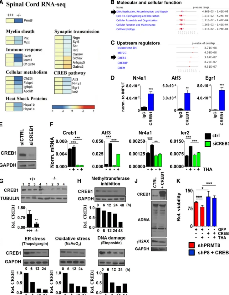
Figure 7. Compromised CREB1-dependent activation of stress signaling pathways.A, RNA-Seq analysis of spinal cord samples isolated from 6-month-old control and PRMT8-null mice (n⫽5 in each group).B,C, Prediction of PRMT8-dependent molecular and cellular functions and upstream regulators using IPA based on RNA-Seq data obtained in control and PRMT8-null spinal cord samples.D, ChIP-qPCR validation of CREB1 binding in differentiated NSC34 cells.E, Analysis of siCREB1 efficiency by immunoblot. Differentiating NSC34 cells were transfected and incubated for 2 d.
F, qRT-PCR analysis ofCreb1and target gene expression in CREB1-depleted cells upon thapsigargin-induced stress.G, Immunoblot analysis of spinal cord samples taken from 12-month-old control and PRMT8-null animals for CREB1 (n⫽4 in each group).H, Immunoblot analysis of CREB1 in ADOX-treated differentiated NSC34 cells.I, Changes of CREB1 protein (Figure legend continues.)
12-month-old animals, respectively), suggesting an impaired axonal transportin PRMT8-null animals (Fig. 5A, right).
Overall, these data suggested that more than one-fourt of ag- ing MNs were defunct in 12-month-old PRMT8-null mice.
The relatively mild impacts of PRMT8 depletion on the num- ber of MNs led us to investigate whether loss of PRMT8 is associated with decreased ADMA level and if these cells are intact. Immunoblot comparison revealed decreased arginine methylation (Fig. 5B), suggesting that, even if other type I arginine methyltransferases (e.g.,
Prmt1) are expressed inadult spinal cord MNs, loss of
Prmt8is not fully compensated by them.
Electron microscopy revealed premature accumulation of abnormal amount of lipofuscin in the MNs (Fig. 5C). Further analysis quantitatively confirmed the increased level of autofluo- rescent aging pigments in the PRMT8 knock-out spinal cord MNs (Fig. 5D), a pathological change that also appears prema- turely in ALS patients (McHolm et al., 1984).
Accumulation of DNA DSBs in MNs of aged PRMT8-null animals
Arginine methylation has been previously linked to DNA DSB repair in proliferating cells (Yu et al., 2009, 2012) and, according to our data, the ADOX-treated NSC34 cells showed accumula- tion of
␥H2AX (Fig. 2B). Recent findings in neurodegenerativediseases revealed that even small changes in the machinery of DNA repair can affect the age of onset of neurodegeneration, raising the possibility that compromised DNA repair is a com- mon risk in the progression of such diseases (Madabhushi et al., 2014). Comparison of spinal cord tissues isolated from 3-month- old and 24-month-old mice revealed the increased level of
␥
H2AX, as well as presence of CHOP (ER stress marker) and increased GFAP level (astrocyte activation) in aging mice, likely as consequences of aging-associated stress (Fig. 6A). ADMA level showed a mild reduction with aging (Fig. 6A). To test how ADMA level changes upon stress in
in vitroconditions, we treated MN cells with thapsigargin (ER stress) and sodium arsenite (oxidative stress).
Strikingly, persistent ER stress or oxidative stress reduced total ADMA levels and in parallel induced DNA DSB response (␥H2AX) (Fig. 6B). To determine whether loss of PRMT8 may lead to prema- ture accumulation of DSBs in spinal cord MNs, we compared
␥
H2AX levels. Immunoblot analysis revealed an increased
␥H2AX level in 12-month-old PRMT8 knock-out mice, indicating insuffi- cient DNA repair (Fig. 6C). Immunohistochemistry confirmed the accumulation of
␥H2AX⫹cells in the PRMT8 knock-out spinal cords, but not in age-matched controls (Fig. 6D).
Previous studies already proposed the role of arginine meth- ylation in DNA repair in proliferating cells. Importantly, the mu- tant form of FUS, in which the mutation affects the arginine methylated site, results in impaired DSB repair (Wang et al., 2013). Using a GFP-FUS-expressing stable cell line, we confirmed the dynamic recruitment of FUS to micro-irradiation-induced DSBs (Fig. 6E). Moreover, we found that ADOX pretreatment of
the cells strongly increased the residence time of FUS at the sites of DSBs, indicating impaired function and altered kinetics of hypomethylated FUS (Fig. 6E). Collectively, these data suggest impaired DNA DSB repair in aging PRMT8-null animals.
Loss of PRMT8 disrupts the CREB1-dependent neuroprotective transcriptional network
In previous studies, profiling of gene expression has been widely used in MN disease models at several stages during the course of disease and revealed early gene expression changes related to in- flammation, apoptosis, ATP biosynthesis, myelination, axonal transport, and synaptic transmission (de Oliveira et al., 2013;
Scekic-Zahirovic et al., 2016). To identify components affected in spinal cords of aged PRMT8-null animals in an unbiased man- ner, we applied RNA-Seq on total RNA extracted from the lum- bar spinal cord regions of five mice per group. We considered those genes differentially expressed that showed at least 1.5-fold change in their expression at least in 4 mice per group. As ex- pected,
Prmt8expression was completely diminished in the knock-out mice (Fig. 7A). Several genes with well established roles in myelinization (e.g.,
Mpz,Prx) were dysregulated (Fig.7A). Genes involved in synaptic plasticity (Nrgn,
Syt5,Sst,Camkv, Gabra2) and cellular metabolism (Ch25h,Igfbp6) were also de-creased in PRMT8-null mice (Fig. 7A). The list of dysregulated genes included many genes previously shown to be differentially expressed in human ALS or FTD (e.g.,
Nrgn, Gabra2, Hspa1b, Gucy1a2) (Lederer et al., 2007;Comley et al., 2015) and several IEGs (e.g.,
Atf3, Egr1, Ier2). Pathway analysis of differentiallyexpressed genes by ingenuity pathway analysis (IPA) indicated DNA replication, recombination, and repair as one of the top cellular function affected in PRMT8 knock-out tissues (Fig. 7B), consistent with the accumulation of
␥H2AX detected in spinal cord lysates of PRMT8 knock-out mice (Fig. 6). To better under- stand the molecular mechanisms impaired in PRMT8-depleted MNs, we used IPA to predict potential upstream transcriptional regulators causing dysregulated gene expression. This analysis revealed that the function of CREB is likely affected in the absence of PRMT8 (Fig. 7C).
CREB1 is a constitutively expressed transcription factor that regulates the expression of genes involved in neuronal survival and function (Sakamoto et al., 2011). To assess whether CREB1 directly regulates the genes differentially expressed in the spinal cord of PRMT8-null mice (e.g.,
Atf3,Nr4a1,Egr1,Ier2;Fig. 7A), we analyzed binding of CREB1 in the proximity of their tran- scription start site by ChIP-qPCR. These results confirmed that CREB1 is enriched at these sites (Fig. 7D). The requirement of CREB1 for the induction of these IEGs upon cellular stress was validated by loss-of-function experiments in differentiated NSC34 cells (Fig. 7
E,F).
To understand why the CREB1-dependent gene expression network is dysregulated in PRMT8-null mice, we next analyzed CREB1 at the protein level. This comparison revealed a reduction of CREB1 protein levels in
in vivoin 12-month-old PRMT8 knock-out spinal cord samples (Fig. 7G). Comparison of CREB1 levels following inhibition of methyltransferase activity by ADOX confirmed a gradual decrease in CREB1 protein levels (Fig. 7H ), formally suggesting that arginine methylation is required to maintain CREB1 level. Exposing NSC34 cells to persistent stress also reduced the level of CREB1 (Fig. 7I) in addition to reducing the ADMA level (Fig. 6B), suggesting a positive correlation be- tween the level of ADMA and CREB1.
We next wanted to determine whether ectopic expression of PRMT8 or CREB1 is sufficient to increase cell survival and stress
4(Figure legend continued.) level upon ER stress (thapsigargin, 1M), oxidative stress (so- dium arsenite, 0.2M), and DNA DSB induction (etoposide, 5M). Representative results of at least three independent experiments are shown.J, Immunoblot analysis of ADMA level in control versus CREB1-overexpressing NSC34 cells. Representative results of at least three inde- pendent immunoblot analyses and their quantification are shown.K, Cellular viability of PRMT8-depleted cells transfected with GFP (control) or CREB1 as measured by MTT assay. Data are presented as mean⫾SEM. ns, Nonsignificant, *pⱕ0.05, **pⱕ0.01, ***pⱕ0.001 (Student’sttest).