III./7.4.: Pathological characteristics, most common types of tumors
The pathological and clinical characteristics of the most common and important nervous system tumors are described in this chapter.
Introduction
A review of the general histological features of benign and malignant tumors is recommended first.
Astrocytomas
The behavior of astrocytomas depend on their grade
Astrocytomas are a heterogeneous group of tumors derived from astrocytes, including tumor types from benign (grade I) pilocytic astrocytoma to malignant (grade IV) glioblastoma. They are also known as diffuse astrocytomas, as they infiltrate the surrounding or remote brain regions, and their boundaries are not well-defined.
Astrocytomas comprise about 60% of all brain tumors, they are the most common adult forms of hemispheric tumors. They mainly occur in the frontal and temporal lobes. The more benign forms are more common in children and in young adults, whereas
glioblastoma is most common in the sixth decade of life.
The only grade I astrocytoma is pilocytic astrocytoma. It occurs primarily in children, mainly in mid-line structures, especially in the cerebellum and also in the optic nerve, in the medial temporal lobe, in the brainstem, in the hypothalamus and in the spinal cord. Imaging tests often show cystic, well circumscribed tumors, with their walls containing contrast enhancing noduli. Its name was given after the pilocytes with a fibrillary structure ("hairy" cells). Malignant transformation is rare, prognosis is very good, 96% of patients survive over 10 years.
Grade II diffuse astrocytomas are slow-growing, well differentiated tumors. Imaging studies show an ill-defined, homogeneous, non-enhancing lesion, which has low density in CT scans, and shows decreased signal intensity in T1-weighted MRI images, and increased in T2-weighted images.
Fig. 9: 28-year-old male patient. Low grade (A2) astrocytoma, located in the left temporal region, showing infiltrative growth, causing no space-occupying effect. MRI scans, FLAIR sequence (Fluid Attenuated
Inversion Recovery).
The average survival time after diagnosis is 6-8 years, showing significant variation depending on malignization.
Anaplastic (grade III) astrocytoma differs from grade II astrocytomas mainly in radiological contrast enhancing. Median survival is 2-3 years, the 5-year survival rate is around 10%.
Glioblastoma is the most malignant astrocytoma
Glioblastomas (formerly glioblastoma multiforme) comprise 12-15% of intracranial tumors, and 50-60% of astrocytomas. Approx. 10% develops from lower-grade tumors, but the tumor is "de novo" discovered in the majority of the cases.
It can occur at any age, but typically between the ages of 40 and 70 years. It occurs commonly in the hemispheres, in the brainstem mainly at a young age, and it is rare in the spinal cord and cerebellum. Glioblastoma is characterized by the extensive infiltration of surrounding tissues, especially along white matter tracts; therefore, complete surgical removal is impossible. The spread through the corpus callosum is typical: tumor masses seen in both hemispheres are connected by a narrow ʽstalk’ in the corpus callosum ("butterfly tumor").
New foci of the tumor often develop by propagation via the comissures, fornix, and optic radiation, and these multiple tumor masses may raise the suspicion of metastases.
Despite this aggressive proliferation in the brain, metastases rarely develop on the meninges, in the CSF space, or outside the nervous system. The radiological analysis of the tumor shows large perifocal edema around the tumor, ring-like enhancement in the marginal parts, and central hypodensity (CT) or decreased signal intensity in
T1-weighted MRI images due to tumor necrosis.
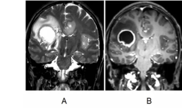
Fig. 10: 49-year-old woman. Histology: glioblastoma.
In T2-weighted MRI images, a right hemispheric cystic tumor appears surrounded by edema, which spreads through the corpus callosum to the opposite side. There is also a secondary tumor focus in the left temporal area. (A) In contrast-enhanced T1-weighted images, the wall of the cystic area shows significant enhancement (B).
Glioblastomas show high mytotic activity, necrosis and vascular proliferation. The immunohistochemical detection of the typical astrocytic protein, GFAP (glial fibrillary acidic protein), helps in identifying the tumor.
Fig. 11: Histology of glioblastoma
A: A hematoxylin-eosin stained image shows the diversity of cells (pleiomorphism), nuclear atypia, and the ʽpseudo-palisade’-like (long arrow) arrangement of cells (created by dividing cells, arranged side by side) around the necrotic area (*), and vascular proliferation (short arrows).
B: GFAP (Glial Fibrillary Acid Protein), with immunohistochemistry. Inside the tumor, the better differentiated areas are stained with brown, while the less differentiated areas show no or barely visible staining.
C: Tumor cells gather around blood vessels (*) forming secondary structures (GFAP- immunohistochemistry).
Certain genetic alterations have also been demonstrated. Prognosis is poor; average survival rate is about one year.
Oligodendroglia derived tumors
Oligodendrogliomas usually contain calcifications, and cause epilepsy
This group comprises oligodendroglioma (grade II) and anaplastic oligodendroglioma (grade III).
Oligodendrogliomas make up approx. 2.5% of primary brain tumors, and 5-6% of gliomas. It can occur at any age but most frequently around 40-50 years of age. CT scan shows a hypo-or isodense, well-defined lesion, which is usually located in the subcortical white matter involving also the surrounding cortex. Calcification of the tumor is frequent and typical.
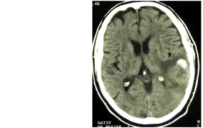
Fig. 12: Oligodendroglioma, non-enhanced CT scan. The tumor is located in the left posterior temporal lobe, and contains calcification, which is also hyperdense in non-enhanced scans.
Contrast enhancement may occur. Microscopically, cells have a typical shape,
resembling fried eggs (the nucleus is pushed aside in the faintly stained cytoplasm). In 80% of the cases, chromosomal deletions (1p and 19q) can be detected.
Anaplastic oligodendrogliomas comprise 1.2% of all brain tumors. In general, the tumor is de novo formed, but may also develop as the malignization of an
oligodendroglioma. The radiological appearance of anaplastic oligodendroglioma is mixed, contrast enhancement is common, but its absence does not rule out this type of tumor. It is interesting that if chromosomal deletions (1p and 19q) are present, the tumor shows a better response to chemotherapy and radiation therapy, and survival is longer.
Ependymomas
Ependymomas are gliomas originating from cells making up the wall of the ventricles or the central spinal canal, which comprise about 6% of gliomas. Ependymomas usually show contrast enhancement, and often cause hydrocephalus due to their location. Grade II ependymomas and grade III anaplastic ependymomas are
well-known. Subependymomas and myxopapillary ependymomas are slowly growing grade I tumors.
Ependymomas may be intracranial or spinal
Ependymomas may develop in the spinal cord as well.
Fig. 13: Intraventricular ependymoma (A) and a spinal form (B,C).
MRI scans.
Tumors of the plexus chorioideus
Tumors of the plexus chorioideus are rare tumors, occurring mainly in childhood.
Radiologically, they show homogeneous enhancement, and may contain cystic, calcified and hemorrhaged areas; because of their location, they may also cause hydrocephalus. They often occur as part of familial tumor syndromes. The benign form is papilloma, while the malignant is plexus carcinoma, which may give metastases via the CSF.
Tumors with neuronal origin
Tumors with neuronal origin are rare tumors in adulthood. Gangliocytoma is a grade I tumor, which most often originates in the temporal lobes, clinically it is manifested in epilepsy. The ganglioglioma contains cells with neuronal origin and glial cells as well, and consequently a malignant form is also known (anaplastic ganglioglioma). Central neurocytoma occurs in young adults; it is a rare tumor, which starts from the region of the foramen of Monroe, with a relatively good prognosis.
Medulloblastoma
Medulloblastoma is one of the most malignant childhood brain tumors
Medulloblastoma is the most common type of the childhood brain tumor, it comprises approximately 20% of all nervous system tumors occurring at this age. It occurs most commonly between the ages of 5-10 years. It is an aggressive, rapidly growing tumor, originating from the undifferentiated cells of the cerebellar vermis. As it spreads, it can infiltrate the brainstem, and leads to occlusive hydrocephalus by occluding the fourth ventricle. Symptoms include headache, nausea, vomiting, dizziness, ataxia, and
abnormal gait. The first symptom of hydrocephalus may be a rapid decrease of the level of consciousness. Tumor cells loosened from the meninges drift away with the CSF, and form metastases anywhere of the surface of the brain and spinal cord. Diagnosis is provided by MRI.
Fig. 14: Medulloblastoma, filling the fourth ventricle. Dilation of the lateral ventricles indicates a developing
hydrocephalus (B). MRI images.
If the brainstem is affected, the success of surgical removal is limited. Survival can be significantly improved with radiotherapy and chemotherapy, but the risk of recurrence remains. To prevent spinal drop metastases, irradiation of the entire neuraxis may help.
Meningiomas
Meningioma is a very common, slowly growing, benign intracranial or spinal tumor
Meningiomas are mesenchymal tumors originating from the meninges, comprising approximately 30% of central nervous system tumors. They are more common in the elderly, especially in the sixth or seventh decade. They are twice as common in females as in males, but the female: male ratio is 9:1 in posterior meningiomas. After cranial irradiation, the risk of meningioma is clearly increased. They may occur anywhere in the nervous system, but particularly often on the parasagittal convexity. In the skull base, they occur more frequently in a frontobasal or parasellar location, and along the sphenoidal wings and the pyramidal bone. Multiple meningiomas may occur in neurofibromatosis type 2. On non-enhanced neuroimaging tests, the density and signal intensity of meningiomas are similar to that of the cortex, but their contrast
enhancement is typically homogeneous, marked, and often extends to the surrounding dura ("dural tail").
Fig. 15: 37-year-old woman. Meningioma originating on the skull base in contrast enhanced (A) and non-enhanced (B) T1-weighted MRI images.
Meningiomas often contain calcifications, which can be best seen on CT scans.
Fig. 16: Meningioma containing calcifications on non-enhanced CT scans.
Larger meningiomas are often surrounded by extensive perifocal edema. Meningiomas with an irregular surface may show infiltration and malignancy. Histological variability is marked. In addition to nine types of grade I meningiomas, four forms of grade II tumors are also known. Malignant meningiomas (grade III) include anaplastic, papillary and rhabdoid meningiomas. Recurrence may occur after removal of a meningioma, and malignant transformation is also possible. Prognosis is determined by the histological subtype.
Schwannoma
Characteristic clinical symptoms
Seven-eight percent of intracranial tumors are schwannomas; 90% of them is vestibular schwannoma, which was formerly called acoustic neurinoma. Vestibular schwannoma is a cerebellopontine angle tumor, which typically causes unilateral (or asymmetric) hearing loss (mainly affecting the higher frequencies), but sudden unilateral hearing loss (due to the compression of the blood vessels supplying the inner ear), tinnitus, dizziness, peripheral facial paresis, and trigeminal nerve damage may also occur.
Vestibular schwannomas are more common in females than males by a factor of 1,5 to 2. Ninety-five percent of cases are unilateral; in bilateral tumors, neurofibromatosis type 2 should be considered. Schwannomas may occur on spinal and peripheral nerves as well. The diagnosis of the vestibular schwannoma is established with contrast -enhanced MRI imaging.
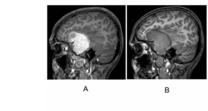
Fig. 17: A 70-year-old female patient. Left-sided large vestibular schwannoma. In contrast- enhanced T1-weighted images, the narrow initial
part of the tumor is well seen in the internal acoustic meatus, from which the tumor originates.
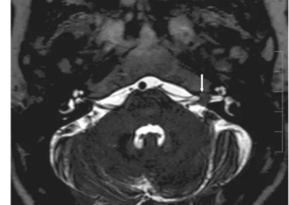
Fig. 18: Cisternography MRI images of a small, intra- meatal left vestibular schwannoma
In the treatment of vestibular schwannomas (especially in small tumors), radiation surgery has a growing role.
Primary central nervous system lymphoma (PCNSL)
PCNSL can be treated easily with radiation surgery
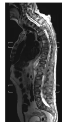
PCNSL is an extranodal non-Hodgkin lymphoma (NHL), however as there is no lymphoid tissue in the nervous system, its origin is unclear. The isolated nervous system manifestation of a systemic disease is supposed. PCNSL comprise 2-4% of CNS tumors, and 2% of NHL presents as PCNSL. Eighty-five percent of PCNSL is large B-cell lymphoma. It is common in immunocompromised patients, especially in AIDS.
Brain lymphoma is suspected if a tumor is seen showing intense enhancement and perifocal edema, which is connected with or located close to CSF spaces, and has a relatively mild space-occupying effect relative to its size.
Fig. 19: Primary Central Nervous System Lymphoma in contrast-enhanced T1-weighted MRI images. There are two nearly symmetrical, contrast enhancing lesions affecting both thalami, located near the third
ventricle, having only minimal space-occupying effect.
PCNSL is suspected in
immunosuppressed patients
Patients show a history of mental decline during the previous months, and cognitive dysfunction can be explored. Vitreous opacity is detected by the ophthalmologist. The condition is more common in immunosuppressed states (transplanted patients, HIV infection), in which case the lesions are often multiple. In the diagnosis of PCNSL, MRI and subsequent brain biopsy are essential. The appearance of the tumor is often
fluctuating in repeated imaging studies. Tumor regression is especially typical after steroid treatment, even complete regression can be seen ("ghost tumor") in some cases.
The first-line treatment of primary brain lymphoma is high-dose methotrexate chemotherapy, which may cause rapid and prolonged remission. Radiotherapy is also very effective, typically used in relapsed cases.
Pituitary tumors
Tumors in the sellar region are common. Depending on their size, they are called micro-and macro-adenoma (above or below 10 mm). Sellar tumors may be hormonally active (i.e. hormone producing) or inactive. Hormone-producing adenomas, by
secreting excessive amount of hormones into the circulation, cause typical hyper- function disorders (galactorrhea, amenorrhea, acromegaly, Cushing's disease, and central hyperthyroidism). Prolactin-producing adenomas are the most common.
However, hormonal disorders can develop in other ways, e.g. a hormonally inactive sellar tumor compresses and damages the pituitary gland, leading to general hypofunction (panhypopituitarism). The tumor’s hormone-producing ability is not related to tumor size, a microadenoma may also cause serious endocrine abnormalities.
However, an excessive production of hormones may be the sign of hyperplasia of the gland, not only of adenomas.
Prognosis is favorable with appropriate treatment
The majority of adenomas are asymptomatic. The complaints are most often caused by hormonal disorders. Since the patients’ tolerance of symptoms is highly variable, diagnosis may be delayed by years.
Only macroadenomas cause focal neurological signs by compressing the surrounding neural structures. The typical symptom is bitemporal heteronymous hemianopia due to the damage of the decussating fibers of the optic chiasma.
Endocrine
disorders: hyper- or hypofunction
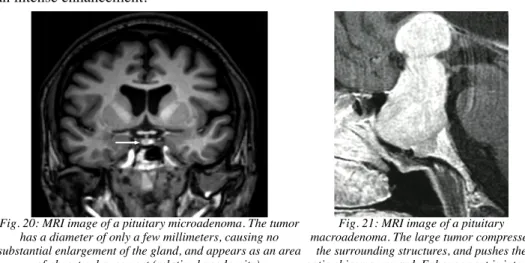
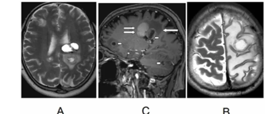
Patient assessment usually starts with laboratory tests of hormone levels. This is followed by a pituitary gland imaging study, which should be a contrast-enhanced MRI targeting the sellar region. Microadenomas appear as an area showing no enhancement within the homogeneously enhancing normal gland. In contrast, macroadenomas show an intense enhancement.
Fig. 20: MRI image of a pituitary microadenoma. The tumor has a diameter of only a few millimeters, causing no substantial enlargement of the gland, and appears as an area
of absent enhancement (relative hypodensity).
Fig. 21: MRI image of a pituitary macroadenoma. The large tumor compresses
the surrounding structures, and pushes the optic chiasma upward. Enhancement is intense
and homogeneous.
Surgical and hormonal therapy
The treatment is determined by the endocrinologist and the neurosurgeon.
Microadenomas do not require surgery, often hormonal therapy (dopamine agonist bromocriptine) results in normalization of the hormonal disorder and also in reduction of tumor size. Macroadenomas causing symptoms due to compression should be removed, regardless of their hormonal activity.
Nervous system metastases
Malignant tumors often give metastases to the nervous system, mainly via a hematogenous route.
Half of IC tumors in adults are metastases, and they are responsible for 20% of cancer- related deaths. Metastases can originate from many different types of malignancies of the body. However, certain tumors show an increased affinity in forming metastases in the nervous system. Therefore, the incidence of the most common extraneural tumors and of the most common intracranial metastases is not the same. Bronchial and breast cancer often give metastases to the nervous system, but the likewise common
gastrointestinal tumors only infrequently. Melanomas, which accounts for only a small part of systemic tumors, are relative common causes of intracranial metastases.
Metastases of different origin prefer different parts of the nervous system.
Most common origin of metastatic brain tumors, in order of frequency:
• Lung
• Breast
• Malignant melanoma
• Urogenital
• Gastrointestinal
• Other (sarcoma, lymphoma, head and neck tumors, tumors of unknown origin)
Localization
Metastatic tumors may be located in the parenchyma itself (brain, spinal cord), on the meninges, and on the bones of the skull and spine. The symptoms of metastatic tumors are the same as in primary brain tumors, but metastases should be suspected if signs of multiple lesions are present.
Symptoms, diagnosis
Brain metastases act as a foreign tissue, they trigger substantial perifocal oedema, and may irritate surrounding structures (e.g. the cortex), manifesting in focal or secondarily generalized epileptic seizures. Metastases should be suspected in partial epilepsy with onset in the elderly. Neuroimaging shows multiple round lesions of various size (commonly in the watershed areas of the hemispheres and in the cerebellum), with typical ring-like enhancement, and with relatively large perifocal white matter oedema.
Cystic change and hemorrhage are also not uncommon.
Fig. 22: Brain metastases. In images A and B, multiple hemispheric and cerebellar metastases are seen, with hemorrhage within the tumor (double arrow). In the contrast-enhanced image, ring-like enhancement is seen (short arrow). Perifocal oedema is also seen (long arrow). In image C, a relatively small metastasis is seen,
which causes edema in the entire left hemisphere.
Searching for the primary tumor
Solitary metastasis is present in 50% of the cases. The morphology of brain metastases may sometimes suggest the initial tumor (e.g. melanoma), but the appearance is not specific in most cases.
In 15% of the patients with brain metastases, the primary tumor is unknown at the time
diagnosis. Brain biopsy may be helpful in such cases, but sometimes it provides only an approximate diagnosis (e.g. adenocarcinoma). The next step is to perform a thorough search for the primary tumor, for which a PET-CT scan may be indicated. It is possible that the primary tumor is not found, even at autopsy. A small bronchial carcinoma is the most likely primary tumor in these cases.
Brain metastases may cause diagnostic problems even in patients with a known tumor, if they originate from another, yet unknown tumor, and a primary brain tumor may resemble a metastasis.
Treatment
The treatment of brain metastases - depending on the number of lesions, location, size and the patient's general oncological status - includes surgery and radiation surgery, irradiation and chemotherapy. Symptomatic treatment (reduction of the brain oedema, anti-epileptic drugs) is also important.
Spinal columnal metastases
Metastases in the spinal column typically grow in an extradural location, in the vertebrae. Multiple occurrences is typical. As an initial symptom, local spinal pain is common, followed by spinal cord and radicular symptoms when the lesion grows out of the vertebra. At the time of diagnosis, MRI of other, asymptomatic segments of the spine should also be performed. Vertebral metastases are most often caused by bronchial carcinoma, breast cancer and melanoma, but prostate cancer should also be considered in males, which often propagates via the venous plexus toward the spine.
With improvement of cancer therapy and longer survival of patients, the incidence of nervous system metastases increases. The diagnosis of brain metastases is a dramatic event, as they may constitute a direct threat to life. Spinal metastases are not directly life threatening, but they lead to serious deterioration of the quality of life by causing severe pain and paralysis as a result of spinal cord compression. In such cases, life expectancy is a key factor in determining treatment. Surgery is not indicated if the expected survival is only a few weeks or months. Radiation therapy often helps in reducing the symptoms.
Why does the number of nervous system metastases increase?
What is the role of oncology?
In summary, the treatment of nervous system metastases should be individualized, it is a complex task, carried out by an onco-team.
References concerning tumor pathology, based on the most recent classification can be found here:
http://www.brainlife.org/who/classification_updates.htm
Spinal tumors
Relationship between tumor location and type
Spinal tumors are classified mainly according to their location. Location correlates with the histological type, i.e. certain types of tumor occur more frequently in certain location. In the diagnosis of spinal tumors, MRI is the most effective method.
Extradural tumors grow outside the spinal cord, usually in the spinal vertebra. Local back pain and sensitivity upon tapping are typical. In case of progression, the spinal roots and the spinal cord may become compressed, segmental pain and spinal cord symptoms appear. Metastases are the most common in this location.
Fig. 23: Multiple vertebral metastases causing spinal cord compression on T2-weighted MRI images
Hemangiomas, benign tumors of the vertebrae are often accidentally discovered by MRI examination, however aggressively growing, symptomatic forms of hemangiomas are also known.
Intradural (extramedullary) tumors develop inside the dural sack, and cause spinal root symptoms. Since the spinal cord is compressed from the outside, superficial fibers are damaged first, which are the longest fibers representing the lower extremities.
Therefore, symptoms (numbness, weakness) start in the distal parts of lower
extremities, and spread slowly upwards. Thus, the upper limit of sensory loss may be misleading, as it may be lower than actual level of the tumor. Local spinal pain may also occur. Distal sensory loss may be an early sign. Motor symptoms are manifested for a long time in the form of slowly progressing spasticity of the legs and stiff gait, with weakness occurring later. At this stage, urinary and sexually dysfunction are usually present, which are often the cause that persuades patients to seek medical attention.
Other patients receive physiotherapy because of local pain for years, until neurological symptoms appear. The spinal cord tolerates slowly developing compression
surprisingly well. Relatively good function and good chances of recovery are seen in case of large tumors with a marked space-occupying effect.
Fig. 24: Meningioma almost completely filling the thoracic spinal canal, causing spinal compression (A). On the axial image, it is seen that the spinal cord is pushed to the side, having the shape of a sickle (B,
arrows).The patient was still able to walk. (Contrast-enhanced, T1-weighted MRI images.)
Most of the intradural tumors are benign, mainly neuromas and meningiomas.
Intramedullary tumors are located inside of the spinal cord. They have different symptoms compared to the spinal tumors mentioned above. Local pain is not typical. In affected segments, segmental symptoms of lower motoneuron lesion and dissociated sensory loss (isolated loss of pain and thermal sensation) develop. Corticospinal tract involvement causes spastic paralysis of the lower limbs, which may be accompanied by autonomic dysfunction. Central (neurogenic) pain is not uncommon.
Fig. 25:
Hemangioblastoma. MRI image of a cystic tumor
with a small solid part located within the spinal
cord (intramedullary tumor),causing bulging of
the spinal cord
Fig. 26: Classification of spinal tumors based on their localization.
Meningeal carcinomatosis
Meningeal carcinomatosis is a serious, progressive disease, caused by tumor infiltration of the leptomeninges and occurring mainly in advanced stages of systemic
malignancies. In most cases, solid brain metastases, often near the surface of the brain, are also seen, but meningeal carcinomatosis may be the first and only neurological manifestation. In addition to hematological malignancies, solid tumors (lung and breast cancer, malignant melanoma and gastrointestinal cancers) may also be the cause.
Sometimes the primary tumor remains unknown.
As the survival time of cancer increases, similarly to brain metastases, the incidence of meningeal carcinomatosis is also increasing. Often this is the direct complication leading to death. Clinical symptoms are caused by the infiltration and compression of spinal and cranial nerve roots, and by the increased intracranial pressure caused by the disturbance of CSF circulation and absorption. Eye movement disorder, bilateral peripheral facial palsy, neural hearing loss, and rarely cauda syndrome may occur.
Headache, confusion and subsequent decreased level of consciousness are explained by increased IC pressure and hydrocephalus. Urinary retention, and motor and sensory symptoms may develop. Excitatory meningeal signs are usually mild or absent.
Meningeal carcinomatosis occurs in the end-stage malignant diseases!
Symptoms!
MRI with contrast-enhancement shows thickened meninges of the brain, spinal cord, cranial nerves and spinal roots, occasionally with nodular enhancement .
Fig. 27: Meningeal carcinomatosis. On T1-weighted enhanced MRI images, intensive enhancement can be seen on the surface of the brain
CSF examination shows elevated pressure and protein content, and an increased cell count. CSF cytology shows the tumor cells and reactive cells, the primary tumor can also be determined in some cases.
Fig. 28: MGG (May-Grünwald-Giemsa) staining of sedimented CSF cells.
In the preparation rich in cells, pleomorphic cells can be seen with nuclei of variable size, often arranged in groups. On the left-sided, an insert with high-magnification shows the typical "signet-ring" cells, which are characteristic for adenocarcinoma: the cytoplasm is filled with vacuoles, and the nucleus is pushed aside. The right-sided insert shows multinucleated tumor cells. Meningeal carcinomatosis due to breast cancer.
Treatment
CSF examination is essential!
Systemic treatments are not effective in meningeal carcinomatosis. In addition to irradiation of the whole neuraxis, intrathecal cytostatic therapy (methotrexate and cytosine arabinosid) can be administered via repeated lumbar punctures or an Ommaya reservoir. If hydrocephalus is present, a shunt surgery can be done as a palliative measure. Prognosis is poor, survival is a few weeks without treatment and a few months with combination therapy.